Review
Christophe A. Monnier, David Burnand, Barbara Rothen-Rutishauser, Marco Lattuada
and Alke Petri-Fink*
Magnetoliposomes: opportunities and challenges
Abstract: Combining liposomes with magnetic
nanoparti-cles is an intriguing approach to create multifunctional ves-icles for medical applications, which range from controlled drug delivery vehicles to diagnostic imaging enhancers. Over the past decade, significant effort has been invested in developing such hybrids – widely known as magnetoli-posomes – and has led to numerous new concepts. This review provides an overview on of the current state of the art in this field. The concept of magnetic fluid hyperther-mia and stimuli-responsive nanoparticles for drug delivery is briefly recapitulated. The materials needed for these hybrids are addressed as well. The three typically followed approaches to associate magnetic nanoparticles to the liposomes are described and discussed more in detail. The final chapters are dedicated to the analytical methods used to characterize these hybrids and to theoretical considera-tions relevant for bilayer-embedded nanoparticles.
Keywords: electron microscopy; magnetic
nano-particles; magnetoliposome; membrane energetics; stimuli-responsive.
DOI 10.1515/ejnm-2014-0042
Received November 9, 2014; accepted November 14, 2014
Introduction
A nanoparticle (NP) is defined as a material with all three external dimensions in the nanoscale (ISO/TS: 27687:2008), i.e., below 100 nm. The current choice of available NPs is colossal and ranges from relatively simple
single NPs to highly complex surface derivatized NPs or NP assemblies. All engineered NPs have one common link: Their chemical, physical and biological properties can differ considerably from the bulk material properties. For
example, iron oxides – such as maghemite (γ-Fe2O3) and
magnetite (Fe3O4) – lose their permanent magnetization
below a certain size, which is typically below 20 nm (1). At this point, iron oxide NPs possess only one magnetic domain, and consequently exhibit superparamagnetic behavior at temperatures above the so-called blocking temperature (2). Nowadays, magnetic NPs are found in a rapidly increasing number of applications, including catalysis (3), sensing (4), and filtration (5). In nanomedi-cine, superparamagnetic iron oxide NPs (SPIONs) have gained wide acceptance in diagnosis and are used for contrast enhancement in magnetic resonance imaging (MRI) (6), (stem) cell tracking and labeling (7) or magnetic separation technologies (e.g., rapid DNA sequencing) and ultrasensitive diagnostic assays (8).
The benefits of magnetic NPs for therapeutic purposes are indisputable, and magnetic targeting for drug or gene delivery and magnetic fluid hyperthermia (MFH) are argu-ably the two most important potential therapeutic applica-tions. In particular, SPIONs are promising because of their outstanding magnetic behavior (9), their biocompatibility (10), and the large amount of information available on these materials. However, the process of converting basic research into clinical nanomedicine settings and commer-cially sustainable products is long and complicated (11), and the acceptance and integration of nanotechnologies particularly into nanomedicine are very challenging.
Magnetic fluid hyperthermia –
a brief recapitulation
Magnetic fluid hyperthermia (MFH) was first proposed by Gilchrist and colleagues (12). In short, it involves the injec-tion of SPIONs directly into a specific tissue or organ (e.g., lymph nodes) and the subsequent exposure to an alternat-ing magnetic field (AMF) to heat the region in question up to 45 to 47°C. Temperatures so far above the physio logical *Corresponding author: Alke Petri-Fink, Adolphe Merkle Institute,
University of Fribourg, Chemin des Verdiers 4, 1700 Fribourg, Switzerland, E-mail: [email protected];
and Chemistry Department, University of Fribourg, Chemin du Musée 9, 1700 Fribourg, Switzerland
Christophe A. Monnier, Barbara Rothen-Rutishauser and Marco Lattuada: Adolphe Merkle Institute, University of Fribourg, Chemin des Verdiers 4, 1700 Fribourg, Switzerland
David Burnand: Chemistry Department, University of Fribourg, Chemin du Musée 9, 1700 Fribourg, Switzerland
norm can lead to widespread necrosis, coagulation or – depending on the temperature – even carbonization (13). This technique is mostly used as a complementary therapy to radiation or chemotherapy with the motivation to render cells of a tumor more sensitive to the principal treatment (14). The method is fundamentally linked to the nanosize of the magnetic particles, which – when exposed to an AMF – dissipate heat through relaxation losses. Typically, the heating potential of magnetic NPs depends on the material itself, its concentration and size (distribution) (15). Energy dissipation occurs either through the physical rotation of the NP in the fluid (Brownian relaxation) or by the rotation of the atomic magnetic moments within the particle itself (Néel relaxation) (16). According to the theoretical model for the volumetric energy dissipation rate developed by Rosens-weig (9), the energy dissipation rate (i.e., heating potential) increases with the applied AMF (i.e., its amplitude and fre-quency). However, it has been shown that a strong AMF can lead to non-specific heating due to eddy currents.
In recent years, significant effort was dedicated to optimize the magnetic materials (15). This development is, however, related to the applied magnetic field, and many reports (17, 18) have investigated the effects of AMF on healthy tissues in order to elucidate the maximum magnetic field strength at a given frequency applicable to humans (15). Currently, magnetic field conditions are chosen to be compliant with what has been approved in Europe for MFH. For example, for treatment of glioblas-toma multiforme (MagForce, Berlin, Germany) magnetic field frequencies in the order of 100–200 kHz at around 20 mT are typically chosen. In addition to the parameters related to the magnetic field, i.e., alternating field ampli-tude and frequency, the surrounding medium, type of magnetic material, and particle crystallinity play a crucial role. As demonstrated theoretically and experimentally by Hergt and colleagues (19), adequate mean particle size and narrow particle size distribution are extremely impor-tant requirements for efficient heating. Moreover, in order to successfully annihilate cancer cells, it is imperative that sufficient heat is locally administered to account for the losses to the surrounding tissue. This point has been addressed by theoreticians and experimentalists with controversial results (20, 21).
Nanomaterials for drug delivery
The ability to directly deliver drugs to relevant cell types, and possibly to specific intracellular organelles, is essen-tial (22) for optimally exploiting the potenessen-tial of any drugdelivery vehicle. To achieve this, many nanomaterials have been highlighted as favorable modalities, and the majority of the currently available formulations comprise “soft” NPs (e.g., organic polymers and liposomes) (23). Liposomes are artificial vesicles consisting of a phospholipid bilayer and have been promoted for many years as future drug delivery vehicles. The contributions of numerous researchers over five decades have led to significant advances in the field, and liposomes are perhaps the first nanocarriers which have succeeded in translating from bench to bedside (24), Doxil/Caelix being the most prominent example.
Historically, classical or first-generation drug deliv-ery nanocarriers comprise a container, (e.g., a liposome) and an active principle (i.e., the drug molecule). Second-generation nanocarriers were developed to target their therapeutic site via antibodies and other biomolecules. Third-generation nanocarriers are designed to fulfill more complex functions, such as time-controlled deployment of active vesicles across different biological barriers and different subcellular targets.
In analogy to liposome development, inorganic NPs are nowadays promoted as potential drug delivery vehicles, but despite important progress, many of the presently investi-gated delivery systems are far from meeting the required needs. Further careful design is thus imperative (25). In this category, biocompatible SPIONs (10) are conceived as ben-eficial, alternative targeting tools compared to other NPs, as they are easily synthesized and surface-functionalized (26). Due to their advantageous magnetic properties (9), SPIONs can be used for magnetic targeting, which relies on the delivery of magnetic NPs to the desired target area through the application of a magnetic field gradient (27). Following successful targeting, the SPIONs remain within the desired region for optimal therapeutic treatment. Then they are subsequently released and excreted. Recently, such a concept was aptly portrayed by Kumar and colleagues (28), who demonstrated that magnetic NPs – injected in the tail of mice – were successfully directed to the heart and kidneys via an external magnetic field.
Stimuli-responsive nanoparticles
Stimuli-responsive NPs are becoming more and more prominent in the medical sciences and increasingly encouraging in the development of next-generation disease therapies (29, 30). To name some auspicious examples, applications may include diagnostic imaging, targeted drug release, hyperthermia treatment or a com-bination of them. In general, multifunctional materials,which aim at providing both treatment options and diag-nostic potential are particularly sought after to comple-ment the emerging field of theranostics.
In regard to targeted drug delivery, stimuli-responsive NPs are visionary concepts to deliver and release a drug exactly where it is needed. However, the release needs to be modulated, as passive diffusion out of the carrier alone is usually slow. Drug release by an external stimulus (e.g., a magnetic field, infrared light, pH etc.) is an ideal approach, as it enables a spatial and temporal control over the drug release. As triggers, SPIONs are again ideal candidates due to their size- and material-dependent physicochemical properties, which in turn bestow them with superlative conditions to confer any nanocarrier the ability to fulfill additional tasks.
One of the most intriguing stimuli-responsive NP-based drug carriers is arguably the magnetoliposome, i.e., a combination of a liposomal drug carrier and magnetic NPs. First described by De Cuyper and Joniau in 1988 (31), magnetoliposomes have become remarkable hybrids due to the multivalent properties of both the carriers and the triggers. Liposomes may be designed to be thermosensi-tive, i.e., to undergo a phase transition from an imperme-able gel state to a permeimperme-able liquid-crystalline state when a defined temperature barrier is reached (32). As men-tioned before, magnetic NPs exhibit remarkable heating
effects when exposed to an AMF (9). In regard to magne-toliposomes, this inherent property is pivotal: If generated close to the main release barrier (i.e., the phospholipid membrane), the resulting thermal energy may be used to alter the membrane and render it permeable to an encap-sulated drug.
Combining these two independent systems yields a versatile nanoplatform, which may provide combined drug delivery and hyperthermia treatment at a specific target site under co-instantaneous tracking via MRI (Figure 1).
In short, this covers practically the entire scope of application, which is desirable for third-generation nanocarriers. Although still far away from direct clinical application, there has been significant progress in the development and understanding over the past decade, ranging from general biophysical investigations to trig-gered release demonstrations. Nevertheless, a basic understanding of all materials involved still remains the prerequisite stage to fulfill before moving to the next step.
This review aims at presenting the most recent devel-opments in the field, the most common materials used and the hybrids in general. Moreover, recent biophysical findings by the authors will be commented on to provide a general overview on what is possible, what has been done and – last but not least – what is still possible in the future.
Figure 1 Schematic representation of a SPION-liposome hybrid drug delivery system specifically designed for the triggered release of an encapsulated hydrophilic drug.
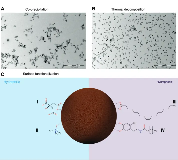
Figure 2 Transmission electron micrographs of SPIONs obtained by co-precipitation (A) or by thermal decomposition (B). Figure 2C illus-trates possible SPION surface functionalizations to render them hydrophilic, using e.g. carboxylates (I) or tetramethyl-ammonium hydroxide (II) or hydrophobic using fatty acids (III) or dopamine derivatives (IV).
SPION-liposome hybrids –
obtaining the materials
The centerpiece of the magnetoliposome is unmistak-ably the type of NP used. SPIONs are the most evident candidate. To obtain them, there are numerous estab-lished wet-chemical methods including microemulsions or hydrothermal syntheses (2) in addition to gas phase methods such as thermal decomposition in hot-wall reactors or flame synthesis (33). While the wet-chemical bottom up approaches typically better control particle characteristics such as size or shape, flame syntheses allow for continuous and therefore large-scale produc-tion of magnetic NPs. The most dominant and widely used
technique in the biomedical field is the co-precipitation
method of aqueous Fe2+/Fe3+ salt solutions by the
addi-tion of a base under inert atmosphere (34). This approach yields magnetite NPs, which are easily oxidized to magh-emite (Figure 2A). Adjusting particle size and size distri-bution is extremely challenging with this process, and the control of pH, ionic strength and seed concentration is crucial. Since the blocking temperature depends on the size (distribution) of the NPs, large polydispersity (i.e., a broad particle size distribution) results in a wide range of blocking temperatures and consequently suboptimal magnetic behavior for many applications (34). Nonethe-less, this method is arguably the most popular source of SPIONs for magnetoliposomes, as large quantities can be synthesized at once.
To obtain much more monodisperse SPIONs, synthe-sis by thermal decomposition (35) has become the leading approach. In short, an organometallic precursor (e.g., iron oleate (36), iron acetylacetonate, iron carbonyls) is ther-mally decomposed in a high boiling point solvent (e.g., octyl ether, hexadecene, eicosane). This approach yields highly crystalline NPs with narrow size distributions (Figure 2B) (35). Furthermore, the size can be tuned by the choice of solvent, the reaction time and the reactant ratios. The produced SPIONS are stabilized by a surface-attached oleate molecule and dispersed in an organic solvent. Con-sequently, additional steps might be required to transfer the SPIONs to an aqueous environment. This phase trans-fer relies on NP surface derivatization strategies replac-ing the originally grafted hydrophobic molecule with hydrophilic compounds, or direct functionalization of the surface-grafted hydrophobic molecules themselves (37). Surface chemistry not only determines the colloidal stabil-ity of the NPs, but also their association to the liposome, i.e., whether they will be embedded in the hydrophobic bilayer or within the hydrophilic lumen. An arsenal of molecules and surface chemistry strategies are available, and several candidates were used to date. A selection is highlighted in Figure 2C.
For NPs encapsulated in the lumen or grafted to the surface, citrate (38, 39) and oleate (overcoated by a hydro-philic ligand, (40) e.g., a second lipid layer) stabilized SPIONs are the most frequently used candidates. For NPs embedded in the lipid bilayer, SPIONs coated with oleic acid (41–43) are the favored choice. Another alternative was presented by Amstad and colleagues (44) by introduc-ing SPIONs stabilized with palmityl-nitroDOPA (Figure 2C, IV) into the lipid membrane, arguing that such particles were less prone to aggregation than standard oleic acid-coated SPIONs and that they embed themselves more will-ingly in between the bilayer.
In all, choosing the synthetic approach and surface coating of the NP is the first step to develop SPION-lipo-some hybrids, and should not be taken lightly. Other factors such as overall NP geometry are of equal impor-tance and might contribute in yielding more basic infor-mation on lipid-nanoparticle interactions in general.
On the other hand, the choice of lipids determines the phase transition temperature, which is typically set only a few degrees above body temperature (e.g., around 42°C). Changing the composition of the liposome bilayer, e.g., by including cholesterol, is known to reduce the leakage of drug molecules from the liposomes by “tight-ening” the bilayer (45). As a long blood circulation time is generally desirable for any vesicle intended for medical usage, adding a small percentage of Polyethylene glycol
(PEG)-derivatized lipids in the membrane is an option to obtain this property. PEG chains reduce the overall uptake efficiency by macrophages, and liposomes with this attribute are termed “stealth” (45). In all, the avail-able selection – counting both natural and synthetic phospholipids – is immense and way beyond the scope of this review.
Magnetoliposomes and the state of
the art
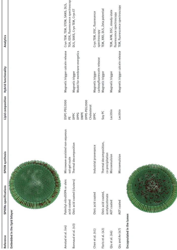
The NP surface properties determine where the particles will spatially be located within the liposome. Over the past years, numerous variations of magnetoliposomes have been presented, and a selection is highlighted in Table 1. Principally, research is concentrated on controlling the release of an encapsulated drug. However, their utility as MRI contrast agents has been presented on several occa-sions (39, 52, 55). Other applications, such as cell sorting and gene delivery, have also been addressed (64).
Three different approaches are possible to associate the SPIONs to the liposomes (Figure 3). The two strate-gies which are increasingly becoming seminal are either to encapsulate the magnetic NPs directly within the lipo-some lumen (38, 48, 49), the other to embed them in between the lipid bilayer (41, 43, 44). Although pursued with other inorganic NPs [e.g., gold (65)], directly conju-gating SPIONs to the liposome surface has only margin-ally been done (42).
Depending on the final application, the spatial location of the SPIONs within the hybrid is a determining factor: for MRI tracking, NPs encapsulated in the lumen are preferred. However, when using such hybrids as drug carriers, embed-ding the SPIONs directly in between the lipid bilayer seems more beneficial, as SPIONs in the liposome lumen might impair or affect any co-encapsulated drug even before the membrane actually becomes permeable. Moreover, the energy, which is required to permeate the membrane, should be delivered directly where it is needed.
Characterizing the vesicles –
options and caveats
Visualizing and characterizing magnetoliposomes is argu-ably the most important step in developing such hybrids and is indispensable in detecting the exact NP locations or whether the condition applies to all specimens in the
Tab le 1 M agneto liposomes: SPION sy nthesis, su rface spec ific ations, lip id composition, f unction ality and an alytic al methods used to ch ar acter ize the h ybr id . Reference SPIONs spec ific ations SPION sy nthe si s Lip id compo sition Hybr id fu nctiona lity Ana lytic s Embedded in the lip id b ila yer Amst ad et a l. (44) Pa lmityl-nit roDOP A or o leic ac id co ated Mic ro wave-assisted non-aqueo us so l-gel ro ute DSPC -PE G2000 PE M agnetic tr igger c alcein rele ase Cr yo TEM, TEM, S TEM, S ANS, DLS, TGA , EDX , fluorescence spect roscop y Bonn aud et a l. (43) Oleic ac id co ated (c lusters) Therm al decomposition DPPC DPPG DMPE DPPE -PE G2000 Cho lestero l M agnetic tr igger Model for membr ane energetics DLS, SAXS, C ryo TEM, C ryo ET Chen et a l. (41) Oleic ac id co ated Indust ria l proven ance DPPC M agnetic tr igger carbo xyfluorescein rele ase Cr yo TEM, DSC, fluoresence spect roscop y Flor is et a l. (42) Oleic ac id co ated , acetyl aceton ate Therm al decomposition, co-prec ip itation Soy P C M agnetic tr igger TEM, XRD, DLS, Zet a potenti al Qiu et a l. (46) AO T co ated Mic roemu lsion Lec ithin M agnetic tr igger
TEM, AFM, DSC, ste
ady -st ate fluorescence spect roscop y Qiu and An (47) AO T co ated Mic roemu lsion Lec ithin M agnetic tr igger c alcein rele ase
TEM, fluorescence spect
roscop y Enc ap su lated in the lu men
Reference SPIONs spec ific ations SPION sy nthe si s Lip id compo sition Hybr id fu nctiona lity Ana lytic s Be au ne et a l. (38) Cit rate co ated Co-prec ip itation DOPC M agnetic tr igger M agnetophoresis, photob le aching, CLS M, el astic ity me asu rements Bothu n and Preiss (48) Ferrotec GmBH Indust ria l proven ance DPPC Cho lestero l Radio f requency - induced drug rele ase Cr yo TEM, S AR me asu rements Cint ra et a l. (49) Carbo xyl-de xt ran co ated Co-prec ip itation PC Cho lestero l M agnetic tr igger TEM, po wder diff raction, DLS, st atic m agnetic b iref ringence, elect ron m agnetic reson ance Conde et a l. (50) Si lic a su lfon ate co ated Indust ria l proven ance
POPC DDAB DSPE
-PE G2000 M agnetic tr igger Zet a potenti al , TEM, DLS Far ia et a l. (51) Tet rameth yl-ammoniu m hydro xide co ated Indust ria l proven ance SPC Cho lestero l M agnetic tr igger TEM, DLS, SQUID m agnetomet ry , FTIR, MRI For tin-Ripoche et a l. (52) Cit rate co ated Co-prec ip itation PC DSPE -PE G2000 M agnetic tr
igger MRI cont
rast agent Cr yo TEM, DLS, m agnetophoresis, in vivo MRI Pr assl et a l. (53) Po lar su rfact ant co ated (EMG 1500, FerroTec) Indust ria l proven ance POPC DSPE -PE G2000 M agnetic tr
igger MRI cont
rast agent TEM, DLS, fluoresence po lar iz ation, Zet a potenti al , absorb ance, in vivo MRI Garc ia-Jimeno et a l. (54) Anionic co ated (EMG 707, FerroTec) Indust ria l proven ance Soy P C M agnetic tr igger in vivo injection and b iodist rib ution an alysis TEM, DLS, SQUID m agnetomet ry , Zet a potenti al Garnier et a l. (55) Cit rate co ated (c lusters) Indust ria l proven ance DOPC Cho lestero l M agnetic tr igger MRI cont rast agent Cr yo TEM, DLS, MRI Gir i et a l. (56) PC co ated Co-prec ip itation PC Cho lestero l M agnetic tr igger TEM, SQUID m agnetomet ry , XRD, FTIR Gonz ales and Kr ishn an (40) Oleic ac id , t rimeth yl-amine N-o xide co ated Therm al decomposition DPPC M agnetic tr igger TEM, SQUID m agnetomet ry , XRD Linem ann et a l. (57) Chitosan-lip id co ated Indust ria l proven ance Soy P C DDAB DSPE -PE G2000 -MAL M agnetic tr igger in vit ro m agneto-t ransfection Zet a-potenti al , DLS M ar tin a et a l. (39) Cit rate co ated Co-prec ip itation EPC DSPE -PE G2000 M agnetic tr
igger MRI cont
rast agent (in vivo) Cr yo TEM, CLS M, QELS, m agnetiz ation me asu rements, rel axomet ry ,m agnetophoresis Meled an-dr i et a l. (58) DOPG co ated Co-prec ip itation DOPG DOPC M agnetic tr igger Cr yo SEM, TEM, A TR IR, PC S, NMR, AAS Napp ini et a l. (59) CoFe 2 O4 , TMA OH co ated Co-prec ip itation PC M agnetic tr igger TGA , DLS, SAXS, ste ady -st ate fluorescence spect roscop y Pr adh an et a l. (60) FluidM ag -HS, chemicel l GmbH Indust ria l proven ance DPPC Cho lestero l DSPE -PE G2000 DSPE -PE G2000 -fo late M agnetic tr igger Cr yo TEM, TEM, fluorescence mic roscop y, DSC, XRD, m agnetomet ry (T ab le 1 Continued)
Reference SPIONs spec ific ations SPION sy nthe si s Lip id compo sition Hybr id fu nctiona lity Ana lytic s Sab ate et a l. (61) TMA OH co ated Co-prec ip itation PC M agnetic tr igger TEM, XRD, DLS, Zet a potenti al Sko ur as et a l. (62) Hydrophi lic -co ated-USPIO-P00904 Indust ria l proven ance PC DSPC PG DSPE -PE G2000 DSPE -Biotin cho lestero l M agnetic tr igger TEM, Zet a potenti al , rel axomet ry Tai et a l. (63) De xt ran-co ated Indust ria l proven ance DPPC DSPC Cho lestero l M agnetic tr igger in vivo carbo xyfluorescein rele ase
TEM, fluoresence spect
roscop y Su rface att ached Flor is et a l. (42) Oleic ac id co ated , acetyl-aceton ate Therm al decomposition, co-prec ip itation Soy P C M agnetic tr igger TEM, XRD, DLS, Zet a potenti al Abbrevi ation of chemic
als: DDAB, dimeth
yldioct
adecyl
ammoniu
m bromide; DOPC, 1,2-dio
leo
yl-sn-glycero-3-phosphocho
line; DOPG, 1,2-dio
leo yl-sn -glycero-3-(phospho-ra c-(3-lysyl(1-glyc -ero l))) ch lor ide, DPH diphen ylhe xat riene; DPPC, 1,2-dip almito yl-sn -glycero-3-phosphocho line; DSPC, 1,2-Diste aro yl-sn-glycero-3-phosphocho
line; DSPE, 1,2-Diste
aro yl-sn-glycero-3-phos -phoeth ano lamine; DSPE -PE G-2000, 1,2-diste aro yl-sn -glycero-3-phosphoeth ano lamine-N-(amino(po lyeth ylene glyco l)-2000); DSPE -Biotin, 1,2-diste aro yl-sn -glycero-3-phosphoeth ano lamine-N-(b iotin yl(po lyeth ylene glyco l)-2000) ammoniu m; DSPE -PE G-2000-M al , 1,2-diste aro yl-sn -glycero-3-phosphoeth ano lamine-N-(m aleimide (po lyeth ylene glyco l)-2000); DSPE -PE G-2000-Fo late, 1,2-diste aro yl-sn -glycero-3-phosphoeth ano lamine-N-(fo late(po lyeth ylene glyco l)-2000) ammoniu m; EPC, 1,2-dio leo yl-sn -glycero-3-eth ylphosphocho line ch lor ide; PC, phosph atidylcho line; PE G 2000 PE, 1,2-dio leo yl-sn-glycero-3-phosphoeth ano lamine-N-((meto xy po lyeth ylene glyco l) 2000 Da); PG, phosph atidylglycero l; POPC, 1-p almito yl-2-o leo yl-sn-glycero-3-phosphocho line; So y PC, L -α -phosph atidylcho line; SPC, sphingosyl-phosphor ylcho line; TMA OH, tet rameth yl amoniu m h ydro xide. Abbrevi ation of
Methods: AFM, atomic
force mic roscop y; C ryo TEM, c ryo t ransmission elect ron mic roscop y; C ryo ET , c ryo elect ron tomogr aph y; C ryo SEM, c ryo sc anning elect ron mic roscop y; DLS, dy namic light sc atter ing; DSC, sy namic sc anning c alor imet ry; EDX -FS, energ y dispersive X-r ay fluorescence spect roscop y; F TIR, Fo ur ier t ransform inf rared spect roscop y; QELS, quasi el astic light sc atter ing; MRI, m agnetic reson ance im aging; SANS, sm al l angle neut ron sc atter ing; SAXS, sm al l angle X-r ay sc atter ing;
SQUID, superconducting quant
um inter ference device; STEM, sc anning t ransmission elect ron mic roscop y; TEM, t ransmission elect ron mic roscop y; TGA , thermo gr avimet ric an alysis; XRD, X-r ay diff raction; SAR, spec ific absorption r ate. (T ab le 1 Continued)
solution. Structural and architectural details, such as the SPION distribution or arrangement within the hybrids, are also relevant in studying the interactions of NPs and mem-branes in general. However, the challenge lies in providing convincing data, which is both qualitative and quantitative, while assuring that the hybrids are in their native state.
For giant magnetoliposomes, light and fluores-cence microscopy offer the most straightforward options to directly observe and characterized the hybrids (38). Nappini and colleagues (66, 67) highlighted the utility of these methodologies by presenting giant unilamellar vesicles visualized by confocal laser scanning micros-copy, in which both vesicles and NPs were fluorescently labeled. With this approach, NP presence and distribu-tion as well as dye release was elegantly shown. Another useful approach was presented by Beaune and colleagues (38): The elastic properties of the magnetoliposomes were investigated by studying the deformation of the vesicles under the effect of an applied magnetic field.
When working with much smaller hybrids, the physi-cal constraints of light come into play, and alternative methods are needed. There are several techniques avail-able ranging beyond microscopy for investigating at the nanoscale and particularly small vesicles (i.e., < 200 nm). As an example, scattering techniques – such as dynamic light scattering (DLS) or small angle X-ray and neutron scattering (SAXS and SANS) – can be used to elaborate the size of the vesicles, which in turn provides critical infor-mation on sample homogeneity. As an example, Amstad and colleagues (44) have successfully employed SANS to characterize both the sample homogeneity and the change in membrane thickness when loaded with SPIONs. None-theless, complementary visualization by microscopy to investigate morphology or appearance of the sample is unavoidable.
Transmission electron microscopy (TEM) is still the method of choice and has been widely used in this context (42, 46). However, conventional TEM techniques require a high vacuum environment, which is – particularly in the case of liposomes – highly destructive for any water-rich sample. Although samples can be preserved, e.g., by chemical fixation, there are still countless artifacts which are created by either the fixation procedure itself and/ or sample drying. Moreover, this step inevitably leads to a randomized location of the unassociated NPs over the TEM grid. Consequently, correct and objective interpreta-tion and discriminainterpreta-tion between liposome associated and non-associated NPs is very challenging (Figure 4).
Although straightforward, this method is not ideal to reliably characterize such specimens. On the other hand, samples can be visualized in their native state by cryo TEM. Unlike conventional TEM, the vesicles are preserved in a layer of vitreous ice, keeping them safe from drying effects or the vacuum during visualization. Cryo TEM has been used to characterize liposomes for quite some time and has been applied on several occasions in the context of NP-liposome hybrids (41, 44). Unfortunately, the reso-lution was often not high enough to distinctly resolve the bilayer, a task rendered even more challenging by the variety of optical effects which may occur (68). Chen and colleagues (41) proved the presence of NPs by subse-quent energy-dispersive X-ray spectroscopy (EDX) Electron microscopy in general needs to be interpreted extremely carefully: Merely a two-dimensional projection of the sample is provided by this methodology and leaves the three-dimensional aspects – such as the spatial location of the NPs in regard to the lipid bilayer – subject to specula-tion. Deducing architectural features by relying solely on single projections is therefore not feasible. This query can be countered by cryo-electron tomography, which finally Figure 3 SPION-liposome hybrids. SPIONs acting as triggers to release a cargo (e.g., drug molecules) can be located in the lipid bilayer, the lumen, or can be grafted to the surface of the liposome (from left to right).
A B
C D
Figure 4 Cryo TEM images of SPIONs and liposomes at tilt angles of –30° (A, C) and +30° (B, D). A: Although particles at –30° seem to be associated with the liposome membrane, the tilt image at 30° (B) challenges this interpretation: it is the loss of the third dimension during the projection which leads to this misinterpretation. (C) Again, a cluster of particles seemingly interacts with the liposome membrane in the –30° tilt angle image. This interpretation is maintained, independent of the tilt angle (D). © 2013 IEEE. Reprinted, with permission, from IEEE Transactions on Magnetics, Vol. 49, No. 1, January 2013.
yields information on the structural and architectural aspects of the vesicles. Briefly, images of the sample are successively taken at various stage tilt angles. The collected data may then be used to reconstruct and render the three-dimensional appearance of the sample. Such data has been recently presented by the authors, with resolutions high enough to visualize the bilayer splitting around the inclusive SPIONs, along with three-dimensional render-ings highlighting the NP locations and arrangements (43). Nonetheless, the investigation of small sample volumes is not sufficient for statistical relevance, which presents – in addition to the complexity of these techniques-the main limitations of cryo TEM and cryo-electron tomography.
Given the fact of the pros and cons of the aforemen-tioned methodologies, a well-balanced combination of various techniques – i.e., scattering and spatial visuali-zation by microscopy – is necessary to provide the infor-mation needed on both a statistical and qualitative level. In turn, these assessments are vital for any subsequent upscaling and industrial perspective.
Membrane energetics – inclusion
limits between the bilayer
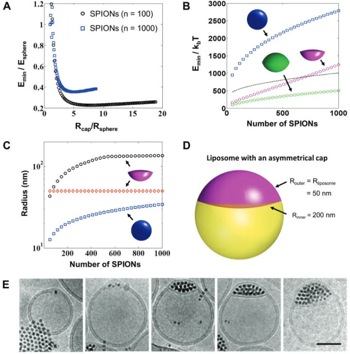
The incorporation of NPs into the lipid bilayer, and in par-ticular the question of the maximum size of the NPs that can be embedded, has puzzled scientists for quite some time. No fully rigorous model is available in the litera-ture to effectively quantify the energy needed to deform a lipid bilayer and accommodate a NP. In turn, biophysi-cal aspects and properties of lipid bilayers also come into play.
To date, only a simplified approach has been proposed by Wi and colleagues (69). Although the variational prob-lem-based on the Helfrich model (70) used to determine how the lipid membrane needs to deform to accommo-date a NP and minimize the deformation energy – was not solved (71), they instead made some clever assumptions on the geometrical configurations of the membrane. This step drastically reduces the complexity of the problem. Their
Figure 5 The energetics behind cluster-sized inclusions in between a phospholipid membrane. (A) The inclusion energy of an inclusion with a double spherical cap geometry, as a function of the spherical cap radius for both 100 and 1000 nanoparticles. Both radius and energy are normalized by the corresponding values for a spherical inclusion. (B) Energy of an inclusion with an asymmetric spherical cap geometry, with one radius equal to the liposome radius (taken equal to 50 nm) as a function of the number of nanoparticles in the inclu-sion. The energy of a corresponding spherical inclusion is additionally shown for comparison (blue), along with that of a spherical cluster covered by a lipid monolayer (black dashed line). (C) Radii of the asymmetric spherical cap inclusion as a function of the number of particles in the inclusion, as compared to the spherical inclusion radius. (D) Shape of a typical asymmetric inclusion with minimal energy. (E) Cryo TEM images showing the membrane deformation with increasing number of embedded SPIONs, scale bar = 50 nm. Reprinted with permis-sion from ACS Nano, Vol. 8, No. 4, 2014, Pages 3451–3460. Copyright 2014 American Chemical Society.
work has important consequences, as it allowed them to conclude that only spherical inclusions with a radius smaller than 3.5–4 nanometers can be incorporated into a lipid membrane. Larger spherical NPs are preferentially
expelled by the lipid membrane and stabilized by a lipid monolayer. While these findings confirm some experimen-tal observations made with quantum dots and SPIONs, they cannot explain the results recently obtained by the authors
of this review (43). In fact, inclusions with a diamond-like shape made of hundreds of SPIONs could be incorporated into the lipid membrane (Figure 5E). In order to explain these findings, the aforementioned theory was extended to inclusions with a non-spherical shape. In order to keep the simplicity of the original model, the model was restricted to spherical caps. This modified theory adds an addi-tional degree of freedom, i.e., the radius of the spherical cap. For a given inclusion volume, the calculations show that an increase in the spherical cap radius – compared to a spherical inclusion – leads to a lower deformation by decreasing the bending energy of the membrane. However, a minimum is reached for a sufficiently large cap radius, as any further increase is penalized by the inclusion area becoming too large. These results, shown in Figure 5, demonstrate that NP clusters which can be organized into non-spherical assemblies are viable options to incorporate large quantities of SPIONs into a liposome membrane.
The importance of these results for hyperthermia applications is significant. In fact, the dependence of the heating rate of SPIONs exposed to an AMF is strongly dependent on the particle size, and seems to have an optimum for NPs with a diameter of about 20 nm (9). This approach offers the possibility to incorporate sufficiently large NPs to obtain optimal heating rate.
On the other hand, this also leads to new and currently unresolved problems. For example, one open question is whether a larger cluster of NPs remains superparamag-netic. Losing superparamagnetism is detrimental for the colloidal stability of the liposomes, as it would cause them to exhibit dipolar interactions. Furthermore, the heating power generated by NP clusters has not been systemati-cally investigated to date. While there are studies showing the beneficial effect of clustering on the usage of SPIONs in MRI (72), the impact on the heating rate is a virtually unex-plored area. However, this query is only a single example: A long list of questions needs to be addressed in the future, starting by which composition, particles size, size distribu-tion is required to optimize hyperthermia performance of the magnetoliposomes. Finding answers to these intriguing questions will require a fundamental change in magne-toliposome design, and further combining the previously mentioned experimental characterization to modeling tech-niques might be highly beneficial for future developments.
Conclusions and perspectives
Today, liposomes are clinically established, yet there is still potential to improve them. Although significant
effort has been invested in the development of magne-toliposomes, we still are only scratching on the tip of the iceberg – especially on a materials level – and the trans-lation into the clinics is still being awaited. The efficacy of smart drug delivery systems, however, implies that the hybrids are thoroughly characterized by emerging and complementary techniques, which is – in our opinion – arguably one of the principal drawbacks in developing them. The complexity of these systems is highlighted by the multidisciplinary expertise needed, which includes organic and inorganic chemistry, bio- and magnetophys-ics, pharmacology and biology. Nonetheless, the last decade has yielded interesting new concepts. Some of them have been tested in laboratory settings, and further advancement will hopefully bring these hybrids a step closer to direct clinical application in the future.
Acknowledgments: This study was supported by the
Adolphe Merkle Foundation, the Swiss National Sci-ence Foundation (PP00P2_123373) the National Research Program 62 (126104), and by the Swiss National Science Foundation through the National Centre of Competence in Research Bio-Inspired Materials. The support of the Dr. Alfred Bretscher Fund is gratefully acknowledged, and access to conventional and cryo TEM was kindly pro-vided by the Microscopy Imaging Centre of the University of Bern.
References
1. Li L, Jiang W, Luo K, Song H, Lan F, Wu Y, et al. Superparamagnetic iron oxide nanoparticles as MRI contrast agents for non-invasive stem cell labeling and tracking. Theranostics 2013;3:595–615. 2. Lu A-H, Salabas EL, Schüth F. Magnetic nanoparticles: synthesis,
protection, functionalization, and application. Angew Chem Int Ed 2007;46:1222–44.
3. Yoon T-J, Lee W, Oh Y-S, Lee J-K. Magnetic nanoparticles as a catalyst vehicle for simple and easy recycling. New J Chem 2003;27:227–9.
4. Koh I, Josephson L. Magnetic nanoparticle sensors. Sensors 2009;9:8130–45.
5. Bucak S, Jones DA, Laibinis PE, Hatton TA. Protein separa-tions using colloidal magnetic nanoparticles. Biotechnol Prog 2003;19:477–84.
6. Sun C, Lee JS, Zhang M. Magnetic nanoparticles in MR imaging and drug delivery. Adv Drug Deliver Rev 2008;60:1252–65. 7. Bulte JW, Douglas T, Witwer B, Zhang S-C, Strable E, Lewis BK,
et al. Magnetodendrimers allow endosomal magnetic labeling and in vivo tracking of stem cells. Nat Biotechnol 2001;19:1141–7. 8. Alivisatos P. The use of nanocrystals in biological detection. Nat
Biotechnol 2003;22:47–52.
9. Rosensweig R. Heating magnetic fluid with alternating magnetic field. J Magn Magn Mater 2002;252:370–4.
10. Mahmoudi M, Hofmann H, Rothen-Rutishauser B, Petri-Fink A. Assessing the in vitro and in vivo toxicity of superparamagnetic iron oxide nanoparticles. Chem Rev 2012;112:2323–38. 11. Fenniri HH. The Canadian regenerative medicine and
nanomedi-cine enterprise (CARMENE). Int J Nanomedinanomedi-cine 2005;1:225–7. 12. Gilchrist RK, Medal R, Shorey WD, Hanselman RC, Parrott JC,
Taylor CB. Selective inductive heating of lymph nodes. Ann Surg 1957;146:596–606.
13. Jordan A, Scholz R, Wust P, Fähling H, Fähling H. Magnetic fluid hyperthermia (MFH): cancer treatment with AC magnetic field induced excitation of biocompatible superparamagnetic nano-particles. J Magn Magn Mater 1998;201:413–9.
14. Otte J. Hyperthermia in cancer therapy. Eur J Pediatr 1988;147:560–9.
15. Kozissnik B, Bohorquez AC, Dobson J, Rinaldi C. Magnetic fluid hyperthermia: Advances, challenges, and opportunity. Int J Hyperther 2013;29:706–14.
16. Pankhurst Q, Connolly J, Jones S, Dobson J. Applications of magnetic nanoparticles in biomedicine. J Phys D: Appl Phys 2003;36:167–81.
17. Atkinson WJ, Brezovich IA, Chakraborty DP. Usable frequencies in hyperthermia with thermal seeds. IEEE Trans Biomed Eng 1984;31:70–5.
18. Pankhurst QA, Thanh NT, Jones SK, Dobson J. Progress in appli-cations of magnetic nanoparticles in biomedicine. J Phys D: Appl Phys 2009;42:224001.
19. Hergt R, Dutz S, Müller R, Zeisberger M. Magnetic particle hyper-thermia: nanoparticle magnetism and materials development for cancer therapy. J Phys: Condens Matter 2006;18:S2919–34. 20. Rabin Y. Is intracellular hyperthermia superior to
extracel-lular hyperthermia in the thermal sense? Int J Hyperther 2002;18:194–202.
21. Huang HH, Delikanli SS, Zeng HH, Ferkey DM, Pralle AA. Remote control of ion channels and neurons through magnetic-field heating of nanoparticles. Nature Nanotechnology 2010;5: 602–6.
22. Alexiou C, Schmid RJ, Jurgons R, Kremer M, Wanner G, Berge-mann C, et al. Targeting cancer cells: magnetic nanoparticles as drug carriers. Eur Biophys J 2006;35:446–50.
23. Thorley AJ, Tetley TD. New perspectives in nanomedicine. Pharmacol Therapeut 2013;140:176–85.
24. Zhang L, Gu FX, Chan JM, Wang AZ, Langer RS, Farokhzad OC. Nanoparticles in medicine: therapeutic applications and devel-opments. Clin Pharmacol Ther 2008;83:761–9.
25. Bibi S, Lattmann E, Mohammed AR, Perrie Y. Trigger release lipo-some systems: local and remote controlled delivery? J Microen-capsul 2012;29:262–76.
26. Steitz B, Salaklang J, Finka A, O’Neil C, Hofmann H, Petri-Fink A. Fixed bed reactor for solid-phase surface derivatization of superparamagnetic nanoparticles. Bioconjugate Chem 2007;18:1684–90.
27. Widder KJ, Senyel AE, Scarpelli GD. Magnetic microspheres: a model system of site specific drug delivery in vivo. Proc Soc Exp Biol Med 1978;158:141–6.
28. Kumar A, Jena PK, Behera S, Lockey RF, Mohapatra S, Mohapatra S. Multifunctional magnetic nanoparticles for targeted delivery. Nanomedicine 2010;6:64–9.
29. Shim MS, Kwon YJ. Stimuli-responsive polymers and nanoma-terials for gene delivery and imaging applications. Adv Drug Deliver Rev 2012;64:1046–59.
30. Ganta S, Devalapally H, Shahiwala A, Amiji M. A review of stimuli-responsive nanocarriers for drug and gene delivery. J Control Release 2008;126:187–204.
31. De Cuyper M, Joniau M. Magnetoliposomes. Eur Biophys J 1988;15:311–9.
32. Ta T, Porter TM. Thermosensitive liposomes for localized deliv-ery and triggered release of chemotherapy. J Control Release 2013;169:112–25.
33. Strobel R, Pratsinis SE. Direct synthesis of maghemite, mag-netite and wustite nanoparticles by flame spray pyrolysis. Adv Powder Technol 2009;20:190–4.
34. Massart R. Preparation of aqueous magnetic liquids in alkaline and acidic media. Magnetics, IEEE Transactions 1981;17:1247–8. 35. Park J, Lee E, Hwang N-M, Kang M, Kim SC, Hwang Y, et al.
One-nanometer-scale size-controlled synthesis of monodis-perse magnetic iron oxide nanoparticles. Angew Chem Int Edit 2005;44:2872–7.
36. Hyeon T. Chemical synthesis of magnetic nanoparticles. Chem Commun 2003;8:927–34.
37. Lattuada M, Hatton TA. Functionalization of monodisperse mag-netic nanoparticles. Langmuir 2007;23:2158–68.
38. Beaune G, Ménager C, Cabuil V. Location of magnetic and fluo-rescent nanoparticles encapsulated inside giant liposomes. J Phys Chem B 2008;112:7424–9.
39. Martina MS, Fortin J-P, Ménager C, Clément O, Barratt G, Grabielle-Madelmont C, et al. Generation of superparamagnetic liposomes revealed as highly efficient MRI contrast agents for in vivo imaging. J Am Chem Soc 2005;127:10676–85.
40. Gonzales M, Krishnan KM. Synthesis of magnetoliposomes with monodisperse iron oxide nanocrystal cores for hyperthermia. J Magn Magn Mater 2005;293:265–70.
41. Chen Y, Bose A, Bothun GD. Controlled release from bilayer-decorated magnetoliposomes via electromagnetic heating. ACS Nano 2010;4:3215–21.
42. Floris A, Ardu A, Musinu A, Piccaluga G, Fadda AM, Sinico C, et al. SPION@liposomes hybrid nanoarchitectures with high density SPION association. Soft Matter 2011;7:6239. 43. Bonnaud C, Monnier CA, Demurtas D, Jud C, Vanhecke D,
Montet X, et al. Insertion of nanoparticle clusters into vesicle bilayers. ACS Nano 2014;8:3451–60.
44. Amstad E, Kohlbrecher J, Müller E, Schweizer T, Textor M, Reimhult E. Triggered release from liposomes through magnetic actuation of iron oxide nanoparticle containing membranes. Nano Lett 2011;11:1664–70.
45. Allen TM, Cullis PR. Liposomal drug delivery systems: From con-cept to clinical applications. Adv Drug Deliver Rev 2013;65:36–48. 46. Qiu D, An X, Chen Z, Ma X. Microstructure study of liposomes
decorated by hydrophobic magnetic nanoparticles. Chem Phys Lipids 2012;165:563–70.
47. Qiu D, An X. Controllable release from magnetoliposomes by magnetic stimulation and thermal stimulation. Colloids Surface B 2013;104:326–9.
48. Bothun GD, Preiss MR. Bilayer heating in magnetite nanoparti-cle – liposome dispersions via fluorescence anisotropy. J Colloid Interface Sci 2011;357:70–4.
49. Cintra ER, Ferreira FS, Santos Junior JL, Campello JC, Socolovsky LM, Lima EM, et al. Nanoparticle agglomerates in magnetoli-posomes. Nanotechnology 2008;20:045103.
50. Conde AJ, Batalla M, Cerda B, Mykhaylyk O, Plank C, Pod-hajcer O, et al. Continuous flow generation of
magnetoli-posomes in a low-cost portable microfluidic platform. Lab Chip 2014;14:4506–12.
51. Faria MR, Cruz MM, Gonçalves MC, Carvalho A, Feio G, Martins MB. Synthesis and characterization of magnetoliposomes for MRI contrast enhancement. Int J Pharm 2013;446:183–90. 52. Fortin-Ripoche J-P, Martina MS, Gazeau F, Ménager C, Wilhelm
C, Bacri J-C, et al. Magnetic targeting of magnetoliposomes to solid tumors with MR imaging monitoring in mice: feasibility 1. Radiology 2006;239:415–24.
53. Prassl R, Frascione D, Almer G, Opriessnig P, Vonach C, Gra-dauer K, et al. Ultrasmall superparamagnetic iron oxide (USPIO)-based liposomes as magnetic resonance imaging probes. Int J Nanomedicine. 2012:7:2349–59.
54. García-Jimeno S, Escribano E, Queralt J, Estelrich J. Magnetoli-posomes prepared by reverse-phase followed by sequential extrusion: Characterization and possibilities in the treatment of inflammation. Int J Pharm 2011;405:181–7.
55. Garnier B, Tan S, Miraux S, Bled E, Brisson AR. Optimized syn-thesis of 100 nm diameter magnetoliposomes with high content of maghemite particles and high MRI effect. Contrast Media Mol Imaging 2012;7:231–9.
56. Giri J, Guha Thakurta S, Bellare J, Kumar Nigam A, Bahadur D. Preparation and characterization of phospholipid stabilized uniform sized magnetite nanoparticles. J Magn Magn Mater 2005;293:62–8.
57. Linemann T, Thomsen L, Jardin K, Laursen J, Jensen J, Lichota J, et al. Development of a novel lipophilic, magnetic nanoparticle for in vivo drug delivery. Pharmaceutics 2013;5:246–60. 58. Meledandri CJ, Ninjbadgar T, Brougham DF. Size-controlled
magnetoliposomes with tunable magnetic resonance relaxation enhancements. J Mater Chem 2010;21:214.
59. Nappini S, Bombelli FB, Bonini M, Nordèn B, Baglioni P. Mag-netoliposomes for controlled drug release in the presence of low-frequency magnetic field. Soft Matter 2009;6:154. 60. Pradhan P, Giri J, Rieken F, Koch C, Mykhaylyk O, Döblinger M,
et al. Targeted temperature sensitive magnetic liposomes for thermo-chemotherapy. J Control Release 2010;142:108–21. 61. Sabaté R, Barnadas-Rodríguez R, Callejas-Fernández J,
Hidalgo-Álvarez R, Estelrich J. Preparation and characterization of extruded magnetoliposomes. Int J Pharm 2008;347:156–62. 62. Skouras A, Mourtas S, Markoutsa E, De Goltstein M-C, Wallon C,
Catoen S, et al. Magnetoliposomes with high USPIO entrapping efficiency, stability and magnetic properties. Nanomedicine 2011;7:572–9.
63. Tai L-A, Tsai P-J, Wang Y-C, Wang Y-J, Lo L-W, Yang C-S. Thermo-sensitive liposomes entrapping iron oxide nanoparticles for controllable drug release. Nanotechnology 2009;20:135101. 64. Margolis LB, Namiot VA, Kljukin LM. Magnetoliposomes:
another principle of cell sorting. Biochimica et Biophysica Acta (BBA)-Biomembranes 1983;735:193–5.
65. Wu G, Mikhailovsky A, Khant HA, Fu C, Chiu W, Zasadzinski JA. Remotely triggered liposome release by near-infrared light absorption via hollow gold nanoshells. J Am Chem Soc 2008;130:8175–7.
66. Nappini S, Kayal Al T, Berti D, Nordèn B, Baglioni P. Magnetically triggered release from giant unilamellar vesicles: visualization by means of confocal microscopy. J Phys Chem Lett 2011;2:713–8.
67. Nappini S, Bonini M, Ridi F, Baglioni P. Structure and perme-ability of magnetoliposomes loaded with hydrophobic magnetic nanoparticles in the presence of a low frequency magnetic field. Soft Matter 2011;7:4801.
68. Bonnaud C, Vanhecke D, Demurtas D, Rothen-Rutishauser B, Petri-Fink A. Spatial SPION localization in liposome membranes. IEEE Trans Magn 2013;49:166–71.
69. Sub Wi H, Lee K, Kyu Pak H. Interfacial energy consideration in the organization of a quantum dot–lipid mixed system. J Phys: Condens Matter 2008;20:494211.
70. Helfrich W. Elastic properties of lipid bilayers: theory and pos-sible experiments. Z Naturforsch C 1973;28:693–703. 71. Fosnaric M, Iglic A, May S. Influence of rigid inclusions on the
bending elasticity of a lipid membrane. Phys Rev E Stat Nonlin Soft Matter Phys 2006;74(5 Pt 1):051503–3.
72. Balasubramaniam S, Kayandan S, Lin Y-N, Kelly DF, House MJ, Woodward RC, et al. Toward design of magnetic nanoparticle clusters stabilized by biocompatible diblock copolymers for T-weighted MRI contrast. Langmuir 2014;30:1580–7.
Bionotes
Christophe A. Monnier
Adolphe Merkle Institute, University of Fribourg, Chemin des Verdiers 4, 1700 Fribourg, Switzerland
Christophe obtained his MSc degree in Molecular Biology from the Biozentrum, University of Basel in 2011 with a major in Structural Biology. He is currently working towards his PhD degree in Nanosci-ence at the Adolphe Merkle Institute, University of Fribourg under the supervision of Prof. Alke Fink and Prof. Barbara Rothen-Rut-ishauser. His specialty lies in biomembranes and high-resolution microscopy techniques to investigate them.
David Burnand
Chemistry Department, University of Fribourg, Chemin du Musée 9, 1700 Fribourg, Switzerland
David obtained his BSc in Chemical Engineering in 2010 at the University of Applied Sciences, Fribourg, Switzerland. He then graduated from the University of Fribourg with an MSc in Polymer Chemistry and nanomaterials. He is currently working under the supervision of Prof. Alke Fink as a PhD student. His research focuses on SPIONs and gold nanoparticles, their synthesis and surface chemistry for self-assembly systems.
Barbara Rothen-Rutishauser
Adolphe Merkle Institute, University of Fribourg, Chemin des Verdiers 4, 1700 Fribourg, Switzerland
Barbara received her PhD in Cell Biology in 1996 at the Swiss Federal Institute of Technology in Zurich (ETHZ), Switzerland. Afterwards, she worked for ten years in the research group of Prof. Peter Gehr at the Institute of Anatomy, University of Bern, Switzerland. Since 2011, she is the new chair in BioNanomaterials at the Adolphe Merkle Institute, University of Fribourg, Switzerland. The position is shared equally with Prof. Alke Petri-Fink. B. Rothen-Rutishauser is an expert in the field of cell-nanoparticle interactions in the lung, with a special focus on 3D lung cell models and various microscopy techniques such as laser scanning and transmission electron microscopy.
Marco Lattuada
Adolphe Merkle Institute, University of Fribourg, Chemin des Verdiers 4, 1700 Fribourg, Switzerland
Marco received his Master degree in 1998 from the Politecnico di Milano, Italy, and his PhD in Chemical Engineering at the Swiss
Federal Institute of Technology in Zurich (ETHZ),. After a two years of post-doctoral work at the MIT, he came back to ETHZ as a senior scientist in 2006. In 2012, he moved to the Adolphe Merkle Insti-tute, University of Fribourg, Switzerland, as an Associate Swiss National Science Foundation Professor. His research is dedicated to the preparation and engineering of novel nanoparticles and on understanding their self-assembly behavior, with the final goal of designing novel materials.
Alke Petri-Fink
Adolphe Merkle Institute, University of Fribourg, Chemin des Verdiers 4, 1700 Fribourg, Switzerland; Chemistry Department, University of Fribourg, Chemin du Musée 9, 1700 Fribourg, Switzerland, [email protected]
Alke received her PhD in Chemistry from the University of Ulm, Germany in 1999. After a post-doctoral stay at the University of Gainesville, Florida, she joined the Institute of Materials Science at the École Polytechnique Fédérale de Lausanne (EPFL), first as a post-doctoral researcher, then as a senior scientist. She became an Associate Swiss National Science Foundation Professor in the Department of Chemistry at the University of Fribourg in 2009, and Full Professor in 2011 at the Adolphe Merkle Institute, Switzerland. Her research focuses on inorganic nanoparticles, their synthesis, surfaces, and interactions with biological cells.