HAL Id: hal-03043988
https://hal.archives-ouvertes.fr/hal-03043988
Submitted on 5 Jan 2021HAL is a multi-disciplinary open access archive for the deposit and dissemination of sci-entific research documents, whether they are pub-lished or not. The documents may come from teaching and research institutions in France or abroad, or from public or private research centers.
L’archive ouverte pluridisciplinaire HAL, est destinée au dépôt et à la diffusion de documents scientifiques de niveau recherche, publiés ou non, émanant des établissements d’enseignement et de recherche français ou étrangers, des laboratoires publics ou privés.
Influence of Surface Compositions on the Reactivity of
Pyrite toward Aqueous U(VI)
Bin Ma, Alejandro Fernandez-Martinez, Mingliang Kang, Kaifeng Wang, Aled
Lewis, Thierry Maffeis, Nathaniel Findling, Eduardo Salas-Colera, Delphine
Tisserand, Sarah Bureau, et al.
To cite this version:
Bin Ma, Alejandro Fernandez-Martinez, Mingliang Kang, Kaifeng Wang, Aled Lewis, et al.. Influence of Surface Compositions on the Reactivity of Pyrite toward Aqueous U(VI). Environmental Science and Technology, American Chemical Society, 2020, 54 (13), pp.8104-8114. �10.1021/acs.est.0c01854�. �hal-03043988�
1
The influence of surface compositions on the reactivity of
pyrite toward aqueous U(VI)
Bin Ma a, Alejandro Fernandez-Martinez a, Mingliang Kang b,*, Kaifeng Wang a,c, Aled R.
Lewis d, Thierry G.G. Maffeis d, Nathaniel Findling a, Eduardo Salas-Colera e,f, Delphine
Tisserand a, Sarah Bureau a, Laurent Charlet a
a
Univ. Grenoble Alpes, Univ. Savoie Mont Blanc, CNRS, IRD, IFSTTAR, ISTerre, 38000 Grenoble, France
b
Sino-French Institute of Nuclear Engineering and Technology, Sun Yat-sen University, Zhuhai 519082, China
c
Decommissioning Engineering Technology Center, China Institute of Atomic Energy, 102413, Beijing, China
d
Systems and Process Engineering Centre, College of Engineering, Swansea University, Fa-bian Way, Swansea SA1 8EN, UK
e
Instituto de Ciencia de Materiales de Madrid, CSIC, Sor Juana Inés de la Cruz 3, 28049, Cantoblanco Madrid, Spain.
f
Spanish CRG BM25 SpLine Beamline at the ESRF, 71 Avenue de Martyrs, F-38043 Greno-ble, France.
2
ABSTRACT
Pyrite plays a significant role in governing the mobility of toxic uranium in anaerobic
envi-ronment via an oxidation-reduction process occurring at the mineral-water interface, but the factors influencing the reaction kinetics remain poorly known. In this study, natural pyrites with different impurities (Pb, As and Si) and different surface pretreatments were used to re-act with aqueous U(VI) from pH ~3.0 to ~9.5. Both aqueous and solid results indicated that
freshly crushed pyrites, which do have more surface Fe2+/Fe3+ and S2- sites that were
generat-ed from breaking Fe(S)-S bonds during ball-milling, exhibitgenerat-ed a much stronger reactivity than those treated with acid-washing. Besides, U(VI) reduction which involves the possible inter-mediate U(V) and the formation of hyperstoichiometric UO2+x(s) was found to preferentially occur on Pb- and As-rich spots on the pyrite surface, suggesting that the impurity doping could act as reactive sites due to the generation of lattice defects and galena- and arsenopyrite-like local configurations. These reactive surface sites can be removed by acid-washing, leav-ing a pyrite surface nearly inert towards aqueous U(VI). Thus, reactivity of pyrite towards U(VI) is largely governed by its surface compositions, which provides an insight on the chem-ical behavior of both pyrite and uranium in various environments.
3
1. INTRODUCTION
Pyrite (FeS2), the Earth’s most abundant sulfide mineral, is known to be one of the important
minerals buffering the reducing conditions in nuclear waste repositories where it is present in
host claystone or granite as well as in near-field bentonite materials.1 Besides, pyrite can also
be present in sulfide-rich blended cement that could be used as repository construction
mate-rial.2 Previous studies have confirmed that pyrite can effectively immobilize redox-sensitive
radionuclides (such as 79Se3,4 and 99Tc5) via reductive precipitation.
Uranium (U), accounting for 96 wt.% of the spent nuclear fuel and receiving extensive
atten-tion in the geological disposal of nuclear waste,6-9 is also redox-sensitive and its solubility is
largely dependent on its oxidation state. U mainly exists as U(VI) and U(IV) or other
aliovalent oxides with U(V)10 in the natural environment. The formation of ternary complexes
of hexavalent uranium ion (UO22+) with CO32- and alkaline earth metals in the
groundwater-rock system can further enhance the solubility of U(VI) 11. In contrast, except for coffinite
(USiO4) nanoparticles12 or U(IV)-bearing colloids,13 UO2 and other aliovalent oxides (e.g.,
U4O9, U3O7, or U3O8) are nearly insoluble under weakly acidic to alkaline conditions. Thus, reductive precipitation, especially by the ubiquitous Fe(II)-bearing minerals in the geologic environment, is considered an effective way to immobilize the toxic uranium. Surface
cataly-sis of U(VI) reduction by Fe(II),14 or abiotic reduction of U(VI) by Fe(II)-bearing minerals
such as magnetite15 and mackinawite,16 has long been recognized in this respect.
Experi-mental and theoretical works have further revealed that intermediate U(V) can be stabilized
by arrangement in a uranate-like octahedral structure.15,17,18
Pyrite is frequently found in association with uranium ore deposits in both metamorphic and
sedimentary settings.19,20 Thermodynamically, UO2 is the most stable mineral in
pyrite-containing systems in a wide pH range,21 in particular at a relatively high U concentration.22
4
(pH 5.1-6.3) after equilibrating for 6-8 days.23 After, the formation of a hyperstoichiometric
UO2+x(s) product on natural pyrite was confirmed at pH 3.6-6.1 over short reaction times (i.e.,
4 or 48 hours).24,25 Besides, freshly polished pyrite surfaces were reported as efficient
scaven-gers for U(VI) from solution at pH 4.8,26 and dissolved organic matter was found to increase
the final uranium solution concentration and decrease the fraction of uranium(IV) on the
py-rite solid surface.27 In contrast, a previous study demonstrated that U(VI) reduction by
syn-thetic and natural pyrite samples with acid-washed surfaces was largely inhibited under most
pH conditions.21 These discrepant observations impel us to clarify the factors controlling the
reactivity of pyrite.
It is generally accepted that the kinetics of redox reactions occurring at the mineral-water
in-terface are largely dependent on the reactivity of surface sites,23 which might be influenced by
the treatment procedures. In addition, natural pyrite typically contains a host of minor and trace elements, such as As, Co, Pb, and Zn. The variability in the chemical composition can
result in variations in physico-chemical properties,28 and in particular, the oxidation rates can
be enhanced for As- and Co-doped pyrite.29 A previous study compared the oxidation kinetics
between varying impurities-doped and undoped pyrites, concluding that pure pyrite is less
re-active than As-containing pyrite.30 To date, the factors influencing the oxidation-reduction
rates of pyrite toward aqueous U(VI) are poorly known and not sufficiently supported by ex-perimental data. In order to better understand the role of pyrite in affecting the mobility of
uranium in the environment, we investigated the influence of S2- species and impurities (Pb,
As and Si) on pyrite surfaces on the reduction rate of aqueous U(VI).
2. MATERIALS AND METHODS 2.1. Materials
Analytical grade UO2(NO3)2 stock solution (1000 ppm U in 2% HNO3), Na2CO3, NaOH, and
5
first boiling and then cooling with continuous argon purging before use for solution prepara-tion. Three types of natural pyrite were collected: (i) a commercial pyrite containing Pb and free of As and Si from Alfa Aesar, named Pb-pyrite; (ii) an As-rich pyrite from the
Jiguanshan ore mine (Tongling, China), called hereafter As-pyrite; and (iii) a SiO2-coexisting
pyrite from the Shizishan ore mine (Tongling, China), named Si-pyrite. The As-pyrite blocks
were crushed, sieved, and magnetically separated in the air, following the procedures
de-scribed previously.4 Similar pretreatment was applied to the Si-pyrite blocks, while no
pre-treatment was carried out for the commercial Pb-pyrite. The sizes of the obtained particles
were 1.5-4.8 mm, 0.13-0.15 mm, and 5.0-10.0 mm for Pb-pyrite, As-pyrite, and Si-pyrite,
re-spectively. After washing with 0.2 M HCl, degassed water and acetone in sequence, the dried pyrite grains were ground with a planetary ball mill (Pulverisette-7, Fritsch GmbH, Germany) under N2-atmosphere protection. Two kinds of pyrite powders were employed for sorption experiments. The first one (labeled as Type I pyrite) was prepared with the same washing procedure again and then dried. The second one (labeled as Type II pyrite) was used directly after ball milling. All preparation procedures for Type I and Type II pyrite powders were
per-formed under N2-atmosphere protection or in a glove box (O2 < 2 ppm). Both were stored
un-der anoxic conditions.
The specific surface area of the ground pyrite powder was measured by the
Brunauer-Emmett-Teller (BET) N2-absorption method. X-ray diffraction (XRD) analysis was used to
characterize the mineral components. The main elements Fe and S as well as minor and trace elements (As, Co, Ni, Cu, Pb, etc.) in the pyrite were identified and quantified by inductively
coupled plasma optical emission spectrometry (ICP-OES, Varian 720-ES apparatus) after
di-gestion in aqua regia. Certified single element standards (Sigma Aldrich) were used for quali-ty control, allowing a deviation of no more than 10% with respect to the certified values.
6
Sorption experiments were conducted at pH ~3.0, ~4.5, ~6.5, and ~9.5 at 25 °C under con-stant shaking in a N2-filled glove box (O2 < 2 ppm). Prior to introducing U(VI), the pyrite powder was equilibrated with 0.01 M NaCl at the given pH values for 2 days. Identical
solid-to-liquid (S/L) ratios of 10 g∙L-1 and initial U(VI) concentrations of 0.1 mM were applied to
all reactors. Spontaneous precipitation of U(VI) tends to occur at a relatively high pH. For the
reactors at pH ~6.5 and ~9.5, an extra 1.0 mM Na2CO3 was added to increase the solubility of
U(VI). After adding a certain portion of the UO2(NO3)2 stock solution, a 5 mL aliquot of sus-pension was sampled and filtered through a 0.22 μm pore size membrane filter. Subsequently, the solution pH was quickly adjusted back to the given reaction pH values using NaOH and HCl solution. At each defined time interval, the pH was measured and readjusted if necessary; then, a 5 mL aliquot of suspension was sampled by filtration. Complementary experiments were performed simultaneously to determine the concentrations of sulfur species before and
after adding U(VI) at pH ~3.0 and ~4.5.
The solution pH was measured by a combined glass Micro-pH electrode (Metrohm 6.0234.100) immediately after calibration by pH standard solutions. The filtrate samples for all reactors were analyzed by ICP-OES to determine the concentrations of total U, Fe, S, As,
Co, Pb, and Ni. Aqueous sulfide was analyzed by the methylene blue method.31 In order to
precipitate and stabilize the aqueous S(-II), a certain amount of 5 wt% zinc acetate solution was added immediately into the freshly obtained filtrates. Elemental sulfur was extracted by perchloroethylene and subsequently analyzed by high-performance liquid chromatography (HPLC) with a Varian ProStar 230 apparatus coupled with a UV/Visible detector (Perkin
Elmer 785A).32 The U(VI)-reacted pyrite in each reactor was collected by vacuum filtration
through a 0.22 µm nitrocellulose membrane and then dried and stored in the glove box. Spe-cial care was taken to prevent any possible oxidation for the subsequent solid characterization.
7
X-ray photoelectron spectroscopy (XPS) measurements were performed on the pristine and the selected U(VI)-reacted pyrites. Samples were transported to the XPS facility in a N2-filled metallic jar, pasted flat on double-face carbon tape, and then rapidly transferred into the XPS chamber under the protection of high-purity nitrogen. The XPS spectra were recorded using a Kratos AXIS Supra electron spectrometer. A monochromated Al Kα source, a hybrid lens
system providing an elliptic analysis area of 0.3×0.8 mm2, and a charge neutralizer were used.
The pass energy was 160 eV for survey scans and 20 eV for regions. The XPS data were pro-cessed using the CasaXPS software. The binding energy (BE) scale was calibrated with the C1s line of aliphatic carbon, which was set at 284.8 eV. The Shirley background algorithm was used to construct the baselines. The Gaussian/Lorentzian product formula, with GL(30) mixing, was applied to describe the line shapes. The U(4f) spectra were fitted with a spin-orbit splitting of 10.89 eV, a fixed intensity ratio of 4:3 (4f7/2:4f5/2), and identical full width at half of maximum (FWHM) values for the doublet peaks. The S(2p) spectra were fitted with
S(2p3/2) and S(2p1/2) spin-orbit doublets, with a fixed 2:1 intensity ratio and a 1.18 eV energy
separation. The uncertainty of BE on the fit peak was ±0.2 eV.
Sorbed U speciation on Pb-pyrite at different pH values was characterized by U L3-edge
(17166 eV) X-ray absorption near edge structure (XANES) spectroscopy analyses. The meas-urements were conducted at the SpLine Spanish CRG Beamline (BM25A) at the European
Synchrotron Radiation Facility (ESRF), Grenoble, France. Energy calibration for the U L3
-edge was achieved by setting the inflection point of an yttrium (Y) foil spectrum to 17038 eV.
Reference spectra of solid U3O8 and U4O910, UO2(NO3)233 and UO234 were used for
compari-son. The schoepite (UO3∙2H2O) reference (Figure S1) was precipitated by neutralizing the
acidic 1000 ppm UO2(NO3)2 solution with 1 M NaOH. The dried samples were sealed double-side using Kapton tape in the glove box and then transferred in an anaerobic metallic jar to the synchrotron facility. All the samples were measured in fluorescence mode, while the
refer-8
ences prepared as pellets by diluting the solids in cellulose were measured in transmission mode. A Sirius liquid nitrogen cooled Si(Li) 13-multielement solid-state X-ray detector from e2v was used for data collection. During the measurement, samples were always under a N2 atmosphere protection. The Demeter software package was used for the data integration and
reduction of XANES (Athena).35 A linear combination fit (LCF) was applied to the XANES
spectra to identify and quantify the U solid speciation. The LCF was performed starting with
the best two references, selected using crystal-chemistry concepts, and the energy shift of ref-erence was fixed. One more refref-erence spectrum (n+1) would be added, only if a reduced χ square (χν2) value of at least 15% lower than that of the best n-component fit was obtained
(i.e., a significantly better fit was achieved).36 The statistical significance of adding a standard
was verified using F-tests.37 We note here that more advanced analyses such as iterative
trans-formation factor analysis were not performed due to the limited number of samples and stand-ards.
Pair distribution function (PDF) analysis of high-energy X-ray scattering (see experimental details in reference38) and sulfur K-edge XANES and extended X-ray absorption fine
struc-ture (EXAFS) analyses (see experimental details in reference2) were performed to check the
crystallinity of pristine pyrites. The morphology of the pristine and U(VI)-reacted pyrite
pow-ders was characterized by scanning electron microscopy (SEM, Hitachi S4800). The fine
powder samples were spread across double-sided carbon tapes, which were attached to silicon substrates. The corresponding chemical composition was analyzed using an energy dispersive X-ray spectrometer (EDX).
3. RESULTS AND DISCUSSION 3.1. Pyrite characterization
XRD patterns indicated that the Pb-pyrite and As-pyrite used for the reaction were mineralogically pure, whereas visible diffraction peaks of crystalline alpha-quartz were
ob-9
served for the Si-pyrite. No other crystalline phase was detected (Figure S2). The sharp XRD
peaks suggested a high and comparable crystallinity for all the pyrites. PDF analysis was fur-ther used to check any existence of amorphous/nano-crystalline phases and of lattice defects. As shown in Figure S3, the three Type II pyrites resulted in almost the same PDF patterns, with all their correlation peaks matching well with the simulated PDF of single-crystal pyrite.39 Besides, S K-edge XANES-EXAFS spectra of Type II Pb-pyrite and As-pyrite were
well-matched with each other (Figure S4), indicating that the atomic arrangement of their bulk phases were nearly identical. The EXAFS spectra were collected up to k ~8 Å-1, allowing
the quantification of at least the first neighbor atoms.40 By the quantitative EXAFS fit (Table
S1), the first shell can be reproduced by coordination number (CN) of ∼1 S atom at a bond length of dS-S = 2.13 Å and CN of ~3 Fe atoms at dS-Fe = 2.24 Å, in accordance with the
per-fect lattice of highly crystalline pyrite. It suggested that the three pyrites possessed a similar high crystallinity. The relatively little lattice defect and impurity doping were invisible in the PDF and S K-edge EXAFS analyses.
The specific surface areas of the ground Pb-, As-, and Si-pyrites were measured as 0.8, 0.6,
and 0.9 m2 g-1, respectively. ICP-OES analysis of the acid digestion solution revealed ∼0.10
wt% Pb for Pb-pyrite (i.e., FeS2Pb0.00060), ∼1.66 wt% As and ∼0.12 wt% Pb for As-pyrite (i.e.,
FeS2As0.027Pb0.00070), and ∼0.10 wt% Pb for Si-pyrite (i.e., FeS2Pb0.00059), while other
ele-ments were below the detection limits of the ICP-OES analysis. As reported previously, arsenian pyrite may contain up to 10% As, and Pb-containing pyrite is generally
arsenic-rich.28 Thus, the Pb and As impurity levels observed in this study are in the range found for
natural pyrites.
10
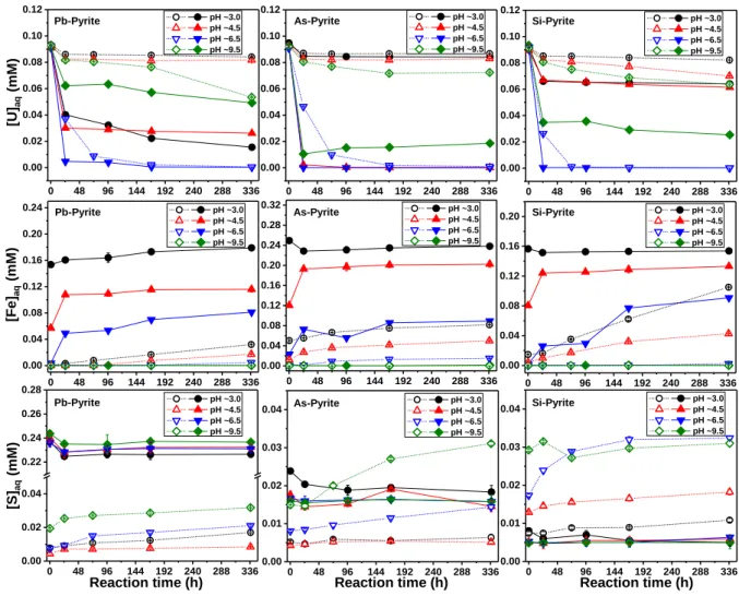
The evolutions of aqueous U, Fe and S in different reactors are illustrated in Figure 1 (detailed
data are shown in Tables S2 and S3). Pb and As were not detected probably due to the
scarci-ty of impurities contained in the pyrites and the noncongruent dissolution of pyrite.
U(VI) concentration. As shown in Figure 1, the decreases of aqueous U(VI) concentration
([U(VI)]aq) at pH ~3.0, ~4.5, and ~9.5 for Type II pyrites are larger than those for Type I
pyri-tes. For Type I Pb- and As-pyrites at pH ~3.0 and ~4.5 and Si-pyrite at pH ~3.0, [U(VI)]aq
remained nearly invariant after a slight decrease in the first 24.5 hours. Conversely, [U(VI)]aq decreased dramatically to a low level within the first 24.5 hours for Type II Pb- and
As-pyrites at pH ~4.5. The rapid uptake of aqueous U(VI) by Type II As-pyrite at pH ~4.5 was
further confirmed by the complementary experiments (Table S4). Compared with the other
two Type II pyrites, Type II As-pyrite immobilized the least amount of U at pH ~3.0. This probably results from the electrostatic repulsion or a relatively weak surface attraction as
cati-onic UO22+ is predominant (Figure S5) and the pyrite surface may be positively charged at
this low pH. Indeed, the point of zero charge (PZC) values of pyrite and arsenopyrite are
ap-proximately pH 2.0 and 5.4, respectively.41 Arsenic substitution is expected to increase the
pHPZC of pyrite since it has a local order similar to that of arsenopyrite.42,43 At pH ~6.5 a
sig-nificant decrease in [U(VI)]aq was observed for both Type I and Type II pyrites, which might be mainly explained by the spontaneous precipitation of U(VI)-containing phases such as
schoepite that shows a relatively low solubility at this pH condition (Figure S5). In addition,
we cannot exclude the possible occurrence of surface sorption on the formed Fe(III) (oxyhydr)oxides at this pH condition (see section 3.3 below). At pH ~9.5, the sorption also
proceeded relatively slowly, especially for Type I pyrites, which could be mainly explained
by the negatively charged pyrite surface and the prevalence of anionic U(VI) species under
11
Fe concentration. The concentration of aqueous Fe ([Fe]aq) was strongly pH-dependent for both types of pyrites. Less aqueous Fe was released from pyrites with increasing pH. Besides, much higher [Fe]aq was detected in Type II pyrite reactors compared to Type I pyrite reactors. In addition to the reaction with U(VI), this could be explained mainly by the reaction of
sur-face Fe3+ with S2- upon contact with solution. Indeed, during the anoxic grinding process, the
breaking of Fe(S)-S bonds would result in an electron transfer from iron to sulfur and thus the
formation of S2- and Fe3+ sites on the pyrite surfaces.44 This is further confirmed by the
gener-ally high [Fe]aq and [S]aq (the concentration of aqueous S) observed in Type II pyrite reactors
before introducing U(VI) (Table S3), although no SO32-/SO42- was detected on the surface of
pristine Type II pyrite (Figure S6).
S concentration. A continuous slight increase in [S]aq with reaction time was observed for Type I pyrites, probably caused mainly by the non-oxidative dissolution of pyrites. The
[S]aq/[Fe]aq ratio in each reactor was quite lower than 2.0 (Tables S2 and S3), indicating a
non-congruent dissolution of the pyrites. A large quantity of aqueous S (~2.4×10⁻⁴ M) was
observed in Type II Pb-pyrite suspensions, which might be mainly ascribed to the oxidation of
surface S2- by surface Fe3+.
For all Type II pyrite reactors, it is worth noting that an obvious decrease of [S]aq at the early stage of reaction can be observed under all pH conditions. This decrease could be explained
by the oxidation of aqueous S(-II) to elemental sulfur (S0) by the added U(VI).45,46 Oxidation
of surface S2-to SO32-/SO42-by the surface Fe3+ at the pyrite-water interface cannot consume
all S2-, which could result in release of the residual surface S2- species to the solution.
Aque-ous sulfide is reactive and can be rapidly oxidized by aqueAque-ous U(VI) to S0 in a homogeneous
system.45,46 This explanation was further verified by the analysis of sulfur species (Table S5),
as [MBS] (represents the concentration of methylene-blue-detectable sulfur, i.e., total S(-II) species) decreased from 11.0 to 1.3 µM at pH ~3.0 and from 8.5 to 0.3 µM at pH ~4.5 after
12
the addition of U(VI) in Type II As-pyrite reactors. Meanwhile, a significant amount of S0
was detected (Table S5). In contrast, the [MBS] values were immeasurable or negligible in
the case of Type I Pb- and As-pyrites (Table S5). The concentrations of sulfur oxyanions were
not measured directly but can be estimated by the determined [S]aq and [MBS] for samples
listed in Tables S4 and S5.
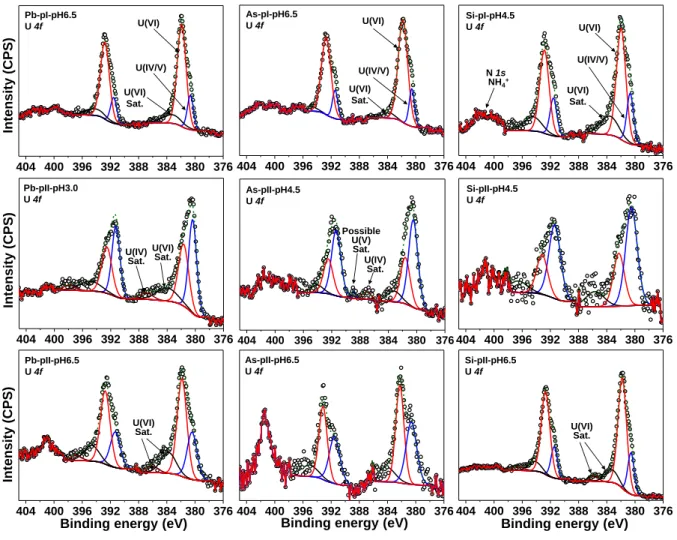
3.3. XPS surface features
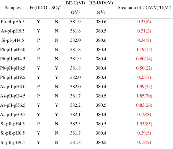
In line with the minor decreases of [U(VI)]aq, solid concentrations of sorbed uranium in a few reacted Type I pyrites were too low to be detected by XPS. The representative XPS U(4f)
spectra of selected samples are shown in Figure 2 and the rest are in Figure S7. The same
no-tation strategy was used for all the samples, e.g., Pb-pI-pH6.5 represents Type I Pb-pyrite at
pH ~6.5. Two main peaks of U(4f7/2) spectrum can be fitted at 381.9±0.4 and 380.5±0.2 eV,
which are corresponding to unreduced U(VI) and reduced uranium (labeled as U(IV/V)),
re-spectively. Area percentages of XPS peaks are shown in Table S6. Previous studies by cyclic
voltammetry verified that U(VI) can be reduced to U(V) on the surface of pyrite.47, 48
Howev-er, the exact reduced U species were difficult to be identified as the binding energies for U(V)
and U(IV) are very close.49 Shoulder peaks fitted at 385.0 ± 1.0 eV were probably ascribed to
the U(VI) satellite signal. The satellite peaks could also help to determine the valences of
re-duced U.17,50,51 However, this was largely limited by the predominance of UO2+x product
formed in this study.51Relatively weak peaks at ~389 eV and ~387 eV, possibly attributing to
U(4f7/2) satellites of U(V) and U(IV), respectively, were observed only for As-pII-pH4.5 and
Pb-pII-pH3.0 that had a relatively high content of reduced uranium.49 At each pH condition,
the U(IV/V)/U(VI) ratios of Type II pyrites are higher than that of Type I pyrites (Table 1),
indicating a more extensive U(VI) reduction on Type II pyrites. Besides, the U(IV/V)/U(VI)
13
(electrostatic repulsion) of cationic U(VI) species at lower pH (Figure S5) and/or the
simulta-neous occurrence of U(VI) precipitation at a relatively high pH.
The Fe(2p) spectra for U(VI)-reacted Type I pyrite can be fitted with three peaks located at 707.2±0.1, 708.0±0.1, and 709.7±0.2 eV, corresponding to Fe(II)-S of pyrite, Fe(II)-O of
hy-drated ferrous iron24 or Fe(III)-S resulting from the breaking of Fe–S bonds at the pyrite
sur-face,44,52 and an Fe(II) satellite peak under acidic conditions or Fe(III)-O probably coming
from ferric oxyhydroxide under neutral to alkaline conditions, respectively. No obvious shape
difference between Type I and Type II pristine pyrites was observed for both Fe(2p) and S(2p) spectra (Figure S6 and Table S6), as the depth probed by XPS (~5 nm) is larger than the thickness of a surface layer (approximately one Fe(S)-S bond length, i.e., a few angstroms).
Compared with the pristine pyrite, the Fe(2p) spectra of the U(VI)-reacted Type I pyrite are
nearly identical (Figure S8). The S(2p) spectra show the presence of oxidized sulfur (e.g.,
polysulfides or thiosulfate) and surface S2- at 164.0±0.6 and 161.9±0.1 eV, respectively,
to-gether with the bulk S22- dimers located at 162.6±0.1 eV. These contributions are also
ob-served for the pristine pyrites. In particular, no peak was obob-served above 166.4 eV
corre-sponding to the BE of SO32-/SO42- for the pristine pyrites (Figure S6), suggesting that there
was no surface oxidation during the anoxic grinding process. After reaction with U(VI), the
characteristic band of SO32-/SO42- located above 166.4 eV is not observed for Type I pyrites.
In contrast, for Type II pyrites of Pb-pII-pH6.5, Pb-pII-pH9.5, pH6.5, and
As-pII-pH9.5 (Figures S7 and S9), the characteristic peak of SO42- at ~168.5 eV appeared.
Simulta-neously, the Fe(2p) spectra showed an apparent increase in the amplitude at ~710.2 eV, indi-cating the occurrence of Fe(III)-O compounds. The precipitation of Fe(III) oxyhydroxide
might account for the SO42- detected at these pH conditions because of its good affinity to
many anions, like SO42-.53 However, these products were not detected for Si-pyrite. The
14
passivation layers on pyrite surfaces and be another reason for the relatively slow reaction ob-served at pH ~9.5.
In addition, a peak at ~402 eV, which can be attributed to NH4+ (N 1s orbit), was observed on
the reacted Type II Pb- and As-pyrites. As reported, both Fe(II) and S(-II) are able to reduce
NO3- into NH4+, and the processes can be appreciably catalyzed by surface sites of Fe
miner-als.54,55 The occurrence of NH4+ also verified the stronger reducing ability of Type II Pb- and
As-pyrites compared with Type I pyrites. However, the signal of NH4+ on reacted Type II
Si-pyrite was not enhanced obviously compared with Type I Si-Si-pyrite (i.e., Si-pI-pH4.5), imply-ing that the presence of Si impurity would not increase the reducimply-ing ability of pyrite.
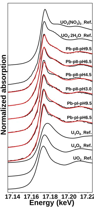
3.4. XANES characterization of uranium speciation
The XANES spectra of U(VI)-reacted Type I and Type II Pb-pyrites and the reference
XANES spectra for UO2(NO3)2, UO3∙2H2O, U3O8, U4O9, and UO2 are shown in Figure 3. A
shift of the absorption edge toward lower energy from U(VI) standards to U(VI)-reacted py-rite samples can be observed, confirming the formation of reduced uranium on pypy-rite surfaces.
The LCF analysis indicates that the combination of UO2and UO3∙2H2O references gives the
best fits to the experimental sample spectra (Table S7). Nevertheless, PHREEQC calculations
using the NEA/OECD thermodynamic database56 suggested that the solubility of schoepite is
higher than 0.1 mM at pH < 4.6 (Figure S10). As the initial U(VI) loadings in the experiments
were identical at 0.1 mM, a schoepite precipitate was therefore unlikely to form for the
sam-ples of pH ~3.0 and ~4.5, which is also evidenced by the almost invariant [U(VI)]aq observed
for Type I Pb- and As-pyrites at these two pH conditions (Figure 1). Additionally, it is worth
noting that UO2 is strongly reactive toward U(VI) to form hyperstoichiometric UO2+x(s),57
although the reaction may be controlled by the relative amounts of these solids. Therefore, the
formation of UO2 is unlikely in the presence of large unreduced U(VI) in this study.
15
similarity of different references can allow us to draw only a general conclusion about the ox-idation states of immobilized U, rather than the exact species. Nevertheless, the LCF analysis of the XANES data clearly demonstrated the occurrence of U(VI) reduction. As shown in
Figure 3, all the powder sample spectra give a weak shoulder signal at ~17180 eV, probably
indicating the multiple scattering within the uranyl moiety58 and thus the possible presence of
complexed uranyl structures on pyrite surfaces. However, adding UO2(NO3)2 as the third
standard in the LCF is not statistically significant enough from the F-test (Table S8). In future, more powerful techniques, e.g., U M4-edge high-energy-resolution fluorescence detection
(HERFD) XANES,33 will be considered to improve the identification of complex U species.
3.5. Reactive surface sites detected by SEM-EDX
Following the XPS and XANES analyses, SEM-EDX was used to image the surfaces of U(VI)-reacted pyrite and to determine the reactive sites where surface precipitates occur. Rep-resentative backscattered electron SEM images for Type II Pb-, As-, and Si-pyrite samples
af-ter reaction with U(VI) are shown in Figure 4. The bright regions correspond to an enrichment
of high Z number elements (i.e., heavy metals). In addition to the main elements of Fe and S, the EDX spectra indicate the presence of U along with Pb for both Pb-pyrite and Si-pyrite and
with As for As-pyrite (Table S9). Bright spots on pristine surfaces of the three Type II pyrites
were also characterized by SEM-EDX analysis (Figure S11), suggesting an absence of U on
the fresh surfaces. Spot explorations in dark areas show only the elements Fe and S (Spectra 2,
4, and 6 in Figure 4). These results indicate that U(VI) reduction preferentially occurred at Pb-
or As-impurity-rich spots on the pyrite surfaces, agreeing well with previous observations that freshly polished pyrite surfaces were efficient scavengers of aqueous U(VI) and that uranium
distribution was heterogeneous on pyrite surfaces.26 The incorporation of Pb and As
impuri-ties into the pyrite structure can introduce additional defect states in the electronic structure of the stoichiometric crystal. The impurity defect states broaden to overlap the FeS2 conduction
16
band, making the donated electrons delocalized.29 These delocalized electrons may lower the
activation energy for the reaction, serving as reactive sites on the pyrite surface. It is likely that the grinding process could expose more impurity-rich sites that could also partially ac-count for the observed U(VI) reduction. Nevertheless, these reactive impurity-rich sites could be removed by several cycles of acid wash, leaving a surface nearly inert toward aqueous U(VI). This situation was further confirmed by the SEM analysis prior to the experiments, as no (or fewer) heavy element regions were observed in the backscattered electron images for
the acid-washed Type I As-pyrite (Figure S12).
3.6. Possible reaction pathways
Results of this work underline the importance of aqueous sulfide in U(VI) reduction. The
sur-face S2- generated from anoxic grinding process would be partially consumed by the
concur-rent surface Fe3+ upon contact with water, and the rest would release into the solution and
re-act with the subsequently added U(VI). The occurrence of surface S2- species could partially
account for the faster reaction kinetics observed for the Type II pyrites, in particular for the rapid U(VI) decrease in the first 24.5 hours (Figure 1).
Apart from the reaction involving surface S2-, the circular oxidation and reduction of Fe2+/Fe3+
species sorbed on pyrite surfaces, could be another important mechanism for U(VI)
reduction.59 The occurrence of Fe(III)-O revealed by the XPS analysis for Type II pyrites of
Pb-pII-pH6.5, Pb-pII-pH9.5, As-pII-pH6.5, and As-pII-pH9.5 also supports this mechanism,
as intermediate Fe3+ is easy to precipitate by OH- at these pH conditions. The more facile
pathway for UO22+ reduction is to produce UO2+ first, followed by the disproportionation of
two intermediate UO2+ ions.60 Ab initio electron transfer calculations reveal that the reduction
of U(VI) to U(V) by Fe(II)aq (adsorbed on pyrite surface), considering an inner-sphere Fe-U
complex electron transfer reaction, is both thermodynamically favored and kinetically rapid
sta-17
bilized in the case of sorption onto mineral surfaces or be structurally incorporated into Fe
(oxyhydr)oxide solid phase and formation of a uranate-type coordination environment,15,17,18
which might occur for the reactions at pH ~6.5 and ~9.5 in this study. Previous study revealed
that U4O9 and U3O8 are predominantly composed of 2U(IV)+2U(V) and 2U(V)+U(VI),
re-spectively.10 The incomplete reduction of U(VI) revealed by the XPS and XANES analyses
supports the possible reaction pathways described by Eqs. (1) and/or (2), which are
thermo-dynamically favorable as indicated by the negative ∆rG0 values.56 Therefore, pyrite surfaces
may serve as “incubators” for transforming the reduced UO2+
ions into aliovalent oxides, such
as U3O8 in combination with unreduced U(VI), or U4O9 in combination with U(IV) generated
from the disproportionation of UO2+ ions.
2UO2+(aq) + UO22+(aq) + 2H2O(l) = U3O8(cr) + 4H+(aq) ∆rG0 = -20.598 kJ/mol (1)
2UO2+(aq) + 2U4+(aq) + 5H2O(l) = U4O9(cr) + 10H+(aq) ∆rG0 = -109.038 kJ/mol (2)
Arsenic substitutions can result in a localized decrease of pyrite crystallinity, giving rise to a localized increase in the solubility and reactivity.62 Besides, the formation of AsS dianion
groups is the most energetically favorable mechanism for arsenic incorporation into pyrite,
leading to a local configuration that is very close to the one in arsenopyrite.43 The presence of
AsS dianion groups can effectively trap electrons and accelerate the charge transfer between
the liquid and pyrite surface, enhancing the oxidation rate of arsenic-containing pyrite.30
Pref-erential release of As impurities via arsenopyrite oxidation has previously been observed for
As-rich pyrite oxidation by aqueous Se(IV).4 Unlike As impurities, the pyrite lattice does not
readily accommodate Pb at high concentrations, and the Pb content of pyrite was attributed to
the presence of nano-scale inclusions of Pb-bearing minerals such as galena.28 Previous
stud-ies indicated that galena has priority over pyrite for U(VI) reduction,23,63 which further
sup-ports our study that the Pb-rich area on the pyrite surface is the reactive site for U(VI) reduc-tion. Given all the above information, arsenopyrite- and galena-like local configurations may
18
be generated at As- and Pb-rich spots, leading to the U(VI) reduction on the pyrite surfaces occurring prevalently via the reaction pathways of arsenopyrite and galena, respectively.
3.7. Environmental implications
Contamination caused by concentrated uranium may occur in many fields, e.g., the leaching and accumulation of uranium from a uranium mill tailing,64,65 or the failure of a nuclear waste
canister in the future. The results of the present study are directly linked to the key geochemi-cal processes that govern the mobility of toxic U in the environment. This study shows that
surface S2- and Fe3+ generated by breaking of Fe(S)-S bonds during surface cleavage, as well
as the minor As-/Pb-impurity doping on pyrite surface, can largely accelerate the rate of aqueous U(VI) reduction by pyrite. These reactive surface species/sites can be removed by ac-id-washing, resulting in an inhibition of U(VI) reduction by pyrite, though thermodynamically favorable. In other words, the pyrite surface freshly polished or doped with trace element
im-purities is more efficient for U(VI) removal by reductive precipitation compared to the bulk
phase or to the weathered/acid-washed surface.
Pyrite frequently found in igneous (e.g., granite), metamorphic and sedimentary (e.g., clay-stone) settings possesses different crystallinities. As illustrated in this study, the reactivity of well-crystalline pyrite is quite weak towards U(VI), leading to a poor effect on U(VI) immo-bilization in igneous host rocks (e.g., granite) of nuclear waste repositories where pyrite is present as an accessory mineral. Situations may differ for granite fractures, in which pyrite could also form under a low temperature groundwater environment and biogenic influence, and probably have various grain sizes, crystallinities, surface compositions, and thus different chemical reactivity towards U(VI).66,67 In comparison, the pyrite precipitating at ambient
tem-perature in flooded soils and sediments, and at former mining and processing sites is usually characterized by poorer crystallinity, smaller size, more lattice defects, more impurity doping, and higher solubility, and thus is expected to be more reactive towards U(VI).13 This kind of
19
pyrite is able to reduce U(VI) and accumulate U effectively in the surroundings if the soil/sediment can stay reducing (i.e. depending on the persistence of redox oscillations68),
which is evidenced by the widespread co-existence of pyrites and U precipitates in sedi-ments.20 Therefore, the mobility of uranium is presumed to be largely attenuated by the pyrite
present in sedimentary materials, e.g., claystone that is also widely considered for a bedrock barrier in nuclear waste disposal. The findings in this study could improve our understanding of the geochemical behavior of U in the presence of pyrite in the relevant environments.
ASSOCIATED CONTENT
Supporting Information Available. Additional materials referenced in the text are available
free of charge.
XRD patterns of synthesized schoepite and natural pyrites. PDFs, S K-edge XAFS spectra,
XPS spectra, BSE images, and EDX spectra of pristine pyrites. XPS spectra of U(VI)-reacted
pyrites. Aqueous U(VI) speciation and schoepite dissolution with pH values. Aqueous results
of reactors with Type I and Type II pyrites. Aqueous results for verifying S(-II) and S(0)
spe-cies. Fitting results of XPS, S K-edge EXAFS, and U L3-edge XANES. F-tests for LCF of U
L3-edge XANES. Elemental contents determined by EDX spectroscopy.
AUTHOR INFORMATION Corresponding Author
*Phone: +86(0) 756 3668392. E-mail: kangml3@mail.sysu.edu.cn (M. KANG).
Notes
The authors declare no competing financial interest.
20
The authors are grateful to Eugene S. Ilton (PNNL, USA) for the XPS data interpretation and
Robin Pauer (Empa, Switzerland) for the EDX spectrum treatment. This work was financially supported by the National Natural Science Foundation of China (NSFC, Nos. 41773095 and 41403075), the Fundamental Research Funds for the Central Universities of Sun Yat-sen Uni-versity (No. 201545000-31610011), ANDRA, and Labex OSUG@2020 (Investissements d’avenir - ANR10 LABX56). Bin Ma also appreciates the financial support from the China Scholarship Council (CSC) and European Union's Horizon 2020 research and innovation pro-gramme under the Marie Skłodowska-Curie grant agreement number 754364. The synchro-tron facilities ESRF (BM25A-25-01-976) are acknowledged for the allocation of beam time.
REFERENCES
(1) Gaucher, É. C.; Blanc, P.; Bardot, F.; Braibant, G.; Buschaert, S.; Crouzet, C.; Gautier, A.; Girard, J.-P.; Jacquot, E.; Lassin, A. Modelling the porewater chemistry of the Callovian– Oxfordian formation at a regional scale. C.R. Geosci. 2006, 338 (12-13), 917-930.
(2) Ma, B.; Fernandez-Martinez, A.; Wang, K.; Madé, B.; Henocq, P.; Tisserand, D.; Bureau, S.; Charlet, L. Selenite Sorption on Hydrated CEM-V/A Cement in the Presence of Steel Corrosion Products: Redox vs. Nonredox Sorption. Environ. Sci. Technol. 2020, 54 (4), 2344-2352.
(3) Badaut, V.; Schlegel, M. L.; Descostes, M.; Moutiers, G. In situ time-resolved X-ray near-edge absorption spectroscopy of selenite reduction by siderite. Environ. Sci. Technol. 2012,
46 (19), 10820-10826.
(4) Kang, M.; Bardelli, F.; Charlet, L.; Géhin, A.; Shchukarev, A.; Chen, F.; Morel, M.-C.; Ma, B.; Liu, C. Redox reaction of aqueous selenite with As-rich pyrite from Jiguanshan ore mine (China): Reaction products and pathway. Appl. Geochem. 2014, 47, 130-140.
21
(5) Huo, L.; Xie, W.; Qian, T.; Guan, X.; Zhao, D. Reductive immobilization of pertechnetate in soil and groundwater using synthetic pyrite nanoparticles. Chemosphere 2017, 174, 456-465.
(6) Townsend, L. T.; Shaw, S.; Ofili, N. E. R.; Kaltsoyannis, N.; Walton, A. S.; Mosselmans, J. F. W.; Neill, T. S.; Lloyd, J. R.; Heath, S.; Hibberd, R.; Morris, K. Formation of a U(VI)– persulfide complex during environmentally relevant sulfidation of iron (oxyhydr)oxides.
Environ. Sci. Technol. 2020, 54 (1), 129-136.
(7) Ma, B.; Charlet, L.; Fernandez-Martinez, A.; Kang, M.; Madé, B. A review of the retention mechanisms of redox-sensitive radionuclides in multi-barrier systems. Appl.
Geochem. 2019, 100, 414-431.
(8) Faybishenko, B.; Birkholzer, J.; Sassani, D.; Swift, P., Eds. International Approaches for
Deep Geological Disposal of Nuclear Waste: Geological Challenges in Radioactive Waste Isolation: Fifth Worlwide Review; Lawrence Berkeley National Laboratory, Sandia National
Laboratories, 2016.
(9) Chen, P.; Ma, Y.; Kang, M.; Shang, C.; Song, Y.; Xu, F.; Wang, J.; Song, G.; Yang, Y.
The redox behavior of uranium on Beishan granite: Effect of Fe2+ and Fe3+ content. J. Environ.
Radioact. 2020, 217, https://doi.org/10.1016/j.jenvrad.2020.106208.
(10) Kvashnina, K.; Butorin, S. M.; Martin, P.; Glatzel, P. Chemical state of complex uranium oxides. Phys. Rev. Lett. 2013, 111 (25), 253002.
(11) Dong, W.; Brooks, S. C. Determination of the formation constants of ternary complexes
of uranyl and carbonate with alkaline earth metals (Mg2+, Ca2+, Sr2+, and Ba2+) using anion
exchange method. Environ. Sci. Technol. 2006, 40 (15), 4689-4695.
(12) Guo, X.; Szenknect, S.; Mesbah, A.; Clavier, N.; Poinssot, C.; Ushakov, S. V.; Curtius, H.; Bosbach, D.; Ewing, R. C.; Burns, P. C. Thermodynamics of formation of coffinite, USiO4. Proc. Natl. Acad. Sci. U.S.A. 2015, 112 (21), 6551-6555.
22
(13) Wang, Y.; Bagnoud, A.; Suvorova, E.; McGivney, E.; Chesaux, L.; Phrommavanh, V.; Descostes, M.; Bernier-Latmani, R. Geochemical control on uranium(IV) mobility in a mining-impacted wetland. Environ. Sci. Technol. 2014, 48 (17), 10062-10070.
(14) Chakraborty, S.; Favre, F.; Banerjee, D.; Scheinost, A. C.; Mullet, M.; Ehrhardt, J.-J.; Brendle, J.; Vidal, L.; Charlet, L. U(VI) sorption and reduction by Fe(II) sorbed on montmorillonite. Environ. Sci. Technol. 2010, 44 (10), 3779-3785.
(15) Ilton, E. S.; Boily, J.-F.; Buck, E. C.; Skomurski, F. N.; Rosso, K. M.; Cahill, C. L.;
Bargar, J. R.; Felmy, A. R. Influence of dynamical conditions on the reduction of UVI at the
magnetite-solution interface. Environ. Sci. Technol. 2010, 44 (1), 170-176.
(16) Veeramani, H.; Scheinost, A. C.; Monsegue, N.; Qafoku, N. P.; Kukkadapu, R.; Newville,
M.; Lanzirotti, A.; Pruden, A.; Murayama, M.; Hochella Jr, M. F. Abiotic reductive immobilization of U(VI) by biogenic mackinawite. Environ. Sci. Technol. 2013, 47 (5), 2361-2369.
(17) Ilton, E. S.; Pacheco, J. S. L.; Bargar, J. R.; Shi, Z.; Liu, J.; Kovarik, L.; Engelhard, M.
H.; Felmy, A. R. Reduction of U(VI) incorporated in the structure of hematite. Environ. Sci.
Technol. 2012, 46 (17), 9428-9436.
(18) Roberts, H. E.; Morris, K.; Law, G. T.; Mosselmans, J. F. W.; Bots, P.; Kvashnina, K.;
Shaw, S. Uranium (V) incorporation mechanisms and stability in Fe(II)/Fe(III)(oxyhydr) oxides. Environ. Sci. Technol. Lett. 2017, 4 (10), 421-426.
(19) England, G. L.; Rasmussen, B. Krape , B.; Groves, D. I. The origin of uraninite,
bitumen nodules, and carbon seams in Witwatersrand gold-uranium-pyrite ore deposits, based on a Permo-Triassic analogue. Econ. Geol. 2001, 96 (8), 1907-1920.
(20) Cumberland, S. A.; Douglas, G.; Grice, K.; Moreau, J. W. Uranium mobility in organic matter-rich sediments: A review of geological and geochemical processes. Earth Sci. Rev.
23
(21) Yang, Z.; Kang, M.; Ma, B.; Xie, J.; Chen, F.; Charlet, L.; Liu, C. Inhibition of U(VI) reduction by synthetic and natural pyrite. Environ. Sci. Technol. 2014, 48 (18), 10716-10724. (22) Bone, S. E.; Dynes, J. J.; Cliff, J.; Bargar, J. R. Uranium (IV) adsorption by natural organic matter in anoxic sediments. Proc. Natl. Acad. Sci. U.S.A. 2017, 114 (4), 711-716. (23) Wersin, P.; Hochella Jr, M. F.; Persson, P.; Redden, G.; Leckie, J. O.; Harris, D. W. Interaction between aqueous uranium (VI) and sulfide minerals: Spectroscopic evidence for sorption and reduction. Geochim. Cosmochim. Acta 1994, 58 (13), 2829-2843.
(24) Descostes, M.; Schlegel, M.; Eglizaud, N.; Descamps, F.; Miserque, F.; Simoni, E.
Uptake of uranium and trace elements in pyrite (FeS2) suspensions. Geochim. Cosmochim.
Acta 2010, 74 (5), 1551-1562.
(25) Eglizaud, N.; Miserque, F.; Simoni, E.; Schlegel, M.; Descostes, M. Uranium (VI) interaction with pyrite (FeS2): Chemical and spectroscopic studies. Radiochim. Acta 2006, 94 (9-11), 651-656.
(26) Scott, T.; Tort, O. R.; Allen, G. Aqueous uptake of uranium onto pyrite surfaces; reactivity of fresh versus weathered material. Geochim. Cosmochim. Acta 2007, 71 (21), 5044-5053.
(27) Bruggeman, C.; Maes, N. Uptake of uranium(VI) by pyrite under Boom Clay conditions: Influence of dissolved organic carbon. Environ. Sci. Technol. 2010, 44 (11), 4210-4216. (28) Abraitis, P.; Pattrick, R.; Vaughan, D. Variations in the compositional, textural and electrical properties of natural pyrite: A review. Int. J. Miner. Process. 2004, 74 (1-4), 41-59. (29) Lehner, S.; Savage, K. The effect of As, Co, and Ni impurities on pyrite oxidation kinetics: Batch and flow-through reactor experiments with synthetic pyrite. Geochim.
24
(30) Lehner, S.; Savage, K.; Ciobanu, M.; Cliffel, D. E. The effect of As, Co, and Ni impurities on pyrite oxidation kinetics: An electrochemical study of synthetic pyrite. Geochim.
Cosmochim. Acta 2007, 71 (10), 2491-2509.
(31) Wan, M.; Shchukarev, A.; Lohmayer, R.; Planer-Friedrich, B.; Peiffer, S. Occurrence of surface polysulfides during the interaction between ferric (hydr)oxides and aqueous sulfide.
Environ. Sci. Technol. 2014, 48 (9), 5076-5084.
(32) McGuire, M. M.; Hamers, R. J. Extraction and quantitative analysis of elemental sulfur from sulfide mineral surfaces by high-performance liquid chromatography. Environ. Sci.
Technol. 2000, 34 (21), 4651-4655.
(33) s, R.; Rivenet, M.; Solari, P.-L.; Kvashnina, K. O.; Scheinost, A. C.; Martin, P. M.
Use of HERFD-XANES at the U L3-and M4-edges to determine the uranium valence state on
[Ni(H2O)4]3[U(OH,H2O)(UO2)8O12(OH)3]. Inorg. Chem. 2016, 55 (9), 4260-4270.
(34) Opel, K.; Weiss, S.; Hübener, S.; Zänker, H.; Bernhard, G. Study of the solubility of amorphous and crystalline uranium dioxide by combined spectroscopic methods. Radiochim.
Acta 2007, 95 (3), 143-149.
(35) Ravel, B.; Newville, M., ATHENA, ARTEMIS, HEPHAESTUS: data analysis for X-ray absorption spectroscopy using IFEFFIT. J. Synchrotron Radiat. 2005, 12 (4), 537-541.
(36) Rimondi, V.; Bardelli, F.; Benvenuti, M.; Costagliola, P.; Gray, J. E.; Lattanzi, P.
Mercu-ry speciation in the Mt. Amiata mining district (Italy): Interplay between urban activities and mercury contamination. Chem. Geol. 2014, 380, 110-118.
(37) Downward, L.; Booth, C.; Lukens, W.; Bridges, F. In A Variation of the F-Test for
Determining Statistical Relevance of Particular Parameters in EXAFS Fits, 13th International Conference on X-ray Absorption Fine Structure, Stanford, CA, USA, July 9-14, 2006; AIP Conf. Proc.; American Institute of Physics (AIP): Melville, NY, 2007; pp 129-131.
25
(38) Ma, B.; Fernandez-Martinez, A.; Grangeon, S.; Tournassat, C.; Findling, N.; Carrero, S.;
Tisserand, D.; Bureau, S.; Elkaïm, E.; Marini, C.; Aquilanti, G.; Koishi, A.; Marty, N. C. M.; Charlet, L. Selenite uptake by Ca-Al LDH: A description of intercalated anion coordination geometries. Environ. Sci. Technol. 2018, 52 (3), 1624-1632.
(39) Fujii, T.; Yoshida, A.; Tanaka, K.; Marumo, F.; Noda, Y. High pressure compressibilities of pyrite and cattierite. Mineral. J. 1986, 13 (4), 202-211.
(40) Vjunov, A.; Fulton, J. L.; Huthwelker, T.; Pin, S.; Mei, D.; Schenter, G. K.; Govind, N.; Camaioni, D. M.; Hu, J. Z.; Lercher, J. A. Quantitatively probing the Al distribution in zeolites. J. Am. Chem. Soc. 2014, 136 (23), 8296-8306.
(41) Kosmulski, M. pH-dependent surface charging and points of zero charge. IV. Update and
new approach. J. Colloid Interface Sci. 2009, 337 (2), 439-448.
(42) Le Pape, P.; Blanchard, M.; Brest, J.; Boulliard, J.-C.; Ikogou, M.; Stetten, L.; Wang, S.;
Landrot, G.; Morin, G. Arsenic incorporation in pyrite at ambient temperature at both
tetrahedral S–I and octahedral FeII sites: Evidence from EXAFS-DFT analysis. Environ. Sci.
Technol. 2017, 51 (1), 150-158.
(43) Blanchard, M.; Alfredsson, M.; Brodholt, J.; Wright, K.; Catlow, C. R. A. Arsenic
incorporation into FeS2 pyrite and its influence on dissolution: A DFT study. Geochim.
Cosmochim. Acta 2007, 71 (3), 624-630.
(44) Nesbitt, H.; Bancroft, G.; Pratt, A.; Scaini, M. Sulfur and iron surface states on fractured
pyrite surfaces. Am. Mineral. 1998, 83 (9-10), 1067-1076.
(45) Hua, B.; Xu, H. F.; Terry, J., Deng, B. L. Kinetics of uranium(VI) reduction by hydrogen sulfide in anoxic aqueous systems. Environ. Sci. Technol. 2006, 40, 4666-4671.
(46) Hyun, S. P.; Davis, J. A.; Hayes, K. F. Abiotic U(VI) reduction by aqueous sulfide. Appl.
26
(47) Luo, M.; Liu, S.; Li, J.; Luo, F.; Lin, H.; Yao, P. Uranium sorption characteristics onto
synthesized pyrite. J. Radioanal. Nucl. Chem. 2016, 307 (1), 305-312.
(48) Renock, D.; Mueller, M.; Yuan, K.; Ewing, R. C.; Becker, U. The energetics and kinetics
of uranyl reduction on pyrite, hematite, and magnetite surfaces: A powder microelectrode study. Geochim. Cosmochim. Acta 2013, 118, 56-71.
(49) Ilton, E. S.; Bagus, P. S. XPS determination of uranium oxidation states. Surf. Interface
Anal. 2011, 43 (13), 1549-1560.
(50) Massey, M. S.; Lezama-Pacheco, J. S.; Jones, M. E.; Ilton, E. S.; Cerrato, J. M.; Bargar, J.
R.; Fendorf, S. Competing retention pathways of uranium upon reaction with Fe(II). Geochim.
Cosmochim. Acta 2014, 142, 166-185.
(51) Ilton, E. S.; Du, Y.; Stubbs, J. E.; Eng, P. J.; Chaka, A. M.; Bargar, J. R.; Nelin, C. J.;
Bagus, P. S. Quantifying small changes in uranium oxidation states using XPS of a shallow core level. Phys. Chem. Chem. Phys. 2017, 19 (45), 30473-30480.
(52) Elsetinow, A. R.; Guevremont, J. M.; Strongin, D. R.; Schoonen, M. A.; Strongin, M.
Oxidation of {100} and {111} surfaces of pyrite: Effects of preparation method. Am. Mineral.
2000, 85 (3-4), 623-626.
(53) Rietra, R. P.; Hiemstra, T.; van Riemsdijk, W. H. Sulfate adsorption on goethite. J.
Colloid Interface Sci. 1999, 218 (2), 511-521.
(54) Ottley, C. J.; Davison, W.; Edmunds, W. M. Chemical catalysis of nitrate reduction by
iron (II). Geochim. Cosmochim. Acta 1997, 61 (9), 1819-1828.
(55) Brunet, R. C.; Garcia-Gil, L. J. Sulfide-induced dissimilatory nitrate reduction to
ammonia in anaerobic freshwater sediments. FEMS Microbiol. Ecol. 1996, 21 (2), 131-138.
(56) Hummel, W.; Mompean, F. J.; Illemassène, M.; Perrone, J. Chemical thermodynamics of
compounds and complexes of U, Np, Pu, Am, Tc, Se, Ni and Zr with selected organic ligands;
27
(57) Cui, D.; Spahiu, K. On the interaction between uranyl carbonate and UO2(s) in anaerobic
solution. J. Nucl. Sci. Technol. 2002, 39 (sup3), 500-503.
(58) Hudson, E.; Allen, P.; Terminello, L.; Denecke, M.; Reich, T. Polarized
X-ray-absorption spectroscopy of the uranyl ion: Comparison of experiment and theory. Phys. Rev.
B: Condens. Matter 1996, 54 (1), 156.
(59) Moses, C. O.; Herman, J. S. Pyrite oxidation at circumneutral pH. Geochim. Cosmochim.
Acta 1991, 55 (2), 471-482.
(60) Yuan, K.; Renock, D.; Ewing, R. C.; Becker, U. Uranium reduction on magnetite:
Probing for pentavalent uranium using electrochemical methods. Geochim. Cosmochim. Acta
2015, 156, 194-206.
(61) Taylor, S.; Marcano, M.; Rosso, K. M.; Becker, U. An experimental and ab initio study
on the abiotic reduction of uranyl by ferrous iron. Geochim. Cosmochim. Acta 2015, 156, 154-172.
(62) Qiu, G.; Gao, T.; Hong, J.; Tan, W.; Liu, F.; Zheng, L. Mechanisms of arsenic-containing
pyrite oxidation by aqueous arsenate under anoxic conditions. Geochim. Cosmochim. Acta
2017, 217, 306-319.
(63) Aubriet, H.; Humbert, B.; Perdicakis, M. Interaction of U(VI) with pyrite, galena and
their mixtures: A theoretical and multitechnique approach. Radiochim. Acta 2006, 94 (9-11), 657-663.
(64) Abdelouas, A. Uranium mill tailings: geochemistry, mineralogy, and environmental impact. Elements 2006, 2 (6), 335-341.
(65) Yin, M.; Tsang, D. C. W.; Sun, J.; Wang, J.; Shang, J.; Fang, F.; Wu, Y.; Liu, J.; Song, G.; Xiao, T.; Chen, D. Critical insight and indication on particle size effects towards uranium release from uranium mill tailings: Geochemical and mineralogical aspects. Chemosphere
28
(66) Nishimoto, S.; Yoshida, H. Hydrothermal alteration of deep fractured granite: Effects of dissolution and precipitation. Lithos 2010, 115 (1), 153-162.
(67) Sahlstedt, E.; Karhu, J. A.; Pitkänen, P.; Whitehouse, M. Implications of sulfur isotope fractionation in fracture-filling sulfides in crystalline bedrock, Olkiluoto, Finland. Appl.
Geochem. 2013, 32, 52-69.
(68) Couture, R.-M.; Charlet, L.; Markelova, E.; Madé, B. t.; Parsons, C. T. On-off mobilization of contaminants in soils during redox oscillations. Environ. Sci. Technol. 2015,
29
Table 1. Summary of the fitted XPS data for U(VI)-reacted pyrite.a The analytical BE
uncer-tainty of the U(4f7/2) spin-orbit peak is ±0.2 eV. Uncertainties of area ratios of U(IV/V)/U(VI)
are given by the numbers in brackets on the last digit, i.e., 0.23(6) represents 0.23 ± 0.06, 1.19(15) represents 1.19 ± 0.15.
Samples Fe(III)-O SO4
2-BE-U(VI) (eV)
BE-U(IV/V) (eV)
Area ratio of U(IV/V)/U(VI)
Pb-pI-pH6.5 Y N 381.9 380.6 0.23(6) As-pI-pH6.5 Y N 381.8 380.5 0.21(2) Si-pI-pH4.5 P N 382.0 380.6 0.34(8) Pb-pII-pH3.0 P N 381.8 380.4 1.19(15) Pb-pII-pH4.5 P N 381.9 380.4 0.60(14) Pb-pII-pH6.5 Y Y 381.8 380.4 0.50(22) Pb-pII-pH9.5 Y Y 382.0 380.4 0.25(7) As-pII-pH3.0 P N 382.0 380.4 1.99(52) As-pII-pH4.5 P N 381.7 380.5 1.85(70) As-pII-pH6.5 Y Y 382.2 380.5 0.83(26) As-pII-pH9.5 Y Y 382.1 380.4 0.19(8) Si-pII-pH4.5 P N 382.3 380.5 1.95(85) Si-pII-pH6.5 Y N 381.7 380.4 0.29(5) Si-pII-pH9.5 Y N 381.8 380.5 0.18(2) a
30
Figure 1. Concentration evolutions of aqueous U, Fe and S under various pH conditions as
functions of time. The hollow symbols linked with short dash lines represents the results for Type I pyrite, while the solid symbols linked with solid lines for Type II pyrite.
0 48 96 144 192 240 288 336 0.00 0.02 0.04 0.06 0.08 0.10 0.12 pH ~3.0 pH ~4.5 pH ~6.5 pH ~9.5 0 48 96 144 192 240 288 336 0.00 0.01 0.02 0.03 0.04 pH ~3.0 pH ~4.5 pH ~6.5 pH ~9.5 0 48 96 144 192 240 288 336 0.00 0.01 0.02 0.03 0.04 pH ~3.0 pH ~4.5 pH ~6.5 pH ~9.5 0 48 96 144 192 240 288 336 0.00 0.02 0.04 0.22 0.24 0.26 0.28 pH ~3.0 pH ~4.5 pH ~6.5 pH ~9.5 0 48 96 144 192 240 288 336 0.00 0.04 0.08 0.12 0.16 0.20 pH ~3.0 pH ~4.5 pH ~6.5 pH ~9.5 0 48 96 144 192 240 288 336 0.00 0.04 0.08 0.12 0.16 0.20 0.24 0.28 0.32 pH ~3.0 pH ~4.5 pH ~6.5 pH ~9.5 0 48 96 144 192 240 288 336 0.00 0.04 0.08 0.12 0.16 0.20 0.24 pH ~3.0 pH ~4.5 pH ~6.5 pH ~9.5 0 48 96 144 192 240 288 336 0.00 0.02 0.04 0.06 0.08 0.10 0.12 pH ~3.0 pH ~4.5 pH ~6.5 pH ~9.5 0 48 96 144 192 240 288 336 0.00 0.02 0.04 0.06 0.08 0.10 0.12 pH ~3.0 pH ~4.5 pH ~6.5 pH ~9.5
Pb-Pyrite As-Pyrite Si-Pyrite
Pb-Pyrite As-Pyrite Si-Pyrite
Pb-Pyrite As-Pyrite Si-Pyrite
Reaction time (h) Reaction time (h) Reaction time (h)
[U ]aq (mM ) [Fe] aq (mM ) [S ]aq (mM )
31
Figure 2. Fitted U(4f) XPS spectra for U(VI)-reacted pyrite. Sat. represents the satellite peak. 404 400 396 392 388 384 380 376 404 400 396 392 388 384 380 376 404 400 396 392 388 384 380 376 404 400 396 392 388 384 380 376 U(VI) U(IV/V) Pb-pI-pH6.5 U 4f As-pI-pH6.5 U 4f Si-pI-pH4.5 U 4f Int ensit y ( CP S ) Int ensit y ( CP S ) Int ensit y ( CP S )
Binding energy (eV) U(VI) Sat. 404 400 396 392 388 384 380 376 404 400 396 392 388 384 380 376 404 400 396 392 388 384 380 376 Pb-pII-pH3.0 U 4f As-pII-pH4.5 U 4f Si-pII-pH6.5 U 4f
Binding energy (eV) U(VI) U(IV/V) U(VI) Sat. Possible U(V) Sat. 404 400 396 392 388 384 380 376 404 400 396 392 388 384 380 376 Pb-pII-pH6.5 U 4f As-pII-pH6.5 U 4f Si-pII-pH4.5 U 4f U(VI) U(IV/V) N 1s NH4+
Binding energy (eV) U(VI) Sat. U(VI) Sat. U(IV) Sat. U(IV) Sat. U(VI)
32
Figure 3. Normalized U L3-edge XANES spectra and the LCF results of U(VI)-reacted
Pb-pyrite samples. Red lines are the fits of the experimental spectra.
17.14 17.16 17.18 17.20 17.22
No
rmaliz
ed
abso
rp
tion
Energy (keV)
Pb-pI-pH6.5 UO2_Ref. U3O8_Ref. U4O9_Ref. Pb-pII-pH4.5 Pb-pII-pH3.0 Pb-pI-pH9.5 Pb-pII-pH9.5 Pb-pII-pH6.5 UO32H2O_Ref. UO2(NO3)2_Ref.33
Figure 4. Backscattered electron (BSE) images of (a) Pb-pII-pH6.5, (b) As-pII-pH3.0, and (c)
Si-pII-pH6.5. The corresponding normalized EDX spectra of selected spots are shown to the
right of each SEM image. Elemental contents of the selected spots are shown in Table S9.
1 2 3 4 5 6 7 8 9 10 0.00 0.04 0.08 0.5 1.0 1 2 3 4 5 6 7 8 9 10 0.00 0.04 0.08 0.5 1.0 1 2 3 4 5 6 7 8 9 10 0.00 0.04 0.08 0.5 1.0 1 2 3 4 5 6 7 8 9 10 0.00 0.04 0.08 0.5 1.0 1 2 3 4 5 6 7 8 9 10 0.00 0.04 0.08 0.5 1.0 1 2 3 4 5 6 7 8 9 10 0.00 0.04 0.08 0.5 1.0 Spectrum 2 Spectrum 1
a
Spectrum 1 Spectrum 2b
Spectrum 3 Spectrum 4 Spectrum 3 Spectrum 4c
Spectrum 5 Spectrum 6 Spectrum 5 Spectrum 6 keV keV keV As Pb S U Fe Fe Fe34
Table of Contents (TOC)
UO22+(aq) UO2+x(s) largely accelerated possible U(V)