Basal Constriction: Shaping the Vertebrate Brain
by
Ellie Graham Graeden
B.S. Microbiology Oregon State University, 2002
ARCHIVES
MASSACHUSETTS INSTITUTEOF TECHNOLOGY
SEP 2 9
2010
LIBRARIES
SUBMITTED TO THE DEPARTMENT OF BIOLOGY
IN PARTIAL FULFILLMENT OF THE REQUIREMENTS FOR THE DEGREE OF DOCTOR OF PHILOSOPHY IN BIOLOGY
AT THE
MASSACHUSETI'S INSTITUTE OF TECHNOLOGY SEPTEMBER 2010
©
2010 Massachusetts Institute of Technology. All rights reserved.Signature of
Author-Department of Biology, MIT September 2010
(NO1
Certified by:\KQ
/ / Accepted by: Hazel L. Sive Member, Whitehead Institute for Biomedical Research Professor of Biology, MIT Associate Dean, School of Science Thesis SupervisorTania A. Baker E. C. Whitehead Professor of Biology, MIT Investigator, Howard Hughes Medical Institute Co-chairperson, Graduate Committee
Basal Constriction: Shaping the Vertebrate Brain
by
Ellie Graham Graeden
Submitted to the Department of Biology
on 10 September 2010 in Partial Fulfillment of the Requirements
for the Degree of Doctor of Philosophy in Biology
ABSTRACT
Organs are primarily formed from epithelia, polarized sheets of cells with an apical surface facing a lumen and basal surface resting on the underlying
extracellular matrix. Cells within a sheet are joined by junctions, and changes in cell shape and size drive epithelial bending and folding during morphogenesis. These shape changes include constriction and expansion of the cell surfaces, elongation or shortening of the apical-basal length, or cell spreading. In this thesis, I present the first description of basal constriction, a process by which cells narrow on their basal surfaces to bend the neuroepithelium. Specifically, I describe morphogenesis of a major conserved bend in the vertebrate neural tube, the midbrain-hindbrain boundary constriction (MHBC). The MHBC forms
between 17 and 24 hours post fertilization in zebrafish, concomitant with, but
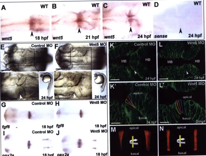
independent of ventricle inflation. Cells shorten to 75% the length of the surrounding cells prior to basal constriction, during which a band of 3-4 cells becomes wedge-shaped. Subsequently, these cells apically expand by twice the width of the surrounding cells. Basal constriction is laminin-dependent, with actin enriched at the basolateral surface of the constricted cells. Wnt5 is highly expressed specifically at the MHBC prior to and during basal constriction and is required for this process. Focal adhesion kinase (FAK) is activated by
phosphorylation at the MHBC and is required for basal constriction. FAK activation at the MHBC is dependent upon Wnt5 function. Loss of basal constriction in Wnt5 and FAK loss-of-function embryos can be rescued by inhibiting Gsk3p. These data suggest a novel pathway in which Wnt5 activates FAK in conjunction with the inhibition of Gsk3P to drive basal constriction at the MHBC. This study is the first to describe basal constriction during epithelial morphogenesis and provides mechanistic insights into a newly described cell shape change required for normal brain development.
Thesis Supervisor: Hazel L. Sive
Title: Member, Whitehead Institute for Biomedical Research Professor of Biology, MIT
BIOGRAPHICAL NOTE
Ellie Graeden was introduced to research in the laboratory of Dr. Phil Youderian at the University of Idaho, studying antibiotic resistance in Myxoccocus xanthus and Salmonella typhimurium. She earned her B.S. in microbiology at Oregon State University where she worked with Dr. Stephen Giovannoni analyzing the phylogeny of novel marine communities. After teaching at a Swiss boarding school, she returned to research in the laboratory of Dr. George Sprague at the University of Oregon where she characterized protein-protein interactions in the filamentous growth pathway of Saccharomyces cerevisiea. Her doctoral work in the Sive laboratory at MIT was supported by an NSF Pre-doctoral Fellowship.
ACKNOWLEDGMENTS
I offer many thanks to my advisor, Hazel Sive, for her support not only of my research, but of my intellectual development; to members of the Sive lab, particularly Jen Gutzman and Amanda Dickinson, for their help, support, and mentorship, and to Olivier Paugois for taking such good care of my fish; to Frank Solomon for his kind support of me, my research, and of the BioREFS program; to the members of the BioREFS, particularly Lourdes Aleman for helping me get it going and Calvin Jan and Shankar Sundar for keeping me going; to Brute Squad - 100% BS; to Kurt Weiss for reminding me that sometimes it is more important to go play in the sun; to Sarah Bagby for her unfailing and dear friendship; to Bec Gianotti and Gin Rich for laughter and perspective; to Julie Mumford who is both friend and family; to my grandparents, parents and
brother, who are both family and friends; to my son, Finn, who I am just getting to know; and, finally, to my husband, Ryan, who makes anything possible.
TABLE OF CONTENTS
1. Introduction: Cellular Mechanisms of Epithelial Morphogenesis
2. Formation of the zebrafish midbrain-hindbrain boundary constriction requires laminin-dependent basal constriction
3. Basal constriction at the midbrain-hindbrain boundary constriction is regulated by Wnt5, FAK and Gsk33
4. Future Directions
5. Appendix 1: The role of microtubules in basal constriction
7
43
83
149
Introduction:
CONTRIBUTIONS
ABSTRACT
Epithelial morphogenesis is a pivotal process by which the developing embryonic body plan and forming organs are shaped. Changes in cell shape and length that bend
epithelia include cell shortening, elongation, and cell spreading or flattening. In addition, cells can undergo apical and basal constriction, which bend a cell sheet in opposite directions. I describe the epithelial organization upon which these cell shape and length changes depend and discuss the mechanisms driving these changes. I then discuss possible mechanisms by which epithelial cell shortening and elongation could be regulated, in the context of cuboidal or columnar epithelia and during cell spreading. Apical constriction is well-established, and I review the current state of the field. Basal constriction is little-studied, but my analysis of embryos suggests that this is a
widespread mechanism of cell sheet bending. Identification of basal constriction and mechanisms underlying this process form the basis of this thesis.
INTRODUCTION
During animal development, epithelial tissue is shaped by bending, folding, elongation and growth to form the organs that compose the embryo. An epithelium is defined as a tissue composed of linked cells of three basic types: squamous, cuboidal, or columnar. The basic structure and development of the epithelium are highly conserved across species and tissues. Cells within epithelia can change shape to become apically or basally constricted, which bends the cell sheet. Here, I address the cell shape and length changes that drive epithelial morphogenesis during development with particular
emphasis on gastrulation (formation of the germ layers in the embryo) and organogenesis.
Organization of epithelia
The hallmark of an epithelium is its apical and basal polarity (Bryant and Mostov,
2008). The apical surface faces the outside of the embryo or the lumen of an organ, as in the lung, stomach, and brain. The basal surface is bound to an underlying stabilizing substrate, the extracellular matrix, which connects the epithelia to other tissues (Schock and Perrimon, 2002). The cell membrane composing the apical and basal domains of each cell differ in function, types of proteins localized or bound to them, and in lipid composition (Schuck and Simons, 2004; van Meer and Simons, 1988). The lateral
domains of neighboring cells are joined via junctions, highly regulated protein complexes that link the cells within the epithelia through the interaction of membrane-bound
proteins at the cell surface (Schock and Perrimon, 2002). Each type of lateral junction has distinct functions. Apico-lateral junctions include tight junctions, adherens
junctions, and gap junctions (Giepmans and van Ijzendoorn, 2009). Adherens junctions link the actin and microtubule cytoskeletons of neighboring cells and function as a membrane barrier, separating the apical and basal membrane domains by preventing
movement of membrane-bound proteins between the domains (Perez-Moreno et al.,
2003). Tight and gap junctions serve opposite purposes, respectively preventing and
allowing diffusion of molecules between cells (Giepmans and van Ijzendoorn, 2009). Tight junctions are additionally responsible for preventing movement of domain-specific lipids between apical and basolateral domains (van Meer and Simons, 1988). In the basolateral membrane, desmosomes connect neighboring cells through binding with intermediate filaments (Giepmans and van Ijzendoorn, 2009). At the basal cell surface,
integral membrane proteins, often within focal adhesions, bind the membrane to the underlying extracellular matrix, a complex network of proteoglycans necessary for structural integrity and signaling (Hynes, 2009; Parsons, 2003) (Fig. 1).
Cell shape changes within epithelia
Cells within epithelia are constrained by their neighbors. This allows local changes in shape of even a few cells to drive the formation of bends and folds in regions of the tissue as a whole (Pilot and Lecuit, 2005). Changes in cell shape may be in width at either or both poles or in length. In a cuboidal or columnar epithelium, constriction of cell width at one pole will form either a conical or wedge-shaped cell. If the force of the constriction is circumferential, then the cell forms a cone, like the tip of a pencil (Fig. 2A
to B). This often occurs in conjunction with cell elongation (Lee and Harland, 2007; Sawyer et al., 2010) (Fig. 2A to D to B). If the force of the constriction is lateral instead of
circumferential, the cell will form a wedge with flat sides that meet at an edge like a piece of pie (Fig. 2A to C). This has been shown to occur in conjunction with cell shortening
(Gutzman et al., 2008) (Fig. 2A to E to C). Changes in cell length, sometimes referred to as cell height, describe an elongation or shortening of the distance between the apical and basal cell surfaces (Fristrom, 1988) (Fig. 2A to D, A to E). Cell elongation can also describe lateral cell extension, in which both the apical and basal surfaces
simultaneously expand, as occurs during flattening or spreading (Solnica-Krezel, 2006)
(Fig. 2A to F)
Conventionally, it is assumed that there is minimal change in cell volume with changes of cell shape or length. This was analyzed explicitly in the fiber cells of the adult vertebrate lens where it was determined that cell length increased significantly more than the cell volume (15-fold versus 4-fold) (Bassnett, 2005). Where it has been analyzed
during development, this has been corroborated. For example, during transition from cuboidal to columnar epithelia, there was no observed change in cell volume in the developing wing imaginal disc in Drosophila (Widmann and Dahmann, 2009).
Therefore, the discussion here will focus on mechanisms of cell shape and length changes independent of cell volume.
Together, these cell shape changes drive the epithelial morphogenesis required for organogenesis and shaping the body plan in developing embryos.
CELL LENGTH CHANGE
Cell elongation and cell shortening are often coupled with apical and basal constrictions during morphogenesis of cuboidal or columnar epithelia, but are also required for cell spreading during embryonic epiboly, or the extension of the enveloping layer over the yolk during gastrulation early in development (Lee et al., 2007; Pilot and
Lecuit, 2005; Solnica-Krezel, 2006). Apical constriction is most often paired with cell elongation, whereas cell shortening has been associated with basal constriction. For example, cells nearly double in length concomitant with apical constriction during neural tube closure in Xenopus (Lee et al., 2007). Cell elongation paired with apical constriction
has also been described in bottle cells during Xenopus gastrulation (Lee and Harland,
2007) and wing imaginal disc development in Drosophila (Schlichting and Dahmann,
necessary for cell intercalation and deep cell rearrangements in Xenopus gastrulation (Keller, 1980), and cell elongation drives sheet spreading during both Drosophila dorsal
closure and zebrafish epiboly (Koppen et al., 2006).
Cell length changes have long been associated with microtubules, and this appears to be tightly related to their organization in these cells (Burnside, 1971; Byers and Porter, 1964; Chisholm and Hardin, 2005; Karfunkel, 1972; Piatigorsky et al., 1973; Solnica-Krezel, 2005). In mesenchymal cells, polarized microtubules are organized in radial arrays, anchored by their minus-ends at the centrosome or microtubule organizing center (MTOC) near the nucleus with their plus-ends extending to the cell cortex
(Bartolini and Gundersen, 2006). In epithelial cells, microtubules become organized in non-centrosomal arrays with their minus ends anchored at the apical surface and their plus-ends anchored at the basal cell surface (Bellett et al., 2009; Reilein and Nelson,
2005; Reilein et al., 2005). Microtubules are nucleated from their minus-ends and grow from their plus-ends; microtubule shrinking is termed catastrophe and occurs from the plus-end at random (Wade, 2009). As epithelial cells become polarized, the plus-ends
are captured by dynein and stabilized by Ebi and APC at the cell cortex to form both non-centrosomal microtubule networks at the cortex as well as parallel apical-basal arrays running the length of the cell (Bartolini and Gundersen, 2006; Bellett et al., 2009;
Byers and Porter, 1964; Lee and Harland, 2007; Reilein and Nelson, 2005; Shaw et al.,
2007; Wen et al., 2004). Dynein and kinesin are force-generating
microtubule-associated motors (Hirokawa et al., 2009; Kardon and Vale, 2009). In the case of cell
elongation in the lens of the sunfish, sliding of parallel microtubule arrays mediated by dynein results in rapid cell elongation (Dearry and Burnside, 1986; Troutt and Burnside,
1988). Kinesin has been shown to mediate microtubule sliding with enough force to
deform membranes (Jolly et al., 2010). Together, these data suggest a possible model of cell length change in which apico-basally oriented microtubules are captured and
stabilized at the cell cortex by plus-end-binding proteins. Once captured, the apical and basal cell surfaces are either pushed away or pulled toward each other by the force generated by dynein- or kinesin-mediated sliding. Alternatively, or in conjunction with this process, the cortical network could function to reshape the membrane and remodel the junctions to accommodate the change in cell length (Bellett et al., 2009).
The actomyosin cytoskeleton is also required for cell length changes. Cell flattening or spreading drives large-scale epithelial sheet migrations such as during zebrafish epiboly and Drosophila dorsal closure (Koppen et al., 2006; Pope and Harris,
2008). Both of these processes require accumulation of actin and non-muscle myosin at the leading edge of the advancing sheet to drive elongation of cells necessary to extend the sheet (Koppen et al., 2006; Pope and Harris, 2008; Solnica-Krezel, 2005). In the case of amnioserosa, one set of cells remodeled during Drosophila dorsal closure, columnar epithelial cells flatten to form a squamous epithelium. This process requires microtubule rotation driven by the actin cytoskeleton as myosin helps remodel the cell-cell junctions (Pope and Harris, 2008). Similarly, C. elegans epidermal elongation along the anterior-posterior axis requires Rho-mediated actomyosin contraction (Chisholm and Hardin, 2005). Rho signaling upstream of the myosin regulatory light chain is also required for cell flattening in Drosophila wing disc epithelia (Widmann and Dahmann,
2009).
Cell intercalation is the process by which cells in apposing sheets extend
membrane protrusions between cells in the other sheet. Cell elongation and movement into the sheet forms a new, longer epithelium (Pilot and Lecuit, 2005). This process is termed convergent extension (Keller, 2002). Myosin regulation of the actin cytoskeleton appears to be a conserved driver of this process as it is required for both vertebrate and
Drosophila gastrulation as well as neurulation and neural tube elongation (Nikolaidou
Signaling in cell length change
The upstream regulation of cell length change varies significantly between
developmental systems. Hedgehog (Hh) and Decapentaplegic (Dpp) drive cell shortening in both the wing and eye imaginal discs in Drosophila (Schlichting and Dahmann,
2008). This signaling was shown to be upstream of myosin during ingression in the eye imaginal disc (Escudero et al., 2007). In Xenopus, the transcription factor, Pitxi, regulates Shroom3 in the developing gut (Chung et al., 2010). Shroom3, in turn, is
required for cell elongation during neural tube closure (Lee et al., 2007). Shroom3 expression during mouse lens placode invagination is dependent on Pax6, a known downstream component of Wnt signaling (Kim et al., 2001; Osumi et al., 1997; Plageman
et al., 2010). This suggests a pathway in which early, localized expression of Pitxi
regulates Wnt-dependent Shroom3 to drive cell shortening. The planar cell polarity branch of Wnt signaling regulates convergent extension in Xenopus, zebrafish, mice,
Drosophila, and ascidians, to name a few (Qian et al., 2007; Torban et al., 2004). Given
the link between cell length and shape changes, it is also possible that the signaling pathways known to regulate cell shape change may be required to regulate the concomitant cell length changes.
APICAL CONSTRICTION
Apical constriction, the constriction of the apical surface of an epithelial cell, bends the epithelium toward an apical lumen or drives invagination, in-pocketing, of the tissue. Apical constriction was described as early as in 1902, and it has been studied
extensively since (Sawyer et al., 2010). It is required for a wide range of developmental
processes. Gastrulation requires apical constriction to invaginate the tissue. Apical constriction has been studied during this process in many organisms including sea urchins, Drosophila, Caenorhabditis elegans (C. elegans), and Xenopus. Apical
constriction is also required to close an opening in the epithelium such as during dorsal closure in Drosophila or wound healing in Xenopus. During organogenesis, apical constriction has been studied during neural tube closure and gut formation in Xenopus and hingepoint formation in chick, among others (Chung et al., 2010; Lee and Harland,
2007; Sawyer et al., 2010). While there are distinctions between the mechanisms that
drive apical constriction in each context, there is significant conservation.
Mechanisms of apical constriction
The process of apical constriction requires contraction of the actomyosin cytoskeleton at the apical cell surface. Actin within epithelia is localized both to a circumferential band linking the apically-localized adherens junctions of neighboring cells, and to a cortical meshwork of highly branched F-actin. Immediately prior to apical constriction, actin is enriched at the apical surface (Anstrom, 1992). This is concomitant
with apical recruitment of non-muscle myosin II (hereafter, myosin), a motor protein required for actin contraction (Pilot and Lecuit, 2005). In most cases, once actin and
myosin are recruited apically, Rho (a small GTPase) triggers contraction (Sawyer et al.,
2010).
For many years, the mechanism of contraction during apical constriction has been described by the purse-string model. The apical actin band links adherens junctions in neighboring cells and transmits tension between them. In this model, myosin
contracts the actin bands, driving apical constriction in a group of cells within the tissue (Baker and Schroeder, 1967; Burnside, 1971; Hildebrand, 2005; Karfunkel, 1972).
However, recent studies suggest that this may not be the most likely mechanism. During wounding in Xenopus, actin is apically enriched in the cells surrounding the wound. If intercellular tension were responsible for pulling the tissue together around the wound, breaking the actin cable would be expected to result in recoil. Davidson et al. tested this
hypothesis and found no evidence of recoil (Davidson et al., 2002). The purse-string
model would also suggest that a square wound would become rounded at the corners as healing progressed and the cells were pulled together. Instead, the authors found that the square shape was retained, and a triangle-shaped wound healed first into at Y-shape before closing. The authors conclude that actin-mediated tension between the cells is insufficient to drive migration during wound healing; such a mechanism may also be insufficient to drive apical constriction in a group of epithelial cells during development.
Martin et al. recently suggested an alternative model of apical constriction (Martin et al., 2009). During gastrulation in Drosophila, cells at the midline apically constrict to form an invagination at the ventral furrow. Analysis of time lapse imaging during the invagination showed a series of pulsed contractions each of which pulled the membranes closer to constrict the apical surface. Remarkably, the points of myosin responsible for these contractions co-localized not with the adherens junctions, but with the cortical actin between the junctions. Cortical actin, unlike the apical band of actin linking the adherens junctions, is composed almost entirely of highly branched actin (Weed and Parsons, 2001). Arp2/3 is an actin-related protein that functions to both nucleate and facilitate branching of actin at the cell cortex (Mullins et al., 1998). In C.
elegans gastrulation, Arp2/3 is required for apical constriction of the two endodermal
cells that initiate internalization (Roh-Johnson and Goldstein, 2009). These data suggest a model in which cortical actin, rather than the apical band of actin at adherens
junctions, may be specifically required for apical constriction.
While the role and regulation of actin in apical constriction has been well parsed, microtubules have also been identified as necessary in some contexts. Formation of bottle cells by apical constriction during Xenopus gastrulation requires intact
microtubules (Lee and Harland, 2007). Apical localization of y-tubulin and stabilization of microtubules is required for apical constriction during Xenopus neural tube closure
(Lee et al., 2007; Suzuki et al.). Microtubules could either be required for trafficking of
components necessary for the apical constriction or could be dynamically involved in the constriction itself. However, in both of these cases, apical constriction occurs subsequent to cell elongation. Microtubules may instead be required for the change in cell length and not the apical constriction itself, as discussed in the section on cell length change.
Basal expansion
Apical constriction is often accompanied by basal expansion, though this latter process has not been studied independently. Xenopus bottle cells are defined as having "dramatically constricted apical sides and enlarged basolateral areas" (Sawyer et al.,
2010), and cells at the ventral furrow in Drosophila are described as undergoing basal
expansion after apical constriction (Sweeton et al., 1991).
The most obvious cellular change during basal expansion is an increase in basal membrane. Such an expansion could be driven by biogenesis of new membrane,
transcytosis of the shrinking apical membrane to the basal surface, or transition in identity from apical to basal or basolateral membrane. Differential rates of biogenesis
could expand the basal surface. However, where it has been described, basal expansion appears to occur rapidly following apical constriction (Sweeton et al., 1991). Membrane biogenesis is closely tied to lipid metabolism (Nohturfft and Zhang, 2009), but the rate of biogenesis may not be rapid enough to drive the membrane expansion. An alternative hypothesis is based on data suggesting that endocytosis and transcytosis can result in the deposition of membrane from one region of the cell to another. This is a Rab-dependent mechanism by which specific cell regions can gain surface area (Pelissier et al., 2003). The hypothesis is particularly appealing given an observation that Rab5- and dynamin-mediated endocytosis is required for apical constriction during bottle cell formation in
re-integrate in bulk on the expanded basal surface, but this may have been due to a lack of resolution and not a reflection of the mechanism. Another possible mechanism of basal expansion is that apical membrane could shift in identity to basal or basolateral. In
Drosophila, downregulation of either apical or basolateral polarity determinants causes
an expansion of the opposite domain type (Kaplan et al., 2009). This could drive basal expansion by reducing the amount of apical membrane in the cell and increasing the amount of basal membrane. If coordinated with apical constriction and basal expansion, this would allow the basal membrane to increase in direct response to a decrease in apical membrane without the need for membrane biogenesis or transport. This remains an open area of study in the context of epithelial cell shape change.
Signaling in apical constriction
The upstream signals that regulate apical constriction vary much more than the physical mechanism of the cell shape change and can differ not only between organisms but between tissues within an organism. For example, in Drosophila, Hedgehog and Bmp signaling signal to form the eye morphogenetic furrow, which requires apical constriction (Schlichting and Dahmann, 2008), while Twist and Snail downstream of Dorsal regulate apical constriction during ventral furrow formation (Martin et al., 2009;
Sawyer et al., 2010). In sea urchin gastrulation, Wnt/Frizzled, Fgf, and calcium signaling
have all been implicated (Croce et al., 2006; Nakajima and Burke, 1996; Rottinger et al.,
2008), while non-canonical-Wnt signaling regulates zebrafish gastrulation (Ulrich et al., 2005). Apical constriction during Xenopus bottle cell formation appears to be regulated by non-canonical-Wnt signaling (Choi and Sokol, 2009), while canonical-Wnt signaling
regulates apical constriction upstream of Rho and myosin in the Drosophila wing imaginal disc (Zimmerman et al., 2010). It is likely that some of these pathways regulate
themselves. Closer analysis including temporally-specific activation or inhibition of signaling may bring to light more conservation in regulating these processes than is currently evident.
BASAL CONSTRICTION
Basal constriction, the constriction of the basal surface of an epithelial cell, is a process by which an epithelial sheet can bend away from an apical lumen or evaginate to form an out-pocketing of the epithelia. Such constriction requires the remodeling of the cell surface bound to the underlying extracellular matrix and expands the apical surface of the tissue. This process, unlike apical constriction, has not been studied in detail, and, in fact, has only been described as a mechanism of development in a few contexts. Basal constriction was first demonstrated in a squamous cell line in culture (Auersperg et al.,
1973) (Fig. 3A). A decade later, this type of cell shape change was speculated to be a
possible mechanism of epithelial morphogenesis, but this was not shown experimentally (Fristrom, 1988). Although apical constriction and apico-basal lengthening drive
invagination during Drosophila ventral furrow formation, Leptin and Grunewald also noted "narrowing" of the basal cell surface in cells adjacent to the invagination though this has not been further explored (Leptin and Grunewald, 1990) (Figure 3B). The regulation of basal constriction was first studied explicitly as a necessary step in
formation of the midbrain-hindbrain boundary constriction (MHBC) in zebrafish as part of this thesis (Fig. 3C). This was followed by a study showing basal constriction to be required during morphogenesis of the optic cup in Medaka (Martinez-Morales et al.,
2009).
Despite the paucity of examples described in the literature, my own analysis of data from previous studies suggests that this may be a much more common mechanism of epithelial morphogenesis than previously considered. Apical constriction initiates
ectodermal invagination during sea urchin invagination; reminiscent of the observations of ventral furrow formation in Drosophila mentioned above, cells with a
basal-constriction-like morphology lie on either side of the invagination (Sweeton et al., 1991). Early during mouse neurulation, electron microscopy and the observation of actin enrichment at the basal cell surfaces also suggest basal constriction (Sadler et al., 1982),
while in the eye imaginal disc of Drosophila, cells appear to constrict basally during tissue ingression (Corrigall et al., 2007)(Fig. 3D). In addition to these examples of epithelial folding, basal constriction appears to occur during evagination of the salivary gland in Drosophila (Fristrom, 1988). A recent study of the evaginating Hydra bud shows cells at the tip of the evagination that appear to be both basally constricted and apically expanded (Philipp et al., 2009) (Fig. 3E). While further experiments will be
required to confirm whether these cell shape changes share other characteristics of basal constriction, these observations from the literature indicate that basal constriction may be a conserved process that warrants greater attention.
Signaling in basal constriction
What signals initiate basal constriction? I considered both Wnt and focal adhesion kinase (FAK) signaling in regulating this process.
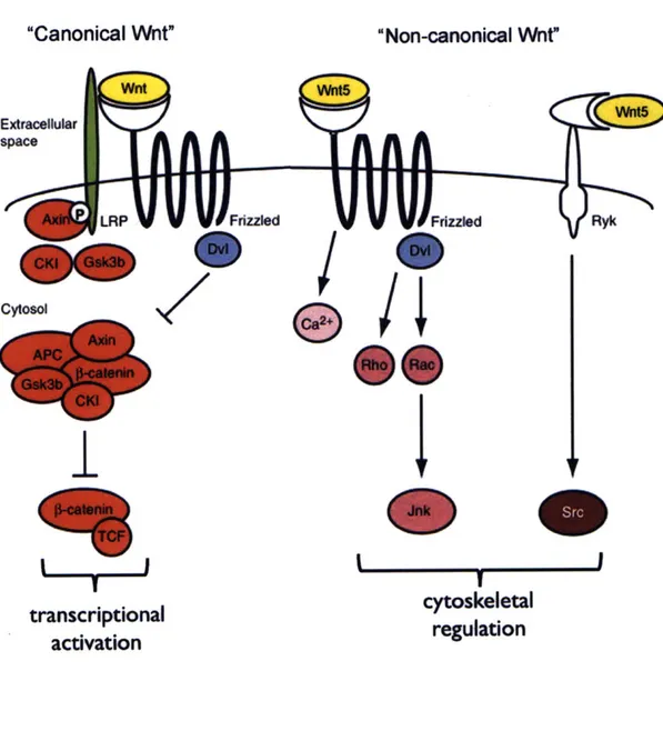
The Wnt signaling pathway is generally considered to include two branches: the canonical, whose activation results in transcriptional activation of downstream target genes, and the non-canonical, the activation of which generally results in cytoskeletal regulation (van Amerongen and Nusse, 2009) (Fig. 4). The canonical pathway, required
for tissue specification during zebrafish MHBC development, is activated by binding of an extracellular Wnt ligand to its receptor, Frizzled. This activation is tied to the activity of a co-receptor, LRP (Verkaar and Zaman, 2010) and results in the inhibition of a destruction complex including Gsk3p, APC, CK-1, Axin and MACF1 by Dishevelled
(Salinas, 2007). In the absence of Wnt ligand, Gsk3p and CKi phosphorylate cytoplasmic
p-catenin, targeting it for degradation. Inhibition of this complex in the presence of Wnt
ligand prevents this phosphorylation, which allows p-catenin to activate transcription ofdownstream targets through binding to TCF/LEF transcription factors in the nucleus (Salinas, 2007). Interestingly, Gsk3p and APC have also recently been shown to regulate the microtubule cytoskeleton in response to Wnt signaling, suggesting that these
components may also play roles outside the traditional canonical pathway (Zumbrunn et
al, 2009; Salinas, 2009).
The non-canonical Wnt signaling pathway is similarly activated by Wnt binding to Frizzled receptors, though in this pathway, Ror often acts as a co-receptor, and the
pathway can also be activated by binding to alternate receptors such as Ryk (van
Amerongen and Nusse, 2010). The signal is transmitted through Dishevelled, an adapter protein that binds to the receptor, sometimes in conjunction with Daami. The small
GTPases Rho or Rac and Jnk are then activated, promoting cytoskeletal rearrangements (Schlessinger et al, 2009). Alternatively, binding of non-canonical Wnt ligands to their receptors can result in activation of calcium signaling or the non-receptor tyrosine kinase, Src, although these pathways are less well understood (van Amerongen and Nusse, 2010). Non-canonical Wnt signaling has been shown to regulate cell shape changes, particularly during convergent extension (Qian, 2007; Torban, 2004).
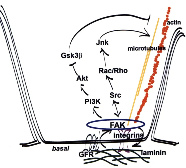
FAK is of particular interest in considering the regulation of basal constriction, as this process is likely to require active regulation of adhesion between the basal cell
surface and the underlying basement membrane. This protein is a well-known regulator of focal adhesions, but has also been shown to play a role in several signaling pathways (Parsons, 2003) (Fig. 5). Among others, FAK has been shown to signal upstream of Akt and P13K in the inhibition of Gsk3p (Huang 2006; Huang 2009), and in signaling
(Parsons, 2003; Igishi 1999). However, the specific role of FAK in these pathways is not
well understood.
A NOTE ABOUT REGION-SPECIFIC MORPHOGENESIS
As described above, epithelial morphogenesis is often driven by changes in cell size and shape. These changes in cell size or shape must occur not only at a specific time during development, but at a specific position in the epithelia. Thus, these changes require components to drive or execute the physical process itself as well as signals to position and initiate the process. In determining what components are necessary for a given cell size or shape change, it is useful to consider how components necessary for execution and initiation differ.
In order for a component to be involved in executing a cell shape change, it must be present at the right time and place and required for the process. This means it must be expressed and localized to the region during the cell shape change, but may also be
present elsewhere in the tissue at the same time. For example, actin is required for apical constriction, and thus, must be expressed and localized to the apical surface of the cells undergoing the cell shape change, but it is also present in epithelial cells not undergoing the cell shape change (Pilot and Lecuit, 2005). In addition, the component must be required for the process, which can be tested by loss of function analysis. Taking the same example, to establish that actin is required for apical constriction, its
depolymerization must prevent the process.
For a component to be characterized as positioning and/or initiating a cell shape change, it must similarly be required for the process. However, its expression and localization must be specific only to the time and place that the process is occurring. For example, during gastrulation, apical constriction occurs in one specific region of the tissue to drive invagination, although the entire tissue is composed of epithelial cells that
might also be competent to constrict apically (Martin et al, 2009). The signal necessary
to position or initiate the cell shape change must be localized specifically and only to the cells that constrict apically. The defining characteristic of a localizing or initiating signal is that it will drive the cell shape change in any cells that are competent. This suggests that an initiating signal expressed or localized in an expanded region of tissue would increase the total number of cells undergoing the cell shape change.
CONCLUSION
In this thesis, I establish basal constriction as a mechanism of epithelial
morphogenesis at the midbrain-hindbrain boundary constriction in zebrafish and begin to address the components required both to execute and initiate this process.
FIGURE LEGENDS
Fig. 1 Schematic of an epithelial cell
The apical and basolateral cell membrane domains are separated by tight junctions, adherens junctions, and gap junctions at the apical cell surface with desmosomes at the basolateral surface. Membrane-bound proteins at the basal surface, sometimes localized to focal adhesions, bind the underlying extracellular matrix. Actin is localized to the cell cortex, links the adherens junctions, and forms stress fibers at focal adhesions.
Microtubules are oriented apico-basally.
Fig. 2 Epithelial cell shape and length changes during morphogenesis
(A) A columnar epithelial cell is depicted though these processes could also be initiated
from cells of other types. (B) Circumferential constriction of one cell membrane will result in a cone-shaped cell. (C) Symmetrical lateral constriction will result in a wedge-shaped cell. (D) Cell elongation as along the apico-basal axis is depicted. This is often coupled with circumferential constriction (Lee et al., 2007; Widmann and Dahmann,
2009). (E) Cell shortening along apico-basal axis is depicted. This has been shown to
occur concomitant with basal constriction (Gutzman et al., 2008). (F) Cell spreading can be described as cell shortening along the apico-basal axis or as lateral elongation and results in a flattened epithelial cell.
Fig. 3 Examples of basal constrictionfrom the literature
Camera lucida drawings taken from the literature show examples of basal constriction either as published or from my own analysis. Red cells appear basally constricted. (A) Squamous cell carcinoma in 3D culture (Auersperg et al., 1973). (B) Ventral furrow as forms during Drosophila gastrulation (Leptin and Grunewald, 1990). (C) Zebrafish
midbrain-hindbrain boundary constriction (Gutzman et al., 2008). (D) Drosophila eye imaginal disc (Corrigall et al., 2007). (E) Tentacle bud in Hydra (Philipp et al., 2009).
Fig. 4 Overview of Wnt signaling
The canonical and non-canonical Wnt signaling pathways promote transcriptional activation and regulation of the cytoskeleton respectively. Wnt5 is shown as one example of a non-canonical Wnt signaling component that may be involved basal constriction at the MHBC. Figure adapted from (van Amerongen and Nusse, 2010).
Fig. 5 Focal Adhesion Kinase as a signaling regulator
Shown are two pathways in which focal adhesion kinase (FAK) acts as a signaling regulator (Huang 2006; Huang, 2009; Igishi, 1999; Parsons, 2003).
FIGURE 1
basal
9
6
96
96
6
9 :.sges:..:.:.::.:.::.:.::.:...
... ... ... ... ... ... ...ts
0
cellsreadng
FIGURE 3
basal basal
Drosophila ventral furrow
D
baIa
Droophila eye Imaginal disc
C
ae.Zebrafish midbrain-hindbrain boundary
E
Hydra tentacle bud
FIGURE 4
"Canonical Wnt"
"Non-canonical Wnt"
Extracellular space LRP Cytosol7
I
L . .Itranscriptional
activation
cytoskeletal
regulation
Ryk I ... ... .. ... ... 1 - 1.1 1 ... .... .... ----- ... ... . ... . OWEFIGURE 5
Jnk
Gsk3p
mco
Rac/Rho
Akt
P13K
basal
... .. .... ... .REFERENCES
Anstrom, J. A. (1992). Microfilaments, cell shape changes, and the formation of
primary mesenchyme in sea urchin embryos. JExp Zool 264, 312-22.
Auersperg, N., Erber, H. and Worth, A. (1973). Histologic variation among poorly
differentiated invasive carcinomas of the human uterine cervix. JNatl Cancer Inst 51,
1461-77.
Baker, P. C. and Schroeder, T. E. (1967). Cytoplasmic filaments and morphogenetic
movement in the amphibian neural tube. Dev Biol 15, 432-50.
Bartolini, F. and Gundersen, G. G. (2006). Generation of noncentrosomal
microtubule arrays. J Cell Sci 119, 4155-63.
Bassnett, S. (2005). Three-dimensional reconstruction of cells in the living lens: the relationship between cell length and volume. Exp Eye Res 81, 716-23.
Bellett, G., Carter, J. M., Keynton, J., Goldspink, D., James, C., Moss, D. K. and Mogensen, M. M. (2009). Microtubule plus-end and minus-end capture at
adherens junctions is involved in the assembly of apico-basal arrays in polarised epithelial cells. Cell Motil Cytoskeleton 66, 893-908.
Brand, M., Heisenberg, C. P., Jiang, Y. J., Beuchle, D., Lun, K., Furutani-Seiki, M., Granato, M., Haffter, P., Hammerschmidt, M., Kane, D. A. et al. (1996). Mutations in zebrafish genes affecting the formation of the boundary between
midbrain and hindbrain. Development 123, 179-90.
Bryant, D. M. and Mostov, K. E. (2008). From cells to organs: building polarized
tissue. Nat Rev Mol Cell Biol 9, 887-901.
Buckles, G. R., Thorpe, C. J., Ramel, M. C. and Lekven, A. C. (2004).
Combinatorial Wnt control of zebrafish midbrain-hindbrain boundary formation. Mech
Dev 121, 437-47.
Burnside, B. (1971). Microtubules and microfilaments in newt neuralation. Dev Biol
26, 416-41.
Byers, B. and Porter, K. R. (1964). Oriented Microtubules in Elongating Cells of the
Developing Lens Rudiment after Induction. Proc Natl Acad Sci USA 52, 1091-9.
Chisholm, A. D. and Hardin, J. (2005). Epidermal morphogenesis. WormBook,
1-22.
Choi, S. C. and Sokol, S. Y. (2009). The involvement of lethal giant larvae and Wnt
signaling in bottle cell formation in Xenopus embryos. Dev Biol 336, 68-75.
Chung, M. I., Nascone-Yoder, N. M., Grover, S. A., Drysdale, T. A. and Wallingford, J. B. (2010). Direct activation of Shroom3 transcription by Pitx proteins
Corrigall, D., Walther, R. F., Rodriguez, L., Fichelson, P. and Pichaud, F. (2007). Hedgehog signaling is a principal inducer of Myosin-II-driven cell ingression in
Drosophila epithelia. Dev Cell 13, 730-42.
Croce, J., Duloquin, L., Lhomond, G., McClay, D. R. and Gache, C. (2006).
Frizzled5/8 is required in secondary mesenchyme cells to initiate archenteron invagination during sea urchin development. Development 133, 547-57.
Davidson, L. A., Ezin, A. M. and Keller, R. (2002). Embryonic wound healing by
apical contraction and ingression in Xenopus laevis. Cell Motil Cytoskeleton 53, 163-76.
Dearry, A. and Burnside, B. (1986). Dopaminergic regulation of cone retinomotor
movement in isolated teleost retinas: I. Induction of cone contraction is mediated by D2 receptors. JNeurochem 46, 1006-21.
Escudero, L. M., Bischoff, M. and Freeman, M. (2007). Myosin II regulates
complex cellular arrangement and epithelial architecture in Drosophila. Dev Cell 13, 717-29.
Fristrom, D. (1988). The cellular basis of epithelial morphogenesis. A review. Tissue
Cell 20, 645-90.
Giepmans, B. N. and van Ijzendoorn, S. C. (2009). Epithelial cell-cell junctions
and plasma membrane domains. Biochim Biophys Acta 1788, 820-31.
Gutzman, J. H., Graeden, E. G., Lowery, L. A., Holley, H. S. and Sive, H. (2008). Formation of the zebrafish midbrain-hindbrain boundary constriction requires
laminin-dependent basal constriction. Mech Dev 125, 974-83.
Hildebrand, J. D. (2005). Shroom regulates epithelial cell shape via the apical
positioning of an actomyosin network. J Cell Sci 118, 5191-203.
Hirokawa, N., Noda, Y., Tanaka, Y. and Niwa, S. (2009). Kinesin superfamily
motor proteins and intracellular transport. Nat Rev Mol Cell Biol 10, 682-96.
Hynes, R. 0. (2009). The extracellular matrix: not just pretty fibrils. Science 326,
1216-9.
Jolly, A. L., Kim, H., Srinivasan, D., Lakonishok, M., Larson, A. G. and Gelfand, V. I. (2010). Kinesin-1 heavy chain mediates microtubule sliding to drive
changes in cell shape. Proc NatlAcad Sci USA 107, 12151-6.
Joyner, A. L. (1996). Engrailed, Wnt and Pax genes regulate midbrain--hindbrain
development. Trends Genet 12, 15-20.
Kaplan, N. A., Liu, X. and Tolwinski, N. S. (2009). Epithelial polarity: interactions
between junctions and apical-basal machinery. Genetics 183, 897-904.
Kardon, J. R. and Vale, R. D. (2009). Regulators of the cytoplasmic dynein motor.
Karfunkel, P. (1972). The activity of microtubules and microfilaments in neurulation
in the chick. JExp Zool 181, 289-30 1.
Keller, R. (2002). Shaping the vertebrate body plan by polarized embryonic cell
movements. Science 298, 1950-4.
Keller, R. E. (1980). The cellular basis of epiboly: an SEM study of deep-cell
rearrangement during gastrulation in Xenopus laevis. JEmbryol Exp Morphol 60, 201-34.
Kim, A. S., Anderson, S. A., Rubenstein, J. L., Lowenstein, D. H. and Pleasure, S. J. (2001). Pax-6 regulates expression of SFRP-2 and Wnt-7b in the
developing CNS. JNeurosci 21, RC132.
Koppen, M., Fernandez, B. G., Carvalho, L., Jacinto, A. and Heisenberg, C.
P. (2006). Coordinated cell-shape changes control epithelial movement in zebrafish and Drosophila. Development 133, 2671-81.
Lee, C., Scherr, H. M. and Wallingford, J. B. (2007). Shroom family proteins
regulate gamma-tubulin distribution and microtubule architecture during epithelial cell shape change. Development 134, 1431-41.
Lee, J. Y. and Harland, R. M. (2007). Actomyosin contractility and microtubules
drive apical constriction in Xenopus bottle cells. Dev Biol 311, 40-52.
Leptin, M. and Grunewald, B. (1990). Cell shape changes during gastrulation in
Drosophila. Development 110, 73-84.
Lun, K. and Brand, M. (1998). A series of no isthmus (noi) alleles of the zebrafish
pax2.1 gene reveals multiple signaling events in development of the midbrain-hindbrain boundary. Development 125, 3049-62.
Martin, A. C., Kaschube, M. and Wieschaus, E. F. (2009). Pulsed contractions of
an actin-myosin network drive apical constriction. Nature 457, 495-9.
Martinez-Morales, J. R., Rembold, M., Greger, K., Simpson, J. C., Brown, K. E., Quiring, R., Pepperkok, R., Martin-Bermudo, M. D., Himmelbauer, H. and Wittbrodt, J. (2009). ojoplano-mediated basal constriction is essential for optic
cup morphogenesis. Development 136, 2165-75.
Mullins, R. D., Heuser, J. A. and Pollard, T. D. (1998). The interaction of Arp2/3
complex with actin: nucleation, high affinity pointed end capping, and formation of branching networks of filaments. Proc Natl Acad Sci USA 95, 6181-6.
Nakajima, Y. and Burke, R. D. (1996). The initial phase of gastrulation in sea
urchins is accompanied by the formation of bottle cells. Dev Biol 179, 436-46.
Nikolaidou, K. K. and Barrett, K. (2004). A Rho GTPase signaling pathway is used
reiteratively in epithelial folding and potentially selects the outcome of Rho activation.
Nohturfft, A. and Zhang, S. C. (2009). Coordination of lipid metabolism in
membrane biogenesis. Annu Rev Cell Dev Biol 25, 539-66.
Osumi, N., Hirota, A., Ohuchi, H., Nakafuku, M., limura, T., Kuratani, S., Fujiwara, M., Noji, S. and Eto, K. (1997). Pax-6 is involved in the specification of
hindbrain motor neuron subtype. Development 124, 2961-72.
Parsons, J. T. (2003). Focal adhesion kinase: the first ten years. J Cell Sci 116,
1409-16.
Pelissier, A., Chauvin, J. P. and Lecuit, T. (2003). Trafficking through Rab11
endosomes is required for cellularization during Drosophila embryogenesis. Curr Biol
13, 1848-57.
Perez-Moreno, M., Jamora, C. and Fuchs, E. (2003). Sticky business:
orchestrating cellular signals at adherens junctions. Cell 112, 535-48.
Philipp, I., Aufschnaiter, R., Ozbek, S., Pontasch, S., Jenewein, M., Watanabe, H., Rentzsch, F., Holstein, T. W. and Hobmayer, B. (2009).
Wnt/beta-catenin and noncanonical Wnt signaling interact in tissue evagination in the simple eumetazoan Hydra. Proc NatlAcad Sci USA io6, 4290-5.
Piatigorsky, J., Rothschild, S. S. and Wollberg, M. (1973). Stimulation by insulin
of cell elongation and microtubule assembly in embryonic chick-lens epithelia. Proc Natl
Acad Sci USA 70, 1195-8.
Pilot, F. and Lecuit, T. (2005). Compartmentalized morphogenesis in epithelia: from
cell to tissue shape. Dev Dyn 232, 685-94.
Plageman, T. F., Jr., Chung, M. I., Lou, M., Smith, A. N., Hildebrand, J. D., Wallingford, J. B. and Lang, R. A. (2010). Pax6-dependent Shroom3 expression
regulates apical constriction during lens placode invagination. Development 137, 405-15.
Pope, K. L. and Harris, T. J. (2008). Control of cell flattening and junctional
remodeling during squamous epithelial morphogenesis in Drosophila. Development 135,
2227-38.
Puelles, L. and Martinez-de-la-Torre, M. (1987). Autoradiographic and Golgi study
on the early development of n. isthmi principalis and adjacent grisea in the chick embryo: a tridimensional viewpoint. Anat Embryol (Berl) 176, 19-34.
Qian, D., Jones, C., Rzadzinska, A., Mark, S., Zhang, X., Steel, K. P., Dai, X. and Chen, P. (2007). Wnt5a functions in planar cell polarity regulation in mice. Dev
Biol 306, 121-33.
Reifers, F., Bohli, H., Walsh, E. C., Crossley, P. H., Stainier, D. Y. and Brand, M. (1998). Fgf8 is mutated in zebrafish acerebellar (ace) mutants and is required for
maintenance of midbrain-hindbrain boundary development and somitogenesis.
Development 125, 2381-95.
Reilein, A. and Nelson, W. J. (2005). APC is a component of an organizing template
Reilein, A., Yamada, S. and Nelson, W. J. (2005). Self-organization of an
acentrosomal microtubule network at the basal cortex of polarized epithelial cells. J Cell
Biol 171, 845-55.
Roh-Johnson, M. and Goldstein, B. (2009). In vivo roles for Arp2/3 in cortical
actin organization during C. elegans gastrulation. J Cell Sci 122, 3983-93.
Rolo, A., Skoglund, P. and Keller, R. (2009). Morphogenetic movements driving
neural tube closure in Xenopus require myosin IIB. Dev Biol 327, 327-38.
Rottinger, E., Saudemont, A., Duboc, V., Besnardeau, L., McClay, D. and Lepage, T. (2008). FGF signals guide migration of mesenchymal cells, control skeletal
morphogenesis [corrected] and regulate gastrulation during sea urchin development.
Development 135, 353-65.
Sadler, T. W., Greenberg, D., Coughlin, P. and Lessard, J. L. (1982). Actin
distribution patterns in the mouse neural tube during neurulation. Science 215, 172-4.
Sato, T., Joyner, A. L. and Nakamura, H. (2004). How does Fgf signaling from the
isthmic organizer induce midbrain and cerebellum development? Dev Growth Differ 46, 487-94.
Sawyer, J. M., Harrell, J. R., Shemer, G., Sullivan-Brown, J., Roh-Johnson, M. and Goldstein, B. (2010). Apical constriction: a cell shape change that can drive
morphogenesis. Dev Biol 341, 5-19.
Schlichting, K. and Dahmann, C. (2008). Hedgehog and Dpp signaling induce
cadherin Cad86C expression in the morphogenetic furrow during Drosophila eye development. Mech Dev 125, 712-28.
Schock, F. and Perrimon, N. (2002). Molecular mechanisms of epithelial
morphogenesis. Annu Rev Cell Dev Biol 18, 463-93.
Schuck, S. and Simons, K. (2004). Polarized sorting in epithelial cells: raft clustering
and the biogenesis of the apical membrane. J Cell Sci 117, 5955-64.
Shaw, R. M., Fay, A. J., Puthenveedu, M. A., von Zastrow, M., Jan, Y. N. and Jan, L. Y. (2007). Microtubule plus-end-tracking proteins target gap junctions directly
from the cell interior to adherens junctions. Cell 128, 547-60.
Solnica-Krezel, L. (2005). Conserved patterns of cell movements during vertebrate
gastrulation. Curr Biol 15, R213-28.
Solnica-Krezel, L. (2006). Gastrulation in zebrafish -- all just about adhesion? Curr Opin Genet Dev 16, 433-41.
Suzuki, M., Hara, Y., Takagi, C., Yamamoto, T. S. and Ueno, N. (2010). MID1
and MID2 are required for Xenopus neural tube closure through the regulation of microtubule organization. Development 137, 2329-39.
Sweeton, D., Parks, S., Costa, M. and Wieschaus, E. (1991). Gastrulation in
Drosophila: the formation of the ventral furrow and posterior midgut invaginations.
Development 112, 775-89.
Torban, E., Kor, C. and Gros, P. (2004). Van Gogh-like2 (Strabismus) and its role in
planar cell polarity and convergent extension in vertebrates. Trends Genet 20, 570-7.
Troutt, L. L. and Burnside, B. (1988). Microtubule polarity and distribution in
teleost photoreceptors. JNeurosci 8, 2371-80.
Ulrich, F., Krieg, M., Schotz, E. M., Link, V., Castanon, I., Schnabel, V., Taubenberger, A., Mueller, D., Puech, P. H. and Heisenberg, C. P. (2005).
Wnt11 functions in gastrulation by controlling cell cohesion through Rab5c and E-cadherin. Dev Cell 9, 555-64.
van Meer, G. and Simons, K. (1988). Lipid polarity and sorting in epithelial cells. J
Cell Biochem 36, 51-8.
Wade, R. H. (2009). On and around microtubules: an overview. Mol Biotechnol 43,
177-91.
Weed, S. A. and Parsons, J. T. (2001). Cortactin: coupling membrane dynamics to
cortical actin assembly. Oncogene 20, 6418-34.
Wen, Y., Eng, C. H., Schmoranzer, J., Cabrera-Poch, N., Morris, E. J., Chen, M., Wallar, B. J., Alberts, A. S. and Gundersen, G. G. (2004). EB1 and APC bind
to mDia to stabilize microtubules downstream of Rho and promote cell migration. Nat
Cell Biol 6, 820-30.
Widmann, T. J. and Dahmann, C. (2009). Wingless signaling and the control of cell
shape in Drosophila wing imaginal discs. Dev Biol 334, 161-73.
Zimmerman, S. G., Thorpe, L. M., Medrano, V. R., Mallozzi, C. A. and McCartney, B. M. (2010). Apical constriction and invagination downstream of the
Formation of the zebrafish midbrain-hindbrain boundary
constriction requires laminin-dependent basal constriction
CONTRIBUTIONS
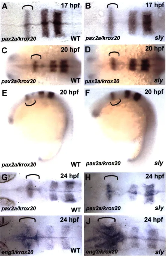
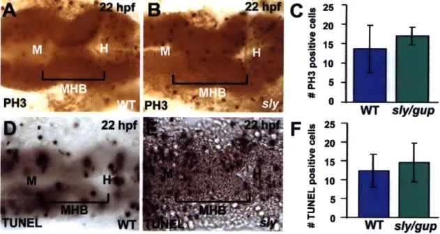
This work was completed in equal collaboration with Jennifer Gutzman, with help from Laura A. Lowery and Heidi S. Holley. Laura A. Lowery first identified sly mutants as lacking a midbrain-hindbrain boundary. Jennifer Gutzman performed the brightfield imaging, antibody staining, and time-lapse confocal microscopy. I characterized basal constriction by live confocal imaging in both wild type and mutant embryos and performed quantification of the phenotypes. I collaborated with Jennifer Gutzman to write the paper. Thanks to Nancy Hopkins and Adam Amsterdam for the guphilll3b
mutant line. This work has been published as:
Gutzman, J. H., Graeden, E. G., Lowery, L. A., Holley, H. S. and Sive, H. (2008). Formation of the zebrafish midbrain-hindbrain boundary constriction requires
laminin-dependent basal constriction. Mech Dev 125, 974-83.
ABSTRACT
The midbrain-hindbrain boundary (MHB) is a highly conserved fold in the vertebrate embryonic brain. We have termed the deepest point of this fold the MHB constriction (MH BC) and have begun to define the mechanisms by which it develops. In the zebrafish, the MHBC is formed soon after neural tube closure, concomitant with
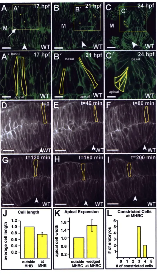
inflation of the brain ventricles. The MHBC is unusual, as it forms by bending the basal side of the neuroepithelium. At single cell resolution, we show that zebrafish MHBC formation involves two steps. The first is a shortening of MHB cells to approximately
75% of the length of surrounding cells. The second is basal constriction, and apical
expansion, of a small group of cells that contribute to the MHBC. In the absence of inflated brain ventricles, basal constriction still occurs, indicating that the MHBC is not formed as a passive consequence of ventricle inflation. In laminin mutants, basal
constriction does not occur, indicating a requirement for the basement membrane in this process. Apical expansion also fails to occur in laminin mutants, suggesting that apical expansion may be dependent on basal constriction. This study demonstrates laminin-dependent basal constriction as a previously undescribed molecular mechanism for brain morphogenesis.
INTRODUCTION
During development of the vertebrate brain, the neural tube assumes a complex structure that includes formation of the brain ventricles and the appearance of conserved folds and bends. These folds and bends delineate functional units of the brain and are likely to shape the brain such that it can pack into the skull. The midbrain-hindbrain boundary (MHB) is the site of one of the earliest bends in the developing brain. In the embryo, the MHB functions as an embryonic organizing center (Brand et al., 1996; Joyner, 1996; Puelles and Martinez-de-la-Torre, 1987; Sato et al., 2004) and later becomes the cerebellum and part of the tectum (Louvi et al., 2003). MHB tissue later
becomes the cerebellum and forms part of the midbrain roof plate, or tectum (Louvi et al., 2003). Patterning of the MHB begins long before neural tube closure (Joyner, 1996).
Subsequent to neural tube closure, and unlike the remainder of the brain, the tissue at the MHB remains apposed at the midline, shows lower levels of cell proliferation than surrounding neuroepithelium, and bends sharply to form the MHBC (Lowery and Sive,
2005). Loss of signaling factors that pattern the MHB results in failure to form the
MHBC (Brand et al., 1996; Sato et al., 2004). However, it is likely that these genes are responsible for specifying the fate of the tissue and, thus, affect the MHBC indirectly.
The brain neuroepithelium has characteristic apico-basal polarity, with apical cell surfaces facing the ventricular lumen, and the basal lamina, surrounding the tube under the basal cell surfaces. While junctions connect the apical and lateral surfaces of
epithelia, the basal surface of these cells is anchored on an extracellular matrix, the basal lamina. This polarity defines the axis of cell division within the epithelium (Fristrom,
1988), coordinates cell movements (Pilot and Lecuit, 2005), and provides spatial
orientation for the larger structural modifications of the entire sheet during extension and bending or folding (Schock and Perrimon, 2002).
The basal lamina plays a critical role in epithelial morphogenesis (Miner and Yurchenco, 2004). One of the major proteins in the basal lamina is laminin, a heterotrimeric protein that interacts with integrins to mediate adhesion of the basal lamina to the cytoskeleton of the overlying cells (Miner and Yurchenco, 2004). Injection of a function-blocking laminin antibody inhibits salivary gland morphogenesis in mice and neural tube closure, somite development, and heart tube formation in chick (Patel et al., 2006; Zagris et al., 2004). Laminin is also critical during mouse embryo
implantation and gastrulation (Miner et al., 2004) and, in zebrafish, is required for proper notochord formation and blood vessel formation (Parsons et al., 2002; Pollard et
al., 2006; Scott and Stemple, 2005; Stemple, 2002). A role for laminin in brain
morphogenesis has not been described.
Another contribution to epithelial morphogenesis is the fluid pressure found in organ lumens. For example, blood flow through the heart modifies the morphology of the atrial and ventricular lumens (Berdougo et al., 2003), and the flow of blood through the ventricles stimulates valve morphogenesis (Hove et al., 2003) while pressure overload can stimulate ventricle hypertrophy (Seidman and Seidman, 2001). During development
of the eye in chick, folds of the ciliary body form due to the hydrostatic pressure and swelling of the vitreous humor (Bard and Ross, 1982a; Bard and Ross, 1982b). During development of the brain, the embryonic cerebrospinal fluid (eCSF) inflates the brain ventricular lumen, and one possible function for the eCSF is to generate pressure in the brain ventricles that contributes to brain morphogenesis (Desmond and Levitan, 2002;
Lowery and Sive, 2005).
We have called the deepest point in the MHB the "midbrain-hindbrain boundary constriction" (MHBC). Here, we ask what processes are necessary for MHBC
morphogenesis, using the zebrafish as a model. In the zebrafish, the MHBC forms between 17 and 24 hours post fertilization (hpf), concomitant with formation of the brain
ventricles. At this stage of development, the neuroepithelium is a pseudostratified-columnar epithelium where apical cell surfaces face the brain ventricle lumen, and basal cell surfaces, on the outside of the tube, abut the basement membrane. Interestingly, the MHBC forms by bending the basal side of the neuroepithelium. This is unusual since essentially all epithelial bends have been described at the apical surface via apical constriction. Basal constriction has rarely been discussed in the literature and has not previously been studied as an independent mechanism of organogenesis. The
organization of the neuroepithelium and correlation with brain ventricle inflation led us to consider three factors that may drive MHBC morphogenesis: (1) fluid pressure on the inside of the neural tube as the brain ventricles inflate (Lowery and Sive, 2005), (2)
changes in cell shape during bending, and (3) interactions of the basal side of the neuroepithelium with the basement membrane.
We show here that MHBC morphogenesis involves two processes, cell shortening at the MHB and basal constriction of the neuroepithelial cells at the MHBC. Basal constriction is dependent upon laminin function, but not upon inflation of the brain ventricles. These data indicate that the MHBC forms through changes in cell shape, dependent on the extracellular matrix, which have not previously been described during brain morphogenesis.
EXPERIMENTAL PROCEDURES
Fish lines and Maintenance
Zebrafish lines were maintained and stages determined as previously described (Kimmel et al., 1995; Westerfield, 1995). Strains used include wild-type AB, slyms6 (Schier et al.,
1996), guphilll3b (Amsterdam et al., 2004), nokm227 (Malicki et al., 1996), and snkt273a
Live imaging
Brain ventricle imaging was carried out as previously described (Lowery and Sive, 2005). For confocal imaging, single cell embryos were micro-injected with CAAX-eGFP mRNA (memGFP) (kindly provided by J. B. Green, Dana-Farber Cancer Institute Boston, MA) transcribed with the mMessage mMachine kit (Ambion). The embryos were mounted inverted in 0.7% agarose (Sigma) and imaged by fluorescent, laser-scanning confocal microscopy (Zeiss LSM510) or with spinning disk confocal microscopy (Perkin Elmer Ultraview RS). Time-lapse data was analyzed using Imaris (Bitplane).
Quantification of cell length and apical cell width
Slices for measurement were chosen based on the ability to outline the entire extent of a cell from the apical to basal surface of the neuroepithelium and by following the cell through a full Z-series. The length of three cells at the MHBC and four cells outside the MHBC were measured using Imaris (Bitplane) software, and the ratio between cell lengths at and outside the MHBC were calculated for each embryo and averaged. The width of two wedge shaped cells at the MHBC and three unconstricted cells outside the MHBC at 24 hpf in each embryo were measured using Imaris (Bitplane). The error bars in Fig. 2 indicate the standard deviation between the ratios found for each embryo.
Immunohistochemistry
Embryos were fixed in 4% paraformaldehyde and dehydrated in methanol. After rehydrating in PBT, embryos were permeabilized with 2.5 mg/ml proteinase K for 1 minute, and blocked in PBT, o.1% Triton X, 1% BSA, and 1% NGS. Embryos were
incubated overnight at room temperature in laminin antibody (laminin rabbit mouse, Sigma L-9393, 1:150), washed, and incubated in secondary antibody, (goat