Coral calcification: insights from inorganic experiments and coral responses to environmental variables
By
Michael Holcomb B.S., University of Idaho, 2004
Submitted in partial fulfillment of the requirements for the degree of Doctor of Philosophy
at the
MASSACHUSETTS INSTITUTE OF TECHNOLOGY and the
WOODS HOLE OCEANOGRAPHIC INSTITUTION February 2010
@ 2010 Michael Holcomb. All rights reserved.
ARC HINES
MASSACHUSEMS INSTRE.OF TECHNOLOGY
MAY 0
5
2010
LIBRARIES
The author hereby grants MIT and WHOI permission to reproduce and to distribute publicly paper and electronic copies of this thesis document in whole or in part in any medium now known
or hereafter created.
Author... - . . . . ... .. ..- ..-.--....- . . .
-.--.-.-.-.--Joint Program in Oceanography/Applied Ocean Science and Engineering Department of Geology and Geophysics Massachusetts Institute of Technology and Woods Hole Oceanographic Institution December 7, 2009
Certified by...--...
Anne L. Cohen Research Specialist, Department of Geology and Geophysics, WHOI Thesis Co-Supervisor
Certified by...
Daniel C. McCorkle Associate Scientist, Department of Geology and Geophysics, WHOI
A ,Thesis Co-Supervisor
Accepted by...
Bradford H. Hager Professor of Earth, Atmospheric, and Planetary Sciences, MIT Chairman, Joint Committee for Geology and Geophysics
Coral calcification: insights from inorganic experiments and coral responses to environmental variables
By
Michael Holcomb
Submitted to the MIT/WHOI Joint Program in Oceanography/Applied Ocean Science and Engineering on November 18, 2009 in partial fulfillment of the requirements for the
degree of Doctor of Philosophy in Marine Geology
Abstract
The mechanisms involved in the formation of coral skeletons are examined using a laboratory model for coral calcification and the growth of living corals under different environmental conditions. Abiogenic aragonite was precipitated from seawater over a range of saturation states (Chapter 1). Abiogenic aragonite formed at high saturation state (Q>-20) had a granular appearance and was enriched in trace elements, similar to the crystals found within the centers of calcification and dark bands in coral skeletons. Abiogenic aragonite formed fibrous crystals at lower saturation states, similar to the crystals which radiate out from the centers of calcification. These similarities suggest that the internal calcifying environment of the coral experiences a wide range of saturation states. To estimate when periods of high or low saturation state occur within the coral, corals were stained to mark the skeleton deposited during specific time intervals (Chapter 2). Dark bands are shown to form between dusk and dawn. A conceptual model is proposed in which daytime saturation state is limited by the
availability of CO2 due to the uptake of CO2 by photosynthesis. To test the potential for photosynthesis to limit CO2 availability to calcification, corals were grown under
experimentally manipulated CO2 and nutrient levels. Elevated CO2 levels were found to
decrease calcification in zooxanthellate colonies of the coral Astrangia poculata, however the addition of inorganic nutrients reduced the negative impact of CO2 (Chapter 3), while reduced calcification rates were associated with elevated nutrients at ambient CO2 levels. Together these results suggest that nutrient availability may limit
photosynthesis under elevated pCO2 conditions, while at ambient conditions additional
stimulation of photosynthesis may limit the CO2 supply to calcification. To further test
the interaction of photosynthesis with calcification, the effects of nutrients, C0 2, and
temperature were tested on both zooxanthellate and azooxanthellate coral colonies (Chapter 4). No clear pattern of nutrient enhancement of photosynthesis or calcification was found. However, a pronounced gender difference was found in the effect of CO2 on calcification in spawning corals, with female corals being more sensitive to elevated CO2.
Abbreviated abstract
Coral calcification is examined using a laboratory model and living corals. In the laboratory model, abiogenic aragonite formed at high saturation state (Q>-20) had a granular appearance and was enriched in trace elements, similar to centers of calcification and dark bands in corals. Abiogenic aragonite formed fibrous crystals at lower saturation states, similar to crystals which radiate out from centers of calcification. These similarities suggest the calcifying environment of the coral experiences a range of saturation states. To estimate when high or low saturation states occur within the coral, living corals were stained, staining patterns suggest dark bands form between dusk and dawn. A model is proposed in which daytime saturation state is limited by the availability of CO2. To test the potential for photosynthesis to limit CO2 availability to
calcification, corals were grown under altered CO2 and nutrient levels. Elevated CO2 levels decreased calcification in zooxanthellate corals, however addition of nutrients reduced the negative impact of CO2. This suggests nutrient availability may limit
photosynthesis under elevated pCO2 conditions. The effects of nutrients, CO2, and
temperature were further tested on both zooxanthellate and azooxanthellate coral colonies. Unexpectedly, a gender difference was found in the effect of CO2 on calcification.
Thesis Supervisors:
Dr. Anne L. Cohen, Research Specialist, WHOI Dr. Daniel C. McCorkle, Associate Scientist, WHOI Defense Chair:
Dr. Laura Robinson, Assistant Scientist, WHOI Thesis Committee:
Dr. Tanja Bosak, Assistant Professor, MIT Dr. Glenn A. Gaetani, Associate Scientist, WHOI Dr. Scott Gallager, Associate Scientist, WHOI Dr. Ann M. Tarrant, Assistant Scientist, WHOI
3-Table of Contents
A b stract... 3 13 Acknowledgements... 7
In tro d uction ... ... ... ... 10
Chapter 1. Compositional and morphological features of aragonite precipitated
experimentally from seawater and biogenically by corals ... 16
Chapter 2. Timing of daily growth band formation in coral skeletons ... 62
Chapter 3. Long-term effects of nutrient and CO2 enrichment on the temperate
coral Astrangia poculata (Ellis and Solander, 1786)... 103
Chapter 4. Coral gender: an unexplored factor in coral calcification? . . . ... 138 Synthesis... 168 Appendices Appendix Al. Appendix A2. Appendix A3. Appendix A4. Appendix A5. Appendix A6.
Supplemental material for Chapter 1... ...
Supplemental experiments for Chapter 2... Additional data for Chapter 3... Additional data for Chapter 4... Nature paper based on Chapter 4...
Supplemental material for Appendix 5...
C5
i 177 196 213 215 260 266Acknowledgements
I must thank my family for allowing me the opportunity to explore my interest in corals
from an early age, for tolerating my numerous 'experiments', the flipped circuit breakers, corroded tools, and occasional floods. Ruby Brackett, Tom Cole, Paul Fischer, Joann Montgomery, and Rusty Taylor, all of whom helped to enrich my education by supporting extracurricular academic activities. Leroy and Sally Jo Headley for maintaining the Geothermal Aquaculture Research Foundation where I had the opportunity to volunteer, work with a wide range of corals in captivity, and overcome any fears I may have had of applying superglue to living corals. The University of Idaho and especially Rolf Ingermann and Joseph Cloud who gave me an incredible opportunity to conduct independent research as an undergraduate, who were extremely supportive, and from whom I learned a great deal - I don't think I could have found a better introduction
to research anywhere. The National Museum of Natural History, and my project advisors: John Pandolfi, Ann Budd, and Ian Macintyre who gave me my first introduction to coral paleo-proxies - particularly banding structures, trace element and isotopic compositions of the skeleton, which have been an interest ever since. The works of Peta Clode, William Fitt, Paola Furla, Joanie Kleypas, Alan Marshall, Francesca Marubini, Anders Meibom, and Lynne Whitehead which fueled my curiosity about coral calcification throughout my undergraduate education and continue to influence my research. The direction my research has taken over the last few years has in large part been shaped by my advisor, Anne Cohen, who has sent me in directions I would have never imagined my research taking when I first came in to the Joint Program. Little did I realize coming in that I would be spending winters diving in Woods Hole while ice floated on the surface of the water just to measure the winter time growth of a local coral, nor had I imagined that I would spend my time looking at crystal morphologies, and using crystal morphology to estimate saturation state within the calcifying environment of the coral and in turn trying to model saturation state variations within the calcifying environment. My advisor, Dan McCorkle also aided my work in ways I would have never imagined - who would have thought I'ld get a 3 month vacation from taking care of some 40 tanks of Astrangia poculata to go spend a winter on a tropical island off the coast of Australia, and doing so would actually be the suggestion of my advisor? If it weren't for Dan, it wouldn't have been possible to do studies in both locations at the same time. Dan also helped me to better understand and characterize carbonate chemistry, which has been critical for all of my projects. Both of my advisors have been very supportive of my work and have helped to improve it, especially the presentation of results. Thanks to the members of my thesis committee, Tanja Bosak, Glenn Gaetani, Ann Tarrant, and Scott Gallager have helped me over the past few years - Glenn helped greatly with inorganic aragonite studies, both with suggestions and use of his lab for the inorganic precipitation experiments and specimen preparation on which so much of my thesis is based, Ann has provided many valuable suggestions for my Astrangia work, and Scott has helped save me countless hours of data processing, made equipment available
to me, not to mention helping to keep ESL running. Everyone involved in keeping ESL running, especially Molly Jacobs, Fred Keller, Kevin Thompson, and Amber York, as well as the security guards (Rick, Bill, Joe, Linda, Rob, John, Gary, Bill, Scot, Norman) who handle many a late night call to Fred or Kevin to take care of repairs, as well providing rides at odd hours. The WHOI dive program and divers - Stace Beaulieu, Lars Behrendt, Michael Brosnahan, Rod Catanach, Diane DiMassa, Scott Gallager, Greg Gerbi, Laura Hmelo, Erich Horgan, Annette M. Hynes, Pat Lohmann, Larry Madin, Scott McCue, Matthew McIlvin, Kelton McMahon, Anna Michel, Christine Mingione, Emily Miller, Todd Morrison, Byron Pedler, Kelly Rakow, Casey Saenger, Peter Schultz, Ann Tarrant, Nan Trowbridge, Kristen Whalen, Sandy Williams, Amber York, and Terry Rioux who have all helped with my various Astrangia projects. The academic programs office for helping to guide me through the logistics of the Joint Program and for financial support. Anne Hoggett, Lyle Vail and everyone at the Lizard Island Research Station who helped to provide one of the most rewarding experiences of my graduate career and in particular Lance and Maryann Pearce, Adel Heenan, Harriet Salomonsen, Dan Bayley, and Monica Gagliano. To Rebecca Gast, Dawn Moran, Mark Dennett and David Beaudoin for helping me with my initial work isolating sequences for aquaporins and calcium binding proteins and for microscopy assistance. To Jeffrey Hutter and Shailesh Nene for helping me to get started with AFM. To Rinat Gabitov for my first introduction to carrying out inorganic precipitation experiments and for samples. And to many others who have allowed me access to equipment, provided training and advice, analyzed samples, given me computer code, checked on my corals, edited rough drafts or helped improve presentations, which include: Fern Gibbons, Janelle Homburg, Greg Hirth, Nobumichi Shimizu, Stan Hart, Scot Birdwhistell, Sheri White, Benjamin Van Mooy, Simon Thorrold, Tom Kleindist, Jason Smith, Jerzy Blusztajn, David Wellwood, Rebecca Belastock, Fred Sayles, Joanne Goudreau, Paul Henderson, Justin Ries, Sharon Lamont, Sonya Dyhrman, Karen Casciotti, Neal Cantin, Ed Boyle, Andrew McDonnell, Jonathan Blythe, Erin Banning, Joanna Gyory, Elizabeth Orchard, Kelton McMahon, Casey Saenger, Meredith White, Evelyn Mervine, Emily Roland, Sharon Hoffman, Louis Kerr, Christina McGraw, Tim McClure, David Housman, Katie Boissonneault, Philip Alatalo, Mark St. Pierre, Darlene Ketten, Summer Praetorius, William Martin, Scott Doney, David Glover, David Kulis, Joan Bernhard, Jerry McManus, Alyson Bodendorf, Hana Keys, Ellen Roosen, Rick Galat, Alfonso Mucci, Holly Wichman, LuAnn Scott, and Susan Humphris. The administrative staff who have helped with so many of the day-day tasks: Christina Cuellar, Maryanne Ferreira, Lynn Stellrecht, Kelly Servant, Suellen Garner. To Katrina Goudkamp, the Great Barrier Reef Marine Park Authority, and other employees of the Australian government for permitting assistance and being responsive to questions. To WHOI computer and information systems personnel who have helped out with so many computer glitches over the years. My office mates who were always there, whether it be 6am or lam, who created many an original song and who proved to me that even I can keep a plant alive, my fellow Joint Program students, my neighbors Ruth and Ryan and the 2004 MIT Biology cohort who provided endless diversions. To Carol Brackett for being there, or not there, and for, well, putting up with me. To all my friends and colleagues who have all helped to enrich my time here. And most
importantly to Astrangia poculata a wonderful, amazing, and utterly confusing coral which has kept me busy for many years and will no doubt occupy my thoughts for years to come.
Funding for this research was provided by a National Science Foundation Graduate Research Fellowship, Academic programs, a MIT Presidential Fellowship, WHOI Interdisciplinary Award #39040300, a WHOI Coastal Ocean Institute Award, the WHOI Ocean Life Institute, National Science Foundation grant #OCE-0648157, a Lizard Island Doctoral Fellowship, and an International Society for Reef Studies/Ocean Conservancy Doctoral Fellowship.
Coral calcification: insights from inorganic experiments and coral responses to environmental variables
Introduction
Despite decades of research, the role of symbiosis in coral calcification remains elusive. It is well established that calcification rates increase in zooxanthellate corals during
periods in which photosynthesis is occurring (Barnes and Chalker 1990). Comparisons of facultatively symbiotic corals with and without zooxanthellae similarly show increased calcification in the presence of symbionts (Jacques et al., 1983). Yet, comparisons of zooxanthellate corals to azooxanthellate corals show that some azooxanthellate corals can grow just as rapidly as zooxanthellate corals, bleached corals are known to continue calcifying for some time at rates similar to those prior to bleaching, and in corals such as
acroporids, the fastest growing regions are largely azooxanthellate (Gladfelter 1982; Marshall 1996; Mortensen and Rapp 1998; Rodrigues and Grottoli 2006). Thus, it is not clear that symbionts per se enhance calcification. There are several mechanisms which could be responsible for the increase in calcification often associated with zooxanthellae, ranging from changes in the synthesis of organic matrix components to changes in carbonate chemistry (e.g., Gattuso et al., 1999); here the focus will be on carbonate chemistry and saturation state. Calcification could increase due to photosynthate
supplied by the zooxanthellae being used to fuel the transport of Ca to the skeleton or protons away from the site of calcification (Goreau 1961), or due to the drawdown in
CO2 as a direct result of photosynthesis, leading to increased pH, shifting the dominate CO2species from bicarbonate to carbonate ion and increasing the saturation state
(Kawaguti and Sakumoto 1948). But, zooxanthellae may also limit calcification by competing for CO2, thus leading to a situation in which increased pH does not correspond
to increased carbonate ion (Marubini and Davies 1996; Langdon and Atkinson 2005). To better understand different aspects of how zooxanthellae influence calcification, four projects were conducted to examine various aspects of the calcification process.
In Chapter 1 abiogenic aragonite precipitates formed under monitored conditions are compared with coral skeletons. The morphology and composition of the abiogenic precipitates are used to infer potential conditions under which the coral skeleton forms
(Holcomb et al., 2009). Abiogenic aragonite precipitates from seawater as spherulites, with clusters of submicron granular materials occupying their centers and elongate (fibrous) needles radiating out to the edge. In each spherulite, the granular material formed first, at the highest fluid saturation state, whereas subsequent needle growth occurred at lower saturation state. The morphology of individual fibers also changes with saturation state, with fibers becoming thinner with increasing saturation state. Coral skeletons are shown to share the same crystal morphologies seen in abiogenic
precipitates, with fine granular materials occupying centers of calcification and 'dark' bands which transect fibers, and fibrous aragonite radiating out from the centers of calcification. In addition to sharing morphological similarities, the elemental
composition (Mg/Ca and Ba/Ca ratios) is shown to have similar patterns in both abiogenic and coral aragonite, with granular regions being enriched in trace elements relative to fibers.
Based on the similar morphologies and compositions of centers of calcification and dark bands in corals, and centers and bands in abiogenic aragonite grains, I propose that granular crystal formation in corals is a result of high saturation state and that 'daily' banding patterns in corals reflect a cyclic change in saturation state.
In Chapter 2 skeletal growth on diurnal cycles is examined to (1) establish the timing of 'daily' band formation, and the relationship between band formation and photosynthesis in zooxanthellate corals and (2) to test the hypothesis that cyclic changes in saturation state could cause the observed banding patterns in corals. Calcium binding dyes were introduced to the seawater surrounding a coral to mark the skeleton formed while the dye was present. Patterns of dye incorporation suggest that crystal nucleation and growth have a diurnal cycle, with nucleation (dark band formation) occurring between dusk and dawn. In conjunction with skeletal extension data (Barnes and Crossland 1980; Vago et al., 1997), staining between dusk and dawn indicates that dark bands form near dusk, coinciding with maximal skeletal extension rates. Together with the abiogenic
precipitation experiments (Chapter 1), this implies a high saturation state near dusk, to drive crystal nucleation. These observations can be explained by a conceptual model of coral calcification in which CO2 is limiting at the site of calcification during the day, and
a non-carbonate buffer system in the calcifying environment allows a CO2 influx,
associated with a decline in photosynthesis near dusk, to generate an increase in saturation state and thus drive crystal nucleation.
In Chapter 3 the effects of CO2 and nutrients on the facultatively symbiotic coral
Astrangia poculata are investigated. In zooxanthellate colonies, calcification is
adversely affected by elevated CO2, but the addition of inorganic nutrients can reduce the
impact of CO2 on calcification. This suggests that under elevated CO2 conditions,
zooxanthellae can become nutrient limited, and thus less efficient at drawing down internal [CO2]aq. In this situation, adding inorganic nutrients could enable zooxanthellae to utilize more CO2, this enhanced photosynthesis may reduce [CO2]aq, and supply energy
or alkalinity to help drive calcification.
In Chapter 4, the effects of CO2, nutrients, and temperature on the facultatively symbiotic coral Astrangia poculata are examined. No positive effect of nutrient addition was found
for corals under elevated pCO2, potentially reflecting a lack of nutrient limitation of the
zooxanthellae due to high ambient nutrient levels. In Chapter 3, ambient nutrient levels were 3±5 pM NH4, 0.8±0.7 pM NO3, 0.5±0.3 pM P04, while for the treatment phase of
Chapter 4, ambient nutrient levels were 1±0.4 pM NH4, 4±0.4 pM NO3, 0.6±0.1 pM P04
for the 240C tank, thus nitrate is consistently higher during the treatment phase of Chapter 4. Phosphate though slightly higher, also shows greater variability in Chapter 3,
reflecting intervals with low and high phosphate concentrations, so the apparent positive effect of nutrients under CO2 enriched conditions found in Chapter 3 may be driven by
intervals in which ambient nutrient levels are low enough to be limiting. In Chapter 4, coral calcification generally decreased in response to CO2, but the pattern differed greatly
more sensitive to CO2 than males. This suggests a greater cost for egg production relative to sperm production which takes resources away from calcification, potentially limiting energy availability for calcification. If this pattern applies to gonochoric coral species under natural conditions, rising atmospheric CO2 levels could lead to a greater
reduction in the growth rates of female corals, making them more vulnerable to disturbance events. With successive disturbance events and reduced recovery rates of female corals, gender ratios could shift toward male dominated reefs, potentially reducing rates of sexual reproduction, making corals more vulnerable to ocean acidification than initial changes in calcification rates would suggest.
References:
Barnes DJ, Chalker BE (1990) Calcification and photosynthesis in reef building corals and algae. in coral reefs ed Z Dubinsky. 109-131
Barnes DJ, Crossland CJ (1980) Diurnal and seasonal variations in the growth of a staghorn coral measured by time-lapse photography. Limnol. Oceanography 25:
1113-1117
Cohen AL, McConnaughey TA (2003) Geochemical perspectives on coral
mineralization. Reviews in Mineralogy Geochem: Biomineralization 54: 151-187 Gattuso JP, Allemand D, Frankignoulle M (1999) Photosynthesis and calcification at
cellular, organismal and community levels in coral reefs: a review of interactions and control by carbonate chemistry. American Zoologist 39: 160-183
Gladfelter EH (1983) Skeletal development in Acropora cervicornis II. diel patterns of calcium carbonate accretion. Coral Reefs 2: 91-100
Goreau TF (1961) On the relation of calcification to primary productivity in reef building organisms. In The biology of hydra and some other coelenterates: Lenhoff, H.M., Loomis, W.H. Ed pp. 269-285
Jacques TG, Marshall N, Pilson MEQ (1983) Experimental ecology of the temperate scleractinian coral Astrangia danae II. Effect of temperature, light intensity and symbiosis with zooxanthellae on metabolic rate and calcification. Marine Biol.
76: 135-148
Kawaguti S, Sakumoto D (1948) The effect of light on the calcium deposition of corals. Bull. Oceanograpaical Inst. Taiwan: 65-70
Kleypas JA, Buddemeier RW, Archer D, Gattuso JP, Langdon C, Opdyke BN (1999) Geochemical consequences of increased atmospheric carbon dioxide on coral reefs. Science 284: 118-120
Langdon C, Atkinson MJ (2005) Effect of elevated pCO2 on photosynthesis and calcification of corals and interactions with seasonal change in
temperature/irradiance and nutrient enrichment. J. Geophys. Res. Ocean Marshall AT (1996) Calcification rates in corals. Science 274: 117-118
Marubini F, Davies PS (1996) Nitrate increases zooxanthellae population density and reduces skeletogenesis in corals. Marine Biol. 127: 319-328
Mortensen PB, Rapp HT (1998) Oxygen and carbon isotope ratios related to growth line patterns in skeletons of Lophelia pertusa (L) (Anthozoa, scleractinia):
implications for determination of linear extension rates. Sarsia 83: 433-446 Muscatine L (1990) The role of symbiotic algae in carbon and energy flux in reef corals.
in coral reefs ed Z Dubinsky. 75-87
Risk MJ, Pearce TH (1992) Interference imaging of daily growth bands in massive corals. Nature 358: 572-573
Rodrigues LJ, Grottoli AG (2006) Calcification rate and the stable carbon, oxygen, and nitrogen isotopes in the skeleton, host tissue, and zooxanthellae of bleached and recovering Hawaiian corals. Geochim. Cosmochim. Acta 70: 2781-2789
Vago R, Gill E, Collingwood JC (1997) Laser measurements of coral growth. Nature
Chapter 1: Compositional and morphological features of aragonite precipitated
experimentally from seawater and biogenically by corals
ABSTRACT
The morphology and composition of abiogenic (synthetic) aragonites precipitated experimentally from seawater and the aragonite accreted by scleractinian corals were characterized at the micron scale. The synthetic aragonites precipitated from
supersaturated seawater solutions as spherulites, typically 20-100 jtm in diameter, with clusters of submicron granular materials occupying their centers and elongate (fibrous) needles radiating out to the edge. Using Sr isotope spikes, the formation of the central granular material was shown to be associated with high fluid pH and saturation state whereas needle growth occurred at lower pH and saturation state. The granular regions have significantly higher Mg/Ca and Ba/Ca ratios than the surrounding fibers. Sr
Two types of crystals are identified in the coral skeleton: sub-micron granular material and elongate (fibrous) crystals that radiate out from the granular regions. Granular materials are found in "centers of calcification" and in fine bands that transect the fibers. They have significantly higher Mg/Ca and Ba/Ca ratios than the surrounding fibers.
The observed relationship between seawater saturation state and crystal morphology and composition in the synthetic aragonites was used as a framework to interpret observations of the coral skeleton. We propose that coral skeletal growth can be viewed as a cyclical process driven by changes in the saturation state of the coral's
calcifying fluids. When saturation state is high, granular crystals precipitate at the tips of the existing skeletal elements forming the centers of calcification. As the saturation state decreases, aragonitic fibers grow, radiating out from the centers of calcification.
Published as: M. Holcomb, A.L. Cohen, R.I. Gabitov and J.L. Hutter, Compositional and morphological features of aragonite precipitated experimentally from seawater and biogenically
by corals. Geochimica et Cosmochimica Acta. v. 73, p. 4166-4179, 2009.
1. INTRODUCTION
Early studies of the ultrastructure of the scleractinian coral skeleton identified two distinct structures: the centers of calcification which appear as dark spots in petrographic thin-section, and the fibrous crystals which radiate out from the centers (e.g., Ogilvie,
1896; Vaughan and Wells, 1943; Wells, 1956). The centers of calcification and their
bundles of fibers are called sclerodermites and are considered to be the basic building blocks of the skeleton (Wells, 1956). Bryan and Hill (1941) noted the striking similarity between the spherulitic morphology observed in a range of mineral systems, and coral sclerodermites. Both exhibit fibrous crystals radiating from a common center.
Spherulitic growth in mineral systems is associated with diffusion-controlled growth from highly supersaturated solutions (Keith and Padden, 1963; Chernov, 1984). Such observations led Barnes (1970) and Constantz (1986), among others, to describe the precipitation of aragonite by scleractinian corals as a process analogous to crystal growth from highly supersaturated solutions.
Over the past few decades, a range of imaging and analytical techniques have been employed to examine coral skeletons. SEM imaging of materials occupying the centers of calcification showed that these are morphologically distinct from the surrounding fibers. The materials at the centers of calcification have been variously described as small, nano-crystals, very fine, or granular (e.g., Wainwright, 1964; Constantz, 1986; Cohen et al., 2001; Clode and Marshall, 2003). Selective analyses of centers of calcification and adjacent fibers indicate that centers of calcification are also
compositionally distinct. Several elements, notably Mg, Sr, S, Ba, and N, are enriched in the centers of calcification (e.g., Cuif et al., 2003; Gagnon et al., 2007; Meibom et al., 2004, 2006, 2007) and several isotope ratios, such as 613C, SIO, and 6"B, are depleted
in centers of calcification (e.g., Adkins et al., 2003; Rollion-Bard et al., 2003; Meibom et al., 2006; Blamart et al., 2007) relative to the surrounding fibers. These findings have been used to support the hypothesis that centers of calcification are formed by a process distinct from that responsible for the formation of fibers (e.g., Meibom et al., 2006) and, thus, the formation of aragonite by corals is a process distinct from the inorganic
precipitation of aragonite from a highly supersaturated solution. Lacking from these studies, however, is a comparison between coral aragonite and synthetic aragonite precipitated from a highly supersaturated solution.
Here, results from such a study are presented. A range of imaging techniques: light microscopy, fluorescence microscopy, scanning electron microscopy (SEM) and atomic force microscopy (AFM), as well as elemental measurements using secondary ion mass spectrometry (SIMS) were employed to characterize the morphology and chemistry of synthetic (i.e. precipitated experimentally from seawater) and biogenic aragonites (i.e. precipitated by tropical corals).
2. METHODS 2.1. Synthetic Aragonite Precipitates
Techniques employed for precipitating synthetic aragonites from seawater were adopted from Kinsman and Holland (1969), as modified by Gaetani and Cohen (2006).
The details for some of the experiments employed in this study differ slightly, so the specifics of each experiment are briefly described below.
Aragonite was precipitated from 0.45 pm filtered Vineyard Sound (Woods Hole, MA, USA) seawater (salinity 30.8-32.1). A PTFE or PET beaker containing 600 ml of filtered seawater was placed into a Lauda RE-106 isothermal bath, and stirred
continuously with a PTFE stirrer. Note that initial experiments showed no effect of the plastic type on the development of a run, so later runs used transparent PET which allowed the onset of crystallization to be more readily assessed. Several different experiments were conducted, each representing one of three categories of experiments, described below.
2.1.1. Timing of Precipitate Formation
To establish the relationship between fluid chemistry, crystal morphology and crystal composition, experiment 1 used a pair of Sr isotope spikes to constrain the timing of precipitation. In experiment 1, seawater was held at 25 0C in a PTFE beaker and
stirred continuously at 40 rpm. Evaporated seawater and a 0.04 M Na2CO3 (Alfa Aesar)
solution were introduced simultaneously at a rate of 4.8 ml h-1 for 37.5 hours.
Evaporated seawater was prepared by placing seawater in a PTFE beaker held in a 800C
water bath (for some experiments, concentrated seawater was prepared in a
polypropylene beaker held at 600C - similar results were obtained with both methods) until half the mass had been lost. The evaporated seawater was used to maintain the salinity at a roughly constant value as the 0.04 M Na2CO3solution was added (initial
salinity 31.85, final salinity 31.88). During this experiment, strontium isotope spikes ( 8Sr and 86Sr) were introduced at separate times into the seawater mixture. These spikes are incorporated into the growing crystals and serve as a marker from which the timing and rate of crystal growth can be determined. After the first 65 ml were added, 100 P1 of a 84Sr solution was added to nearly double the 84Sr concentration. After the final Na2CO3 addition, 100 pl of 86Sr was added to approximately double the 86Sr concentration.
Precipitation of aragonite was allowed to proceed for an additional 24 hours after pumping stopped. Precipitates were separated from solution by filtration through a 0.7 pm glass fiber filter and rinsed briefly with distilled water and ethanol. Sr isotopes were purchased from Oak Ridge National Lab.
2.1.2. Cyclic Saturation States
In experiments 2 and 3, the saturation state was cycled over the course of each run
by pulsed addition of Na2CO3 in an attempt to produce precipitates with alternating
regions of growth formed at high and low saturation states (bands). Experiment 2 was conducted under variable salinity conditions at either 55 C or 65 0C. A 0.01 M Na
2CO3
solution was added to the beaker of seawater in two 125 ml steps, 1 day apart, at a rate of -1ml min- and the solution was stirred at 120 rpm. This experiment is described in detail by Gaetani and Cohen (2006). In Experiment 3, a -0.04 M Na2CO3 solution and
evaporated seawater were added in two steps to seawater (initial salinity 31.3, final 32.1) held in a PET beaker in a 25'C water bath. The solution was stirred continuously at 120 rpm. 84Sr, 86Sr and 137Ba isotope spikes were added at different times over the course of
each run. Pumping durations, volumes, times of isotope spike addition and solution chemistry are given in the electronic annex Table EA1, Fig. EA1 and EA2.
2.1.3. Effect of Pumping Rate on Morphology
To examine the effects of saturation state on the morphology of precipitates, experiments 4, 5, 6 and 7 were conducted using different pumping rates to generate different 'steady state' conditions under which the bulk of the precipitate formed in each experiment. In experiments 4, 5 and 6, a syringe pump was used to continuously add a
-0.04M Na2CO3 solution and evaporated seawater to seawater in a PTFE beaker (final
salinities ranged from 31.4-32.8). Experiments 4-6 were conducted at 25 C while
stirring at 120rpm. Pumping rates for the bulk of each experiment were: high = 360 ml h~
for experiment 4, intermediate =2 ml h-1 for experiment 5, and low = 0.2 ml h-1 for experiment 6. Injection volumes ranged from 76 to 181ml. Aragonite seed crystals (fish
otoliths, cleaned by sonication and ground to 5-300 pm) were added to experiment 6 in an effort to speed nucleation. Precipitation was allowed to continue for 21-35 h after pumping stopped.
In experiment 7, a -0.04 M Na2CO3 solution and evaporated seawater were added
continuously at a rate of 0.4 ml/hr to seawater (initial salinity 31.3, final 33.5) held in a PET beaker in a 25C water bath. The solution was stirred continuously at 120 rpm. Isotope spikes (8 Sr, 16Sr, and 13'Ba) were added at different times over the course of the run. Pumping durations, volumes, times of isotope spike addition and solution chemistry
2.1.4. Growth Conditions of the Synthetic Aragonites
Solution pH (NBS scale) was monitored throughout aragonite synthesis
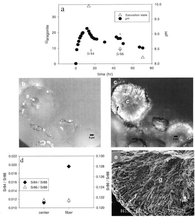
experiments 1, 3, 4, 5, 6, and 7 (Table 1, Fig. Ia, electronic annex Table 1, Fig.
EA-1). Saturation state was also determined periodically by measuring alkalinity and
calcium concentration, data are presented in Fig. la, Table EA-i and Figure EA-1. Solution pH increased as Na2CO3 was added until nucleation occurred. Following
nucleation, solution pH fell to a quasi-steady state value related to pumping rate (Fig.
EA-1, EA-2). At the end of each experiment, when pumping of the Na2CO3 solution
ceased, the pH of each experimental solution dropped but remained above the initial value (-8.0). Saturation states follow a trend similar to that seen in pH data (Table.
EA-1). Solution chemistry for some of the runs and starting materials are provided in Table EA-1. Raman spectra of the precipitates reveal a peak at -705 cm- , consistent with the
presence of aragonite (e.g., Clarkson et al., 1992). Raman data are presented in Fig.
EA-3.
2.2. Coral Samples
The skeletons of three coral species Diploria labyrinthiformis (brain coral),
Porites lutea and Porites solida were examined. D. labyrinthiformis (sample #BER002)
was collected live from John Smith's Bay, Bermuda at a depth of 13 m (Cohen et al.,
2004). Porites lutea (sample #JA4) was collected live from Johnston Atoll, north central
David Barnes (AIMS). Porites solida was collected live in June 1989 from the Great Barrier Reef, Australia (151'E, 21'S) (Cohen and Hart, 1997).
2.3. Polishing of Synthetic and Coral Aragonites
All samples were placed inside 2.5 cm Al rings and imbedded in Epo-thin epoxy (Buehler) prior to polishing. Double sided adhesive tape (Buehler) was used to hold specimens and rings in place as epoxy was added. Corals were prepared by breaking -1 cm2 sections of coral skeleton, roughly parallel to the axis of growth, and placing the
sections on the adhesive. Synthetic aragonites were mounted by applying a light dusting of grains to the adhesive. Polishing was done on nylon cloths with alumina (Mark V Laboratories) suspended in heptane (Alfa Aesar), except for the coral used for light
microscopy and SIMS analysis, which was prepared commercially (OMNI Laboratories).
A small amount of water was used to initially apply the alumina grit. Each specimen was
polished through a range of grit sizes down to 0.3 ptm, or 0.05 pm for specimens imaged with Atomic Force Microscopy. Following mechanical polishing, a final polish with 0.02 pm colloidal silica was applied to all samples, with the exception of those used in the acridine staining experiments (see below). Colloidal silica is a water-based polishing compound, hence there is dissolution of the aragonite in addition to mechanical abrasion. An initial comparison of different techniques revealed that a colloidal silica etch on the synthetic grains gave good AFM images (Fig. EA-4). In addition to the dissolution induced by the colloidal silica, corals were etched for 30-50 s prior to AFM imaging using 0.1% formic acid and 3% gluteraldehyde in water (Cuif et al., 2003). Etching was
stopped by rinsing with water. This additional etching step improved contrast between features in coral samples, as observed in preliminary comparisons of different techniques
(Fig. EA-5). Samples for SEM imaging were either mounted on SEM stubs without any
further preparation, or were polished as above and etched for 10s with 0. IN HCI.
2.4. Imaging:
2.4.1. Light Microscopy and Scanning Electron Microscopy (SEM)
Polished sections of corals and synthetic aragonites were examined in reflected and transmitted light with a Nikon Eclipse E 600 Polarizing microscope equipped with a Spot Insight color CCD camera. SEM imaging of Au/Pd coated samples (both polished and unpolished) was done using a JEOL 840 scanning electron microscope. Accelerating voltage was 15kV unless otherwise specified.
2.4.2. Acridine Orange Staining
Acridine orange staining of polished sections of corals and synthetic aragonites was carried out following the protocol of Stolarski (2003). Acridine orange is a dye used to stain coral skeletons, and regions retaining acridine orange are thought to have high concentrations of organic materials. Corals were etched prior to staining by placing samples in distilled water overnight to lightly etch the surface. Samples were overlain with a 1% acridine orange (Alfa Aesar) aqueous solution, allowed to sit for 5 minutes, briefly rinsed, blotted dry and imaged on a Zeiss Axiovert inverted microscope using a mercury vapor UV source and FITC short pass filter set. Images were captured using either a Sony color CCD camera or a Canon Digital Rebel XTi camera.
2.4.3. Atomic Force Microscopy (AFM)
AFM imaging (which measures small changes in the height of a surface) of
polished sections of corals and synthetic aragonites was conducted at The University of Western Ontario using a Veeco MultiMode AFM and at Woods Hole Oceanographic Institution using a Veeco Dimension 3100 AFM equipped with silicon nitride tips (Veeco
NP-S, with a -10 nm radius of curvature, and BudgetSensors, with a -20 nm radius of
curvature, respectively). All images were acquired in contact mode. Image capture and processing were performed using Digital Instruments NanoScope software.
2.5. Secondary Ion Mass Spectrometry (SIMS ion microprobe)
Metal (M)/Ca (Mg/Ca, Sr/Ca, and Ba/Ca) ratios of the coral Porites lutea and synthetic aragonite formed in experiment 1 were analyzed with a Cameca 3f Ion
Microprobe. Following a 3-minute pre-burn to remove the gold coating, a single spot on the coral sample was illuminated with the primary ion beam while measuring secondary ion intensities for 24Mg, 88Sr, 138Ba and 42Ca (Gaetani and Cohen, 2006). A 2.5 nA
0-primary ion beam, -10 pm in diameter, was accelerated at 12.7 keV. Secondary ion intensities were measured using a -80 eV offset from the peak of the energy distribution. This energy filtering reduces molecular interferences to <0.1% (Hart and Cohen, 1996).
In addition to the M/Ca ratios, 84Sr/"Sr and 86Sr/"Sr ratios were also determined in the synthetic aragonite grains in order to locate the isotope spikes. Individual synthetic
aragonite grains were targeted with a 4 nA 0- primary ion beam, -7 pm in diameter, accelerated at 12.7 keV. Secondary ion intensities (24 Mg, 84Sr, Sr, 8Sr, 138Ba and 44Ca)
intensity ratios were converted to molar ratios using the carbonatite standard OKA assuming an intercept of zero and a linear response. OKA was assumed to be
homogeneous with a Mg/Ca ratio of 4.47 mmol/mol, a Sr/Ca ratio of 19.3 mmol/mol and a Ba/Ca ratio of 1.61 mmol/mol (Gaetani and Cohen, 2006), isotopic abundances were assumed to be similar for both OKA and aragonite. At least eight measurements of the OKA standard were made each day samples were measured, average intensity ratios measured at the time of coral data collection were: 0.202, 2.62 and 0.16 for 24Mg/42 Ca, 88Sr/42 Ca and 18Ba/42 Ca respectively; at the time of synthetic aragonite measurements, values were: 0.00 18, 0.014 and 0.00072 for 24Mg/4 Ca, 8Sr/4 Ca and 138Ba/40Ca
respectively. In all cases, standard errors were less 3%. Means were compared using a t-test (Zar, 1984).
3. RESULTS 3.1. Synthetic Spherulites
The individual grains formed in experiment I are roughly circular; in polarized light, the center (c) of each grain is distinguished by a dark region (Fig Ic). Radiating out from this center are aragonite fibers (f). The morphology of the grains is consistent with the spherulitic morphology found in a range of minerals i.e. a radially disposed array of acicular crystals that emerge from a common center or nucleation region (e.g., Cross,
1891; Iddings 1891), typical of crystals formed rapidly from a supersaturated or
supercooled solution (Lofgren, 1971; Sunagawa, 1987; Lowenstam and Weiner, 1989). Using SEM (Fig le), it can be seen that the material found in the center is sub-micron in
size and has a granular (g) appearance, while the fibers (f) radiating out from the center are -1 m wide and several microns long.
3.1.1. Timing of Growth
The Sr isotope ratios in the center of each grain formed during experiment 1 reflect natural abundances (Fig. Id), indicating that the centers formed prior to the addition of the 84Sr isotope spike. The 84Sr spike is present in the fibrous aragonite between the centers and the edge of the spherulite, indicating that fibers formed after the centers but prior to the addition of 86Sr (Fig. Id). High 86Sr was found only at the edges of a few spherulites, indicating that very little aragonite precipitated following the addition of 86Sr (data not shown).
The presence of spikes allows the crystal morphology to be correlated with the solution chemistry. In experiment 1, solution pH increased as Na2CO3 was added until
nucleation occurred (Fig. la). The granular centers of the aragonite grains formed during this high pH period (pH -9.2), as indicated by the absence of the 84Sr spike. Following nucleation, solution pH fell to a quasi-steady state value related to pumping rate. Fibrous aragonite grew during this period when pH was -8.9, as indicated by the presence of the
84Sr but absence of 86Sr spikes (Fig. 1b, c and d). Very little aragonite was deposited near
the end of the experiment after pumping stopped when pH was -8.6, as indicated by the scarcity of elevated 86Sr.
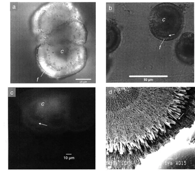
Fig. 2a represents a cross section through a composite of three synthetic aragonite
grains formed in experiment 2 at 65'C. Fig. 2b shows a cross section through a single synthetic aragonite grain formed in experiment 3 at 25'C. The two pumping cycles used in each of these experiments lead to two cycles of high and low saturation state over the course of aragonite precipitation. The synthetic aragonite grains precipitated in these experiments are similar to those in Fig. 1c, but an additional feature, a single dark band running perpendicular to the axis of fiber growth, is present (Fig. 2a,b). In these
spherulites, the dark band is located -15 pm from the outer edge of each grain formed in experiment 2, and -4 pm from the edge for grains formed in experiment 3.
Fig. 2c shows a fluorescence image of a synthetic aragonite grain, grown in
experiment 2 by cyclic pumping, stained with acridine orange. Addition of acridine orange results in increased fluorescence associated with the center (c) and dark bands (arrow) observed in light microscopy.
Fig. 2d shows an SEM image of a polished, HCl etched aragonite grain formed in experiment 3 by cyclic pumping. The central region is composed of granular to finely fibrous material. Fine fibers radiate out from the center and become larger till reaching the band (arrow). Following the band, fibers again radiate out to the edge of the
aragonite grain.
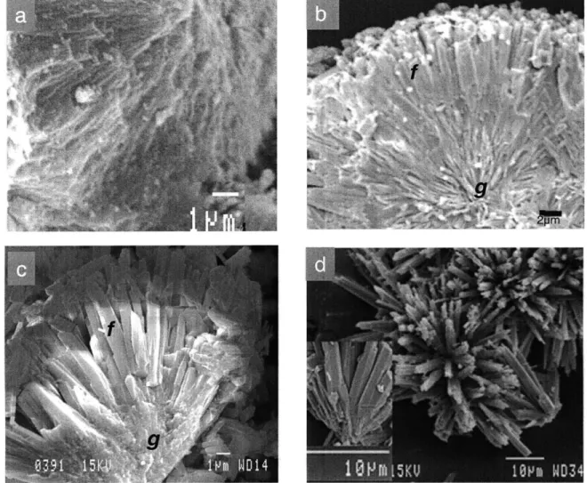
3.1.3 SEM Imaging
SEM images of synthetic aragonites precipitated in experiment 4 (Fig. 3a), 5 (Fig 3b), 6 (seeded, Fig. 3d) and 7 (Fig. 3c) at high (Fig. 3a), medium (Fig. 3b), and low (Fig.
3c,d) pumping rates reveal a systematic change in crystal morphology with pumping rate, and thus pH (Fig. EA 1,2). Synthetic aragonite grains formed in the high pumping rate run (-pH 9.5) lack well-defined crystals, and are composed of very fine fibers (Fig. 3a). Synthetic grains grown at intermediate pumping rates (pH -8.8) are composed of well-defined aragonite blades, 1-2 pm wide and several microns long and there are clear grain boundaries between individual fibers (Fig. 3b). Synthetic aragonites precipitated in the
low pumping rate experiments (pH -8.2 for Fig. 3c, -8.3 for Fig. 3d) are composed of
broad (-2 pm wide), highly faceted fibers that are widely separated (Fig. 3c,d). The fibers emerge from a common center, as shown in Fig. 3c and the inset in Fig. 3d.
3.1.4. AFM Imaging
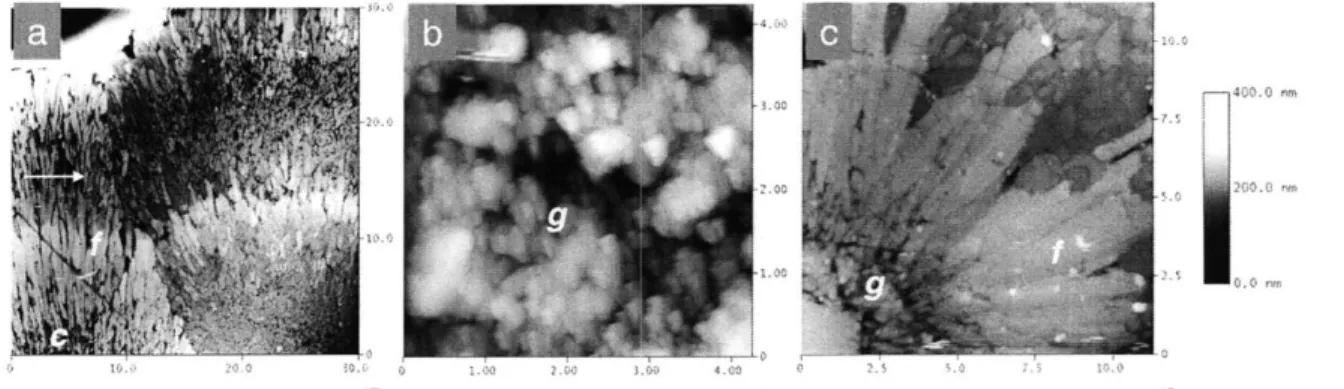
AFM height images of synthetic aragonite grains etched with colloidal silica show similar changes in crystal morphology with pumping rate to that seen with SEM (Fig. 4). In all images, height is on a scale of 0-400nm, with the highest regions shown as white,
lowest as black. The length-scale, in microns, is shown on the x and y axes of each image.
In Fig. 4a, an AFM image of two synthetic aragonite grains precipitated with two stepped additions of sodium carbonate (experiment 2) is shown. The grains have roughly circular centers (c) (only partly visible at the base of the image) surrounded by two layers
of fibrous aragonite (f). A darker region of granular material, -10pLm wide (arrow),
separates the inner and outer fibrous layers. Granular materials are sub-micron in size, while fibers are micron scale features - typically 8pim long, and 0.7[m wide.
Fig. 4b shows a synthetic aragonite formed in a high pumping rate run (pH -9.5,
Exp. 4). The entire surface of the grain is rough and granular (g), lacking defined fibers. In Fig. 4c, a synthetic aragonite formed in a low pumping rate (pH -8.3, Exp. 6) run is imaged. Granular material (g) is restricted to near the center of the grain. The bulk of the grain is made up of broad well-defined fibers
(f)
that emerge from the granular materialsand radiate outward to the edge (Fig 4c).
3.1.5. M/Ca Ratios
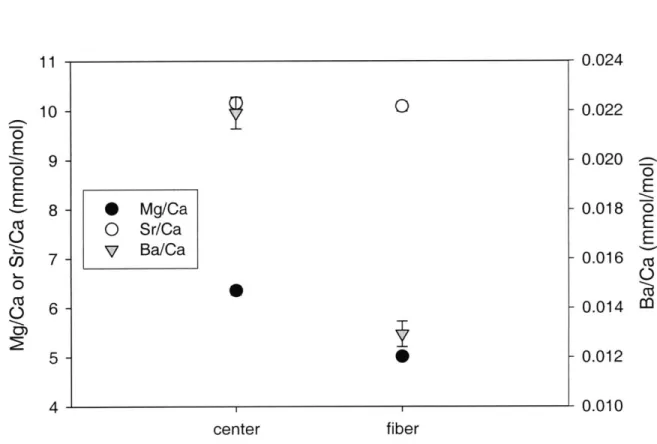
Mg/Ca, Sr/Ca, and Ba/Ca ratios in centers and fibers of synthetic aragonites grown in experiment 1 were measured by SIMS ion microprobe (Fig. 5). Discrete analyses of centers of synthetic grains were possible with the 7 Pm diameter analytical spot. Similarly, discrete analyses of aragonite fibers emerging from the centers of synthetic aragonite grains were made. The Mg/Ca and Ba/Ca ratios are higher in the centers of the grains (6.35 ±0.09 and 0.022 ±0.001 mmol/mol respectively) than in the surrounding fibers (5.01 ±0.07 and 0.013 ±0.001 mmol/mol respectively) (p<0.01). Sr/Ca ratios show no significant difference between centers (10.2 ±0.1 mmol/mol) and fibers (10.1 ±0.1 mmol/mol).
3.2. Coral aragonite
3.2.1. Light Microscopy
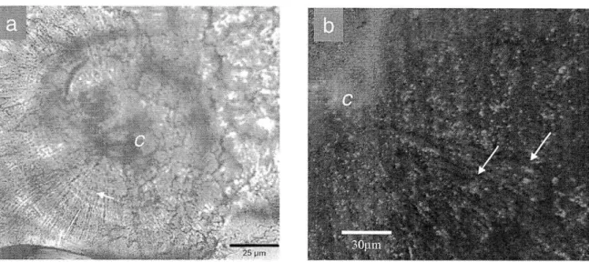
In Fig. 6a, the arrangement of aragonite crystals in a coral skeleton is seen in a cross section through a synapticulum or horizontal cross-bar of Porites solida. In
polarized light, the center of calcification (c) appears dark with poorly defined edges. Radiating outward from the center of calcification are aragonite fibers (f). This
observation is consistent with the description of coral sclerodermites (Wells, 1956). In this section, the fibers are interrupted by fine dark bands (arrow), -2 pm apart and
aligned perpendicular to the axis of crystal extension.
3.2.2 Fluorescence Imaging
Fig. 6b shows a fluorescence image of a section of the coral Diploria labyrinthiformis stained with acridine orange. There is an increase in fluorescence
associated with the centers of calcification (c) and dark bands (arrow) following acridine staining. This observation is consistent with that of Stolarski (2003).
3.2.3. SEM Imaging
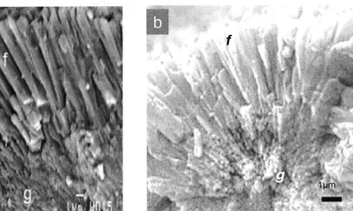
Fig. 7 shows SEM images of dissepiments (horizontal sheets) in the skeletons of Diploria labyrinthiformis (Fig. 7a) and Porites lutea (Fig. 7b). The dissepiments are
composed of two layers (as identified by Barnes, 1971). The primary (base) layer is built of small granular materials (g), <1 pm diameter. The secondary layer is composed of broad blade-like fibers (f), each -1-2 pm wide and several microns long.
3.2.4. AFM Imaging
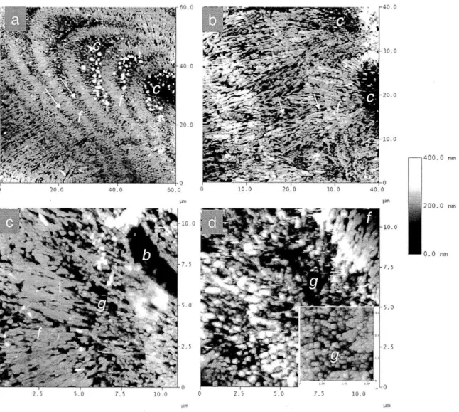
In Figure 8, AFM height images of skeletal cross sections of Diploria (Fig. 8a,c)
and Porites (Fig. 8b,d) reveal centers of calcification as regions of low relief (dark) and
the aragonite fibers are also visible as regions of low relief. Materials occupying centers of calcification (Fig. 8d) and dark bands (Fig. 8c) are small and granular in texture. Conversely, in both species, the light bands are packed with larger elongated fibers In Figure 8a, a cross section through a Diploria septotheca, the direction of vertical growth is from the lower right to the upper left of the image. Centers of
calcification (c) appear crescent-shaped, and form a line of discrete dark crescents up the middle of the septum. Centers of calcification are continuous with fine dark bands (arrow) that extend toward the edge of the septotheca. The width of the fine bands
decreases with distance from the centers of calcification. Near the centers of calcification, the fine bands are 5.3±0.4pm (n=10) wide. At the outer edge of the septotheca, the width of the bands is not discernible. As the width of the dark bands decrease, the distinction between light bands of aragonite fibers and the dark bands that interrupt them becomes
less distinct. Near the center of the septotheca, the transition between light fibrous bands and fine dark bands is abrupt. Toward the edge of the septotheca, the fibers appear continuous, cutting across dark bands.
In the Porites septum, the centers of calcification are oval rather than crescent shaped (Fig. 8b). Fine dark bands (arrows) are present in the fibers but these are not as clearly defined in this Porites specimen as they are in the Diploria skeleton.
3.2.5. MICa Ratios
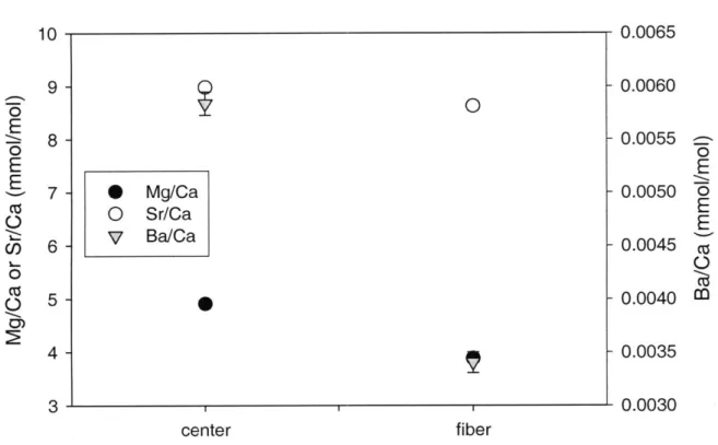
Selective analysis of Mg/Ca, Sr/Ca, and Ba/Ca ratios in centers of calcification and surrounding fibers of a Porites skeleton are shown in Figure 9. Mg/Ca, Sr/Ca, and Ba/Ca ratios are higher in the centers of calcification (4.91 ±0.02, 8.98 +0.03, and 0.0058
+0.0001 mmol/mol respectively) than in adjacent fibers (3.88 +0.01, 8.63 +0.03, and
0.0034 ±0.0001 mmol/mol respectively). For all M/Ca ratios measured, the value for the centers was significantly higher than for the fibers (p<0.01).
4. DISCUSSION 4.1. Synthetic Precipitates
Synthetic aragonite crystal morphology and composition are coupled to the pH and saturation state of the fluid from which the crystals grew. Within a single spherulite, the centers packed with sub-micron sized granular materials form when the saturation
state of the fluid is very high. Growth of fibrous crystals outward from the centers occurs when the saturation state of the fluid has decreased following nucleation. The
supersaturation achieved prior to nucleation depends on the rate at which Na2CO3 is
added; the faster the addition of Na2CO3, the higher the saturation state achieved prior to
nucleation, consistent with the work of Prieto and others (1989, 1994). In addition, the morphology of the spherulite (i.e., open, coarse versus closed, fine) and the size and
shape of the aragonite needles within the spherulites change systematically with the pH (saturation state) of the seawater in which they grew. In the high pH, high saturation state experiments, fine closed spherulites form that contain densely packed fibers with ill-defined grain boundaries (Fig 3a). Conversely, spherulites formed at low pH are typically
open and coarse, containing fewer, broad, faceted fibers (Fig 3b). This observation is consistent with systematic variations in crystal morphologies observed in non-CaCO3 minerals and polymers with increasing degrees of supercooling or with increasing supersaturation (e.g., Keith and Padden, 1963; Lofgren, 1971, 1974; Chernov, 1984;
Sunagawa, 1987), and reflects the crystal morphology that allows the maximum growth rate under those conditions (e.g., Tiller, 1964). The formation of smooth sides and development of facets in the lower pumping rate experiments likely reflects a change in growth mechanism, with a rough interface associated with spherulitic growth
transitioning to a smooth interface dominated by dislocation growth controlling step flow on faceted crystals (e.g., Sunagawa, 1981, 1987; Prieto et al., 1989).
Measurements of Mg/Ca, Sr/Ca, and Ba/Ca show that the Mg/Ca and Ba/Ca ratios are significantly elevated at the center of the spherulite relative to the fibers (Fig 5). In each experiment, the highest saturation state occurs at the onset of nucleation, thus the center of each spherulite contains crystals expected to have the highest
growth/precipitation rate (Burton and Walter, 1987) (though it should be noted that growth mechanism and relative areas of different crystal faces also change, and could influence composition as well). This relationship between crystal growth rate and M/Ca ratio is consistent with the growth rate dependency expected for Mg/Ca and Ba/Ca, as all MI/Ca ratios are expected to increase with higher growth rates due to more efficient entrapment of an impurity enriched mineral surface layer composition at higher crystal growth rates (Watson 2004; Gaetani and Cohen, 2006). Gabitov et al. (2006; 2008)
showed that the growth dependence of Mg/Ca in aragonite is much higher than that of Sr/Ca. Therefore, the absence of a significant elevation of Sr/Ca in the centers found in this study is consistent with their data.
Data presented here concur with earlier observations that coral sclerodermites consist of aragonite needles radiating out from regions of fine granular materials or
'nanocrystals' (Fig 6, 7, 8) (Vaughan and Wells, 1943; Wainwright, 1964; Constantz,
1986; Cohen et al., 2001; Clode and Marshall, 2003). The sub-micron-sized granular
materials are found at the base of dissepiments (Fig. 7a,b), in the centers of calcification
(Fig. 8) and in the fine bands that cut across aragonite fibers (Fig. 8).
Regions associated with granular materials are also associated with an increase in fluorescence following acridine orange staining (Fig. 6b), which is consistent with the results of Gautret et al. (2000) and Stolarski (2003). Mg/Ca, Sr/Ca, and Ba/Ca ratios are found to increase at the centers of calcification relative to adjacent fibers (Fig. 9), which is consistent with the findings of Meibom et al. (2004, 2006).
4.3. Results of Inorganic Precipitation Experiments in Relation to Coral Skeleton Morphology and Composition
Aragonite crystals formed by experimental precipitation from a supersaturated seawater solution and the aragonite crystals formed by living corals during skeletogenesis share several morphological and compositional features. Both the synthetic aragonites precipitated in this study and the coral sclerodermites are composed of two distinct types
of crystals: submicron-sized granular materials and larger elongate fibrous crystals that radiate out from the granular materials. Granular materials are found near the centers of synthetic grains, in the centers of calcification of corals, and in dark bands found both in corals and in the synthetic aragonites formed by stepped additions of Na2CO3. Higher
Mg/Ca and Ba/Ca ratios and acridine orange staining correspond to regions of granular materials in both synthetic aragonite (Fig. 2c, 5) and corals (Fig. 6b, 9), consistent with the results of Gautret et al. (2000) and Meibom et al. (2006) for corals.
Since the composition and morphology of crystals provide insights into the conditions under which they grew (e.g., Lofgren, 1971; Reddy and Nancollas, 1976; Marsh, 1988; Prieto et al., 1997; Marsh, 1998; Cohen and McConnaughey, 2003; Tong et al., 2004; Wasylenski et al., 2005), the observed crystal morphologies and compositions associated with known conditions in the synthetic experiments may be useful in assigning possible conditions of formation of naturally formed precipitates.
The similarity between the finely fibrous to granular materials at the centers of spherulites (Fig. 4a), grains formed at very high pH (Fig 4b) and materials occupying centers of calcification, fine bands, and the base layer of dissepiments in coral skeletons
(Fig. 8), suggests that these regions in the coral represent material formed at substantially
elevated saturation states. The morphology of fibrous aragonite in coral skeletons (Fig. 7a, b) is consistent with that of aragonites grown in the mid pumping (moderate
precipitation) rate experiment (Fig. 3b): tightly spaced but with distinct boundaries
between fibers. This may suggest that fibrous growth in corals occurs at a saturation state below that needed to induce nucleation but substantially above that of ambient seawater.
4.3.1. Band Formation in Corals and Synthetic Aragonite
The formation of alternating bands of fibrous crystals and granular materials in synthetic aragonites precipitated in experiments 2 and 3 is consistent with the stepped
addition of Na2CO3 which caused the saturation state of the seawater solution to cycle
during the experiment. Addition of the first volume of Na2CO3 elevated the solution pH
(supersaturation), initiating nucleation and the formation of granular materials at the center of the spherulites. Following nucleation, the solution pH dropped, enabling fibers to grow and radiate outward from the center. Addition of a second volume of Na2CO3
solution elevated the solution pH, favoring nucleation over elongation of pre-existing crystals, forming a fine band of granular materials followed again by radial growth (Fig 4a).
Such a cycling of fluid saturation state may explain the formation of microscale bands of alternating fibers and granular materials within the coral skeleton (Jell, 1974;
Sorauf and Jell, 1977; Risk and Pearce, 1992). The crystal morphologies reported here (Fig 8a,b,c) are consistent with repeated cycles of high and moderate saturation states. Zooxanthellate coral calcification rates (e.g., Kawaguti and Sakumoto, 1948; Goreau,
1959; Barnes and Chalker, 1990), and the internal pH of the coral (Al-Horani et al., 2003), are both known to increase substantially during the day and fall at night. Daytime pH values within the coral have been measured in excess of 9.0 (Al-Horani et al., 2003).
Thus, the microscale bands (often called daily growth bands) within zooxanthellate coral skeletons may be the product of substantially elevated daytime saturation states and more moderate night time saturation states.
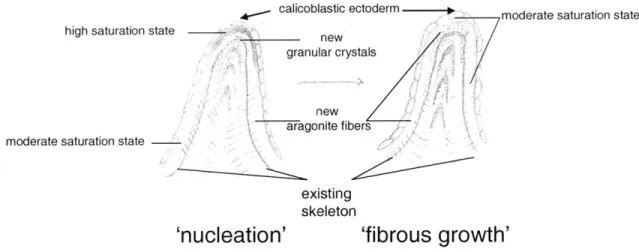
A model to explain the banding patterns seen in Diploria is presented in Fig. 10. In Diploria, the banding patterns are characterized by thick regions of granular material
from the center, transitioning to fibrous growth and a loss of bands (Fig. 8a). This pattern could be explained by the presence of a highly supersaturated fluid adjacent to where the granular centers form, transitioning to a moderate saturation state far from the centers, where fibers are continuous. The region of highest saturation state would be associated with the fastest growth and, thus, the thickest band of granular material. Moving down that saturation state gradient, growth would slow, shifting from granular to fibrous
material. At different times (perhaps at night), a more uniform, moderate, saturation state could exist throughout the calcifying environment, generating a band of fibrous crystals. Variations in rates of ion pumping or internal fluid flow could generate both spatial and temporal variations in saturation state.
The proposed cycle in saturation state could account for both the high Mg/Ca ratios at centers of calcification, and the alternating micron-scale bands of high and low Mg/Ca ratios reported by Meibom et al. (2004). This is consistent with the suggestion of Tsukamoto and Tsukamoto (1996) that growth rate variation could account for some of the variability in Mg/Ca ratios and is similar to the explanation proposed by Eggins et al. (2004), which attributes daily Mg/Ca bands in foraminifera to cycles in pH.
Cycles in the saturation state of the calcifying environment can also give rise to fine-scale heterogeneity in isotope ratios. Differences in 613C and 6180 between centers and fibers, as well as fine scale heterogeneity in the composition of coral fibers may, in part, be due to diffusion kinetics favoring the lighter isotope in the faster growing regions
(Rollion-Bard et al., 2003; Meibom et al., 2006). The calcification model proposed by Adkins et al. (2003) expands upon the diffusion model to incorporate multiple carbon