pH-Responsive Nano-Self-Assemblies of the Anticancer Drug
2
‑Hydroxyoleic Acid
Rama Prajapati,
†,∥Mark Gontsarik,
‡,∥Anan Yaghmur,*
,†and Stefan Salentinig*
,§,‡†
Department of Pharmacy, Faculty of Health and Medical Sciences, University of Copenhagen, Universitetsparken 2, DK-2100
Copenhagen Ø, Denmark
‡
Laboratory for Biointerfaces, Empa, Swiss Federal Laboratories for Materials Science and Technology, Lerchenfeldstrasse 5, 9014
St. Gallen, Switzerland
§
Department of Chemistry, University of Fribourg, Chemin du Musée 9, 1700 Fribourg, Switzerland
*
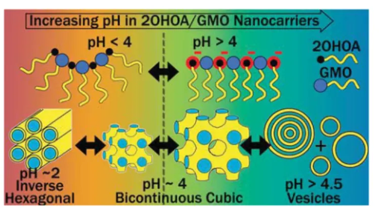
S Supporting InformationABSTRACT:
pH-responsive lipid nanocarriers have the
potential to selectively target the acidic extracellular pH
environment of cancer tissues and may further improve the
e
fficacy of chemotherapeutics by minimizing their toxic
side-e
ffects. Here, we present the design and characterization of
pH-sensitive nano-self-assemblies of the poorly water-soluble
anticancer drug 2-hydroxyoleic acid (2OHOA) with glycerol
monooleate (GMO). pH-triggered nanostructural
trans-formations from 2OHOA/GMO nanoparticles with an
internal inverse hexagonal structure (hexosomes) at pH
around 2.0
−3.0, via nanocarriers with an internal inverse
bicontinuous cubic structure (cubosomes) at pH 2.0
−4.5, to
vesicles at pH 4.5
−7.4 were observed with synchrotron
small-angle X-ray scattering, and cryogenic transmission electron microscopy.
ζ-potential measurements highlight that the pH-driven
deprotonation of the carboxylic group of 2OHOA, and the resulting charge-repulsions at the lipid
−water interface account for
these nanostructural alterations. The study provides detailed insight into the pH-dependent self-assembly of 2OHOA with
GMO in excess bu
ffer at physiologically relevant pH values, and discusses the effects of pH alterations on modulating their
nanostructure. The results may guide the further development of pH-responsive anticancer nanocarriers for the targeted
delivery of chemotherapeutics to the local microenvironment of tumor cells.
■
INTRODUCTION
Cancer is one of the major public health problems
world-wide.
1−3Along with the current cancer therapies, including
surgical intervention and radiation therapy, chemotherapy is
characterized as the least invasive cancer treatment approach
with a clinical success in tumor shrinking and cancer relapse
reduction.
4−6However, chemotherapy is often associated with
side-e
ffects caused by the off-site toxicity due to the lack of
drug speci
ficity.
5Hence, the design of novel and more e
fficient
cancer nanomedicines that can selectively target the tumor
sites, avoiding o
ff-target side-effects, may improve current
therapies and also patients
’ quality of life.
5,7−10Among
di
fferent suggested nanomedicines, pH-triggered
chemother-apeutic delivery systems may be of interest in cancer treatment,
as the extracellular microenvironment of tumor cells with pH
of around 5.5
−6.5 is more acidic than that of healthy cells with
pH of 7.4.
10−12The occurrence of such acidic local tumor
microenvironment is attributed to an increased glycolysis rate,
a higher production level of lactic acid, and an insu
fficient
vasculature for the removal of acidic byproducts.
11,13−18This study describes the formation and characterization of
pH-responsive self-assembled nanocarriers based on glycerol
monooleate (GMO), and the anticancer drug 2-hydroxyoleic
acid (2OHOA). The molecular structures of both lipids are
presented in
Figure 1
. The poorly water-soluble anticancer
drug 2OHOA is a derivative of oleic acid (OA) with an
additional hydroxyl group on the
α-carbon of the fatty acid
backbone.
19It is a potent anticancer agent that induces cell
Figure 1. Molecular structures of the lipids: (A) glycerol monooleate (GMO) and (B) 2-hydroxyoleic acid (2OHOA). The carboxylic group of 2OHOA is highlighted in red color in panel B.
http://doc.rero.ch
cycle arrest and apoptosis, cellular di
fferentiation, and
autophagy in a wide range of human cancer cells, including
leukemia, glioma, lung, colon, and breast cancer cells.
19−27GMO-based nanocarriers have been previously used for
enhancing the solubilization of poorly water-soluble
therapeu-tic agents.
28−35Among these nanocarriers, dispersions of
inverse bicontinuous cubic, hexagonal, and micellar cubic
structures (cubosomes, hexosomes, and micellar cubosomes,
respectively), emulsi
fied microemulsions, vesicles, and sponge
phases are gaining increasing attention for their drug delivery
applications. The biological relevance and the relatively large
extent of the internal lipid
−water interfacial area render these
colloidal nanosystems attractive for delivering hydrophilic,
hydrophobic, and amphiphilic drugs and peptide
mole-cules.
36−48For targeted delivery applications, they can be
surface nanoengineered to respond to an external stimulus,
such as pH, for triggering the release of therapeutic molecules
or enhancing the interactions with cells on demand, owing to
structural alterations in these nanocarriers.
33,49−55Despite the
attractiveness of cubosomes, hexosomes, and related
non-lamellar liquid crystalline nanoparticles in the development of
injectable drug nanocarriers, there is still to date no
FDA-approved formulation based on these nanostructured
emul-sions.
56,57Nevertheless, recent in vivo studies showed that
these nanoparticles are promising candidates in the
develop-ment of nanocarriers for anticancer drug delivery.
58,59The major goal of this study is to investigate the
composition and pH dependence of 2OHOA/GMO
nano-self-assemblies and gain further insight into their pH-triggered
structural, morphological, and size alterations by using
synchrotron small-angle X-ray scattering (SAXS), cryogenic
transmission electron microscopy (cryo-TEM), and
ζ-potential
measurements. An improved understanding of the structural
mechanism involved in these pH-triggered transitions is an
important keystone toward the development of pH-responsive
anticancer nanomedicines. Based on the presented
exper-imental
findings, the pH-induced phase transformations in the
interiors of 2OHOA/GMO nanoparticles are most likely
attributed to the protonation/deprotonation of the carboxylic
group of 2OHOA at the lipid
−water interface. The presented
results shed light on the molecular interactions during the
self-assembly of a cell membrane active anticancer agent with a
biologically relevant lipid in excess bu
ffer and could potentially
be applied for the future design of pH-guided nanocarriers for
tumor-targeted delivery of anticancer drugs.
■
EXPERIMENTAL SECTION
Preparation of 2OHOA/GMO Nanodispersions. Sodium salt of 2-hydroxyoleic acid (2OHOA; Avanti polar lipids, Alabama, U.S.A.) and glycerol monooleate (GMO;≥90% purity, Riken Vitamin co., Ltd. company, Japan) were weighed out in glass vials at 3/7 and 1/1 2OHOA/GMO mass ratios and dispersed in 149 mM Dulbecco’s Phosphate-buffered saline (PBS) at pH 7.4 (Sigma-Aldrich, Poole, UK), containing 2 mg/mL Pluronic F127 (gift from BASF SE, Ludwigshafen, Germany), to achieve a total lipid concentration of 20 mg/mL. These raw lipid emulsions were then homogenized by means of ultrasonication (Ultrasonic Processor Qsonica 500, Qsonica LLC, Newtown, CT, U.S.A.) for 4 min in pulse mode (3 s pulse, 5 s break) at 27% of its maximum power (500 W). The pH of the prepared nanodispersions was then adjusted with 1 M NaOH (≥99% purity, Carl Roth GmbH, Karlsruhe, Germany) or 1 M HCl prepared from 37% HCl stock solution (ACS reagent grade, Sigma-Alrdich, Buchs, Switzerland) and allowed to equilibrate at 25°C for at least 1 h before carrying out the planned experiments. In this study, the pH of the
continuous aqueous medium of the dispersion with 1/1 2OHOA/ GMO mass ratio wasfirst gradually decreased from 7.4 to 2.0 and then increased back to 5.0, while taking out samples at different selected pH points for SAXS structural characterization. This pH cycle was applied to study whether the structural alterations associated with changing pH can be reversed. Double distilled water was used for the preparation of all samples. All ingredients were used without further purification.
Synchrotron Small-Angle X-ray Scattering (SAXS). SAXS patterns were recorded at the Austrian SAXS beamline at the synchrotron light source ELETTRA (Trieste, Italy). An X-ray beam with a wavelength of 1.54 Å (8 keV) was used, with a sample-to-detector distance of 1314 mm, covering a q-range from about 0.18 to 5.00 nm−1, where q is the length of the scattering vector, defined by q = 4π/λ sin(θ/2), λ being the wavelength, and θ the scattering angle. The samples were sealed in thin-walled quartz capillaries and five frames with an exposure time of 20 s per frame were collected and averaged at room temperature. The 2D SAXS patterns were acquired using a Pilatus3 1 M detector (Dectris Ltd., Baden, Switzerland; active area 169× 179 mm2with a pixel size of 172× 172 μm2), integrated
into one-dimensional (1-D) scattering function I(q) using Fit2D60 (European Synchrotron Radiation Facility, Grenoble, France) and then analyzed with IGOR pro (Wavemetrics, Inc., Lake Oswego, U.S.A.). The scattering curves were plotted as a function of intensity, I(q), averaged over the five repeated exposures versus q. The scattering from PBS was subtracted as background from all measurements before further data analysis. The calculation of the lattice parameters of the corresponding lyotropic nonlamellar liquid crystalline phases is described inSI(eqs S2−S4).
Cryogenic-Transmission Electron Microscopy (cryo-TEM). The morphological characterization of F127-stabilized 2OHOA/ GMO nanoparticles was done in their vitrified states by cryo-TEM imaging. Briefly, lacey carbon 300 mesh copper grid (Ted Pella Inc., CA, U.S.A.) enforced by a silicon monoxide coating was subjected to glow discharge treatment and treated with 3μL of the sample. The excess sample on the grid was then blotted with filter paper at a blotting time of 3.5 s, a blotting force 0, temperature 25°C, and 100% humidity (FEI Vitrobot IV, Holland) and was rapidly plunged into liquid nitrogen-cooled ethane close to its melting point (−174 °C). A Gatan 626 cryo-holder (Gatan, U.K.) was used to transfer the plunged grid with the samples in a frozen glassy state into the Tecnai G2 20 transmission electron microscope (FEI, Holland). The images were digitally recorded at a voltage of 200 kV, under low-dose conditions (∼5 e/Å2s), and at a magnification of ×29 000.
ζ-Potential Measurements. The ζ-potential of the 2OHOA/ GMO nanodispersions was measured at different pH using Zetasizer Nano ZS equipped with a 633 nm laser (Malvern Instruments, Worcestershire, U.K.). Prior to measurements at 25 °C and 173° scattering angle, the samples were diluted 1/100 in PBS and their pH was readjusted.ζ-potential was calculated by Zetasizer Software 7.11 (Malvern Instruments) using Smoluchowski equation:61
μ ε ε ζ
η =
e r 0 (1)
whereμeis the electrophoretic mobility,εris the dielectric constant of
water,εois the permittivity of vacuum, andη is the viscosity of the
solvent (water). The measurements were done in triplicate and averaged. Estimations of 2OHOA’s pKaappfrom theζ-potential values
are described in theSI.
■
RESULTS AND DISCUSSION
The GMO dispersed in water forms cubosomes with an
internal bicontinuous cubic phase of the Pn3m or Im3m type
symmetry depending on the F127 stabilizer concentration.
62,63The integration of the anticancer drug 2OHOA into this
dispersion led to composition- and pH-dependent
nano-structural transformations in the nanocarriers.
Figure 2
demonstrates the pH-dependent self-assembly of the binary
2OHOA/GMO self-assemblies in excess bu
ffer at a mass ratio
of 1/1. The appearance of three dominating Bragg re
flections
in the SAXS curves at q values of 1.26, 2.17, and 2.51 nm
−1,
corresponding to a peak spacing-ratio of 1:
√3:√4, indicate
the formation of hexosomes with an inverse hexagonal (H
2)
phase at pH 2.0. The corresponding lattice parameter, a
H2, is
5.8 nm. Additional weak re
flections, occurring most
prom-inently around q
≈ 1.1 nm
−1, indicate a coexisting
bicontinuous cubic structure with Pn3m symmetry having a
lattice parameter, a
Pn3m, of 8.0 nm. These results demonstrate
that the 2OHOA mostly integrates into the hydrophobic
domains of the self-assembled nanostructure at pH 2.0. This
integration increases the apparent volume of the hydrophobic
domains, which ultimately leads to the nanostructural
transition from cubosomes to hexosomes. This observation
agrees with theoretical considerations from the critical packing
parameter model (see
SI
). The trend is also consistent with
2OHOA-induced lamellar-nonlamellar structural transitions in
dielaidoylphosphatidylethanolamine dispersion.
24,64Upon increasing the pH from 2.0 to 3.0, the Bragg peaks of
the H
2phase shifted to lower q values, corresponding to an
increase in a
H2to 5.9 nm (
Figure 2
). In addition to the
characteristic re
flections of the H
2phase, additional Bragg
peaks with peak-ratios of
√2:√3:√4:√6:√8:√9 were
detected. These peaks could be indexed as the (110), (111),
(200), (211), (220), and (221) re
flection of the bicontinuous
Pn3m-type cubic structure with a
Pn3m= 8.3 nm. In this phase
assignment, it should be noted that the characteristic re
flection
(220) of the cubic Pn3m phase at pH 2.0 and 3.0 appears to
overlap with the characteristic re
flection (11) of the coexisting
H
2phase. A single peak around q
≈ 0.8 nm
−1was also
observed, likely resulting from traces of a coexisting Im3m-type
bicontinuous cubic structure. The identi
fication of this
coexisting Im3m structure was based on its gradual evolvement
with a further increase in pH. At pH 3.5, the Bragg re
flections
of the H
2phase diminished and peaks indicating a coexistence
of Pn3m and Im3m phase were observed. The corresponding
lattice parameters for the biphasic Pn3m/Im3m feature were
9.1 and 12.0 nm, respectively. Further increase in pH to 4.0
caused a shift in the peak positions of the Pn3m and Im3m
phases to lower q values, resulting in an increase in a
Pn3mto 9.9
nm and a
Im3mto 12.9 nm. At pH 4.5, the lattice parameters of
Pn3m and Im3m increased to 12.3 and 15.3 nm, respectively.
The ratio of a
Im3m/a
Pn3mwas calculated and found to be 1.32,
1.30, and 1.24 at pH 3.5, 4.0, and 4.5. This ratio is consistent
with the reported theoretical Bonnet ratio of 1.279 for these
structures
36,65(
Table S1
). At pH 5.0 and 6.0, two Bragg peaks
at q values of about 1.56 and 3.12 nm
−1were observed,
showing the formation of multilamellar vesicles (MLVs) with
an interlamellar d-spacing of about 4.0 nm, in agreement with
the previously reported bilayer dimensions in GMO-based
self-assemblies.
66,67At pH 7.4, the diffuse-dominating scattering
patterns indicated the formation of unilamellar vesicles
(ULVs). The additional broad correlation peaks in the low-q
region of the SAXS curves at pH 6.0 and 7.4, with maxima
around q
≈ 0.45 and 0.90 nm
−1are most likely attributed to
weak bilayer correlations in a coexisting population of swollen
bi- or multilamellar vesicles with an estimated d of around
∼14
nm. The formation of highly swollen lamellar structures with
similar bilayer-to-bilayer spacing and low repeat numbers was
also previously reported for nano-self-assemblies of GMO with
the anionic phospholipid dioleoyl-phosphatidylglycerol.
67To evaluate the potential to reverse the colloidal
trans-formations in this system, the pH in the sample at 1/1
2OHOA/GMO mass ratio was decreased from pH 7.4, where
vesicles were the dominating species, to pH 2.0 where
cubosomes and hexosomes dominated (
Figure 2
). The
following pH increase to 5.0 in this sample led to the
reformation of vesicles, demonstrating the pH-induced
trans-formations from nonlamellar structures to vesicles and back
upon circulating the pH of the continuous water phase. The
pH-triggered swelling of nonlamellar liquid crystalline
structures and the gradual nonlamellar-lamellar transitions
have been previously reported for self-assemblies containing
the analogue OA
50,52,69,70and other fatty acids.
49,71The cryo-TEM images of the 2OHOA/GMO
nano-self-assemblies at 1/1 mass ratio confirmed the pH-triggered
nanostructural transitions observed by SAXS. Hexosomes were
observed at pH 2.0, and internally structured nanoparticles
coexisting with vesicles were found at pH 4.0 (
Figure 3
A,B). At
pH 6.0, the cryo-TEM image (
Figure 3
C) indicated the
Figure 2. SAXS patterns for the F127-stabilized nano-self-assemblies prepared at 2OHOA/GMO mass ratio of 1/1 as the pH of the dispersion was gradually decreased from 7.4 to 2.0 (red curves) and then increased back to 5.0 (black curves). The characteristic Bragg peaks and the corresponding Miller indices of the cubic Pn3m (red), Im3m (black), and H2(blue) phases are marked with arrows. Low
intensity peak attributed to a potential coexistence of Im3m phase is marked with a black asterisk at pH 3.0. Thefirst two Bragg reflections from the interlamellar distance of the MLVs are marked with Lα(h=1)
and Lα h=2)(green). Broad correlation peaks most likely
correspond-ing to weak bilayer correlations in a coexistcorrespond-ing population of swollen MLVs are marked with green asterisks.
presence of MLVs coexisting with smaller ULVs. The image at
pH 7.4 (
Figure 3
D) was dominated by ULVs mostly below
100 nm in diameter, coexisting with bilamellar vesicles. The
bilayer-to-bilayer distance of around 10 nm in the cryo-TEM of
multilamellar vesicles at pH 7.4 (
Figure 3
D) correlates
reasonably well with the estimated interlamellar distance
from SAXS (
Figure 2
).
Figure 4
shows the SAXS patterns for the 2OHOA/GMO
nano-self-assemblies at a 2OHOA/GMO mass ratio of 3/7, in
the pH range of 2.0
−7.4. At pH 2.0, the SAXS curve shows the
characteristic peak-spacing of the Pn3m type cubic phase with
a
Pn3mof 8.39 nm. Hence, compared to the dispersion that was
prepared at a mass ratio of 1/1, the 2OHOA content in this
sample is insu
fficient to induce a Pn3m-to-H
2phase transition
even at pH 2.0. On increasing pH to 3.0 and 3.5, the calculated
lattice parameter of the cubic Pn3m phase increased to 8.6 and
9.3 nm, respectively. However, three additional Bragg peaks at
q values of 0.72, 1.02, and 1.25 nm
−1with peak spacing-ratio of
√2:√4:√6 were observed at pH 3.5, which can be attributed
to a coexisting Im3m phase with a
Im3mof 12.3 nm. The
calculated ratio between the lattice parameters of Im3m and
Pn3m phases was 1.33 and fairly consistent with the theoretical
Bonnet ratio (
Table S1
). The SAXS curve at pH 4.0 was
dominated by the Im3m phase with a
Im3mof about 14.6 nm, as
the three Im3m peaks were shifted to lower q values. Further
increase in pH to 4.5
−7.4 led to the loss of the highly ordered
nonlamellar structures and the appearance of di
ffusive SAXS
curves, indicating a transformation to vesicles. The correlation
peaks observed in the low-q region of curves at pH 6.0 and 7.4,
with their maxima around 0.48 and 0.96 nm
−1, may correspond
to the weak bilayer correlations of a coexisting population of
highly swollen MLVs with an estimated d of around 13 nm in
agreement with the discussions above.
The structural transformations of the 2OHOA/GMO
nano-self-assemblies upon increasing pH from 2.0 to 7.4 are
attributed to the charge repulsions between the gradually
deprotonating carboxylic groups of 2OHOA embedded at the
lipid
−water interface. Such repulsions would increase the
e
ffective headgroup area of the embedded 2OHOA molecules
at the lipid
−water interface, modifying its spontaneous
curvature.
Figure 5
summarizes the pH-induced phase
transitions and changes in the lattice parameters for all phases
detected on increasing pH from 2.0 to 4.5 for both 2OHOA/
GMO samples prepared at mass ratios of 3/7 and 1/1. The
cubic Im3m phases in both 2OHOA/GMO dispersions
summarized in
Table S1
have smaller lattice parameters at
pH 3.0 and 3.5 than pristine GMO cubosomes with a
Im3m=
13.1 nm around the same pH (see
Figure S1
). This is
consistent with the integration of 2OHOA into the
hydro-phobic domains of the structure at this pH value. On the other
hand, upon reaching pH 4.0 and 4.5 in dispersions with
2OHOA/GMO mass ratios of 3/7 and 1/1, respectively, the
a
Im3mwas found to be greater than that of GMO cubosomes in
Figure 3. Representative cryo-TEM images of the nanodispersions prepared at 2OHOA/GMO mass ratio of 1/1 and the following pH: (A) pH 2.0, (B) pH 4.0, (C) pH 6.0, and (D) pH 7.4. The insert in panel A shows the fast Fourier transformation (FFT) applied to the structured part of the image indicating the formation of hexosomes. Colloidal transitions from (A) hexosomes to (B) cubosomes, and (C) MLVs (marked with black arrows) coexisting with relatively small nano-self-assemblies were observed with increasing pH. Small ULVs of variable sizes from around 100 to 15 nm in diameter were observed at pH 7.4, coexisting with multilamellar vesicles (marked with black
arrows) with a bilayer-to-bilayer distance of around∼10 nm (D). Figure 4. SAXS patterns of the F127-stabilized nanoself-assemblies prepared at 2OHOA/GMO mass ratio of 3/7 and pH in the range of 2.0−7.4. The Bragg peaks of the cubic Pn3m and Im3m phases and their corresponding Miller indices are indicated with red and black arrows, respectively. Low intensity peaks potentially from a coexisting Im3m phase are marked with black asterisks at pH of 2.0 and 3.0. Broad correlation peaks, most likely corresponding to the weak bilayer correlations in coexisting swollen MLVs, are marked with green asterisks at pH 6.0 and 7.4.
absence of 2OHOA (
Figure S1
). The later dispersion,
measured as control, showed only a slight structural alteration
in response to pH change between 3.0 and 7.0. The increase in
a
Im3mfrom 13.1 nm at pH 3.0 to 13.5 nm at pH 7.0 may be
attributed to the potential presence of trace concentrations of
OA as a hydrolysis product in the sample.
68However, this
change in lattice parameter in the 2OHOA free cubosomes is
insigni
ficant compared to that in the 2OHOA/GMO system
reported above. The larger pH-induced increase in the lattice
parameters of the internal liquid crystalline phases of the
2OHOA/GMO nanocarriers at pH 4.0
−4.5, as compared to
that at pH 3.0
−3.5, reflected a higher degree of 2OHOA
deprotonation and indicated the closer proximity to the
apparent pK
a(pK
aapp) of the embedded 2OHOA (
Table S1
).
Upon increasing the pH to >5.0, the 2OHOA/GMO
cubosomes eventually transformed into vesicles.
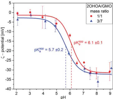
The pH e
ffect on the ζ-potential of 2OHOA/GMO
nano-self-assemblies is demonstrated in
Figure 6
. At pH 2.0, the
measurements for the dispersions with 1/1 and 3/7 2OHOA/
GMO mass ratios indicated the occurrence of rather neutral
surfaces with
ζ-potential values of −0.4 ± 0.7 and −3.8 ± 2.9
mV, respectively. The
ζ-potential of these dispersions was
observed to gradually decrease upon increasing pH, with the
largest drop in
ζ-potential occurring around pH 6.0, and
reaching
−30.9 ± 4.8 and −29.1 ± 3.5 mV at pH 9.0 for the
samples with 1/1 and 3/7 2OHOA/GMO mass ratios,
respectively. This decrease in
ζ-potential is mostly caused by
the deprotonation of 2OHOA, and correlated well with the
discussed nanostructural transitions observed by SAXS and
cryo-TEM.
For the system at 2OHOA/GMO mass ratio of 1/1, the
decrease in the
ζ-potential from −5.8 ± 2.8 to −31.3 ± 1.7 mV
on increasing pH from 5.0 to pH 7.5 could be associated with
an increase in the electrostatic repulsions between the
negatively charged 2OHOA/GMO bilayers in the MLVs.
This charge repulsion between bilayer sheets could then
induce the formation of the swollen MLVs with bilayer
−
bilayer distances above 10 nm at pH 6.0 and 7.4, as reported
above.
The pK
aappvalues of 2OHOA in the nano-self-assemblies at
2OHOA/GMO mass ratios of 1/1 and 3/7 were calculated
from the pH-dependent
ζ-potential values (see
Figure 6
and
SI
). The resulting pK
aappvalues are 6.1
± 0.1 and 5.7 ± 0.2,
respectively. These pK
aappvalues are slightly larger than the
previously reported pK
aappof 5.4 for 2OHOA in Triton X-100
micelles.
64This may be caused by the di
fferent 2OHOA
content in the 2OHOA/GMO nano-self-assemblies
(2OHOA/lipid mass ratios of 1/1 and 3/7) as compared to
a mass ratio 1/9 in 2OHOA/Triton X-100 micelles. The
higher concentration of ionizable carboxylic groups on the oil
−
water interface increases the negative surface-charge density,
which could enhance the accumulation of protons, leading to
an increase in pK
aapp.
52,72The results suggest that the pK
aappof
2OHOA in the designed 2OHOA/GMO nanocarriers may be
controlled through changes in the lipid composition. This
could potentially be used to tune the phase boundaries of these
self-assemblies to desired pH ranges for speci
fic delivery
applications.
The detected pH-triggered structural transformations in this
study can be of interest in the development of 2OHOA/GMO
nanocarriers for tumor-targeted delivery applications. In the
investigated nanocarriers, the major changes in the protonation
state of 2OHOA occurred within the typical pH range of the
extracellular tumor environment of pH 5.5
−6.5.
11,12The
modi
fications of the 2OHOA/GMO ratio in these nanocarriers
was further found to modulate the pK
aappof 2OHOA. Thus, the
nanocarriers could be
fine-tuned to obtain pH-triggered phase
transitions at speci
fic pH values by optimizing the lipid
composition. Additionally, the introduction of hydrophobic
additives, such as vitamin E or triglycerides, could increase the
hydrophobic tail volume.
73−78This could also shift the phase
boundaries between cubosomes and vesicles to higher pH
values, even close to the physiological pH of 7.4. The reported
pH-triggered nanostructural transformations could also trigger
Figure 5. pH-dependent changes in the lattice parameters of the H2 (■), Pn3m (●), and Im3m (▲) phases as derived from SAXS patterns of the two samples prepared at 1/1 (red) and 3/7 (blue) 2OHOA/GMO mass ratios presented in Figures 2 and 4. For numerical values seeTable S1.
Figure 6. pH-dependent ζ-potential values for the dispersions prepared at 1/1 (red) and 3/7 (blue) 2OHOA/GMO mass ratios. Fits ofeq S5to the data (seeSI) are represented as lines and used to estimate the apparent pKaof 2OHOA in the samples prepared at two
different lipid compositions.
speci
fic interactions of the nanocarriers with the cells, similar
to previous observations on antimicrobial peptide
nano-carriers.
70This would improve the targeted delivery of this
anticancer agent and may focus its exposure to the acidic
tumor microenvironment, minimizing o
ff-target side-effects.
Further in vitro and in vivo studies are required to determine
the practical application of these nano-self-assemblies in cancer
therapy.
■
CONCLUSIONS
The design and characterization of pH-responsive nanocarriers
based on mixtures of 2OHOA with GMO are presented. In a
concentration and pH dependent manner, 2OHOA was found
to actively participate and modulate the structural features of
GMO dispersion. The protonation state of the embedded
2OHOA at the lipid
−water interfacial area dictated the
structural and morphological characteristics of 2OHOA/
GMO nanocarriers. At pH <4.0, the mostly protonated
2OHOA integrated into the hydrophobic domains of the
cubosomes, shrinking their internal channel dimensions and
promoting the formation of the H
2phase. However, at pH
≥4.0, the gradual deprotonation of the 2OHOA molecules on
increasing pH resulted in electrostatic repulsions among its
deprotonated carboxyl-headgroups at the lipid
−water interface.
This caused the swelling of the cubic Pn3m and Im3m phases
and eventually, at pH
≥4.5 the colloidal transformation from
cubosomes to vesicles.
The results contribute to the fundamental understanding of
the self-assembly of pH-responsive and surfactant-like lipids.
The presented experimental
findings on the designed
pH-responsive 2OHOA/GMO nano-self-assemblies could guide
the development of advanced cancer nanomedicines for
targeted delivery of chemotherapeutics.
■
ASSOCIATED CONTENT
S
*
Supporting InformationThe
Supporting Information is available
Details on the critical packing parameter model, SAXS
data analysis, and calculation of the pK
aappfrom the
ζ-potential data (
)
■
AUTHOR INFORMATION
Corresponding Authors*E-mail:
[email protected]
.
*E-mail:
[email protected]
.
ORCIDMark Gontsarik:
0000-0001-7613-5137Anan Yaghmur:
0000-0003-1608-773XStefan Salentinig:
0000-0002-7541-2734 Author Contributions∥
R.P. and M.G. contributed equally to this work.
Notes
The authors declare no competing
financial interest.
■
ACKNOWLEDGMENTS
Authors are grateful to Heinz Amenitsch and Jianing Li for
technical support during synchrotron SAXS experiments and
Tillmann Pape (Core Facility for Integrated Microscopy,
University of Copenhagen) for technical assistance with
cryo-TEM imaging. The authors acknowledge the Swiss National
Science Foundation (Project 200021_169513 held by StS) for
funding this research. Financial support to AY by the Danish
Council for Independent Research, Technology and
Produc-tion Sciences, reference 1335
−00150b, is gratefully
acknowl-edged. AY further acknowledges
financial support from the
Danish Natural Sciences Research Council (DanScatt) for
SAXS experiments.
■
ABBREVIATIONS
2-OHOA
2-hydroxyoleic acid
GMO
glycerol monooleate
SAXS
small-angle X-ray scattering
cryo-TEM cryogenic-transmission electron microscopy
H
2inverted type hexagonal phase
PBS
phosphate bu
ffer saline
MLVs
multilamellar vesicles
ULVs
unilamellar vesicles
■
REFERENCES
(1) Siegel, R. L.; Miller, K. D.; Jemal, A. Cancer Statistics, 2018. Ca-Cancer J. Clin. 2018, 68 (1), 7−30.
(2) Wang, H.; Naghavi, M.; Allen, C.; Barber, R. M.; Carter, A.; Casey, D. C.; Charlson, F. J.; Chen, A. Z.; Coates, M. M.; Coggeshall, M.; et al. Global, Regional, and National Life Expectancy, All-Cause Mortality, and Cause-Specific Mortality for 249 Causes of Death, 1980−2015: A Systematic Analysis for the Global Burden of Disease Study 2015. Lancet 2016, 388, 1459−1544.
(3) Fitzmaurice, C.; Allen, C.; Barber, R. M.; Barregard, L.; Bhutta, Z. A.; Brenner, H.; Dicker, D. J.; Chimed-Orchir, O.; Dandona, R.; Dandona, L.; et al. Global, Regional, and National Cancer Incidence, Mortality, Years of Life Lost, Years Lived with Disability, and Disability-Adjusted Life-Years for 32 Cancer Groups, 1990 to 2015: A Systematic Analysis for the Global Burden of Disease Study. JAMA Oncol. 2017, 3 (4), 524−548.
(4) Terry, A. R.; Plotkin, S. R. Chemotherapy: Present and Future. Otolaryngol. Clin. North Am. 2012, 45 (2), 471−486.
(5) Sawyers, C. Targeted Cancer Therapy. Nature 2004, 432 (7015), 294−297.
(6) Brannon-Peppas, L.; Blanchette, J. O. Nanoparticle and Targeted Systems for Cancer Therapy. Adv. Drug Delivery Rev. 2012, 64, 206− 212.
(7) Peer, D.; Karp, J. M.; Hong, S.; Farokhzad, O. C.; Margalit, R.; Langer, R. Nanocarriers as an Emerging Platform for Cancer Therapy. Nat. Nanotechnol. 2007, 2 (12), 751−760.
(8) Bae, Y. H.; Park, K. Targeted Drug Delivery to Tumors: Myths, Reality and Possibility. J. Controlled Release 2011, 153 (3), 198−205. (9) Dai, L.; Liu, J.; Luo, Z.; Li, M.; Cai, K. Tumor Therapy: Targeted Drug Delivery Systems. J. Mater. Chem. B 2016, 4 (42), 6758−6772.
(10) Bor, G.; Azmi, I. D. M.; Yaghmur, A. Nanomedicines for Cancer Therapy: Current Status, Challenges and Future Prospects. Ther. Delivery 2019, 10 (2), 113−132.
(11) Tannock, I. F.; Rotin, D. Acid PH in Tumors and Its Potential for Therpeutic Exploitation. Cancer Res. 1989, 49 (16), 4373−4384. (12) Kato, Y.; Ozawa, S.; Miyamoto, C.; Maehata, Y.; Suzuki, A.; Maeda, T.; Baba, Y. Acidic Extracellular Microenvironment and Cancer. Cancer Cell Int. 2013, 13 (1), 1−8.
(13) Negrini, R.; Fong, W.; Boyd, B. J.; Mezzenga, R. PH-Responsive Lyotropic Liquid Crystals and Their Potential Ther-apeutic Role in Cancer Treatment. Chem. Commun. 2015, 51 (30), 6671−6674.
(14) Chen, W.; Meng, F.; Cheng, R.; Zhong, Z. PH-Sensitive Degradable Polymersomes for Triggered Release of Anticancer Drugs: A Comparative Study with Micelles. J. Controlled Release 2010, 142 (1), 40−46.
(15) Sim, T.; Lim, C.; Hoang, N. H.; Oh, K. T. Recent Advance of PH-Sensitive Nanocarriers Targeting Solid Tumors. J. Pharm. Invest. 2017, 47 (5), 383−394.
(16) Maeda, H.; Wu, J.; Sawa, T.; Matsumura, Y.; Hori, K. Tumor Vascular Permeability and the EPR Effect in Macromolecular Therapeutics: A Review. J. Controlled Release 2000, 65 (1), 271−284. (17) Cammas, S.; Suzuki, K.; Sone, C.; Sakurai, Y.; Kataoka, K.; Okano, T. Thermo-Responsive Polymer Nanoparticles with a Core-Shell Micelle Structure as Site-Specific Drug Carriers. J. Controlled Release 1997, 48 (2−3), 157−164.
(18) Soga, O.; Van Nostrum, C. F.; Fens, M.; Rijcken, C. J. F.; Schiffelers, R. M.; Storm, G.; Hennink, W. E. Thermosensitive and Biodegradable Polymeric Micelles for Paclitaxel Delivery. J. Controlled Release 2005, 103 (2), 341−353.
(19) Jang, E. J.; Choi, W. R.; Kim, S. Y.; Hong, S. S.; Rhee, I.; Lee, S. J.; Choi, S. W.; Choi, H. G.; Lim, S. J. 2-Hydroxyoleic Acid-Inserted Liposomes as a Multifunctional Carrier of Anticancer Drugs. Drug Delivery 2017, 24 (1), 1587−1597.
(20) Escribá, P.; Busquets, X.; Inokuchi, J.; Balogh, G.; Török, Z.; Horváth, I.; Harwood, J. L.; Vígh, L. Membrane Lipid Therapy: Modulation of the Cell Membrane Composition and Structure as a Molecular Base for Drug Discovery and New Disease Treatment. Prog. Lipid Res. 2015, 59, 38−53.
(21) Martin, M. L.; Barceló-Coblijn, G.; De Almeida, R. F. M.; Noguera-Salvà, M. A.; Terés, S.; Higuera, M.; Liebisch, G.; Schmitz, G.; Busquets, X.; Escribá, P. V. The Role of Membrane Fatty Acid Remodeling in the Antitumor Mechanism of Action of 2-Hydroxyoleic Acid. Biochim. Biophys. Acta, Biomembr. 2013, 1828 (5), 1405−1413.
(22) Teres, S.; Llado, V.; Higuera, M.; Barcelo-Coblijn, G.; Martin, M. L.; Noguera-Salva, M. A.; Marcilla-Etxenike, A.; Garcia-Verdugo, J. M.; Soriano-Navarro, M.; Saus, C.; et al. 2-Hydroxyoleate, a Nontoxic Membrane Binding Anticancer Drug, Induces Glioma Cell Differ-entiation and Autophagy. Proc. Natl. Acad. Sci. U. S. A. 2012, 109 (22), 8489−8494.
(23) Llado, V.; Gutierrez, A.; Martínez, J.; Casas, J.; Terés, S.; Higuera, M.; Galmés, A.; Saus, C.; Besalduch, J.; Busquets, X.; et al. Minerval Induces Apoptosis in Jurkat and Other Cancer Cells. J. Cell. Mol. Med. 2009, 14 (3), 659−670.
(24) Martinez, J.; Vogler, O.; Casas, J.; Barcelo, F.; Alemany, R.; Prades, J.; Nagy, T.; Baamonde, C.; Kasprzyk, P. G.; Teres, S.; et al. Membrane Structure Modulation, Protein Kinase c Activation, and Anticancer Activity of Minerval. Mol. Pharmacol. 2004, 67 (2), 531− 540.
(25) Llado, V.; Teres, S.; Higuera, M.; Alvarez, R.; Noguera-Salva, M. A.; Halver, J. E.; Escriba, P. V.; Busquets, X. Pivotal Role of Dihydrofolate Reductase Knockdown in the Anticancer Activity of 2-Hydroxyoleic Acid. Proc. Natl. Acad. Sci. U. S. A. 2009, 106 (33), 13754−13758.
(26) Marcilla-Etxenike, A.; Martın, M. L.; Noguera-Salva, M. A.; Garcı, M.; Anto, M.; Busquets, X.; Soriano-navarro, M.; Dey, I.; Escriba, P. V. 2-Hydroxyoleic Acid Induces ER Stress and Autophagy in Various Human Glioma Cell Lines. PLoS One 2012, 7 (10), e48235.
(27) Torgersen, M. L.; Klokk, T. I.; Kavaliauskiene, S.; Klose, C.; Simons, K.; Skotland, T.; Sandvig, K. The Anti-Tumor Drug 2-Hydroxyoleic Acid (Minerval) Stimulates Signaling and Retrograde Transport. Oncotarget 2016, 7 (52), 86871−86888.
(28) Chen, Y.; Ma, P.; Gui, S. Cubic and Hexagonal Liquid Crystals as Drug Delivery Systems. BioMed Res. Int. 2014, 2014, 1−12.
(29) Gontsarik, M.; Buhmann, M. T.; Yaghmur, A.; Ren, Q.; Maniura-weber, K.; Salentinig, S. Antimicrobial Peptide-Driven Colloidal Transformations in Liquid- Crystalline Nanocarriers. J. Phys. Chem. Lett. 2016, 7 (17), 3482−3486.
(30) Milak, S.; Zimmer, A. Glycerol Monooleate Liquid Crystalline Phases Used in Drug Delivery Systems. Int. J. Pharm. 2015, 478 (2), 569−587.
(31) Nazaruk, E.; Majkowska-Pilip, A.; Bilewicz, R. Lipidic Cubic-Phase Nanoparticles-Cubosomes for Efficient Drug Delivery to Cancer Cells. ChemPlusChem 2017, 82 (4), 570−575.
(32) Larsson, K. Cubic Lipid-Water Phases: Structures and Biomembrane Aspects. J. Phys. Chem. 1989, 93 (21), 7304−7314.
(33) Salentinig, S.; Tangso, K. J.; Hawley, A.; Boyd, B. J. PH-Driven Colloidal Transformations Based on the Vasoactive Drug Nicergoline. Langmuir 2014, 30 (49), 14776−14781.
(34) Azmi, I. D. M.; Moghimi, S. M.; Yaghmur, A. Cubosomes and Hexosomes as Versatile Platforms for Drug Delivery. Ther. Delivery 2015, 6 (12), 1347−1364.
(35) Yaghmur, A.; Glatter, O. Characterization and Potential Applications of Nanostructured Aqueous Dispersions. Adv. Colloid Interface Sci. 2009, 147−148, 333−342.
(36) Hyde, S. T. Bicontinuous Structures in Lyotropic Liquid Crystals and Crystalline Hyperbolic Surfaces. Curr. Opin. Solid State Mater. Sci. 1996, 1 (5), 653−662.
(37) Seddon, J. M. Structure of the Inverted Hexagonal (HII) Phase, and Non-Lamellar Phase Transitions of Lipids. Biochim. Biophys. Acta, Rev. Biomembr. 1990, 1031 (1), 1−69.
(38) Guo, C.; Wang, J.; Cao, F.; Lee, R. J.; Zhai, G. Lyotropic Liquid Crystal Systems in Drug Delivery. Drug Discovery Today 2010, 15 (23−24), 1032−1040.
(39) Shao, X.; Bor, G.; Al-Hosayni, S.; Salentinig, S.; Yaghmur, A. Structural Characterization of Self-Assemblies of New Omega-3 Lipids: Docosahexaenoic Acid and Docosapentaenoic Acid Mono-glycerides. Phys. Chem. Chem. Phys. 2018, 20, 23928−23941.
(40) Yaghmur, A.; Al-hosayni, S.; Amenitsch, H.; Salentinig, S. Structural Investigation of Bulk and Dispersed Inverse Lyotropic Hexagonal Liquid Crystalline Phases of Eicosapentaenoic Acid Monoglyceride. Langmuir 2017, 33 (49), 14045−14057.
(41) Luzzati, V. The Structure of the Liquid-Crystalline Phases of Lipid-Water Systems. J. Cell Biol. 1962, 12 (2), 207−219.
(42) Barauskas, J.; Johnsson, M.; Tiberg, F. Self-Assembled Lipid Superstructures : Beyond Vesicles and Liposomes. Nano Lett. 2005, 5 (8), 1615−1619.
(43) Kaasgaard, T.; Drummond, C. J. Ordered 2-D and 3-D Nanostructured Amphiphile Self-Assembly Materials Stable in Excess Solvent. Phys. Chem. Chem. Phys. 2006, 8 (43), 4957−4975.
(44) Fong, C.; Le, T.; Drummond, C. J. Lyotropic Liquid Crystal Engineering−ordered Nanostructured Small Molecule Amphiphile Self-Assembly Materials by Design. Chem. Soc. Rev. 2012, 41 (3), 1297−1322.
(45) Tiberg, F.; Johnsson, M. Drug Delivery Applications of Non-Lamellar Liquid Crystalline Phases and Nanoparticles. J. Drug Delivery Sci. Technol. 2011, 21 (1), 101−109.
(46) Drummond, C. J.; Fong, C. Surfactant Self-Assembly Objects as Novel Drug Delivery Vehicles. Curr. Opin. Colloid Interface Sci. 1999, 4 (6), 449−456.
(47) Malmsten, M. Phase Transformations in Self-Assembly Systems for Drug Delivery Applications. J. Dispersion Sci. Technol. 2007, 28 (1), 63−72.
(48) Yaghmur, A.; Rappolt, M.; Larsen, S. W. In Situ Forming Drug Delivery Systems Based on Lyotropic Liquid Crystalline Phases : Structural Characterization and Release Properties. J. Drug Delivery Sci. Technol. 2013, 23 (4), 325−332.
(49) Negrini, R.; Mezzenga, R. PH-Responsive Lyotropic Liquid Crystals for Controlled Drug Delivery. Langmuir 2011, 27 (9), 5296− 5303.
(50) Gontsarik, M.; Mohammadtaheri, M.; Yaghmur, A.; Salentinig, S. PH-Triggered Nanostructural Transformations in Antimicrobial Peptide/Oleic Acid Self-Assemblies. Biomater. Sci. 2018, 6 (4), 803− 812.
(51) Fong, W.; Hanley, T.; Boyd, B. J. Stimuli Responsive Liquid Crystals Provide‘on-Demand’ Drug Delivery in Vitro and in Vivo. J. Controlled Release 2009, 135 (3), 218−226.
(52) Salentinig, S.; Sagalowicz, L.; Glatter, O. Self-Assembled Structures and PKa Value of Oleic Acid in Systems of Biological Relevance. Langmuir 2010, 26 (14), 11670−11679.
(53) Agasti, S. S.; Chompoosor, A.; You, C.; Ghosh, P.; Kim, C. K.; Rotello, V. M. Photoregulated Release of Caged Anticancer Drugs from Gold Nanoparticles to Regulate Drug Release, Minimizing Side Effects and Improving. J. Am. Chem. Soc. 2009, 131, 5728−5729.
(54) Bayer, C. L.; Peppas, N. A. Advances in Recognitive, Conductive and Responsive Delivery Systems. J. Controlled Release 2008, 132 (3), 216−221.
(55) Gao, Z. G.; Fain, H. D.; Rapoport, N. Controlled and Targeted Tumor Chemotherapy by Micellar-Encapsulated Drug and Ultra-sound. J. Controlled Release 2005, 102 (1), 203−222.
(56) Barriga, H. M. G.; Holme, M. N.; Stevens, M. M. Cubosomes : The Next Generation of Smart Lipid Nanoparticles ? Angew. Chem., Int. Ed. 2019, 58, 2958−2978.
(57) Otte, A.; Soh, B.; Yoon, G.; Park, K. Liquid Crystalline Drug Delivery Vehicles for Oral and IV/Subcutaneous Administration of Poorly Soluble (and Soluble) Drugs. Int. J. Pharm. 2018, 539, 175− 183.
(58) Nasr, M.; Ghorab, M. K.; Abdelazem, A. In Vitro and in Vivo Evaluation of Cubosomes Containing 5-Fluorouracil for Liver Targeting. Acta Pharm. Sin. B 2015, 5 (1), 79−88.
(59) Jain, V.; Swarnakar, N. K.; Mishra, P. R.; Verma, A.; Kaul, A.; Mishra, A. K.; Jain, N. K. Paclitaxel Loaded PEGylated Gleceryl Monooleate Based Nanoparticulate Carriers in Chemotherapy. Biomaterials 2012, 33 (29), 7206−7220.
(60) Hammersley, A. P.; Svensson, S. O.; Hanfland, M.; Fitch, A. N.; Hausermann, D. Two-Dimensional Detector Software: From Real Detector to Idealised Image or Two-Theta Scan. High Pressure Res. 1996, 14, 235−248.
(61) Smoluchowski, M. Handbuch Der Electrizität Und Des Magnetismus (Graetz). Leipzig, Ger. Barth 1921, 2, 366.
(62) Gustafsson, J.; Ljusberg-wahren, H.; Almgren, M.; Larsson, K. Submicron Particles of Reversed Lipid Phases in Water Stabilized by a Nonionic Amphiphilic Polymer. Langmuir 1997, 13 (26), 6964− 6971.
(63) Landh, T. Phase Behavior in the System Pine Needle Oil Monoglycerides-Poloxamer 407-Water at 20.Degree. J. Phys. Chem. 1994, 98, 8453−8467.
(64) Barceló, F.; Prades, J.; Funari, S. S.; Frau, J.; Alemany, R.; Escribá, P. V. The Hypotensive Drug 2-Hydroxyoleic Acid Modifies the Structural Properties of Model Membranes of Model Membranes. Mol. Membr. Biol. 2004, 21, 261−268.
(65) Hyde, S. T. Microstructure of Bicontinuous Surfactant Aggregates. J. Phys. Chem. 1989, 93, 1458−1464.
(66) Chung, H.; Caffrey, M. The Neutral Area Surface of the Cubic Mesophase: Location and Properties. Biophys. J. 1994, 66, 377−381. (67) Yaghmur, A.; Laggner, P.; Sartori, B.; Rappolt, M. Calcium Triggered Lα-H2 Phase Transition Monitored by Combined Rapid Mixing and Time-Resolved Synchrotron SAXS. PLoS One 2008, 3 (4), e2072.
(68) Murgia, S.; Caboi, F.; Monduzzi, M.; Ljusberg-Wahren, H.; Nylander, T. Acyl Migration and Hydrolysis in Monoolein-Based Systems. Springer Berlin Heidelberg 2002, 120, 41−46.
(69) Suga, K.; Kondo, D.; Otsuka, Y.; Okamoto, Y.; Umakoshi, H. Characterization of Aqueous Oleic Acid/Oleate Dispersions by Fluorescent Probes and Raman Spectroscopy. Langmuir 2016, 32, 7606−7612.
(70) Gontsarik, M.; Yaghmur, A.; Ren, Q.; Maniura-Weber, K.; Salentinig, S. From Structure to Function: PH-Switchable Antimicro-bial Nano-Self-Assemblies. ACS Appl. Mater. Interfaces 2019, 11, 2821−2829.
(71) Salentinig, S.; Phan, S.; Darwish, T. A.; Kirby, N.; Boyd, B. J.; Gilbert, E. P. PH-Responsive Micelles Based on Caprylic Acid. Langmuir 2014, 30, 7296−7303.
(72) Cistola, D. P.; Hamilton, J. A.; Jackson, D.; Small, D. M. Ionization and Phase Behavior of Fatty Acids in Water: Application of the Gibbs Phase Rule. Biochemistry 1988, 27 (6), 1881−1888.
(73) Nilsson, C.; Edwards, K.; Eriksson, J.; Larsen, S. W.; Østergaard, J.; Larsen, C.; Urtti, A.; Yaghmur, A. Characterization of Oil-Free and Oil-Loaded Liquid-Crystalline Particles Stabilized by
Negatively Charged Stabilizer Citrem. Langmuir 2012, 28 (32), 11755−11766.
(74) Yaghmur, A.; De Campo, L.; Sagalowicz, L.; Leser, M. E.; Glatter, O. Emulsified Microemulsions and Oil-Containing Liquid Crystalline Phases. Langmuir 2005, 21 (2), 569−577.
(75) Yaghmur, A.; De Campo, L.; Salentinig, S.; Sagalowicz, L.; Leser, M. E.; Glatter, O. Oil-Loaded Monolinolein-Based Particles with Confined Inverse Discontinuous Cubic Structure (Fd3m). Langmuir 2006, 22 (2), 517−521.
(76) Yaghmur, A.; Sartori, B.; Rappolt, M. Self-Assembled Nanostructures of Fully Hydrated Monoelaidin− Elaidic Acid and Monoelaidin-Oleic Acid Systems. Langmuir 2012, 28, 10105−10119. (77) Amar-yuli, I.; Garti, N. Transitions Induced by Solubilized Fat into Reverse Hexagonal Mesophases. Colloids Surf., B 2005, 43, 72− 82.
(78) Prades, J.; Funari, S. S.; Escribá, P. V.; Barceló, F. Effects of Unsaturated Fatty Acids and Triacylglycerols on Phosphatidylethanol-amine Membrane Structure. J. Lipid Res. 2003, 44, 1720−1727.