HAL Id: pastel-00005010
https://pastel.archives-ouvertes.fr/pastel-00005010
Submitted on 21 Apr 2009HAL is a multi-disciplinary open access
archive for the deposit and dissemination of sci-entific research documents, whether they are pub-lished or not. The documents may come from
L’archive ouverte pluridisciplinaire HAL, est destinée au dépôt et à la diffusion de documents scientifiques de niveau recherche, publiés ou non, émanant des établissements d’enseignement et de
keratoplasty
Valeria Nuzzo
To cite this version:
Valeria Nuzzo. Mechanisms of femtosecond laser-cornea interaction in keratoplasty. Physics [physics]. Ecole Polytechnique X, 2008. English. �pastel-00005010�
présentée pour obtenir le titre de
Docteur de l’École Polytechnique
Spécialité: Physique
parValeria Nuzzo
Mechanisms of femtosecond
laser-cornea interaction in keratoplasty
soutenue le 1erfévrier 2008 devant le Jury composé de
Isabelle Brunette rapporteur, présidente du jury
Nicolas Château Benoît C. Forget Jean-Marc Legeais
Holger Lubatschowski rapporteur
Gérard Mourou
Karsten Plamann directeur de thèse
Marie-Claire Schanne-Klein
préparée au
Ce jour, où j’aurais écrit les remerciements, me semblait très loin, voire utopique, quand, après avoir travaillé pendant deux ans et demi dans une société privée en tant qu’ingénieur, j’ai voulu faire une thèse. Chose un peu insolite dans un système éducatif assez normatif, mais fi-nalement pas impossible, grâce à un brin de chance et à la persévérance de Karsten et moi dans la recherche d’une allocation de recherche. Le choix de quitter le connu pour l’inconnu n’a pas été facile, mais aujourd’hui je suis très contente de l’avoir fait car cette thèse a représenté pour moi une expérience exceptionnelle, stimulante et enrichissante. J’ai pris beaucoup de plaisir à essayer de comprendre de plus près les lois d’interaction entre la nature et la technologie, ainsi qu’a découvrir le monde des chercheurs, si varié.
Mes collaborateurs et collègues au Laboratore d’Optique Appliquée et à l’Hôtel Dieu ont contribué avec leurs compétences, leur disponibilité et leur accueil chaleureux à rendre les trois années de mon doctorat très constructives d’un point de vue scientifique et humainement agréables. Pour cela, je tiens à les remercier.
Je ne peux que commencer par exprimer ma plus grande gratitude à Karsten Plamann, mon directeur de thèse. Je lui suis reconnaissante de la confiance qu’il m’a accordée, de la qualité de son encadrement, franche et rigoureuse, de sa capacité à me conseiller aux moments oppor-tuns et de son soutien pendant la rédaction de cette thèse.
Je remercie également :
Gerard Mourou pour m’avoir accueillie au sein du LOA et avoir permis que cette allocation de recherche me soit attribuée ;
les membres du Jury, en particulier Isabelle Brunette et Holger Lubatschowski, pour leurs remarques précieuses sur mon manuscrit et l’intérêt qu’ils ont montré envers mon travail ;
l’Hôpital Hôtel Dieu de Paris, pour sa disponibilité et la pertinence de ses suggestions ainsi que Michèle Savoldelli, qui, avec beaucoup de passion, m’a fait découvrir le monde de la cornée de très près. Je la remercie pour sa patience et pour la très grande qualité du travail fourni. Jean-Marc et Michèle ont joué un rôle clé en apportant une importante valeur ajoutée à mon travail de thèse ;
M. Rosso, directeur de l’École doctorale de l’École Polytechnique, et toute son équipe, accueillante et compétante ;
David Donate et Florent Aptel pour leur apport médical à mon travail, le Pr. Pouliquen pour son soutien, ainsi que Françoise Dagonet et le secrétariat du LBO pour leur disponibilité et leur sympathie ;
ceux qui m’ont soutenue pendant l’écriture du manuscrit et la préparation de la soutenance, ainsi que les nombreux relecteurs de mes chapitres de thèse et les auditeurs des répétitions orales, je pense surtout à Ciro, Davide, Christoph, Alexandre, Evangelos, Gilles, Rahul, Florent et Donald ;
les équipes techniques, en particulier Thierry, Marc, Charly, Mikael, Alain, Fatima, Pierre et Arnaud, qui ont contribué à l’avancement et la réussite de ce projet, en étant toujours très efficaces et patients, ainsi que Dolores, Cathy, Sandrine, Patricia, Régis, Octavie et Valérie, pour leur aide au secrétariat ;
Alain, qui m’a encouragée pendant les derniers mois et qui m’a acceptée comme voisine de bureau (le calme pour favoriser la concentration, l’espace pour éparpiller les papiers et les tablettes de chocolat ont été des éléments essentiels pour la rédaction de la thèse), Fatima qui avait son bureau toujours ouvert et Charly grâce à qui je ne devrais plus perdre de clé.
Cela a été un plaisir de travailler avec Olivier Albert, dont j’ai beaucoup apprécié les con-seils scientifiques et les remarques qui m’ont permis d’améliorer la présentation de mes travaux de thèse. Je suis également reconnaissante à Michele Merano, qui a su me transmettre une par-tie de son savoir-faire pendant les expériences effectuées ensemble.
Je garderai un bon souvenir des cafés commentés en italien avec Laura ou Alessandro ; des trajets en RER avec Thomas, que je devais toujours attendre avec le risque de louper le train ; des trois ans de partage de bureau avec Guillaume ; des premières expériences réalisées avec Pedro.
envie de faire un doctorat et mes anciens collègues d’ICADE, Fabienne, Isabelle, Magalie, Nasr et Gérard, qui m’ont soutenue dans cette démarche, m ˆeme s’ils auraient aimé que je reste tra-vailler avec eux.
Un merci à Mauricette Ortu, dont les enseignements de français m’ont été si précieux. Un ringraziamento particolare va a Christophe, che mi ha incoraggiata, aiutata e "soppor-tata" durante questi tre anni.
The objective of the present research work has been to study the femtosecond laser-tissue inter-action in order to define optimal conditions for performing keratoplasty of pathological corneas by femtosecond laser. This is the first thesis on the subject accomplished at the Laboratoire d’Optique Appliquée (Ecole Polytechnique, ENSTA, CNRS in Palaiseau) and it has been car-ried out in close collaboration with the Laboratoire Biotechnologie et Œil (Université Paris Descartes, EA 4063, Hospital Hôtel Dieu de Paris).
The femtosecond laser is currently used in refractive surgery for cutting tissue and recently it has been introduced for substituting traditional mechanical instruments for corneal transplant or keratoplasty, which consists in replacing a pathological cornea of a patient with a healthy cornea from a donor. Approximately 100 000 procedures are performed per year worldwide. Leading indications for keratoplasty involve opacification of corneal tissue due to a dysfunc-tion of the endothelial cells. The cornea becomes permeable and looses its transparency. The oedema degree increases progressively and, if not interrupted by transplantation, it can cause total opacification of the cornea and a drastic decrease of the visual acuity. Traditional tech-niques for keratoplasty present a high risk of rejection, insufficient visual recovery and/or a risk of failure because technically delicate. In this context, femtosecond lasers represent a safe, accurate, and reliable surgical tool. However, clinical systems commercially available are very successful in cutting transparent corneal tissue (donor) but present technical limitations when inducing dissection in pathological corneas (receiver).
At the interface between the physics and the ophthalmology, this research work has allowed to assess the potential and the limitations of the present clinical systems and it has brought solu-tions for optimizing the laser-tissue interaction, also indicating direcsolu-tions for the development of a further high performing system.
This research work received the "Fondation Dalloz - Institut de France" Prize 2007 and the "La Recherche" Prize 2008.
Remerciements iv
Abstract vii
Table of contents ix
Introduction 1
1 The cornea 5
1.1 The human eye . . . 5
1.2 The anatomy of the cornea . . . 6
1.3 Optical properties of the cornea . . . 11
1.3.1 Refractive index . . . 11
1.3.2 Birefringence . . . 11
1.3.3 Optical absorption . . . 12
1.3.4 Transparency of healthy corneas . . . 13
1.3.5 Optical scattering of opaque corneas . . . 14
2 Ultrafast lasers and their application in corneal surgery 19 2.1 Ultrashort lasers . . . 20
2.1.1 Ultrashort pulses . . . 20
2.1.2 Femtosecond laser chains . . . 22
2.1.3 Laboratory femtosecond lasers . . . 26
2.2 Clinical laser systems and surgical applications . . . 28
2.2.1 Corneal surgery by femtosecond lasers . . . 28
3 Interaction of femtosecond lasers with corneal tissue 33
3.1 Origin of nonlinearities . . . 35
3.2 Laser-induced optical breakdown . . . 36
3.2.1 Nonlinear ionization . . . 36
3.2.2 Critical free-electron density . . . 39
3.2.3 Laser-induced ablation and disruption . . . 39
3.3 Self-focusing . . . 42
3.3.1 Nonlinear index of refraction . . . 42
3.3.2 Critical power for self-focusing . . . 43
3.4 Second harmonic generation . . . 46
3.4.1 Nonlinear response of tissue . . . 46
3.4.2 Physical mechanism of frequency doubling . . . 49
3.4.3 Second harmonic generation in collagen . . . 50
3.4.4 Application of SHG in biology . . . 54
4 Experimental set-up and sample preparation 57 4.1 Experimental set-up . . . 57
4.2 Sample preparation . . . 65
5 Femtosecond laser corneal surgery 67 5.1 Introduction . . . 67
5.2 Dissection with the clinical laser system . . . 68
5.2.1 Methods . . . 68
5.2.2 Results . . . 70
5.3 Dissections with the experimental laser system . . . 75
5.3.1 Experimental threshold determination . . . 75
5.3.2 Methods . . . 76
5.3.3 Results . . . 77
5.4 Discussion . . . 82
5.5 Self-focusing effects . . . 85
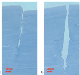
5.6 Deposit of glass fragments during keratoplasty . . . 93
5.6.1 Methods . . . 93
5.6.2 Results . . . 94
6 In situ evaluation and correction of laser energy density attenuation in opaque
corneas 101
6.1 Introduction . . . 101
6.2 Identification of the nonlinear emission . . . 102
6.2.1 Line spectrum of backward emission . . . 103
6.2.2 Dependance of the SHG on the incident light . . . 104
6.3 SHG imaging of cornea . . . 105
6.4 Evaluation of the laser beam attenuation . . . 108
6.4.1 Methods and results . . . 109
6.4.2 Discussion . . . 113
6.5 Correction of the laser beam attenuation . . . 114
6.5.1 Methods and results . . . 115
6.5.2 Discussion . . . 123
Conclusion and outlook 124 RÉSUMÉ EN FRANÇAIS 128 Résumé 129 Introduction 131 7 Notions théoriques 137 7.1 La cornée . . . 137 7.1.1 Anatomie . . . 137 7.1.2 Propriétés optiques . . . 139
7.2 Les lasers ultrarapides et leurs applications en chirurgie ophtalmologique . . . 142
7.2.1 Les chaines laser femtoseconde . . . 142
7.2.2 Applications chirurgicales et systèmes cliniques commercialisés . . . . 144
7.3 Interaction laser-tissu . . . 147
7.3.1 Photodisruption du tissu cornéen par laser femtoseconde . . . 148
7.3.2 Autofocalisation et défocalisation du faisceau . . . 150
7.3.3 Génération de seconde harmonique . . . 151
8 Dispositif expérimental et préparation des échantillons 153 8.1 Dispositif expérimental . . . 153
8.2 Préparation des échantillons . . . 160
9 Chirurgie cornéenne par laser femtoseconde 161 9.1 Introduction . . . 161
9.2 Dissection du tissu cornéen avec le laser femtoseconde . . . 162
9.2.1 Dissection avec le système laser clinique . . . 162
9.2.2 Dissections avec le système laser expérimental . . . 165
9.2.3 Discussion . . . 170
9.3 Effets d’autofocalisation . . . 170
9.4 Dépôt de particules de verre lors de la keratoplastie transfixiante . . . 172
10 Contrôle in situ de l’atténuation de la densité d’énergie laser dans une cornée opaque 175 10.1 Évaluation de l’atténuation laser . . . 175
10.1.1 Détermination de la nature du signal non-linéaire . . . 175
10.1.2 Quantification de l’atténuation laser par GSH . . . 176
10.2 Correction de l’atténuation . . . 180
10.2.1 Compensation de l’atténuation laser . . . 184
10.2.2 Discussion . . . 185
Conclusions et perspectives 186
Bibliography 188
Scientific context
The advent of ultrashort pulsed lasers has represented a major breakthrough in corneal surgery. The first clinical application of femtosecond lasers in ophthalmology has been their use in re-fractive surgery over the last decade. The most common technique for correcting ametropia is called LASIK (laser in situ keratomileusis). It consists of three main steps: first a hinged flap is cut and folded back, revealing the corneal stroma. In the second step, an excimer laser remodels the stromal layer tissue in order to correct focusing errors of the eye. Finally, the flap is unfolded back. Traditionally, the flap is created by a mechanical microkeratome equipped with a metal blade. Imperfect cutting related to the use of a microkeratome may lead to vari-ous intraoperative and/or postoperative complications. Femtosecond lasers have proven to be a useful and potential tool in tissue dissection and tend to replace traditional handled surgical blades. Advantages in using a femtosecond laser include a greater accuracy in flap size, shape, and thickness and therefore, a lower probability of complications. Since they represent a safe, reliable and flexible tool, although costly alternative, femtosecond systems are now currently employed in LASIK procedures.
Recently, the applications of ultrashort pulsed lasers have extended to corneal transplantation or keratoplasty. Leading indications for keratoplasty involve opacification of corneal tissue due to a dysfunction of the endothelial cells. Approximately 100 000 procedures are performed per year worldwide. Different transplantation techniques are currently performed, which are divided into full-thickness penetrating and partial lamellar keratoplasties. In penetrating kerato-plasty, a full-thickness cornel button is replaced. It is the less technically demanding and the most common transplantation procedure. However, it presents a risk of rejection and involves a button anesthesia due to the 360otissue denervation of the sensitive nerves which run mostly
in the anterior layers of the cornea. Recovery of vision may also be slow and not satisfactory because astigmatism and other optical aberrations are often induced by sutures and interface imperfections of the dissected tissue, thus reducing the connection of the donor button and the recipient bed. Part of these problems may significantly be reduced by partial corneal trans-plantation. Anterior lamellar and posterior lamellar keratoplasty are performed by selectively replacing either the anterior layer or the posterior layer of the cornea in patients with localized corneal disease; the Descemet’s stripping automated endothelial keratoplasty (DSAEK) is a rel-atively new surgical technique which involves the replacement of the endothelium through a limbal incision. It presents further clinical advantages compared to traditional lamellar tech-niques. However, it is a very delicate surgery which presents a risk of failure even when per-formed by experienced surgeons.
Femtosecond lasers ensure cutting with accuracy and reproducibility, and matching between the donor graft and the recipient eye, regardless of the type of keratoplasty performed. This leads to reduced astigmatism and a rapid visual recovery. Clinical systems commercially available are successful in dissection protocols of transparent corneas, however they present technical limita-tions when inducing dissection in pathological corneal tissue. The laser beam experiences scat-tering and optical aberrations when propagating through edematous tissue. The highly ordered arrangement of corneal layers is broken and variations in the refractive index are not negligible as in transparent corneas. Moreover, posterior surgery requires to focus at high depths in the cornea, which introduces additional spherical aberrations which contribute to beam broaden-ing. These effects, resulting in attenuation of the laser energy into the bulk of the tissue, are not taken quantitatively into account by daily femtosecond laser system. The quality of the cutting decreases in the volume of the cornea, thus prohibiting them from being routinely employed in keratoplasty.
In this context, a new laser system efficient in cutting pathological corneas safely, and enabling localized dissection or total removing of the diseased tissue, would represent a real advance-ment. It would allow performing "customized" and possibly sutureless keratoplasties.
Objectives
The objective of the present research work is to define optimal conditions for performing ker-atoplasty of pathological corneas by femtosecond lasers. The project has been executed by covering the following phases:
• the investigation of the interaction mechanisms of laser with healthy and pathological
corneal tissue in the femtosecond regime. The purpose was to identify side effects and evaluate their dependance on laser parameters;
• the in situ analysis and quantification of the optical properties of pathological corneas.
This required the development of an in situ method for optical measurements;
• the determination of the optimal laser parameters when dissecting opaque corneal tissue; • the elaboration of strategies for adapting the laser parameters to the tissue properties.
The project has included the development of an experimental set-up for performing corneal surgery by a femtosecond laser.
This is the first thesis on the subject accomplished at the Laboratoire d’Optique Appliquée. The work has been carried out in close collaboration with the Laboratoire Biotechnologie et Œil1.
Organisation
The dissertation is composed of two main parts. First, a literature review on the subject is pro-vided. The second part focuses on the experiments.
Chapter 1 provides a description of the anatomy of the cornea and its optical properties. In
particular, I review the different theories advanced for explaining corneal transparency, and modification of the optical properties in pathological opaque corneas.
Chapter 2 briefly introduces principles and techniques for generating ultrashort laser pulses,
in-cluding detailed description of the two laser systems used in our experiments. Finally, I present the state of the art in applications of femtosecond lasers in corneal surgery and compare clinical systems on the market.
Chapter 3 considers the nonlinear mechanisms which govern the interaction of ultrashort pulsed
lasers with transparent materials. Special emphasis is on phenomena observed in the corneal tissue, when exposed to an infrared femtosecond irradiation: laser-induced optical breakdown,
1Université René Descartes - Paris V, EA 4063, Hospital Hôtel Dieu, 1 place du parvis Notre Dame, 75181
chemical alteration due to low-density plasmas, self-focusing, and second harmonic generation. In Chapter 4, I describe the experimental set-up that we have developed and summarize char-acteristics of the clinical system that we used in our experiments. Sample preparation is also detailed.
In Chapter 5, I present our study on the interaction mechanisms of femtosecond pulses with corneal tissue as function of various parameters: corneal optical properties, pulse energy, nu-merical aperture of the focusing optics, laser systems. All experiments have been performed on human corneal grafts delivered by the Banque Française des Yeux for scientific use. Using a histological and ultrastructural examination of the irradiated corneal tissue, we compare tissue which underwent keratoplasty by a clinical system with tissue dissected by our experimental approach. The parameters outside of which undesirable effects become significant are evalu-ated. In edematous cornea, the laser beam is attenuated during propagation and the real energy delivered to the tissue at a determined depth is a priori unknown. In this case, side effects are more likely to be produced.
In Chapter 6 we propose an in situ and nondestructive method for quantifying the attenuation of the laser beam into the volume of pathological corneas. It is based on the acquisition of the backscattered second harmonic generation. We estimate the contribution of the spherical aberration and the scattering of the beam broadening, when focusing the laser into the bulk of the tissue. Thereby, we determine the optimal numerical aperture for corneal surgery. Finally, incisions performed with optimized laser parameters, with respect to the performances of our experimental set-up, are shown.
Chapter 7 summarizes the work contained in this thesis and gives an outlook on possible further
The cornea
In this chapter, I summarize the composition and structure of the cornea as well as its function. The optical properties of the corneal tissue are then described. Finally, I elucidate the basis for the transparency of normal healthy corneas and the structural changes related to pathological opacification of the cornea.
1.1 The human eye
The eye is a complex organ, which serves the important function of detecting light and give the sense of sight. Vision is the most used human sense to gather information from our environment. The eyeball has approximately an axial length of 23 mm and horizontal and vertical dimensions of 24 mm. Figure 1.1 displays a section of the anatomy of the eye. The outer layer maintains the shape of the eye and consists of the cornea and the sclera, which are connective tissues composed essentially of collagen. Whereas the sclera has a typical white color, the cornea is transparent or clear. After entering the eye through the cornea, light passes through the pupil which acts as a diaphragm; it then reaches the crystalline lens and focuses on the retina where it is converted into neuronal signals. The cornea is responsible for most of the optical power of the eye and the crystalline lens fine-tunes the focus. Inside the eye there are two fluid-filled sec-tions separated by the lens: the anterior segment, including the cornea, the angle structures, and
Figure 1.1: Section of the anatomy of the eye (from www.jirehdesign.com)
the iris, and a clear, watery substance called aqueous humor; the posterior segment containing a clear, gel-like material called the vitreous humor; the retina, and the optic nerve.
1.2 The anatomy of the cornea
The cornea is a transparent and avascular ocular tissue. It is exposed to the external environ-ment as it forms the outer shell of the eyeball covering the iris and the anterior chamber. Its major functions are refracting and transmitting light to the lens, the retina and the choroid as well as protecting the inner components of the eye against physical injury and potential harmful substances.
The cornea has a convex and aspherical shape. Its anterior surface curvature is steeper in the center and flattens towards the periphery. Its horizontal dimensions are 11 to 12 mm, whereas its vertical dimensions are 9 to 10 mm. Its thickness varies from 0.5 mm at the center to 0.7 mm at the periphery. The refractive power of the cornea is 40 to 44 dioptries and accounts for about two-thirds of the total refractive power of the eye. Therefore, small changes in the corneal shape or thickness can result in important visual distortions and refractive errors. Transparency is also a unique characteristic of the corneal tissue, which is necessary to maintain its optical prop-erties. This is ensured by the highly ordered stratified structure of the tissue coupled to small variations in the refractive index of the cornea.
The structure of the cornea has been studied by scanning electron microscopy, transmission electron microscopy and, more recently, synchrotron X-ray diffraction. This last technique does not require sample preparation including fixing, dehydration or staining, and therefore better respects the physiological hydration of the cornea.
The human cornea is composed of five layers, which are the epithelium, Bowman’s membrane, the stroma, Descemet’s membrane and the endothelium. The anterior layer is covered by a tear film and the posterior layer is bathed in the aqueous humor [35, 70, 71, 81, 90]. A histological section of a human cornea is shown in figure 7.1.
The epithelium has five to seven layers of cells for a total thickness of 50 to 52 µm and special-ized metabolic characteristics that allow its survival over an avascular tissue. It is self-renewing and presents the ability to respond to wound healing. The function of light refraction proper
Figure 1.2: Histological section of a human cornea [from Michèle Savoldelli, Hôpital Hôtel Dieu de Paris]
to cornea is brought about by its perfectly smooth and wet surface and its regular thickness. Loss of the smoothness of the epithelial surface results in degradation of the optical image and blurred vision. Another important physiological function of the epithelium is to protect the deeper layers of the cornea from external biological and chemical attacks and to prevent the penetration of the tear liquid into the stroma which could be responsible for edema and
inflam-mation.
The basement membrane is the inner surface of the epithelium. It provides anchorage to the adjacent Bowman’s membrane and stroma. Bowman’s layer is a 8-12 µm acellular membrane formed of a random arrangement of collagen fibers and proteins like proteoglycans. It exists in primates, including humans, but is thin or nonexistent in many mammals. The physiological role of Bowman’s layer remains still unclear, but it is believed that the network of fibrils ensures adhesion between epithelial cells and stromal matrix and contributes to the smoothness of the corneal surface.
The stroma constitutes about 90 % of the corneal thickness. The structure and properties of the stroma are responsible for important characteristics of the cornea, such as transparency and me-chanical strength. The stromal tissue is formed by cells which occupy only 2 to 3 % of the total volume, and by an extracellular matrix whose main components are collagen, water, proteogly-cans, glycosaminoglyproteogly-cans, and other proteins, which can absorb water in amounts equivalent to 1 000 times their volume. Collagen itself is a strongly hydrophilic protein and constitutes about 90 % of the stromal volume. In the stroma, collagen forms fibrils, which get organized in fibers. These last are in turn arranged in lamellae, generating a hierarchic structure. The basilar unit of collagen fibrils is the tropocollagen (TC) molecule, which is 1.5 nm in diameter, and 300 nm long. It consists of three polypeptide chains associated in a triple-helix structure with a periodicity of 8.6 nm, as shown in figure 1.3. Six TC molecules self-assemble around a
com-Figure 1.3: Schematic drawing of the tropocollagen (TC) molecule (modified from [118])
mon center in a nearly hexagonal manner to form fibrils, which in turn settle down with lateral and end-to-end binds in order to compose fibers with uniform diameter. Spaced at a regular distance, the fibers pack into layers. X-ray diffraction measurements yielded a fibril diameter close to 31 nm and a center-to-center distance of 62 nm [38, 97], whereas other investigators reported values of 24-26 nm for the diameter and 64-67 nm for the distance [14] using
transmis-sion electron microscopy. Since TC molecules within the fibril are staggered by approximately one quarter of their length, fibrils display a band structure in which regions with gaps and re-gions with overlaps of TC molecules alternate, as schematized in figure 1.4. Surrounded by
Figure 1.4: Spatial organization of collagen from the molecular level to the fiber level (modified from [118])
a ground substance consisting largely of proteins and nonfibrillar collagens, fibers associate with a regular distance of about 41 nm to form layers or lamellae. In the center of the cornea, the human stroma contains over 300 stacked thick lamellae. Thickness of each lamella is 1 to 2 µm (figure 1.5). Adjacent lamellae lay at different angles, between 0◦and 90◦, with preferred
orientations in the inferior-superior and nasal-temporal directions, and run uninterrupted from limbus to limbus. Different collagens are present in the lamellar stroma: corneal fibers consist essentially of type I collagen; however, they also contain a significant proportion of type V col-lagen. The latter, within a type I fibril, limits the ultimate diameter that fibrils can reach, which may contribute to explain transparency. Type VI and XII filaments bind collagen fibers in the shape of fibril network. They ensure the regularity of the stromal structure, essential to maintain the corneal optical transparency at visible wavelengths. After the collagens, the second major group of extracellular proteins in the stroma is proteoglycans, which are molecules composed of a core protein joined to one or more polysaccharide chains called glycosaminoglycans. Pro-teoglycans are also considered responsible for maintaining the relative position of the fibrils and restricting their growth. The extracellular matrix is secreted by cellular components, the
kera-tocytes, which are elongated cells laying between stromal lamellae and parallel to the corneal surface. Keratocytes are thought to contribute to the stability of the corneal shape and regular organization.
Descemet’s membrane is located between the stroma and the endothelium and constitutes a pro-tective barrier. In adult humans it is 8 to 10 µm thick and is mostly composed of collagen. The endothelium is the inner part of the cornea. It is composed of a single layer of cells, 4 to 6 µm thick, uniformly arranged, with a density of about 3500 cells/mm2. The endothelium
has two important physiological functions: the barrier and the pump function. Adjacent cells are linked so that they constitute a leaky barrier to aqueous humor permitting the transport of nutrients to the cornea while preventing the fluid from flowing into the stroma. Acting as an ionic pump, the endothelium regulates hydration of the cornea and maintains it at low levels. A failing endothelium leads to a stromal swelling and a consequential loss of the corneal trans-parency. It is therefore critical to avoid damaging endothelial cells during any surgery, yet in some cases a possible loss of cells may cause the remaining cells to enlarge until they cover the defective area.
Figure 1.5: TEM micrograph of corneal stroma showing the lamellar organization. Lamellae are parallel to the corneal surface and lay at different angles [from Michèle Savoldelli, Hôpital Hôtel Dieu de Paris]
1.3 Optical properties of the cornea
1.3.1 Refractive index
X-ray diffraction experiments performed by Leonard et al. [60] have unambiguously demon-strated that there is a difference in the refractive indices of the collagen fibrils and of the extra-cellular matrix. Since the fibrils cannot be separated from the ground substance, it is difficult to measure directly the respective indices of refraction. Nevertheless, X-ray diffraction techniques give information about the volume of the compounds and enable estimating the total refractive index using the Gladstone-Dale law of mixture which applies to composite materials. The total refractive index of a composite material ntot, can be expressed as the partial sum of the refractive
indices of its components, n1, n2, ..., nN, weighted by the volume fraction of each component,
f1, f2, ..., fN:
ntot= n1f1+ n2f2+ ... + nNfN . (1.1)
For a human cornea, Leonard et al. calculated a refractive index of 1.41 for fibrils and of 1.36 for extrafibrillar material and a fibril volume fraction of 0.22. By applying the Gladstone-Dale law, this set of values leads to a refractive index of the stroma of 1.37 in agreement with mea-surements provided by Maurice in a previous study [69].
1.3.2 Birefringence
Corneal tissue shows birefringent properties which can be separated into intrinsic birefringence and form birefringence ( [7], review). The former is due to the structure of collagen fibers com-posed of molecules longitudinally elongated. Light linearly polarized along the axis of the fibril travels at a slow velocity from light polarized perpendicularly. Form birefringence is attributed to the anisotropy of the stroma, characterized by a lamellar organization of the collagen fibrils. Each lamella may be considered as a thin linear retarder with its slow axis oriented along the fibril direction; corneal birefringence would represents the cumulative action of the lamellae of the entire stroma. A different approach has likened the cornea to a curved biaxial crystal with its fastest principal axis perpendicular to the surface. In many clinical situations (surgery
or diagnostic), light is incident nearly perpendicular to the surface, the perpendicular axis has little influence on the polarization of the state, and corneal birefringence is governed by the two principal axes that lie tangential to the surface.
The birefringent properties of the corneal tissue has been observed by measuring the cross-polarized transmission for a rabbit cornea as the incident polarization direction is rotated through 360o[25]. Results are presented in figure 1.6 and reflect the behavior of a birefringent material
except that the minimum transmission is higher than zero.
Figure 1.6: Cross-polarized transmission power through a rabbit cornea, recorded as the inci-dent polarization is rotated through 360◦. The solid line represents a fit of the data [25]
1.3.3 Optical absorption
Optical absorption in a tissue takes place when the frequency of the incident light corresponds to energy transitions of the molecules which compose the tissue. Figure 7.2 shows the absorp-tion of proteins, DNA, melanin, hemoglobin and water, which are the main tissue components, as a function of the wavelength.
The optical transmission of the cornea is essentially governed by the absorption properties of water and proteins. In the wavelength range of 400 to 780 nm, the most important chromophores are melanin and hemoglobin that are absent from the cornea. Hence, light is transmitted by the corneal tissue, which is why this spectral range is called the visible region of the optical spec-trum. In the ultraviolet region, proteins display significant absorption and in the infrared both proteins and water contribute to the absorption of the light, with certain transmission windows between 1 and 2 µm.
Figure 1.7: Optical absorption coefficients of principal tissue chromophores in the 0.1-12 µm spectral region [118]
1.3.4 Transparency of healthy corneas
The characteristic property of the cornea of neither absorbing nor appreciably scattering visible light is referred to as transparency. This remarkable property distinguishes the cornea from the other human connective tissues. In the previous section we have seen that the corneal tissue does not absorb light in the 400-780 nm wavelength range. The reasons why light is not scat-tered either are still not fully understood, although it has been a subject of interest for many decades. However, a number of different theories have been put forward.
The simplest model is based on the assumption that the refractive index of the cornea is uni-form, therefore, the light is not scattered when propagating through the tissue. This theory is generally rejected as it contradicts results evidencing a difference in the refractive indices of the fibrils and of the ground substance in which they are embedded. Maurice [69] first pro-posed that the secret of the corneal transparency may lay rather in the regular arrangement of the collagen fibrils in the corneal tissue. He showed that if collagen fibrils acted as independent scatterers, almost 90 % of the incident light would be scattered. Hence, he suggested that if fibrils are arranged in the form of a perfect crystalline-like hexagonal lattice, correlations in their relative positions lead to destructive interferences of light scattered in all direction other than the forward direction. However, electron microscopy and X-ray diffraction investigations
failed to demonstrate crystalline order. Moreover, a perfect crystal-like structure would produce a total transparency, which is not observed as the cornea is visible in slit lamps. Later research showed that lattice structure is not essential to account for transparency. Feuk [26] formulated a theory based on a long range order model with small, random displacements from the ideal lattice organization. Nevertheless, further investigations, based on the Fourier analysis of the corneal structure, were consistent with a short range order rather than a long range order found in crystals [36]. Hart and Farrell [41] computed a distribution function expressing a probability of correlation between fibrils from electron micrographs of corneal stroma. They found that the position of pairs of collagen fibers remains correlated only over distances equivalent to the spacing between two neighboring fibers at most (figure 7.3a). This translates to a short-range correlation of about 200 nm. They made a precise mathematical summation of the fields scat-tered by such a partially ordered array. The calculated corneal scattering was in accordance with that found experimentally. With a similar but simpler mathematical model, Benedeck [3] demonstrated the Bragg diffraction principle. He advanced that scattering of light is produced only by those fluctuations in the refractive index whose Fourier components are larger than one-half of the light wavelength in the medium. In transparent cornea, scattering is then reduced by the densely packed structure of the stroma with low fluctuations in the density of the fibers. In summary, even though there is still no universally accepted explanation to the corneal trans-parency, all theories recognize that three factors are determinant [25]:
1. each individual fibril has a very small scattering cross-section;
2. despite fibril scattering inefficiency, the large number of fibrils requires that the waves scattered by different fibrils must interfere destructively with one other in all directions but the forward one;
3. the cornea is thin, approximately 0.5 mm in humans.
1.3.5 Optical scattering of opaque corneas
Corneal transparency is partially dependent on the ability of the cornea to maintain a constant rate of hydration. When the endothelial cell density falls below a critical density (200 to 400 cells/cm2), following trauma, inflammation or dystrophy, the endothelium fails to regulate the
strongly deteriorate the vision, leading eventually to blindness. Edema are associated with a swelling of the stroma in the posterior direction and with an increase of the corneal thickness. Subsequently, the tissue becomes opaque and the incident light is scattered. Even though many complex models exist describing the attenuation of the light irradiance propagating through a scattering tissue, we limit our study to the Beer-Lambert law, whereby the transmitted irradiance
It can be expressed as
It= I0e−αsz (1.2)
where I0is the incident irradiance, αsis the scattering attenuation coefficient and z is the
thick-ness in the direction of the light path. In the case of a corneal lamella, which consists of parallel fibrils, αs= ρσs, where ρ is the number of fibrils per unit area in a cross section and σs is the
scattering cross section. This last indicates the scattering capability and is related to its cross-sectional area σgthrough the scattering efficiency Qsby the relation σs= Qsσg. The scattering
coefficient αsis defined as the probability of photon scattering per unit length in a medium. The
reciprocal of αs is referred to as the scattering mean free path l. It measures how deep a light
can penetrate according to the relation
l = 1
αs . (1.3)
The loss of transparency in a swollen cornea is often associated to a perturbation of the order in the position of the fibrils forming the collagen matrix [36]. Electron micrographs have revealed that in swollen corneas fibril density is decreased to a point that some regions of the collagen are completely devoid of fibrils (figure 7.3b). Benedek [3] called these defective regions "lakes" in the assumption that they contain excess fluid. Lakes have dimensions comparable to the wavelength of light and introduce fluctuations in the refractive index over a length scale that is equivalent or larger than half the wavelength of the light. They therefore may act as scattering centers [3, 49].
Meek et al. [72] explored theoretically the influence of different parameters on the loss of the light transmission by the cornea. In figure 1.9, the transmission of the light is plotted against the wavelength as function of individual parameters. The graphs clearly demonstrate that light scattering is increased when:
1. the fibril diameter increases. Reducing the diameter has an opposite effect, which means the transmission is augmented, however very thin fibrils would not ensure mechanical strength;
Figure 1.8: Comparison of a transparent cornea characterized by a short range order (a) with an edematous cornea in which the regular arrangement of the fibers is perturbed (b) [from Michèle Savoldelli, Hôpital Hôtel Dieu de Paris]
2. the corneal thickness is increased;
3. the mismatch in the refractive indices of the collagen fibrils and the extracellular matrix is increased.
All these factors reflect either a disorder in the organization of the fibrils in the corneal collagen or an increased scattering efficiency for fibrils. Additionally, recent investigations suggested that keratocytes contain a special protein cell, which is responsible for matching the refractive index of the cellular cytoplasm to that of the surrounding matrix in healthy corneas. When keratocytes are affected by a disorder, they do not ensure this function anymore and can become efficient scatterers [75].
Scattering of the light by spherical particles of any size can be modeled by the Mie theory. It takes into account the reflecting or absorbing properties of the particle material and enables to define the characteristic scattering cross-section. The Mie theory can be reduced to the Rayleigh theory if the spherical particle is much smaller than the wavelength [114]. In this approximation, the scattering cross-section is inversely related to the forth power of the wavelength of the incident light. In swollen corneas, the exact nature of the scattering centers has not yet been determined and scatterers are probably not spherical, therefore a quantitative theoretical model is difficult to derive. However, it could be proved that the modified structure of the collagen fibrils adds a term to the total scattering cross section which varies as the inverse square of the
Figure 1.9: Theoretical effects of varying individual parameters of light transmission by cornea [72]
light wavelength, so that
σ = σnormal+λB2 (1.4)
where σnormal is the scattering cross section for normal corneas, which is proportional to 1/λ3,
Ultrafast lasers and their application in
corneal surgery
The 1980s - 1990s were marked by important technological improvements in the field of ultra-short lasers:
• in 1986, Moulton [76] discovered a new laser material, the titanium-sapphire, which is a
sapphire crystal doped with titanium ions (Al2O3). It is characterized by a large spectral
gain bandwidth enabling the generation of ultrashort pulses, and a good thermal conduc-tivity reducing thermal effects even for high laser powers. Its nonlinear optical properties allow the generation of pulses via Kerr-lens mode-locking [104];
• Strickland and Mourou [107] introduced an amplification technique for the generation of
high energetic laser pulses, called the chirped pulse amplification (CPA);
• in parallel, the progress in high-power diode laser technology enabled to generate pulses
directly by pumping rare-earth-doped glasses.
These developments led to the successive replacement of dye lasers by more efficient ultrafast laser sources: Ti:sapphire lasers, which are nowadays mostly employed as laboratory systems, and diode-pumped-solid-state femtosecond lasers [45, 58]. Despite their slightly less spectac-ular performance with respect to pulse duration and energy, these latter are more adapted to
medical applications. Indeed, a surgical laser device needs to be turnkey, extremely reliable, easy to maintain, compact and affordable. Diode-pumped solid state lasers meet these require-ments.
In particular, the characteristics of neodymium or ytterbium doped glass lasers were soon con-sidered interesting for applications in ophthalmic surgery, like corneal dissection [53]. Fem-tosecond lasers are nowadays commonly employed in refractive surgery and tend to replace conventional techniques in cornea transplantation, due to their safety and reproducibility.
In this chapter, the characteristics and technology of ultrashort pulsed lasers are briefly intro-duced. The laser systems used in the experiments are also described. In the second section, I present current applications of ultrafast lasers in corneal surgery and I review recent experi-mental and clinical findings. Finally, a short overview of femtosecond laser systems commer-cially available is provided.
2.1 Ultrashort lasers
2.1.1 Ultrashort pulses
The time dependence of the complex electric field
E
of a light pulse generated by a laser can be written as [102]E
(t) = E0e−Γt2
e−iω0t . (2.1)
It results from the product of a carrier wave oscillating at an angular frequency of ω0
corre-sponding to the central wavelength of the pulse and an envelope A(t) = E0e−Γt
2
, assumed to have a Gaussian shape. The temporal intensity associated to this pulse is given by
I(t) = 2 η0· |
E
(t)| 2= I 0e−2Γt 2 (2.2) where η0 = pµ0/ε0= 377 Ω is the impedance of free space and I0 = 2E02/η0 is the peak
intensity. The Γ parameter is related to the pulse temporal width by the following relations
• at the 1/e2intensity: ∆t1 e2 =
2
√
• at the full width at half maximum intensity: ∆t1 2 =
2 ln 2
Γ .
By assuming the following Fourier transform convention
E
(ω) =F
[E
(t)] = Z +∞ −∞E
(t)e iωtdt (2.3)E
(t) =F
−1[E
(ω)] = 1 2π Z +∞ −∞E
(ω)e −iωtdω (2.4)the Fourier transform of
E
(t) can be expressed asE
(ω) = E0 2πF
[e −Γt2] ⊗F
[e−iω0t] (2.5a) = E0 2πτ √ 2π(e−Γt2) ⊗ 2πδ(ω − ω0) (2.5b) = E0τ √ 2π µ e−(ω−ω0)24Γ ¶ (2.5c) where the convolution theorem has been used. The Fourier transform of a Gaussian pulse in time is a Gaussian spectrum in frequency. The spectral intensity is therefore given byI(ω) = 2 η0· |
E
(ω)| 2= 2πτ2I 0e− (ω−ω0)2 2Γ (2.6)and the bandwidths are expressed by
• at the 1/e2intensity: ∆ω1 e2 = 4
√
Γ
• at the full width at half maximum intensity: ∆ω1 2 =
√
8 ln 2√Γ .
Hence, for a Gaussian pulse, the time-bandwidth product, that is the product of FWHM in time and frequency domain, satisfies the inequality
∆t∆ω ≥ 4 ln 2 . (2.7)
If the pulse verifies the identity, it is called Fourier transform-limited, which means that the shortest possible duration corresponding to its spectrum is obtained.
Relation 2.7 implies that a very short pulse has a broad spectrum. It is therefore necessary to take into account that pulses are subjected to dispersion when travelling through optical materials. The propagation time delay of the spectral components of the pulse is defined by the relation
T (ω) = dφ(ω)
dω (2.8)
where φ(ω) stands for the spectral phase, defined as the phase of the electric field in the fre-quency domain. When expressing φ(ω) as a Taylor series
φ(ω) = φ0+ µ dφ dω ¶ ω0 (ω − ω0) +12 µ d2φ dω2 ¶ ω0 (ω − ω0)2+ (2.9a) +1 6 µ d3φ dω3 ¶ ω0 (ω − ω0)3+ ... (2.9b) φ(ω) = φ0+ φ(1)(ω − ω0) +12φ(2)(ω − ω0)2+16φ(3)(ω − ω0)3+ ... (2.9c)
the following relation can be obtained
T (ω) = φ(1)+ φ(2)(ω − ω0) +1
2φ
(3)(ω − ω
0)2+ ... (2.10)
In particular, φ(1) represents a constant delay in the pulse propagation and does not modify the pulse characteristics; φ(2) is called the group velocity dispersion and introduces a time delay varying linearly with the frequency, which stretches the pulse duration.
2.1.2 Femtosecond laser chains
The peak intensity of a laser pulse is expressed in W/cm2and can be estimated by
I = E
∆t × S (2.11)
where E is the pules energy, ∆t is the pulse duration and S is the surface of the beam.
Femtosecond pulses are created in a laser cavity, where they typically gain only a few nanojoules of energy. High peak intensities are obtained by amplification using the CPA technique. The principle is to temporally stretch out the pulses to a much longer duration by means of a strongly
dispersive element, before amplification. Reducing the intensity enables avoiding nonlinear effects and optical damage during amplification. After being amplified, pulses are recompressed to their initial duration (figure 7.4).
Figure 2.1: Chirped pulse amplification (modified from www.llnl.gov/str/pdfs/09_95.2.pdf)
Oscillator
The longitudinal modes existing in a laser resonator oscillate independently if there are no rela-tions between the phase of the modes. The resulting laser operates in a free multimode regime and there is a competition between the different modes to be amplified by stimulated emission. If these modes are added in phase by constructive interference, wave packets are formed and a short pulse is generated. In order to obtain ultrashort pulses, many modes are required to interfere and the gain medium is needed to have a large emission bandwidth. Passive mode-locking is the most frequently used technique to hold the modes in phase. It is based on the principle of selectively modulating laser losses, in order to favor the pulsed regime. Passive mode-locking can be achieved by inserting into the cavity a semiconductor saturable absorber mirror or SESAM. It incorporates a saturable absorbing medium, whose optical absorption is
constant at low intensities, but saturates and decreases to lower values as the intensity rises. In this configuration weaker pulses are rejected and only the most energetic pulse propagates in the cavity. The active medium can also generate passive mode-locking by acting as a Kerr-lens which focuses the beam and increases its intensity. The pulsed operation is favored. Further-more, a hard aperture may be used to attenuate the continuous wave as the spatial distributions of the cw and pulsed modes differ. A set of chirped mirrors or a pair of prisms are added in order to compensate for the beam group velocity dispersion (figure 2.2). The shortest pules duration that has been presently obtained is of the order of 5 fs [24].
Figure 2.2: Schematic diagram of a self-mode-locked Ti:Sapphire oscillator
Pulse stretcher and compressor
Before being amplified, a pulse needs to be temporally stretched. The principle is to modify the optical path of the individual frequencies composing the spectrum of the pulse by means of dispersive optical elements. The duration of the incoming pulses is typically stretched from a few femtoseconds to a few hundreds picoseconds. Introducing a combination of gratings and lenses into the system induces a positive dispersion causing lower frequencies travelling faster than higher frequencies. The initial short pulse is therefore broadened or chirped. After being amplified, the pulse is recompressed back to its original duration by travelling through a conjugate dispersion line with opposite group velocity dispersion [87, 120]. The compressor has also the role of compensating for the beam dispersion associated to the propagation in the amplification chain.
Regenerative and multipass amplifier
Common approaches to pulse amplification are the regenerative and the multipass technique. The regenerative amplifier is a resonator in which the pulse travels several times through the gain medium, building up its energy at each roundtrip until gain saturation is obtained. The injection and ejection of the pulse are regulated by an optical switch composed of one or more polarizers coupled to a Pockels cell. Each pass introduces losses and dispersion, however the beam has typically a very good spatial quality, since it consists of a single mode amplified in a laser cavity. A schematic drawing of a regenerative amplifier is presented in figure 2.3a. The multipass amplification device includes a set of mirrors which allows the beam to pass through the gain medium at different angles, as illustrated in figure 2.3b. Compared to the regenerative amplification technique, in the multipass amplifier the beam experiences less dis-persion and losses because of the reduced number of optical elements and passes. However, care has to be taken in order to maintain its spatial quality as aberrations are caused by the tilt of the optical axis of the beam relative to the surface of the gain medium.
2.1.3 Laboratory femtosecond lasers
Neodymium:glass laser
The neodymium:glass femtosecond laser described in this section corresponds to the laser used to perform the experiments at the Laboratoire Biotechnologie et Œil. The experimental set-up is described in chapter 4.
The laser consists of a passively mode-locked diode pumped neodymium doped glass (Nd:glass) oscillator, whose output is amplified by a CPA system with a regenerative amplifier. Figure 7.5 shows a picture of the laboratory Nd:glass laser. The main elements of the system are indicated by numbered labels and specified in the figure caption.
The system includes a Nd:glass oscillator [46]. Neodymium doped glass presents a relatively broad emission bandwidth of 22 nm, an emission cross section of 4 × 10⁻²⁰cm2, an absorption
band at 808 nm enabling diode laser pumping, and it can be relatively easily manufactured. On the other hand, its thermal conductivity is low, in the order of 1 Wm−1K−1 [18]. This last
characteristic requires to design thin glasses to evacuate the heat and to pump at low powers in order to avoid thermal damage. The gain medium is inserted in a z-fold cavity and is pumped by a single-stripe diode laser with a power between 1 and 1.2 W. The pulse propagates in the cavity with a solitonlike shape. The soliton is formed as a result of negative group velocity dispersion, properly introduced by a chirped mirror, and self-phase modulation, experienced by the pulse during propagation through the Nd:glass medium [58]. It is then kept stable by a semiconductor saturable absorber mirror [55]. Inserting a knife-edge into the laser beam path contributes to select the pulsed mode over the continuous one. Output pulses have a duration shorter than 100 fs, a central wavelength of 1.064 µm, an average power of about 20 mW, and are delivered at a repetition rate of 80 MHz.
Before entering the regenerative amplification system, pulses are stretched propagating twice through a transmission grating. The beam path between the stretcher and amplifier is controlled by changing its polarization. The beam first reaches a Glan prism that allows p-polarized light to pass and rejects s-polarized light, then it travels through a Faraday rotator and a λ/2 wave plate, which both rotate its polarization. In the forward direction, the polarization is unchanged and the beam is injected into the regenerative amplifier, where it is amplified by means of a diode pumped Nd:glass, as used in the oscillator. After amplification, it passes again through the Faraday rotator and the λ/2 wave plate and is rejected by the Glan prism because of its polarization, which in the backward direction has experienced a 90◦ rotation. The amplified
pulsed light is therefore separated from the injection beam line and is sent to the compressor composed of the same transmission grating as the stretcher.
Pulses leaving the compression system have a duration of about 500 fs, a repetition rate variable from 1 to 10 kHz, a central wavelength of 1.06 µm, and a maximum average energy of about 6 µJ (at 10 kHz). The pulse peak-to-peak stability is of about 1%.
Figure 2.4: Picture of the laboratory Nd:glass laser, installed at the Laboratoire Biotechnologie et Œil. The main elements are labelled as follows: 1) Nd:silicate; 2a and 2b) cavity mirrors; 3) pump diode; 4) semiconductor saturable absorber mirror; 5) knife-edge; 6) chirped mirror; 7) output coupler; 8) grating; 9) Faraday isolator; 10) amplifier entry; 11) Nd:silicate; 12) exit of the laser
Ti:Sapphire laser
The Ti:Sapphire femtosecond laser described in this section corresponds to the laser used to perform experiments at the Laboratoire d’Optique Appliquée.
The oscillator is a Mira1 Ti:Sapphire Kerr-lens mode-locked oscillator pumped by an Argon
laser. It delivers pulses with a sub-100 fs duration, at a repetition rate of 80 MHz, a central wavelength of 805 nm, and an average power up to 64 mW. Pulses are stretched and recom-pressed after amplification in a CPA system by means of a grating. As in the case of the Nd:silicate laser, the beam path from the stretcher to the amplifier and from the amplifier to the compressor is regulated by a Faraday isolator. Pulse energy is increased in a regenerative amplifier, whose gain medium is a Ti:sapphire crystal pumped by a frequency-doubled yttrium lithium fluoride (YLF) laser. Following compression, pulses have a duration of 170 ± 20 fs, a central wavelength of 800 nm, a maximum pulse energy of 200 ± 20 µJ, and are delivered at a repetition rate of 1 kHz.
2.2 Clinical laser systems and surgical applications
2.2.1 Corneal surgery by femtosecond lasers
Corneal surgery has greatly profited from the technological improvements that pulsed lasers have known over the last decades. Femtosecond lasers enable obtaining very fine disruptive effects in tissue. Compared to nanosecond lasers used for other ophthalmic applications, the reduction of the pulse duration allows reducing the quantity of the energy to be delivered to the tissue for disruption, with strongly diminished mechanical and thermal collateral damages. Therefore, ultrashort pulsed lasers have widely replaced conventional techniques for tissue dis-section, because of their precision, safety and predictability.
The first clinical (and commercially available) application of femtosecond laser systems in oph-thalmology has concerned refractive surgery, to correct ametropia. Refractive errors typically treated by surgery are myopia and hyperopia. Myopia is a condition of the eye whereby the image is focused in front of the retina resulting in a blurred distance vision. It occurs when
the eyeball is too long or the cornea curvature is too strong. The opposite defect of myopia is hyperopia, which exists when light from a distant object comes into focus behind the retina, because of a too short eye or a too weak refractive power of the cornea or the lens.
In 1989, Pallikaris et al. [85] developed a surgical technique called the laser in situ keratomileu-sis (LASIK) which combines the use of an excimer laser and a mechanical microkeratome in order to modify the shape of the cornea. In the traditional LASIK surgery, the so-called micro-keratome, which is a mechanical device, is employed for cutting a corneal flap with a thickness of approximately 150 µm. This layer is created with a portion of the cornea left uncut to provide a hinge (figure 7.6). The flap is then folded back on the hinge exposing the middle portion of the cornea. In a second step, the excimer laser is used to reshape the center of the cornea by remov-ing a certain quantity of stroma. The amount of tissue removed is dependent upon the degree of myopia or hyperopia that is being corrected. Finally, the flap is repositioned over the stromal area fitting without sutures by means of adhesion forces. Since then, the technique has been
Figure 2.5: LASIK procedure steps (from www.ophtalis.tm.fr)
greatly improved with the introduction of automated and more sophisticated microkeratomes. However, complications may still occur in the creation of the flap with the mechanical instru-ments, which are delicate to manipulate. These include buttonholing or tearing of the flap, incomplete flap, free cap rather than hinged flap, and thinner or thicker flaps than expected. These problems have been partially or completely overcome by replacing the microkeratome with the femtosecond pulsed laser in cutting the superficial cornel flap. Since first laboratory ex-periments [53,64,67], fs-LASIK has been widely developed and it represents today a worldwide popular surgical procedure. A growing number of studies agree in stating that advantages in us-ing a femtosecond laser include more precise positionus-ing of the flap, more accurate thickness of the flap, and a lower probability of intraoperative complications [4, 57, 83, 106]. Additional works have shown improved astigmatism neutrality and lower wavefront aberrations [22, 113]. When using a femtosecond laser, depending on the laser system, the cut of the corneal layer
is achieved by applying successive beam spots in small adjacent zones, or a single-pass raster pattern, or a spiral pattern from the periphery to the center of the cornea at a fixed depth. This operation is followed by a movement of the spots along the circumference of the flap in order to incise through the anterior stroma up to the tissue surface.
A cornea requiring a refractive correction is a transparent material and the femtosecond laser interaction with transparent tissues in the ultashort pulsed regime has been widely investigated in both laboratory and clinical studies. From a physical point of view, we believe that the key parameter for a safe, precise and successful procedure is represented by the choice of a proper radiant exposure of the laser, when focused into the bulk of the cornea. As detailed in chapter 3, in the femtosecond regime optical breakdown inducing disruption of collagen fibers occurs at a determined fluence. Pulses with a radiant exposure lower than the threshold will not cause any cut, whereas pulses much more energetic than required may induce unwanted secondary effects. By taking into account the threshold value of the radiant exposure, which has been determined in different studies [34, 79, 109, 116, 118], commercial femtosecond lasers are pre-programmed to deliver the correct energy required for proper collagen disruption in transparent corneas. Recently, ultrafast lasers are increasingly used to perform keratoplasty, which is also referred to as cornea transplantation. The common manual or semi-automated surgical techniques for total grafting or penetrating keratoplasty are critical and not always reproducible considering the depth of the incisions and the difficulty in dimensioning the donor graft to match exactly the geometry of the corneal disc to be replaced in the recipient eye. The flexibility and the precision of the femtosecond lasers ensure connection between the donor and the recipient corneas. In addition, commercial femtosecond lasers offer a wide variety of cut patterns, which may favor wound healing. They include top hat, mushroom and Z- shapes (figure 7.7). The potential of ultrafast lasers to cut corneal tissue in transplant procedures was first assessed in experimental studies. Trephination of corneal tissue [73], inverse mushroom [99], and top hat-shaped [105] penetrating keratoplasties were successfully achieved when using clinical systems. Two re-cent interventional case series have reported neither interoperative nor postoperative complica-tions when performing penetrating keratoplasty in patients with various corneal diseases [9,44]. Partial thickness transplantation can also be carried out. Posterior lamellar keratoplasty consists in grafting only the posterior part of the cornea, including the endothelium, Descemet’s layer, and a layer of stroma of variable thickness. This is a quite delicate surgery where a major advantage of the femtosecond laser over mechanical techniques consists in performing precise lamellar incisions in the volume of the cornea, preserving the anterior layers and reducing the risk of irregular cuts, perforations and tears.
Figure 2.6: Keratoplasty cut patterns: top hat, mushroom and Z-shaped (from left to right)
The feasibility of fs laser posterior lamellar keratoplasty was evaluated in in vitro studies. Tissue dissection was overall efficiently obtained and light and scanning electron microscopy displayed relatively smooth cut surfaces, yet tissue bridges remained [96,100,103]. However, when treat-ing opaque corneas, energies up to three to four times the threshold energy for disruption of healthy corneal tissue were required.
2.2.2 Commercially available femtosecond laser systems
Four femtosecond laser devices are available today for corneal surgery (figure 7.8):
• Intralase FS60, produced by the US company AMO (Advanced Medical Optics, Inc.). Its
main characteristics are: wavelength 1,05 µm, repetition rate 60 kHZ, applanation lens flat. A keratoplasty unit is available.
• Femtec, designed by the German company 20/10 Perfect Visionr. It is characterized by a Nd:glass laser medium, a repetition rate of 40 kHZ and a curved applanation lens.
• Da Vinci, manufactured by the company Ziemer Ophtalmic Systems (Ziemer Group
Cor-porate Structure), installed in Switzerland. It is the only laser system delivering pulses at a repetition rate of approximately 1 MHz.
• VisuMax, recently commercialized by Carl Zeiss Meditec, Inc. It operates at a repetition
rate of 100 kHz.
All systems incorporate a diode-pumped femtosecond laser (solid-state or fiber laser in the case of the VisuMax). They deliver pulses at an energy of a few µJ and a few nJ in the case of the Da Vinci, in the near-infrared wavelength range, with a duration between 400 and 800 fs. The main
Figure 2.7: Commercially available femtosecond lasers (trademarks and model designations are protected)
difference between the four devices consists in the repetition rate and/or the shape of the appla-nation lens. The repetition rate influences the laser-tissue interaction process. When operating at the MHz regime, temperature accumulation effects may occur [116]. However, no clinical studies exist reporting differences in the post-operative visual acuity or related long-term com-plications.
The confidentiality on technical characteristics prevents access to complete specifications, such as the focused spot size or numerical aperture, or the spots separation. These data would allow to fully characterize the system and determine the key parameters in the laser-tissue interaction.
Interaction of femtosecond lasers with
corneal tissue
The physical mechanisms which rule the interaction of the light with the tissue differ strongly depending on the laser operation regime. This allows a variety of surgical applications. The key parameters defining the specific modes of interaction of the laser with the tissue are the wavelength and the duration of the exposure, characterizing the energy deposition time. The interplay between these factors induces mechanical, thermal and/or chemical modifications and may result in hemostatic effects, molecular denaturation, structural changes, tissue removal or cutting.
In the context of ophthalmological surgery, interaction times from 10 ms to 100 ms are still considered quasi-continuous. They are commonly achieved by using argon lasers, which are focused on spot sizes of 30-100 µm2. This leads to irradiances up to 105 W/cm2 which are characteristic for photocoagulation, a thermal process associated to protein denaturation due to an increased temperature of the tissue [6, 119]. Laser photocoagulation is a technique widely employed for the treatment of retinal disorders affecting blood vessels [61, 74, 77]. Long, short, and ultrashort regimes refer to pulse durations from the nanosecond to the femtosecond range. The transition from one regime to the other is gradual; however, pulses lasting from one fem-tosecond to a few picoseconds may be considered ultrashort. When properly focused on a target, ultrashort pulses create irradiances in the order of 1013 W/cm2, which locally generate electric fields comparable to the atomic and intramolecular Coulomb electric field. Pulse widths
![Figure 1.4: Spatial organization of collagen from the molecular level to the fiber level (modified from [118])](https://thumb-eu.123doks.com/thumbv2/123doknet/2845585.69988/24.892.256.640.244.570/figure-spatial-organization-collagen-molecular-level-fiber-modified.webp)
![Figure 1.7: Optical absorption coefficients of principal tissue chromophores in the 0.1-12 µm spectral region [118]](https://thumb-eu.123doks.com/thumbv2/123doknet/2845585.69988/28.892.236.655.162.461/figure-optical-absorption-coefficients-principal-tissue-chromophores-spectral.webp)
![Figure 1.8: Comparison of a transparent cornea characterized by a short range order (a) with an edematous cornea in which the regular arrangement of the fibers is perturbed (b) [from Michèle Savoldelli, Hôpital Hôtel Dieu de Paris]](https://thumb-eu.123doks.com/thumbv2/123doknet/2845585.69988/31.892.110.786.159.391/comparison-transparent-characterized-edematous-arrangement-michèle-savoldelli-hôpital.webp)
![Figure 1.9: Theoretical effects of varying individual parameters of light transmission by cornea [72]](https://thumb-eu.123doks.com/thumbv2/123doknet/2845585.69988/32.892.189.697.160.586/figure-theoretical-effects-varying-individual-parameters-transmission-cornea.webp)
![Figure 3.3: Calculated irradiance (a) and radiant exposure (b) threshold for producing the criti- criti-cal free electron density of 10 21 cm −3 for optical breakdown [116]](https://thumb-eu.123doks.com/thumbv2/123doknet/2845585.69988/56.892.160.737.314.513/figure-calculated-irradiance-exposure-threshold-producing-electron-breakdown.webp)