A new species of the large-headed coastal
marine turtle Solnho
fia (Testudinata,
Thalassochelydia) from the Late Jurassic of
NW Switzerland
Jérémy Anquetin1,2and Christian Püntener3,4 1JURASSICA Museum, Porrentruy, Switzerland
2Department of Geosciences, University of Fribourg, Fribourg, Switzerland 3Naturmuseum Solothurn, Solothurn, Switzerland
4Section d’archéologie et paléontologie, Office de la culture, République et Canton du Jura,
Porrentruy, Switzerland
ABSTRACT
Background:The large-headed turtle Solnhofia parsonsi is known by a handful of specimens from the Late Jurassic of Germany and Switzerland (maybe also France). Solnhofia parsonsi is traditionally regarded as a “eurysternid” Thalassochelydia, a group of small to medium sized, mostly lagoonal or marginal turtles found almost exclusively in the Late Jurassic of Europe. More recently, Solnhofia parsonsi has been proposed to be a close relative of Sandownidae, an enigmatic group of Cretaceous to Paleogene turtles characterized by a derived cranial anatomy and a wider
geographical distribution. Sandownids may therefore have evolved from thalassochelydian ancestors such as Solnhofia parsonsi.
Methods:We herein describe new material of Solnhofia from the Kimmeridgian (Late Jurassic) of Porrentruy, NW Switzerland. The bulk of the material consists of an association of a cranium and over 180 shell bones found together in a block of marly limestone. A second cranium and a mandible from slightly younger, but nearby localities are also described.
Results:We refer the new material to Solnhofia brachyrhyncha n. sp. The new species shares with Solnhofia parsonsi a relatively large head, an extensive secondary palate formed primarily by the maxillae, a greatly developed processus trochlearis oticum with a contribution from the parietal and quadratojugal, a large jugal-palatine contact in the floor of the fossa orbitalis, and a posteromedial process of the jugal running on the dorsal surface of the maxilla and pterygoid. Some of these
characteristics are also present in sandownids, but our morphological study clearly shows that Solnhofia brachyrhyncha is closer to Solnhofia parsonsi than to any sandownids.
Discussion:Solnhofia brachyrhyncha differs from Solnhofia parsonsi in many aspects, notably: a shortened and broader cranium, a shorter and posteriorly broader upper triturating surface with a slightly sinusoidal lateral margin and without contribution from the palatine, a processus trochlearis oticum more oblique in dorsal or ventral view and less concave in anterior view, choanae that do not extend posteriorly on the pterygoids, a more developed processus pterygoideus externus, a condylus mandibularis situated anterior to the level of the occipital plane, a greater ventral exposure of the parabasisphenoid, a mandible about as wide as long, Submitted 11 May 2020 Accepted 22 August 2020 Published 12 November 2020 Corresponding author Jérémy Anquetin, jeremy.anquetin@jurassica.ch Academic editor Mark Young
Additional Information and Declarations can be found on page 47
DOI 10.7717/peerj.9931 Copyright
2020 Anquetin and Püntener Distributed under
a relatively short symphysis, a lower triturating surface widened posterolaterally thanks to the presence of large laterally projecting dentary tubercles, a stouter and shorter coronoid process, a splenial positioned more anteriorly along the mandibular ramus, costo-peripheral fontanelles extending more anteriorly and posteriorly along the costal series, and an escutcheon shaped central plastral fontanelle formed mostly by the hypoplastra. In addition to the morphology of the new species, we also briefly discuss about observable ontogenetic variations and possible taphonomic origin of the assemblage.
Subjects Evolutionary Studies, Paleontology, Taxonomy, Zoology
Keywords Solnhofia, Thalassochelydia, Testudinata, Kimmeridgian, Late Jurassic, Switzerland
INTRODUCTION
Solnhofia parsonsi is a remarkable Late Jurassic turtle characterized notably by a
proportionally large head and the presence of a secondary palate formed primarily by the maxillae (Gaffney, 1975;Lapparent de Broin, Lange-Badré & Dutrieux, 1996;Joyce, 2000). This species was erected byGaffney (1975)based on two skulls. The holotype (TM 4023), a specimen initially described but not named byParsons & Williams (1961), consists of a cranium and associated mandible from an unknown locality (probably from the Solnhofen region). The second, referred specimen (NMS 8741, previously numbered NMS 137) is a deformed cranium originating from the Kimmeridgian of Solothurn, Switzerland. This specimen was briefly mentioned byBräm (1965). Only a handful of specimens have since been referred to Solnhofia.
A juvenile specimen with a large head and roundish shell (MNHN CNJ 76) from the Tithonian lithographic limestones of Canjuers, France was initially attributed to aff. Solnhofia sp. (Fabre et al., 1982), but this identification was later questioned due to the
poor quality of the preservation (Broin, 1994). Another specimen from Canjuers (MNHN CNJ 82) consisting of a cranium and associated scattered elements of the postcranium can be more confidently assigned to Solnhofia sp. based on the large head and elongation of the snout (Broin, 1994), but this specimen was never described in detail. Later, Lapparent de Broin, Lange-Badré & Dutrieux (1996)referred the posterior part of a cranium (ICHLU 005.4.20) from the Kimmeridgian of Labastide-Murat, France to Solnhofia aff. parsonsi. Finally, a well-preserved specimen (JM SCHA 70) from the Kimmeridgian/Tithonian boundary of Schamhaupten, Germany represents thefirst complete skeleton of Solnhofia parsonsi and documents the postcranial morphology of this taxon (Joyce, 2000).
Solnhofia parsonsi is usually considered to be a representative of Thalassochelydia, a clade of coastal marine turtles almost exclusively known from the Late Jurassic of western and central Europe (but at least one species reached Argentina, while at least another one survived into the Early Cretaceous) and that represents thefirst radiation of chelonians into marine environments (Anquetin, Püntener & Joyce, 2017;Anquetin & André, 2020;
González Ruiz, De la Fuente & Fernández, 2020). Thalassochelydians are traditionally
challenged this conclusion by recovering them as stem Testudines, sister group to Pleurodira, or stem Chelonioidea (Evers & Benson, 2019;Evers, Barrett & Benson,
2019;Gentry, Ebersole & Kiernan, 2019). These recent studies consistently found
Thalassochelydia to form a clade with Sandownidae, a group of unique coastal marine turtles characterized by the presence of an extensive secondary palate and known from the Early Cretaceous to the Paleocene (Tong & Meylan, 2013). Possible relationships between sandownids and thalassochelydians, Solnhofia parsonsi in particular, were discussed several times in the literature (Joyce, 2007;Mateus et al., 2009;Evers & Benson, 2019). This hypothesis was further substantiated recently based on morphological arguments by
Evers & Joyce (2020)who suggested that Solnhofia parsonsi could represent the earliest
sandownid. However, these results have not yet been tested in a phylogenetic framework and the respective relationships of sandownids, thalassochelydians and Solnhofia parsonsi remain uncertain. For example, it is unclear whether sandownids and thalassochelydians are just sister clades and Solnhofia only belongs to one of them or if sandownids are included within thalassochelydians and derive from Solnhofia-like taxa. For the time being, we conservatively assign Solnhofia to Thalassochelydia herein.
In the present study, we report new material of Solnhofia from the Kimmeridgian of Porrentruy, Switzerland. The bulk of this new material consists of a dense assemblage containing a cranium and over 180 shell bones. This cranium and the best preserved shell bones are herein used to define the new taxon Solnhofia brachyrhyncha n. sp., which notably differs from the type species Solnhofia parsonsi in its shorter snout, posteriorly broad triturating surface, and escutcheon shaped central plastral fontanelle. A second cranium and an isolated mandible from nearby localities, but different stratigraphical layers are also confidently referred to Solnhofia brachyrhyncha n. sp.
MATERIALS AND METHODS
MaterialThe new material presented herein was unearthed during the construction of the A16 Transjurane highway in NW Switzerland. Thanks to generousfinancial support from the Swiss Confederation, the archeological and paleontological material discovered during this construction was systematically collected and documented. In recent years, several studies focussing on the rich Kimmeridgian strata in the region of Porrentruy (Fig. 1) were published, including articles on plant remains (Philippe et al., 2010), invertebrates
(Schudack et al., 2013;Koppka, 2015),fishes (Leuzinger et al., 2015,2017;Leuzinger,
Püntener & Billon-Bruyat, 2017;Leuzinger et al., 2020), dinosaur tracks (Razzolini et al.,
2017;Marty et al., 2018;Castanera et al., 2018), and turtles (Püntener et al., 2014;
Anquetin, Püntener & Billon-Bruyat, 2015;Püntener, Anquetin & Billon-Bruyat, 2015,
2017a,2020).
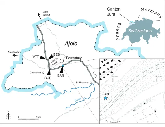
MJSN BAN001-2 originally consisted of a large block of marly limestone which was found entangled in the roots of a deracinated tree in 2001 on the northern slope of the Banné locality (BAN; 47 24′ 23.52″N 7 4′ 20.35″E). “Le Banné” corresponds to a wooded hill that borders the southwest residential area in Porrentruy. Under this hill runs the
Banné tunnel of the A16 highway (Fig. 1). The block contained an assemblage of numerous turtle shell bones, which were at the time interpreted as remains of juveniles because of their relatively small size. Some of these shell bones were initially prepared and removed from the matrix, but many were simply surface prepared and left in the block of
matrix. Several years later, the identification of a turtle cranium among the remains still embedded in the matrix prompted the preparation of the complete block. MJSN BAN001-2 now consists of a collection of over 180 disarticulated shell bones and a cranium. A selection of the best preserved remains were given a specific second level identification number for the purpose of this study (Table 1). MJSN BAN001-2.1 (the cranium) and MJSN BAN001-2.2 to MJSN BAN001-2.28 (shell remains) are referred to the new species Solnhofia brachyrhyncha. A pair of hypoplastra (MJSN BAN001-2.29) is tentatively assigned to a juvenile specimen of Tropidemys? langii? (see below). All other bones from this assemblage are referred to Thalassochelydia indet.
MJSN SCR010-1214 consists of a severely crushed turtle skull lacking most of the ventral and anterior parts. MJSN BEB011-13 is a relatively small turtle mandible that was found in a dinosaur track infilling (see Geological settings). This is one of the rare vertebrate remains found in the dinosaur-track-bearing layers of the Porrentruy region
(Püntener, Anquetin & Billon-Bruyat, 2017a;Püntener et al., 2019).
Figure 1 Geographical map of the Ajoie region, Canton of Jura, Switzerland.The position of the different localities mentioned in the present study is indicated along the A16 Highway (see text for abbreviations). Inset: the Banné (BAN) site is located in a forest on the Banné hill, southwest of
Geological settings
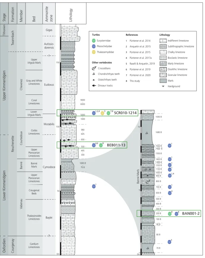
The Mesozoic sediments of the Porrentruy region belong to the tabular portion of the Jura Mountains (Marty et al., 2007). During the Kimmeridgian, this region was part of a SW-NE trending carbonate platform with various depositional environments such as lagoons, channels, and the littoral zone (Marty & Hug, 2003;Colombié & Strasser, 2005;
Marty, 2008). These rapidly changing sedimentation systems led to the formation of the
fossil-rich layers of the Banné Marls, the Courtedoux Member, and the Lower Virgula Marls (Marty & Hug, 2003;Comment et al., 2015;Fig. 2).
Table 1 Identified and figured elements of the assemblage MJSN BAN001-2. This table summarizes the bones of the assemblage MJSN BAN001-2 that were given second level identification numbers (format MJSN BAN001-2.x). For each element, we provide an identification and a reference to the figure in which the element is illustrated.
Specimen number Identification Figures
MJSN BAN001-2.1 Skull 3,4Aand4B
MJSN BAN001-2.2 Costal 1 (left) 8A
MJSN BAN001-2.3 Costal 1 (left) 8B
MJSN BAN001-2.4 Costal 1 (right) 8C
MJSN BAN001-2.5 Costal 1 (right) 8D
MJSN BAN001-2.6 Costal 1 (right) 8E
MJSN BAN001-2.7 Costal 1 (right) 8F
MJSN BAN001-2.8 Costal 5 (left) 10A
MJSN BAN001-2.9 Costal 5 (left) 10B
MJSN BAN001-2.10 Costal 5 (left) 10C
MJSN BAN001-2.11 Costal 5 (right) 10D
MJSN BAN001-2.12 Costal 5 (right) 10E
MJSN BAN001-2.13 Costal 5 (right) 10F
MJSN BAN001-2.14 Peripheral (bridge) 12A–12C
MJSN BAN001-2.15 Peripheral (bridge) 12D–12F
MJSN BAN001-2.16 Peripheral (bridge) 12G–12I
MJSN BAN001-2.17 Peripheral (bridge) 12J–12L
MJSN BAN001-2.18 Hyoplastron (right) 13A
MJSN BAN001-2.19 Hyoplastron (right) 13B
MJSN BAN001-2.20 Hyoplastron (left) 13C
MJSN BAN001-2.21 Hyoplastron (left) 13D
MJSN BAN001-2.22 Hyoplastron (left) 13E
MJSN BAN001-2.23 Hyoplastron (left) 13F
MJSN BAN001-2.24 Hyoplastron (left) 13G
MJSN BAN001-2.25 Hypoplastron (right) 14A
MJSN BAN001-2.26 Hypoplastron (right) 14B
MJSN BAN001-2.27 Hypoplastron (left) 14C
MJSN BAN001-2.28 Hypoplastron (left) 14D
Figure 2 Stratigraphic section of the Reuchenette Formation in Ajoie, Canton of Jura, Switzerland, with a close-up on the Banné Marls.The specimens described in this study are indicated by their collection number and circled in green. Main publishedfindings of “Eurysternidae” (green), “Thalassemydidae” (yellow) and “Plesiochelyidae” (blue) in the area are figured by colored turtle shell icons. Corresponding literature is indicated. Scheme modified afterComment et al. (2015)andRaselli & Anquetin (2019). Full-size DOI: 10.7717/peerj.9931/fig-2
The assemblage MJSN BAN001-2 was extracted from the Banné Marls in the Banné hill (SW of Porrentruy;Fig. 1). The approximately 10-m thick Banné Marls (early Kimmeridgian, Reuchenette Formation, Banné Member, Cymodoce Ammonite Zone;
Fig. 2;Comment et al., 2015) consist of“grey, dm-thick layers of marlstones, calcarenitic
marls, and marly limestones” (Jank, Wetzel & Meyer, 2006). They are especially rich in invertebrates (Koppka, 2015), but also contain remains of vertebrates such as turtles
(Püntener et al., 2014;Püntener, Anquetin & Billon-Bruyat, 2017a,2017b), crocodilians
(Schaefer, Püntener & Billon-Bruyat, 2018), andfishes (Leuzinger et al., 2017;Leuzinger,
Püntener & Billon-Bruyat, 2017). The turtle fauna of the Banné Marls is dominated by
the“plesiochelyids” Tropidemys langii and (less frequent) Plesiochelys bigleri (Püntener
et al., 2014;Püntener, Anquetin & Billon-Bruyat, 2017a;Raselli & Anquetin, 2019).
However, most of the turtle material of the Banné Marls does not originate from the Banné (BAN) site, but comes from stratigraphically younger layers excavated at the Vâ Tche Tchâ (VTT) site, which is situated about four kilometers northwest of the Banné (BAN) site (Figs 1and2).
The poorly preserved cranium MJSN SCR010-1214 originates from the Lower Virgula Marls (late Kimmeridgian, Reuchenette Formation, Chevenez Member, Eudoxus
Ammonite Zone; Fig. 2;Comment et al., 2015). This level is rich in vertebrate and wood remains (Philippe et al., 2010;Leuzinger et al., 2017;Leuzinger, Püntener & Billon-Bruyat,
2017;Püntener, Anquetin & Billon-Bruyat, 2017b;Schaefer, Püntener & Billon-Bruyat,
2018). The better part of the turtle remains from the Porrentruy region comes from the Lower Virgula Marls (Anquetin, Püntener & Billon-Bruyat, 2015;Püntener, Anquetin &
Billon-Bruyat, 2015,2017a,2020).
The mandible MJSN BEB011-13 was found in layer 501 at the base of the Corbis Limestones (early Kimmeridgian, Reuchenette Formation, Courtedoux Member, Cymodoce Ammonite Zone;Fig. 2;Comment et al., 2015). The Courtedoux Member is situated between the Banné Marls and the Lower Virgula Marls. This member is especially renowned for its dinosaur footprint bearing layers (Marty & Hug, 2003;Marty et al., 2007,
2018;Razzolini et al., 2017).
Anatomical comparisons and 3D models
The new material described herein was primarily compared to the available material of Solnhofia parsonsi. For the cranium and mandible, the three main specimens we used for comparison were TM 4023 (holotype of Solnhofia parsonsi), NMS 8741 and JM SCHA 70. The mandible is only known in TM 4023 and JM SCHA 70. For TM 4023, we used the 3D model ofEvers & Benson (2018)as primary source of data, completed by the available literature (Parsons & Williams, 1961;Gaffney, 1975;Evers & Joyce, 2020). We had NMS 8741 at hand during this study.Joyce (2000)was used for JM SCHA 70. In the following description, we refer to the relevant specimens when comparing Solnhofia brachyrhyncha to Solnhofia parsonsi. On rare occasions, we also used the specimens MNHN CNJ 82 and ICHLU 005.4.20 (see Introduction) based on personal observations. Following the work ofEvers & Joyce (2020), the new material was also compared with
sandownids when appropriate. These comparisons were made based on the published literature (Mateus et al., 2009;Tong & Meylan, 2013;Cadena, 2015;Evers & Joyce, 2020).
The 3D model of the cranium and mandible TM 4023 (Solnhofia parsonsi) was kindly provided byEvers & Benson (2018)and is available on Morphosource (see Additional Information). A textured 3D surface model of the cranium MJSN BAN001-2.1 (herein designated as the holotype of the new species Solnhofia brachyrhyncha) was also produced by photogrammetry (Anquetin & Püntener, 2020). This model can be downloaded from the MorphoMuseuM platform (see Additional Information).
For the shell, JM SCHA 70 is the only known specimen referred to Solnhofia
parsonsi in which the shell is sufficiently known. For this specimen, we used mainly the original description ofJoyce (2000). Concerning the plastral elements herein referred to Tropidemys? langii?, we used comparative material from different Kimmeridgian localities in the region of Porrentruy, Switzerland, as described previously by one of us (Püntener
et al., 2014). When needed, comparisons were extended to other thalassochelydians,
notably material from Solothurn and Porrentruy following some of our recent works
(Anquetin, Püntener & Billon-Bruyat, 2014;Püntener, Anquetin & Billon-Bruyat, 2017a).
Nomenclatural act
The electronic version of this article in Portable Document Format (PDF) will represent a published work according to the International Commission on Zoological Nomenclature (ICZN), and hence the new names contained in the electronic version are effectively published under that Code from the electronic edition alone. This published work and the nomenclatural acts it contains have been registered in ZooBank, the online registration system for the ICZN. The ZooBank LSIDs (Life Science Identifiers) can be resolved and the associated information viewed through any standard web browser by appending the LSID to the prefixhttp://zoobank.org/. The LSID for this publication is: [urn:lsid:zoobank. org:pub:C999A877-3080-490B-8202-6E98B621366E]. The online version of this work is archived and available from the following digital repositories: PeerJ, PubMed Central and CLOCKSS.
DESCRIPTION OF SOLNHOFIA BRACHYRHYNCHA N. SP.
CraniumGeneral description
The cranium MJSN BAN001-2.1 is relatively complete, but has suffered an intense postmortem deformation (Figs. 3and4). The skull was compressed obliquely from the anterodorsal left side toward the posteroventral right side, causing much of the right side of the skull to collapse ventrolaterally and posteriorly. Most of the bones are crisscrossed by numerous cracks that make many sutures difficult to see. The tip of the snout and the area around the apertura narium externa, most of the upper and lower temporal regions, and the squamosals are missing. Disarticulation occurred in several places, notably between the left otic chamber and the roof of the cavum cranii, along the left postorbital region, and over much of the right palatoquadrate area and right otic chamber
A B C D E F G 10 mm pf fr mx pal ju ju po pr pa pa so qu op fst qj ct ex co bo cs sq? op qu so fst pr po ju pm pf fr po mx ju ju mx mx pal pal ju fpp ppe pr pto qj qu pt cm qu pbs pt ptf ? op fpcci ex bo co excs pt qu op qu pt pr pa ppe fnt pto lar na? pa fas
Figure 3 MJSN BAN001-2.1, holotype of Solnhofia brachyrhyncha (Kimmeridgian, Porrentruy, Switzerland).(A) Cranium in dorsal view; (B) interpretative drawing of the cranium in dorsal view; (C) cranium in ventral view; (D) interpretative drawing of the cranium in ventral view; (E) cranium in anterior view; (F) cranium in right lateral view; (G) cranium in left lateral view. In the drawings, hatchings correspond to damaged areas, while the light brown color represents the remaining matrix. Abbreviations: bo, basioccipital; cm, condylus mandibularis; co, condylus occipitalis; cs, crista supraoccipitalis; ct, cavum tympani; ex, exoccipital; fas, foramen alveolare superius; fnt, foramen nervi trigemini; fpcci, foramen posterius canalis carotici interni; fpp, foramen palatinum posterius; fr, fontal; fst, foramen stapedio-temporale; ju, jugal; lar, labial ridge; mx, maxilla; na, nasal; op, opisthotic; pa, parietal; pal, palatine; pbs, parabasisphenoid; pf, prefrontal; pm, premaxilla; po, postorbital; ppe, pro-cessus pterygoideus externus; pr, prootic; pt, pterygoid; ptf, pterygoid fossa; pto, propro-cessus trochlearis oticum; qj, quadratojugal; qu, quadrate; so, supraoccipital; sq, squamosal; ?, unnamed foramen.
when interpreting the morphology of this specimen. The left side of the cranium is generally the one that is closer to the natural condition. No scute sulcus is preserved on the skull roof.
The general impression is that the snout of MJSN BAN001-2.1 is not as elongated as the one of TM 4023, NMS 8741 or JM SCHA 70. The triturating surface also appears to be broader posteriorly (Figs. 3Cand3D; see Maxilla below). These differences in cranial shapes are reflected in the general dimensions of the available specimens (Table 1). Skulls referred to Solnhofia parsonsi are longer than wide (length/width ratio ranging
approximatively from 1.30 to 1.45), while the skull of Solnhofia brachyrhyncha is only slightly longer than wide (length/width ration of about 1.10). Since most available specimens are either incomplete or deformed somehow, these measurements and proportions must be handled with care, but the general trend remains true. Solnhofia parsonsi (JM SCHA 70) is characterized by a large head representing 40% of the carapace length (Joyce, 2000). Unfortunately, the cranium MJSN BAN001-2.1 is not associated with particular shell remains so its relative size cannot be determined with precision. However, if the estimated size range of the specimens represented in the assemblage MJSN BAN001-2 (150–250 mm in carapace length) is taken into account, we can surmise
5 mm A B C D mx pal pt pa pr qu ex bo mx qu pr pt epi pa ju pal po epi fnt ppe bo fnt ppe pto pto
Figure 4 Comparison of the region of the processus trochlearis oticum and foramen nervi trigemini in Solnhofia brachyrhyncha and Solnhofia parsonsi. (A) Photograph of the cranium MJSN BAN001-2.1 (So. brachyrhyncha) in right anteroventral view; (B) same as in (A) with bones and sutures highlighted; (C) three-dimensional rendering of the cranium TM 4023 (So. parsonsi) in right anteroventral view; (D) same as in (C) with bones highlighted. (C) and (D) fromEvers & Benson (2018). Abbreviations: bo, basioccipital; epi, epipterygoid; ex, exoccipital; fnt, foramen nervi trigemini; mx, maxilla; pa, parietal; pal, palatine; ppe, processus pterygoideus externus; pr, prootic; pt, pterygoid; pto, processus trochlearis oti-cum; qu, quadrate. Full-size DOI: 10.7717/peerj.9931/fig-4
that Solnhofia brachyrhyncha is also a large-headed species with a skull representing between 25.7% and 42.8% of the carapace length.
The preservation state of the cranium MJSN SCR010-1214 is relatively poor (Fig. 5). The cranium is severely crushed dorsoventrally (Figs. 5Eand5F) and most of its ventral aspect is eroded (Figs. 5Cand5D). The palatal, maxillary, nasal, and most of the orbital regions are missing. Most of the preserved bones are densely fractured, except those forming the dorsal part of the otic chambers. Despite its poor preservation state, this skull documents areas that are badly preserved or absent in the holotype cranium, as highlighted in the following detailed description.
Nasal
Nasals are well developed in most thalassochelydians, including Solnhofia parsonsi (NMS 8741 and JM SCHA 70). Therefore, they were likely present in Solnhofia brachyrhyncha as well, but the relatively poor preservation of the region of the apertura narium externa in MJSN BAN001-2.1 prevents any confirmation. Fragments of bones lying anterior to the prefrontals might correspond to remnants of the nasals (Figs. 3Aand3B). The nasal region is missing in MJSN SCR010-1214 (Fig. 5).
Prefrontal
The prefrontals are badly preserved in MJSN BAN001-2.1 and missing in MJSN SCR010-1214. The right prefrontal is broken into several pieces, but appears to be a roughly quadrangular element contacting the nasal anteriorly, the ascending process of the maxilla anterolaterally, the frontal posteriorly, and the other prefrontal medially along its entire length (Fig. 3Aand3B). The left prefrontal is preserved as an elongate element forming the anterodorsal margin of the orbit. The anterior and medial parts of the left prefrontal are damaged. Only the posterior contact with the left frontal and part of the medial contact with the right prefrontal are preserved. The posterior suture with the frontal and the anterior suture with the nasal were probably mostly transverse, but it is difficult to be sure. The descending process of the prefrontal is broken on both sides, so nothing can be said about the contacts of the prefrontal in the anterior part of the fossa orbitalis and region of the foramen orbito-nasale.
Frontal
The frontals are large elements forming most of the dorsal margin of the orbits. Each of the frontals contacts the prefrontal anteriorly, the postorbital posterolaterally, the parietal posteriorly, and its counterpart medially (Fig. 3Aand3B). The suture with the parietal is transverse and strongly interdigitating. The suture with the postorbital is V-shaped and concave relative to the frontal, which reminds the condition in Solnhofia parsonsi (TM 4023 and NMS 8741). The anterior suture with the prefrontal is probably transverse, but the preservation in that area is imperfect. In many thalassochelydians, including Solnhofia parsonsi, the frontals extend anteromedially below the prefrontal and contact the nasals in the roof of the fossa nasalis. The state of preservation of MJSN BAN001-2.1 prevents us from telling whether this is also a condition present in Solnhofia
Parietal
On the skull roof, the parietal meets the frontal anteriorly, the postorbital laterally and the other parietal medially (Figs. 3Aand3B). In both crania, the posterior part of the bone is shattered in many fragments and it is unclear whether the parietal contacted the squamosal behind the postorbital, although it seems unlikely (see Squamosal). Of the posterior margin of
A
B
E
F
10 mmD
C
fr po pa po pr pa pto so op qu sq qj fst pa po cs op sq sq qj qu so po fr pa pa pr qu qu pt qj pr sq op qu sq ex cs qu qj fpcci pto pto fnt pr po ctFigure 5 MJSN SCR010-1214, cranium of Solnhofia brachyrhyncha (Kimmeridgian, Porrentruy, Switzerland). (A) cranium in dorsal view; (B) interpretative drawing of the cranium in dorsal view; (C) cranium in ventral view; (D) interpretative drawing of the cranium in ventral view; (E) cranium in posterior view; (F) cranium in right lateral view. Note that the ventral aspect of the basicranium is severely eroded, while the palatal and orbital regions are missing. Hatchings correspond to damaged areas. Abbreviations: cs, crista supraoccipitalis; ct, cavum tympani; ex, exoccipital; fnt, foramen nervi trigemini; fr, frontal; fpcci, foramen posterius canalis carotici interni; fst, foramen stapedio-temporale; op, opisthotic; pa, parietal; po, postorbital; pr, prootic; pt, pterygoid; pto, processus trochlearis oticum; qj, quadratojugal; qu, quadrate; so, supraoccipital; sq, squamosal.
the parietal, only the anteriormost embayment of the right upper temporal emargination is preserved in MJSN BAN001-2.1 (Figs. 3Aand3B). The development and shape of the upper temporal emargination appear to be similar to what is known (also imperfectly) in Solnhofia parsonsi (NMS 8741 and MNHN CNJ 82).
As usual, the parietals form the anterior half of the roof of the cavum cranii and send a well-developed process ventrally to form the lateral braincase wall. Like in Solnhofia parsonsi, the parietal contributes significantly to the large processus trochlearis oticum
(Figs. 3A–3D,4A,4B and5A–5D).Evers & Joyce (2020)recently described a similar
condition in the sandownid Sandownia harrisi, but the contribution of the parietal to the processus trochlearis oticum in that taxon is much more reduced than in So. parsonsi and So. brachyrhyncha. The descending process of the parietal forms most of the posterior and anterior margins of the foramen nervi trigemini (Figs. 4Aand4B). The ventral margin of the foramen nervi trigemini is formed by the pterygoid (see Pterygoid). In So. parsonsi (TM 4023), the contribution of the parietal is limited to the dorsal margin of the foramen nervi trigemini, whereas the pterygoid forms the anteroventral, ventral and most of the posterior margins of the foramen (Fig. 4Cand4D). Posterior to the foramen nervi trigemini, the parietal has a broad contact with the pterygoid in
So. brachyrhyncha, clearly excluding the prootic from the posterior margin of the foramen nervi trigemini (Figs. 4Aand4B). In So. parsonsi (TM 4023), the parietal-pterygoid contact is much more reduced and the prootic extends closer to the posterior margin of the foramen nervi trigemini (Figs. 4Cand4D). Anterior to the foramen nervi trigemini, the descending process of the parietal and the pterygoid are fully separated by the epipterygoid in lateral view, but there is apparently a parietal-pterygoid point contact medial to the epipterygoid along the anterior margin of the foramen nervi trigemini (Figs. 4Aand4B). It is unknown whether this contact is more extensive on the medial surface of the lateral braincase wall. In So. parsonsi (TM 4023), the epipterygoid apparently fully separates the parietal and pterygoid in both lateral and medial views.
Jugal
The jugal is only preserved in MJSN BAN001-2.1, but it is damaged on both sides so that the limits and contacts of the bone are somewhat unclear. It appears that the jugal forms the posteroventral margin of the orbit and contacts the postorbital dorsally, the maxilla anteroventrally, and the pterygoid posteroventrally (Figs. 3A–3D). As in Solnhofia parsonsi
(TM 4023, NMS 8741, JM SCHA 70), the jugal also has an anteromedial contact with the palatine in thefloor of the fossa orbitalis, but in contrast to the aforementioned species the jugal does not extend as far anteriorly as the level of the foramen orbito-nasale in Solnhofia brachyrhyncha (Figs. 3Aand3B). As in So. parsonsi (TM 4023) and Sandownia harrisi (Evers & Joyce, 2020), the medial process of the jugal forms a long posteromedial extension along the posteromedial border of the maxilla and the anterolateral border of the pterygoid (Figs. 3Cand3D). As preserved in MJSN BAN001-2.1, this posteromedial extension of the jugal is partly visible in ventral view, and appears to contact the maxilla and pterygoid laterally. However, parts of the bone still in place indicate that this extension of the jugal was in contact with the dorsal surface of the maxilla and pterygoid and therefore
was not visible in ventral view, similar to So. parsonsi (TM 4023). This differs from Sa. harrisi where the posteromedial extension of the medial process of the jugal extends laterally to the pterygoid and contributes to the secondary palate (Evers & Joyce, 2020). The more lateral part of the medial process of the jugal forms a vertical ridge that partly delimits the fossa orbitalis posteriorly and extends dorsally on the medial surface of the main part of the jugal, as preserved on the right side of MJSN BAN001-2.1 (see 3D Model; Additional Information;
Anquetin & Püntener (2020)). A similar ridge is present in So. parsonsi (TM 4023, NMS
8741) and Sa. harrisi (Evers & Joyce, 2020). Posteriorly, the medial process of the jugal reaches the anterodorsal corner of the processus pterygoideus externus. The medial process of the jugal reaches as far posteriorly in So. parsonsi (TM 4023, NMS 8741), but does not really contact the more reduced processus pterygoideus externus (see Pterygoid).
Quadratojugal
In MJSN BAN001-2.1, only a very small fragment of the left quadratojugal is preserved along the anterior margin of the cavum tympani (Figs. 3A–3Dand3G). In MJSN SCR010-1214, more of the quadratojugal is preserved, but the bone is quite fragmented
(Figs. 5A–5Dand5F). As preserved, the quadratojugal contacts the squamosal above the
cavum tympani and the quadrate ventrally and medially. The quadratojugal appears to brace the cavum tympani anteriorly without entering its margin. Dorsally, there is apparently a contact between the quadratojugal and the postorbital, but this area is much fragmented on both sides. As in Solnhofia parsonsi (TM 4023) and most sandownids
(Evers & Joyce, 2020), the quadratojugal forms the lateralmost part of the extensively
developed processus trochlearis oticum.
Squamosal
Except maybe for a small fragment on the right side (Figs. 3Aand3B), the squamosal is entirely missing in MJSN BAN001-2.1. The two squamosals are present in MJSN SCR010-1214, but they are incompletely preserved and broken into several pieces (Fig. 5). The squamosal contacts the quadratojugal anterolaterally above the cavum tympani, the quadrate anteriorly and ventrally, and the opisthotic anteromedially. As in most
turtles, the squamosal apparently formed the antrum postoticum, although this area is poorly preserved here. The posterior process of the squamosal forms a subvertical (or slightly oblique) lamina extending far behind the level of the occipital plane (Fig. 5). This apparently corresponds to what is known in Solnhofia parsonsi (TM 4023, NMS 8741, JM SCHA 70), although the posteriormost part of the squamosal is not fully preserved or accessible in any of these specimens. Anterodorsally, the lamina curves medially and forms the posterolateral margin of the upper temporal emargination. More dorsally and anteriorly, the squamosal is broken on either side so that the contacts with other bones in that area remain unclear. However, it seems unlikely that the squamosal contacted the parietal in Solnhofia brachyrhyncha.
Postorbital
The postorbital is incompletely preserved in both of the available crania. In MJSN BAN001-2.1, only the anteriormost part of the left postorbital is preserved, while the right
postorbital is represented by a larger portion extending from the orbit to the anteriormost part of the upper temporal emargination (Figs. 3A,3Band3F). A comparable portion of the left postorbital is preserved in MJSN SCR010-1214, but its dorsal aspect is heavily fragmented (Figs. 5A–5D). A triangular, fragmented piece of bone lying on top of the right processus trochlearis oticum probably corresponds to the posterior part of the right postorbital, but it is impossible to confirm. The postorbital forms the posterodorsal margin of the orbit. As preserved, the postorbital contacts the frontal anteromedially, the parietal medially, and the jugal ventrally. A contact with the quadratojugal was probably also present posteroventrally (see left side of MJSN SCR010-1214;Figs. 5Aand5B), but the area is poorly preserved. A strong ridge runs on the medial surface of the postorbital
(Figs. 5Cand5D). A similar ridge is present in Solnhofia parsonsi (TM 4023, NMS 8741)
and Sandownia harrisi, and continues on the medial surface of the jugal (Evers & Joyce, 2020). Therefore, it appears likely that the ridges observed on the medial surface of the postorbital (MJSN SCR010-1214) and jugal (MJSN BAN001-2.1) of Solnhofia brachyrhyncha were also continuous.
Premaxilla
The premaxilla is poorly known in Solnhofia brachyrhyncha. Only the palatal part of the two premaxillae is preserved in MJSN BAN001-2.1 (Figs. 3Cand3D). There, the premaxilla contacts the maxilla laterally and the other premaxilla medially. The vomer is missing in that specimen, so nothing can be said about the relation beween this bone and the premaxilla, or the presence/absence of the foramen preapalatinum. More anterior portions of the premaxillae such as their contribution to the labial ridge and to the ventral border of the apertura narium externa are entirely missing. It is therefore impossible to tell whether the premaxillae projected anteriorly as they do so characteristically in Solnhofia parsonsi (TM 4023). In the latter, the palatal part of the premaxilla is flat and contributes fully to the triturating surface. In Solnhofia brachyrhyncha, the palatal part of the premaxilla is apparently divided into a flat lateral surface continuous with the triturating surface of the maxilla and a dorsally arched medial surface delimiting a midline trough. However, this observation may as well be the result of deformation and
disarticulation in MJSN BAN001-2.1. More material is needed to confirm or contradict this potential difference between So. brachyrhyncha and So. parsonsi.
Maxilla
The maxilla is only preserved in MJSN BAN001-2.1. It contacts the premaxilla anteromedially, the prefrontal (and probably also the nasal) anterodorsally, the jugal posterodorsally, the pterygoid posteriorly, and the palatine posteromedially (Figs. 3A–3D). The maxilla probably also contacted the vomer medially, but this bone is lost in MJSN BAN001-2.1 (see Vomer). The maxilla forms the ventral and anteroventral margins of the orbit. As noted above, the jugal does not extend as far anteriorly in thefloor of the fossa orbitalis as it does in Solnhofia parsonsi. As a result, the maxilla forms a larger portion of the floor of the fossa orbitalis in So. brachyrhyncha. Since the ventral process of the prefrontal is damaged on both sides, the foramen orbito-nasale is not readily observable in
MJSN BAN001-2.1. However, the foramen alveolare superius, which opens just medial to the lateral margin of the foramen orbito-nasale in most turtles including So. parsonsi, is visible in the left side of that specimen. In many thalassochelydians including Plesiochelys etalloni, Portlandemys mcdowelli and Jurassichelon oleronensis (Gaffney, 1976;Anquetin,
Püntener & Billon-Bruyat, 2015), as well as So. parsonsi (TM 4023, NMS 8741), a small
unnamed foramen is present lateral to the foramen orbito-nasale just inside (as it is the case for So. parsonsi) or outside of the anteroventral corner of the orbit. This area is damaged in each side of MJSN BAN001-2.1, so that the presence or absence of this unnamed foramen in So. brachyrhyncha cannot be elucidated. However, a foramen occurs more posteriorly in thefloor of the fossa orbitalis approximately at midway along the ventral margin of the orbit. This foramen is only visible in the right side of MJSN BAN001-2.1, but its presence on the left side cannot be confirmed as the corresponding area is damaged and partly covered by matrix. This foramen may correspond to the unnamed foramen of other thalassochelydians.
As in So. parsonsi (TM 4023, NMS 8741, JM SCHA 70), the secondary palate of So. brachyrhyncha is formed primarily by the maxillae (Figs. 3Cand3D). The following characteristics are also similar in the two species: the labial ridge is acute, moderately high, and lined with moderately sized nutritive foramina; the triturating surface itself is broadly arched dorsally and pierced by a few large nutritive foramina; the lingual ridge is nonexistent. However, the shape of the triturating surface is different. In So. parsonsi (TM 4023, NMS 8741, JM SCHA 70), the triturating surface is V-shaped with straight lateral margins, and the width of each ramus of the V remains the same anteroposteriorly. The triturating surface is also much elongated in this species in relation with the great elongation of the snout. In So. brachyrhyncha, each ramus of the triturating surface is very wide posteriorly and reduces much in width anteriorly, notably from the level of the middle of the orbit. As a result, the lateral margins of the triturating surface are slightly sinusoidal in ventral view, the anterior half being concave laterally and the posterior half concave medially (Figs. 3Cand3D).
Vomer
The vomer is apparently disarticulated and lost in MJSN BAN001-2.1 (Figs. 3Cand3D). A minute fragment of bone between the two maxillae may be a remnant of the vomer, but reveals nothing noteworthy. The palate, including the vomer, is entirely missing in MSJN SCR010-1214.
Palatine
The palatines are preserved only in MJSN BAN001-2.1, but their state of preservation is quite poor (Figs. 3A–3D). The palatine contacts the maxilla anterolaterally, the jugal laterally in thefloor of the fossa orbitalis, and the pterygoid posteriorly. Anterior contacts with the vomer and descending process of the prefrontal were probably also present, but these elements are lost or damaged in that specimen. The palatine borders the apertura narium interna and forms the roof of the narial passage. In Solnhofia parsonsi (TM 4023, NMS 8741, JM SCHA 70), the palatine contributes to the triturating surface and forms
an incompletefloor to the narial passage. This contribution of the palatine to the triturating surface and partialflooring of the narial passage are absent in So. brachyrhyncha
(Figs. 3Cand3D). The posterior half of the palatine is poorly preserved, but zigzagging
sutural marks on the pterygoid indicate that the palatine extended as far posteriorly as the posterior part of the processus pterygoideus externus, which reminds the condition in So. parsonsi (TM 4023, JM SCHA 70). The foramen palatinum posterius (only preserved on the left side of MJSN BAN001-2.1) is a small oval opening located anterior to the level of the processus pterygoideus externus. As preserved, the foramen palatinum posterius appears to be formed by the palatine medially and the pterygoid laterally. In So. parsonsi (TM 4023), the foramen palatinum posterius consists in a short canal formed primarily by the palatine with a modest lateral contribution from the pterygoid, but the ventral opening of this canal is formed entirely by the palatine (Evers & Joyce, 2020). However, this is not readily observable from external examination because previous authors have described the foramen palatinum posterius as being formed by the palatine and pterygoid in So. parsonsi (Parsons & Williams, 1961,Gaffney, 1975;Joyce, 2000). Therefore, we consider that the state of preservation of MJSN BAN001-2.1 prevents proper comparison between So. brachyrhyncha and So. parsonsi regarding which bones form the foramen palatinum posterius.
Quadrate
The quadrate can be observed in both MJSN BAN001-2.1 and MSJN SCR010-1214, but with variable degrees of preservation depending on specimen and side (Figs. 3–5). As usual, the quadrate forms most of the lateral part of the processus trochlearis oticum (with a lateral contribution from the quadratojugal), the posterolateral half of the foramen stapedio-temporale, the cavum tympani and the incisura columellae auris (which remained open posteroventrally), and the condylus mandibularis. In dorsal view, the quadrate contacts the prootic anteromedially, the opisthotic posteromedially, the
squamosal posteriorly, and the quadratojugal laterally (Figs. 3A,3B,5Aand5B). Below the processus trochlearis oticum, the quadrate contacts the prootic anteromedially, the pterygoid medially and ventrally, and the quadratojugal laterally (Figs. 3C,3D,4,5Cand
5D). In the roof of the cavum acustico-jugulare and posterior to the incisura columellae auris, the quadrate contacts the prootic anteromedially (forming the aditus canalis stapedio-temporalis), the opisthotic posteromedially, and the squamosal posterolaterally
(Figs. 3C,3D,5Cand5D). The quadrate contributes more to the processus trochlearis
oticum than in Solnhofia parsonsi (TM 4023, NMS 8741) to the detriment of the prootic
(Fig. 4; see Prootic). The condylus mandibularis is best preserved in the left side of MJSN
BAN001-2.1, but the condyle is not complete. The articular surface appears to be just slightly concave. As in So. parsoni (TM 4023, JM SCHA 70), the lateral lobe of the condyle articular surface seems to be larger than the medial lobe. The condylus mandibularis is located anterior to the level of the occipital plane in So. brachyrhyncha as in most other turtles, which differs from the condition in So. parsonsi (TM 4023, NMS 8741, JM SCHA 70) where the condylus mandibularis is situated at the level of the occipital plane. As in all thalassochelydians, and to a lesser extent sandownids, a strong ventrally infolding
ridge is developed on the posterior surface of the quadrate articular process below the incisura columellae auris. This ridge prolongates the posterior margin of the pterygoid laterally, passes below the incisura columellae auris, then bends strongly ventrally and terminates at the lateroventral corner of the mandibular process of the quadrate (see 3D Model; Additional Information;Anquetin & Püntener (2020)).
Epipterygoid
The epipterygoid is only preserved in MJSN BAN001-2.1. It contacts the parietal dorsally and the pterygoid ventrally (Figs. 4Aand4B). It is possible that the ventral part of the bone is broken revealing the crista pterygoidea medial to it. But in any case, the lateral exposure of the epipterygoid is more limited in Solnhofia brachyrhyncha reaching only as high as the center of the foramen nervi trigemini, whereas it extends as high as the top of the foramen nervi trigemini in So. parsonsi (TM 4023). The contribution of the epiterygoid to the anterior margin of the foramen nervi trigemini appears to be limited to a point contact (Figs. 4Aand4B), but this may be an artefact due to the breaking of the lower part of the bone. In So. parsonsi (TM 4023), the contribution of the epipterygoid to the anterior margin of the foramen nervi trigemini is more extensive and located more dorsally along the anterior margin of the foramen (Figs. 4Cand4D). As described above (see Parietal), the parietal and pterygoid are contacting one another medial to the epipterygoid along the anterior margin of the foramen nervi trigemini, but it is unknown whether this contact is more extensive on the medial aspect of the lateral braincase wall. In So. parsonsi (TM 4023), the epipterygoid fully separates the parietal from the pterygoid in both lateral and medial view. Finally, So. brachyrhyncha lacks the well-developed blunt ventral process on the lateral surface of the epipterygoid that is present in So. parsonsi (TM 4023, NMS 8741;Figs. 4Cand4D).
Pterygoid
The pterygoid is best preserved in MJSN BAN001-2.1 (Figs. 3A–3D,4Aand4B). In MJSN SCR010-1214, only a part of the ventral aspect of the pterygoid is present (Figs. 5Cand
5D). In ventral view, the pterygoid contacts the jugal anterodorsally, the maxilla anterolaterally, the palatine anteriorly, the other pterygoid medially for its anterior half, the parabasisphenoid medially for its posterior half, and the quadrate posterolaterally. The suture between the parabasisphenoid and basioccipital is not clear in MJSN BAN001-2.1 so it is uncertain whether the pterygoid contacted the basioccipital in Solnhofia brachyrhyncha. The condition in So. parsonsi is actually somewhat ambiguous. Based on external examination previous authors have described a pterygoid-basioccipital contact in this species (Parsons & Williams, 1961,Gaffney, 1975;Joyce, 2000). However, CT scan data reveal that the parabasisphenoid is intercalated between the pterygoid ventrally and the basioccipital dorsally in TM 4023 (Evers & Benson, 2018). If present, the contact between the pterygoid and basioccipital is probably limited. As much as can be interpreted from MJSN BAN001-2.1, the condition in So. brachyrhyncha does not appear to be much different from the one in So. parsonsi (Figs. 3Cand3D).
Anteriorly, the suture with the palatine is somewhat unclear, but zigzagging marks on the pterygoid indicate the posterior extension of the sutural contact with the palatine (see Palatine). In So. parsonsi (TM 4023, NMS 8741, JM SCHA 70), the choanae extend much posteriorly over the pterygoids well beyond the level of the processus pterygoideus externus. In So. brachyrhyncha, the choanae are limited to the palatine as common in most turtles (Figs. 3Cand3D). In So. parsonsi (TM 4023, NMS 8741, JM SCHA 70), the processus pterygoideus externus is reduced to a small nubbin. In So. brachyrhyncha, the processus pterygoideus externus is more developed, albeit much reduced compared to most turtles. The processus pterygoideus externus is a short process ovoid in section projecting laterally from the pterygoid (Figs. 3C,3D,4A and4B). As noted above (see Jugal), the anterodorsal corner of the processus pterygoideus externus contacts the posterior tip of the medial process of the jugal. Although the jugal reaches as posteriorly in So. parsonsi (TM 4023, NMS 8741), it does not contact the much reduced processus pterygoideus externus. An arched ridge runs on the ventral surface of the pterygoid from the posterior part of the processus pterygoideus externus to the posterior limit of the bone very close to the suture with the parabasisphenoid. In constrast to So. parsonsi (TM 4023, NMS 8741, JM SCHA 70), this ridge is weakly developed in So. brachyrhyncha.
The foramen posterius canalis carotici interni opens in the posteromedial corner of the pterygoid and is apparent in ventral view, as preserved (Figs. 3Cand3D). However, this area is somewhat damaged in the two available specimens. In So. parsonsi (TM 4023), the foramen posterius canalis carotici interni opens slightly more posteriorly and dorsal to the arched ridge running on the ventral surface of the pterygoid, so that the foramen is partly hidden in ventral view. Therefore, the condition in the two species is maybe slightly different. On the left side of MJSN BAN001-2.1, a small foramen opens a short distance anterior to the foramen posterius canalis carotici interni, just medial to the arched ridge running on the pterygoid and approximately halfway along the suture between the pterygoid and parabasisphenoid (Figs. 3Cand 3D). A similar foramen is present in So. parsonsi (TM 4023, NMS 8741). The nature of this foramen is
controversial (Gaffney, 1975). The pterygoid fossa is similarly deep in So. parsonsi (TM 4023, NMS 8741) and So. brachyrhyncha, but the fossa is more pinched mediolaterally in the former species.
Below the processus trochlearis oticum and in the ethmoid region, the pterygoid contacts the quadrate posterolaterally, the prootic posterodorsally, the parietal
posteromedially, and the epipterygoid anteromedially (Figs. 4Aand4B). Compared to So. parsonsi (TM 4023;Figs. 4Cand4D), the contact of the pterygoid with the prootic and its contribution to the foramen nervi trigemini are reduced due to the greater posteroventral extension of the parietal behind the foramen nervi trigemini. The pterygoid forms the ventral margin of the foramen nervi trigemini. As noted above (see Parietal), the pterygoid apparently also contacts the parietal anterior to the foramen nervi trigemini and medial to the epipterygoid. The extent of this contact on the medial surface of the lateral braincase wall is uncertain.
Supraoccipital
The supraoccipital can be observed in both MJSN BAN001-2.1 and MJSN SCR010-1214
(Figs. 3and5). It contacts the parietal anterodorsally, the prootic anterolaterally, the
opisthotic posterolaterally, and the exoccipital posteroventrally. A contact between the prootic and opisthotic prevents the supraoccipital from contacting the quadrate posterior to the foramen stapedio-temporale. The contacts of the supraoccipital are similar in Solnhofia parsonsi (TM 4023). The crista supraoccipitalis extends greatly posterior to the occipital plane. The posterior tip of the crista supraoccipitalis is apparently missing in MJSN SCR010-1214, but the crista reaches almost as far posteriorly as the squamosals
(Figs. 5A–5D). This also corresponds to the condition in So. parsonsi (JM SCHA 70).
Exoccipital
The exoccipitals are best preserved in MJSN BAN001-2.1 (Figs. 3A–3D). The exoccipital contacts the supraoccipital dorsally, the opisthotic laterally and the basioccipital ventrally. There is no contact between the exoccipital and pterygoid. The exoccipital forms the lateral margin of the foramen magnum and the dorsolateral lobe of the three-lobed condylus occipitalis. The exoccipitals appear to meet one another in the ventral margin of the foramen magnum. Ventrally, the exoccipital is pierced by a pair of foramina nervi hypoglossi. This morphology is similar to that of Solnhofia parsonsi (TM 4023).
Basioccipital
The basioccipital is only observable in MJSN BAN001-2.1, but its preservation is rather poor (Figs. 3A–3D). It contacts the parabasisphenoid anteriorly along a more or less transverse suture and the exoccipital dorsally. An anterolateral contact with the pterygoid was possibly also present on the ventral aspect of the skull, but cannot really be confirmed based on the available material (see Pterygoid). The ventral exposure of the basioccipital is somewhat quadrangular and wider than long. However, the ventral surface of the bone is eroded in MJSN BAN001-2.1. The basioccipital forms the ventral lobe of the three-lobed condylus occipitalis.
Prootic
The prootic can observed in both MJSN BAN001-2.1 (Figs. 3,4Aand4B) and MJSN SCR010-1214 (Fig. 5). In the dorsal surface of the otic chamber, the prootic contacts the parietal anteromedially, the supraoccipital posteromedially, the opisthotic posteriorly, and the quadrate posterolaterally. It forms the anteromedial half of the foramen
stapedio-temporale. Medial to this foramen, the dorsal surface of the prootic is deeply incised by a medially oriented trough marking the passage of the arteria stapedialis.
Below the processus trochlearis oticum, the prootic contacts the quadrate laterally, the pterygoid ventrally, and the parietal medially. As noted for most thalassochelydians
(Evers & Benson, 2019), the processus trochlearis oticum is bordered laterally by a deep
recess (Figs. 3and5). As in Solnhofia parsonsi (TM 4023, NMS 8741, JM SCHA 70) and
sandownids, the processus trochlearis oticum of Solnhofia brachyrhyncha is greatly developed and projecting much forward, hiding the foramen nervi trigemini in lateral view. However, a number of differences can be observed between the two species of
Solnhofia. In dorsal or ventral view, the processus trochlearis oticum is more oblique in So. brachyrhyncha, whereas it is oriented more transversely in So. parsonsi. The contribution of the prootic to the processus trochlearis oticum is reduced in
So. brachyrhyncha compared to So. parsonsi (Fig. 4). As noted above, the quadrate and the parietal (especially ventrally) contribute more to the processus trochlearis oticum in So. brachyrhyncha. Finally, the processus trochlearis oticum, notably the part formed by the prootic, is not as deeply concave in So. brachyrhyncha (Fig. 4). In So. parsonsi (TM 4023), the prootic is prevented from entering the posterior margin of the foramen nervi trigemini only by a short contact between the parietal and pterygoid (Figs. 4Cand
4D). In So. brachyrhyncha, the parietal-pterygoid contact is much broader and the prootic is more clearly excluded from the posterior margin of the foramen nervi trigemini in lateral view (Figs. 4Aand4B).
In the cavum acustico-jugulare, the prootic contacts the pterygoid ventrally, the opisthotic dorsomedially, and the quadrate dorsolaterally. There was probably also a medioventral contact with the parabasisphenoid, but this contact is not preserved in MJSN BAN001-2.1. The prootic forms the medial half of the aditus canalis stapedio-temporalis and the roof of the posterior part of the canalis cavernosus, the latter opening just ventromedial to the former. The morphology of this area is similar in So. parsonsi (TM 4023).
Opisthotic
The opisthotic contacts the exoccipital posteromedially, the supraoccipital anteromedially, the prootic anteriorly, the quadrate laterally, and the squamosal posterolaterally (Figs. 3
and5). The anterior contact with the prootic prevents the quadrate from meeting the supraoccipital on the dorsal surface of the otic chamber. The paroccipital process is oriented mostly posteriorly and extends much beyond the level of the occipital plane, more so in ventral aspect. The occipital part of the opisthotic forms a broad, moderately concave triangular surface delimited dorsally and ventrally by a pronounced, vertical ridge. This is similar to the condition in Solnhofia parsonsi (TM 4023), but differs for example from the rod-like structure observed in Plesiochelys planiceps or the ridge-like structure in Sandownia harrisi (Evers & Joyce, 2020). In the cavum acustico-jugulare, the opisthotic contacts the exoccipital posteromedially, the prootic anteriorly, and the quadrate laterally. The processus interfenestralis is only preserved (partly) on the right side of the MJSN BAN001-2.1, the side most affected by deformation. It seems that the processus
interfenestralis does not contact the pterygoid or the basioccipital ventrally, which is also the condition in So. parsonsi (TM 4023). The foramen externum nervi glossopharyngei pierces the dorsal base of the processus interfenestralis of the opisthotic close to its lateral margin.
Parabasisphenoid
The parabasisphenoid can be observed only in MJSN BAN001-2.1 (Figs. 3Cand3D). Only the ventral aspect of the bone can be seen because the interior of the cranium isfilled up with matrix. The parabasisphenoid contacts the pterygoid anteriorly and laterally, and
the basioccipital posteriorly. The ventral exposure of the parabasisphenoid is triangular and more elongated than in Sonhofia parsonsi (TM 4023, NMS 8741, JM SCHA 70). Anteriorly, the parabasisphenoid reaches almost the level of the posterior part of the processus pterygoideus externus in So. brachyrhyncha, whereas it barely goes beyond the level of the anterior limit of the pterygoid fossa in So. parsonsi (TM 4023, NMS 8741).
Mandible
General description
MJSN BEB011-13 is a relatively small mandible (Fig. 6). The right ramus is complete, but the left one is broken at the level of the anterior margin of the fossa meckelii and the posterior portion of this ramus is missing. The mandible suffered some deformation during fossilization: the dorsal aspect of the mandible is shifted slightly to the left compared to the ventral aspect. The bones are crisscrossed by many small fractures that sometimes complicate the interpretation of sutures. These fractures and other areas, such as the anterior tip of the symphysis and the right articular surface are encrusted by iron oxides (Fig. 6).
The anterior part of the mandible has a relativelyflat and low profile (Figs. 6Eand6F). Posteriorly, the processus coronoideus is greatly developed. This somewhat reminds the condition in Solnhofia parsonsi (TM 4023). However, the outline of the mandible in dorsal or ventral view is remarkable by the presence of large dentary tubercles projecting laterally on each side. This is radically different from the mandible of So. parsonsi (TM 4023, JM SCHA 70), which is V-shaped and has straight sides (Parsons & Williams, 1961;Gaffney,
1975;Joyce, 2000). With a total length of 27.75 mm and a maximal width of 26.3 mm
(Table 2), the mandible MJSN BEB011-13 is about as wide as long, which contrasts with the
clearly longer-than-wide mandible of So. parsonsi (TM 4023, JM SCHA 70).
Dentary
The dentary contacts the coronoid posterodorsally, the splenial posteromedially, the angular posteroventrally, and the surangular posterolaterally (Fig. 6). The dentaries are fused anteriorly and form a short symphysis. The symphysis represents about 41–44% of the length of the mandible in Solnhofia parsonsi (TM 4023, JM SCHA 70), whereas it represents only 27.6% of the mandible length in Solnhofia brachyrhyncha (Table 2). In So. parsonsi (TM 4023, JM SCHA 70), the triturating surface extends for the total length of the symphysis. In contrast, the triturating surface only covers the anterior half of the symphysis in So. brachyrhyncha (Figs. 6Aand6B). The posterior half of the symphysis consists of a posteroventrally slopping furrow between the two mandibular rami.
The dentaries form most of the triturating surface, with a small posteromedial contribution from the coronoids. As in So. parsonsi (TM 4023), the triturating surface isflat and bears no labial or lingual ridge. In lateral view, the anterior tip of the mandible is slightly turned upward. Each ramus of the triturating surface widens posteriorly and reaches its greatest width at the level of a large, laterally projecting dentary tubercle (Figs. 6Aand6B). In So. parsonsi (TM 4023, JM SCHA 70), the triturating surface is wider anteriorly because of the extended symphysis, and there is no trace of a dentary tubercle.
A B C D E F G H 10 mm de dt cor pco sur fme pra art sp de dt sp ang pco fic pra sur pco de de dt fnau art cor pco fme sur art pra sp ang fic cof de fim dt fdm
Figure 6 MJSN BEB011-13, mandible of Solnhofia brachyrhyncha (Kimmeridgian, Porrentruy, Switzerland). (A) Mandible in dorsal view; (B) interpretative drawing of the mandible in dorsal view; (C) mandible in ventral view; (D) interpretative drawing of the mandible in ventral view; (E) mandible in right lateral view; (F) interpretative drawing of the mandible in right lateral view; (G) right ramus of the mandible in lingual view; (H) interpretative drawing of the right ramus of the mandible in lingual view. In the drawings, hatchings correspond to damaged areas, whereas the criss-cross pattern indicates areas heavily covered by iron oxides. Abbreviations: ang, angular; art, articular; cof, coronoid foramen; cor, coronoid; de, dentary; dt, dentary tubercle; fdm, foramen dentofaciale majus;fic, foramen intermandibularis caudalis; fim, foramen intermandibularis medius; fme, fossa meckelii; fnau, foramen nervi auriculotemporalis; pco, processus cor-onoideus; pra, prearticular; sp, splenial; sur, surangular. Full-size DOI: 10.7717/peerj.9931/fig-6
The greatly developed dentary turbercles of So. brachyrhyncha are highly distinctive among thalassochelydians. Comparable structures occur in other more or less distantly related taxa, notably in some sandownids like Sandownia harrisi (Meylan et al., 2000;
Evers & Joyce, 2020) and possibly Brachyopsemys tingitana (Tong & Meylan, 2013).
Dentary tubercles are also developed in some baenids (Archibald & Hutchison, 1979;
Gaffney, 1982;Lyson & Joyce, 2009a,2009b), some bothremydid pleurodires (Gaffney,
Tong & Meylan, 2006), as well as kinosternids (Bourque, 2013). The dentary tubercle
probably served for muscular attachment. In So. brachyrhyncha, the dentary tubercle could also be a way to expand the width of the posterior part of the triturating surface on the mandible, matching the shape of the upper triturating surface (Figs. 3and6).
Posterior to the large dentary tubercle, the lateral surface of the dentary is deeply recessed. The foramen dentofaciale majus apparently opens within that recess, but it is difficult to locate it with precision in MJSN BEB-011-13 (Figs. 6E–6H). Posterolaterally, the dentary has a long zigzagging suture with the surangular, but the length and shape of the posteroventral extension of the dentary between the surangular and the angular are unknown because the sutures are poorly preserved in that area (Figs. 6Cand6D). On the medial surface of the mandibular ramus, the dentary forms the anterolateral margin of the relatively small foramen intermandibularis medius, the other half being formed only by the splenial. This foramen is located much anteriorly due to the long anterior extension of the splenial. The foramen intermandibularis medius actually opens just behind the level of the most posterior part of the mandibular symphysis. In So. parsonsi (TM 4023), the foramen intermandibularis medius is located more posteriorly (but still just behind the level of the the posterior part of the symphysis) and formed by the dentary, the splenial and the coronoid.
Angular
The right angular is apparently mostly complete in MJSN BEB011-13, but only the sutures on the medial surface of the right ramus can be traced with some certainty (Figs. 6G
and6H). On the medial surface of the ramus, the angular contacts the dentary anteroventrally, the splenial anterodorsally, the prearticular dorsally, and the articular posteriorly. An oval foramen intermandibularis caudalis opens in the anterior part of the suture between the angular and prearticular level with the anterior part of the fossa meckelii. In Solnhofia parsonsi (TM 4023), the proportionally smaller foramen intermandibularis caudalis is located more posteriorly (level with the posterior part of the fossa meckelii) and may have been formed mostly by the angular.
Surangular
The surangular forms the posterior third of the lateral surface of the mandible
(Figs. 6Eand6F) as well as the lateral wall of the fossa meckelii (Figs. 6Aand6B).
The surangular contacts the dentary anteriorly, the coronoid anterodorsally, and the articular posterodorsally. There was probably also a ventromedial contact with the angular, but sutures are difficult to see in that area. A small foramen nervi auricolotemporalis opens in the middle of the bone (Figs. 6Eand6F). The mandibular ramus is very high at the
level of the processus coronoideus. As a result, the anterodorsal part of the surangular projects much dorsally and reaches the base of the processus coronoideus. In Solnhofia parsonsi (TM 4023), the surangular does not reach as high anterodorsally and the coronoid is exposed posteroventral to the processus coronoideus in lateral view (see Coronoid). Posteriorly, the surangular forms a lateral lip defining a posterodorsally facing surface that probably formed the anterolateral part of the area articularis mandibularis, as in most turtles. This part of the bone is poorly preserved. In So. parsonsi (TM 4023, possibly also JM SCHA 70), an anteroposteriorly directed ridge is present on the lateral surface of the surangular just anterior to the lateral lip contributing to the area articularis
mandibularis. This ridge likely served for muscular attachment. There is apparently no such ridge in MJSN BEB011-13 (Fig. 6).
Coronoid
The coronoid contacts the dentary anterolaterally and anteromedially, the splenial ventromedially, the prearticular posteromedially, and the surangular posterolaterally
(Fig. 6). As in Solnhofia parsonsi (TM 4023, JM SCHA 70) and sandownids (Evers & Joyce,
2020), the coronoid forms the posteromedial part of the triturating surface. Posteriorly, the coronoid forms the anterior wall of the fossa meckelii. In So. parsonsi (TM 4023), the posteroventral process of the coronoid on the medial side forms a large part of the medial wall of the fossa meckelii. The state of preservation of MJSN BEB011-13 prevents us from checking whether this is also the case in So. brachyrhyncha. The anterodorsal process of the surangular hides the main body of the coronoid in lateral view leaving only the coronoid process visible (see Surangular). In So. parsonsi (TM 4023), the surangular does not reach as high and the body of the coronoid is exposed laterally posteroventral to the processus coronoideus. As in many thalassochelydians (Anquetin, Püntener &
Billon-Bruyat, 2015) and possibly Sandownia harrisi (Evers & Joyce, 2020), the medial
surface of the coronoid is pierced by an unnamed foramen (Figs. 6Gand6H). As in So. parsonsi (TM 4023), the coronoid foramen lies close to the suture with the splenial. In contrast to So. parsonsi (TM 4023), the coronoid does not contribute to the formation of
Table 2 Measurements of crania and mandibles referred to Solnhofia spp. Measurements for TM 4023, JM SCHA 70 and ICHLU 005.4.20 were taken fromGaffney (1975),Joyce (2000), andLapparent de Broin, Lange-Badré & Dutrieux (1996), respectively.Measurements in parentheses are estimates that were derived indirectly from a 3D model for TM 4023 and photographs for JM SCHA 70. Maximum skull length was measured either at the level of the crista supraoccipitalis or at the level of the maximal posterior extension of the squamosals, depending on the preservation of each specimen.
Solnhofia parsonsi Solnhofia sp. Solnhofia aff. parsonsi Solnhofia brachyrhyncha
TM 4023 NMS 8741 JM SCHA 70 MNHN CNJ 82 ICHLU 005.4.20 MJSN BAN001-2.1 MJSN BEB011-13
Skull length (mm) 73 58.2 59 49.3 53.3
Skull length, max (mm) 70.3 71 55.9 64.2
Skull width, max (mm) (52.8) 45.3 40 35 60 48.75
Mandible length (mm) (62.7) (59.2) 27.75
Mandible width (mm) (47.5) (41.5) 26.3
the foramen intermandibularis medius in So. brachyrhyncha. As in thalassochelydians and sandownids (Evers & Benson, 2019), the coronoid process is greatly developed and recurved posteriorly. The processus coronoideus of So. brachyrhyncha is somewhat stouter and less high than in So. parsonsi (TM 4023). The upper part of the processus coronoideus is striated.
Articular
Some of the right articular is preserved, but much of its dorsal aspect is eroded and encrusted by iron oxides (Fig. 6). The articular contacts the surangular anterolaterally, as well as the prearticular anterodorsally and the angular anteroventrally on the medial surface of the ramus. Most of these sutures are difficult to follow. Nothing meaningful can be said about the articular surface. The morphology of the articular is also little known in Solnhofia parsonsi (TM 4023, JM SCHA 70).
Prearticular
The prearticular contacts the coronoid anterodorsally, the splenial anteriorly, the angular ventrally, and the articular posteriorly (Figs. 6Gand6H). The sutures with the coronoid, splenial and articular are difficult to see. The prearticular probably contributes to the medial wall of the fossa meckelii, but the degree of contribution of the coronoid to this wall is uncertain in Solnhofia brachyrhyncha (see Coronoid). Ventrally, the prearticular forms the dorsal half of the foramen intermandibularis caudalis (see Angular). Posterodorsally, the prearticular forms a medial lip that was covered dorsally by the articular during life.
Splenial
As in Solnhofia parsonsi (TM 4023), the splenial is a long element covering a large part of the medial surface of the mandibular ramus and forming the medial wall of the sulcus cartilaginis meckelii (Figs. 6Gand6H). The splenial contacts the coronoid dorsally, the dentary anterodorsally and ventrally, the angular posteroventrally, and the prearticular posteriorly. In So. parsonsi (TM 4023), the splenial does not contact the dentary
anterodorsally because the coronoid enters the margin of the foramen intermandibularis medius. In both So. parsonsi (TM 4023) and So. brachyrhyncha, the splenial extends anteriorly almost to the mandibular symphysis making the medially open part of the sulcus cartilaginis meckelii very short. The splenial is proportionally about the same size in the two species, but as the symphysis is much longer in So. parsonsi (TM 4023) the splenial is located more posteriorly along the mandibular ramus in that species. As a result, the splenial reaches posteriorly to the level of the middle of the dorsal opening of the fossa meckelii in So. parsonsi (TM 4023). In contrast, the splenial only reaches posteriorly to the level of the anteriormost point of the dorsal opening of the fossa meckelii in So. brachyrhyncha. This difference in the position of the splenial along the mandibular ramus in the two species is also apparent in the relative length of the ventral contacts of the splenial with the dentary and the angular. The contacts are about equal in length in So. parsonsi (TM 4023), whereas the anteroventral contact with the dentary is clearly longer than the posteroventral one with the angular in So. brachyrhyncha (Figs. 6Gand6H).