HAL Id: hal-00452255
https://hal.univ-brest.fr/hal-00452255
Submitted on 11 Feb 2010HAL is a multi-disciplinary open access archive for the deposit and dissemination of sci-entific research documents, whether they are pub-lished or not. The documents may come from teaching and research institutions in France or abroad, or from public or private research centers.
L’archive ouverte pluridisciplinaire HAL, est destinée au dépôt et à la diffusion de documents scientifiques de niveau recherche, publiés ou non, émanant des établissements d’enseignement et de recherche français ou étrangers, des laboratoires publics ou privés.
Variability of the hemocyte parameters of Ruditapes
philippinarum in the field during an annual cycle
Jonathan Flye-Sainte-Marie, Philippe Soudant, Christophe Lambert, Nelly Le
Goïc, Madeleine Goncalvez, Marie-Agnès Travers, Christine Paillard, Fred
Jean
To cite this version:
Jonathan Flye-Sainte-Marie, Philippe Soudant, Christophe Lambert, Nelly Le Goïc, Madeleine Goncalvez, et al.. Variability of the hemocyte parameters of Ruditapes philippinarum in the field during an annual cycle. Journal of Experimental Marine Biology and Ecology, Elsevier, 2009, 377 (1), pp.1-11. �10.1016/j.jembe.2009.06.003�. �hal-00452255�
Variability of the hemocyte parameters of Ruditapes
philippinarum in the field during an annual cycle.
Jonathan Flye–Sainte–Marie
a,b, Philippe Soudant
a,b,
Christophe Lambert
a,b, Nelly Le Goïc
a,b, Madeleine Goncalvez
a,b,
Marie–Agnès Travers
a,b, Christine Paillard
a,b, Fred Jean
a,b,∗.
a
Université Européenne de Bretagne, France
b
Université de Brest ; CNRS (CNRS/INSU) UMR 6539 LEMAR, IUEM, Place N. Copernic, 29280 Plouzané, France
Abstract
A field monitoring of hemocyte parameters of the Manila clam Ruditapes philippinarum was conducted from July 2004 to September 2005 in Gulf of Morbihan (France), in order to assess (1) the factors controlling the hemocyte parameters of the Manila clam and (2) their relative contribution to the overall variability of these parameters. Monthly, sixty clams were sampled and Total Hemocyte Count (THC), granulocyte and hyalinocyte counts, phagocytosis, phenoloxidase specific activity, length, flesh dry weight, and condition index were measured individually. Perkinsus sp. infection and Brown Ring Disease symptoms were also monitored.Temperature and trophic resource were also monitored.
Results indicate that temperature controls granulocyte counts and subsequently THC. Other environmental factors had no direct influence on the measured hemocyte parame-ters. Almost all measured parameters were significantly affected by size/age and condition index. There were poor relationships between both pathologies and hemocyte parameters presumably because of low infection intensities. Nevertheless, high Perkinsus sp. infection intensity significantly increased total and granulocyte counts and decreased phagocytosis.
An interesting result of this study is that the measured biotic and abiotic factors poorly contribute to the explanation of the total variability of hemocyte parameters. Granulocyte concentration was the best explained parameter. However, only 16.4% of its variance was explained by cumulating temperature, length, condition index and Perkinsus sp. infection effects. This study emphasizes the need for a better understanding of hemocyte functions and the factors modulating these functions.
Key words: Hemocyte parameters, Environmental factors, Variability, Manila clam,
Brown Ring Disease, Perkinsus
1 Introduction
The Manila clam Ruditapes (=Tapes, =Venerupis) philippinarum is one of the most extensively cultivated bivalve molluscs. This species was originally endemic to Indo-Pacific waters and its high adaptive capacity to various rearing environments made of it a target species for aquaculture. In Europe, this species was first in-troduced in France between 1972 and 1975 for aquaculture purposes and later in England, Spain and Italy (Flassch and Leborgne, 1992). In the late 1980s, natural population have developed in Italy (Marin et al., 2003), in England (Jensen et al., 2004; Humphreys et al., 2007) and in most embayments along the French Atlantic coast, resulting in a fishery of ca. 1500 tons in the Gulf of Morbihan at the end of the 1990s. This species is mainly affected by two pathologies: Brown Ring Disease (BRD) and Perkinsosis (see e.g. Paillard, 2004b; Villalba et al., 2004). Brown ring disease is caused by the bacterium Vibrio tapetis (Paillard and Maes, 1990; Bor-rego et al., 1996) which disrupts the production of periostracal lamina and causes an anomalous deposition of periostracum on the inner shell (Paillard and Maes, 1995a,b). Perkinsosis is induced by the protozoan parasite Perkinsus sp. and can affect both Ruditapes decussatus and R. philippinarum (see Villalba et al., 2004, for a review). Both pathologies can interfere with host energy balance (Ngo and Choi, 2004; Park et al., 2006; Leite et al., 2004; Flye-Sainte-Marie et al., 2007b) and can be responsible for mass mortalities (see e.g. Paillard et al., 1989; Castro et al., 1992; Paillard, 1992, 2004b; Villalba et al., 2005, 2004). Epidemiological surveys also showed that both pathologies have also been shown to be influenced by environment factors (Paillard et al., 1997; Villalba et al., 2005). Different labora-tory experiments have been performed to assess the effect of environmental factors (temperature and salinity) on celluar-defence related parameters (Reid et al., 2003; Paillard et al., 2004) in link with pathologies and few field studies assessed the seasonal variation of these parameters (Matozzo et al., 2003; Soudant et al., 2004). Collection of field data is needed to better understand the relationships between en-vironmental factors, defence–related parameters, physiological status and disease development.
Mainly as a result of aquaculture and fisheries industry and associated disease events, hemocyte system, thought to be involved in immune response of bivalves, were extensively studied during the past 30 years. More recently appeared the inter-est in using bivalve hemocyte parameters as biomarkers of environmental perturba-tions. Numbers of studies allowed to show that hemocyte parameters are controlled by numerous factors such as environmental factors (temperature and salinity), par-asites and internal factors (reproduction; see review in Chu, 2000). These factors may contribute to explain the high degree of variability of the hemocyte responses and activities which has been reported only in few studies (see e.g. Ashton-Alcox and Ford, 1998; Ford and Paillard, 2007). In the aim of better understanding the linkages between environment, host physiology and disease development, the rela-tive contribution of biotic and abiotic factors to the overall variability of the
cyte parameters in the field is a key question and remains poorly known.
A multiparametric study was designed to assess the relative effect of environmen-tal and internal factors and diseases on hemocyte parameters in the field. Gulf of Morbihan is one of the largest Manila clam fisheries of Brittany. Meanwhile, in-formation on the physiology and reproduction of the Manila clam of Gulf of Mor-bihan are available (Laruelle et al., 1994; Laruelle, 1999; Calvez, 2003). Manila clams populations from this site are known to be moderately affected by both BRD and perkinsosis (Paillard et al., 1997; Paillard, 2004a,b; Lassalle et al., 2007). Sea-sonal variations were taken into account by monthly sampling over a 1–year period. Flow cytometry methods were applied to determine hemocyte counts, viability and phagocytosis activity.
2 Materials and methods
2.1 Clam sampling
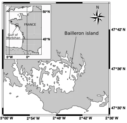
Fig. 1. Location of Bailleron island in Gulf of Morbihan, Southern Brittany, France.
From July 2004 to September 2005, 60 R. philippinarum ranging from 20 mm to 50 mm were monthly sampled at low tide from the natural clam bed of Bailleron island in Gulf of Morbihan, southern Brittany, France (Fig. 1). Clams were stored
in an icebox until being processed in the laboratory. A total of 1020 individuals was sampled from 17 sampling dates. At each sampling date, 60 clams were processed individually according to the protocol that follows.
2.2 Analysis of hemocyte parameters by flow cytometry
Hemolymph sampling
A minimum of 450 µL of hemolymph was withdrawn from the adductor muscle of individual clams using a 1 mL plastic syringe fitted with a 25-gauge needle and ob-served under microscope to control the sample and sampling qualities. Hemolymph samples were filtered through a 80 µm mesh in order to eliminate large debris and were stored individually in 1.5 mL micro–tubes held on ice.
Instrumentation
Analysis of hemocyte parameters were performed using a FACScalibur flow cy-tometer (Becton-Dickinson, San Diego, CA, USA) equipped with a 488 nm argon laser. The light scattered by particles indicated (1) their size through the FSC de-tector (Forward SCatter height) and (2) their internal complexity through the SSC detector (Side SCatter height). The flow cytometer is equipped with three specific fluorescence sensors: FL1 (green, 500-530 nm), FL2 (orange, 550-600 nm) and FL3 (red, >630 nm) allowing the detection of autofluorescence or fluorescent dyes.
Hemocyte viability, total and differential hemocyte counts (THC and DHC)
These parameters were measured following the protocol developed by Delaporte et al. (2003). Briefly, 100 µL of hemolymph from each individual were added in a tube containing 200 µL of anti-aggregant solution for bivalve hemocytes (AASH; Auffret and Oubella, 1994) and 100 µL of filtered sterile seawater (FSSW). Sam-ples were incubated 2 h at 18◦C in dark conditions with 4 µL of SYBR Green
working solution (obtained by diluting 10 x the commercial solution; Molecular probes, Oregon, USA) and propidium iodide (PI, Sigma) at a final concentration of 10 µg mL−1. Live and dead cells containing DNA are stained by SYBR Green;
whereas, dead cells are only stained by PI. SYBR Green fluorescence is detected by the FL1 detector of the flow cytometer, and PI fluorescence is detected by the FL3 detector. By using a density plot visualisation of FL1 vs FL3, it was possible to estimate precisely the percentage of dead cells in each sample.
A density plot visualisation of SSC vs FL1 allowed differentiation and gating of hemocytes stained by SYBR green from other particles in the hemolymph. This
allowed to calculate THC by taking into account the flow rate of the cytometer calculated according to the method of Marie et al. (1999).
Similarly to Allam et al. (2002a), two distinct sub-populations could be identified on a FSC vs SSC density plot: granulocytes (high SSC and high FSC), hyalinocytes (low SSC and high FSC). Results of THC, granulocyte and hyalinocyte counts are expressed as number of cells per mL of hemolymph.
Phagocytosis assays
Phagocytic activity of hemocytes was measured following the protocol described in Delaporte et al. (2003) and Labreuche et al. (2006) using 2 µm diameter latex fluorescent beads (fluoresbrite microspheres YG 2.0 microns, polysciences, Eppel-heim, Germany). A 150 µL sub-sample of hemolymph, primarily diluted with 150
µL of FSSW, was brought in contact with 30 µL of the working solution of
fluo-rescent beads (obtained by diluting 50 x the commercial solution) in micro–tubes. Tubes were incubated for 2 h at 18◦C in dark condition. Analysis by flow cytometry
allowed to detect hemocytes containing fluorescent beads on the FL1 detector. The phagocytic activity of hemocytes was calculated as the percentage of hemocytes that have ingested three fluorescent beads or more.
Phenoloxidase activity
Ninety-six-well plates containing 100 µL hemolymph samples were thawed and phenoloxidase activity measured as described by Reid et al. (2003). Briefly, 50
µL of Tris–HCl buffer (0.2 M, pH = 8) with 100 µL of L–DOPA (20 mM, L-3,4-dihydrophenyl-alanine, Sigma D9628) were added to each well. The microplate was rapidly mixed for 10 s. The reaction was then measured at ambient temper-ature with colour change recorded every 5 min, at 492 nm, over a period of 1 h. The microplate was mixed again prior to each measurement. Controls, without hemolymph, but containing L–DOPA and Tris–SDS buffer, were run in parallel and the values subtracted from test values to correct for possible auto-oxidation of theL-DOPA. Hemolymph protein concentration of each individual was determined from thawed 50-µL hemolymph samples using a BCA Protein Assay Kit (Pierce, USA) with bovine serum albumin as a standard, following the manufacturer’s guide for the Microwell plate protocol. Results of the specific PO activity were expressed in arbitrary units : 1 A.U.= ∆ DO490 nmmin−1mg protein−1.
2.3 Diseases
Detection and quantification of Perkinsus sp. infection
Detection and quantification of Perkinsus sp. were performed in gills since Choi et al. (2002) showed that the total number of Perkinsus sp. cells in the whole clams is linearly correlated with the number of Perkinsus cells in the gill tissue. After hemolymph sampling, clams were opened using a scalpel, gills were dissected and wet weighted. Perkinsus sp. presence and infection intensity in the gills were as-sessed according to the quantitative method of Ray (1966) as modified by Choi et al. (1989). Gills of individual clams incubated in 10 mL of fluid thioglycollate medium (FTM, Difco) supplemented with 67 µg of streptomycin (Sigma) and 32 µg of peni-cilin G (Sigma) dissolved in 100 µL in distilled water to limit bacterial growth. Vials were then incubated at room temperature over one week in the dark.
Perkin-sus cells were then counted after dissolving the FTM cultivated clam gills with 2 M
NaOH according to Choi et al. (1989).
Characterisation and classification of Brown Ring Disease (BRD) syndrome
BRD symptoms were monitored on the inner surface of the clams shells according to the description of Paillard and Maes (1994): conchiolin deposit stage (CDS) range from microscopic brown spots on the inner face of the shell in the earliest stage (CDS 1), to a thick brown deposit covering most of the inner shell in the most advanced stage (CDS 7).
2.4 Biometric measurements
After gill dissection, remaining flesh were removed from the shell and placed in pre-weighted aluminium capsules. Capsules were then freeze-dried for 48 h and dry flesh were weighted. As gills were removed for Perkinsus sp. diagnosis, total flesh dry weight were calculated by adding dry flesh weight and gills dry weight. Gills dry weight were estimated from wet gills weight using the coefficient 0.153 g dry g wet estimated from clams collected in Bailleron island (additional samples, n = 25; S.D. = 0.008).
Shells were air dried and weighted. Length following the maximum length axis was then measured using an electronic caliper and shells were stored until BRD diagnosis.
Condition index was calculated using the following formula:
CI = F lesh Dry W eight Shell Dry W eight × 100
2.5 Environmental factors
Temperature and salinity In situ sediment temperature was measured using an
autonomous temperature data logger (EBI-85A, Ebro, Germany) embedded at 5 cm in the sediment, the depth at which Manila clam are usually found. The probe mea-sured temperature every 20 min. A daily average was then calculated. Salinity data were provided byIFREMERlaboratory of La Trinité-sur-Mer (LER-MPL) and were measured using a Micrel probe in Fort Espagnol were salinity variation are sup-posed to be correlated to that in Bailleron island.
Trophic resource The monitoring of trophic resource begun in September 2004. Water samples were collected weekly at 50 cm above the sediment, and stored in a freezer (-18◦C) until further analysis. For each water sample, six sub-samples
were filtered through pre-weighted GF/F filters (25 mm) and freeze-dried to con-stant weight, and allowed to measure total suspended particulate matter (TPM). Three of the six filters were burnt (4h, 450◦C) and allowed to measure suspended
particulate inorganic matter (PIM). Particulate organic matter (POM, mg L−1) was
calculated by subtracting TPM and PIM. Burnt and unburnt filters were analysed for total carbon and nitrogen on a CE Instruments NC2500 elemental analyser (CE Elantech, USA). Particulate organic carbon (POC, mg L−1) and nitrogen (PON,
mg L−1) were then calculated by subtracting particulate carbon from unburnt and
burnt filters. The choice of characterisation of suspended organic matter quantity (POM) and quality (COP and NOP) rather than chlorophylla to estimate trophic resource for the Manila clam was motivated by a modelling study that emphasized that chlorophylla was not a good estimator of trophic resource for the Manila clam (Flye-Sainte-Marie et al., 2007a).
2.6 Statistical analysis
Statistical analysis were performed using the R software (R Development Core Team, 2006). Differences in hemocyte parameters among sampling dates were tested using Kruskal–Wallis test, differences in prevalence of both disease among sampling dates were tested using a χ2 test. Relationships between environmental factors and hemocyte parameters were assessed by the mean of linear models.
When seasonal effect on a biological variable was significant (i.e. hemocyte param-eters and condition index), it was removed to test the relationships between those variables and between hemocyte parameters and length or sex. Seasonal trend was considered to be represented by the variations of the mean of the considered bio-logical variable in healthy individuals (i.e. BRD-asymptomatic and null Perkinsus sp. clams), as along sampling dates. The season detrended value of the variable was calculated for each individual as:
Ds = Xtn−X¯tH
Where Dsis the season detrended residual, Xtnis the original value of the variable
on sampling date t for individual n and ¯XtH is the mean of the variable on sampling
date t calculated for healthy clams.
Length of clams had a significant effect on THC, concentration of both granulocytes and hyalinocytes, and on proportion of dead cells in healthy clams. Size of clams also had a significant effect on prevalence for both pathologies. In order to test for the effect of pathologies independently of size, a linear model was used to calculate the effect of length on each of those variables among healthy clams; residuals of these models were used in further calculations for testing the potential effect of pathologies on those hemocyte parameters.
To assess effect of categorical factors (i.e. infected versus non infected), t–test was used for normal–distributed data. Fisher test for homogeneity of variance was per-formed, if variances were significantly different Welsch approximation to the de-grees of freedom was used. Wilcoxon test was used for non–normal data. Effect of continuous factors (i.e. length) on hemocyte parameters were assessed using linear models.
3 Results
3.1 Environmental factors
Average daily temperature in the sediment varied between 6 and 23◦C (Fig. 2 A)
during the sampling period. Temperature and salinity profiles were highly cor-related (Fig. 2 A; Pearson r = 0.66, p–value = 6 10−3). Particulate organic matter
(POM, Fig. 2 B) was highly correlated to particulate organic carbon (POC) and particulate organic nitrogen (PON) (Pearson r>0.94, p–value<10−3).
A
B
Fig. 2. Evolution of environmental factors during the studied period. A: daily average tem-perature in the sediment and salinity at the sampling dates. B: evolution of particulate or-ganic matter (POM).
3.2 Influence of environmental factors on hemocyte parameters
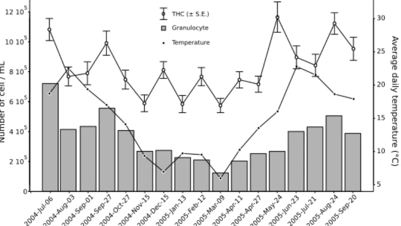
Fig. 3. Evolution of Total Hemocyte Count (THC, bars indicate standard error ), granulo-cyte concentrations and temperature during the studied period.
Evolution of hemocyte counts (total hemocyte and granulocyte counts) are shown in Fig. 3. All hemocyte parameters significantly varied during the sampling pe-riod (Kruskal–Wallis test; p-values < 0.005). Nevertheless, there were few signifi-cant relationships between hemocyte parameters and measured environmental
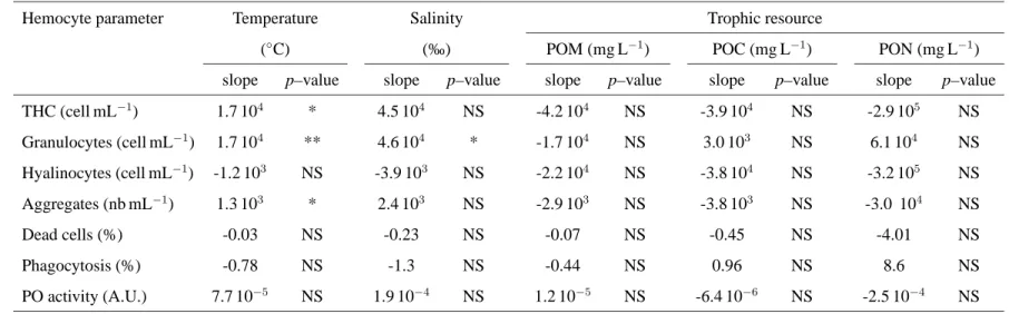
fac-Table 1. Relationships between environmental factors and hemocyte parameters tested using linear models. POM: particulate organic matter, POC particulate organic carbon, PON, particulate organic nitrogen.
,
Hemocyte parameter Temperature Salinity Trophic resource
(◦C) (‰) POM (mg L−1) POC (mg L−1) PON (mg L−1)
slope p–value slope p–value slope p–value slope p–value slope p–value
THC (cell mL−1) 1.7 104 * 4.5 104 NS -4.2 104 NS -3.9 104 NS -2.9 105 NS Granulocytes (cell mL−1) 1.7 104 ** 4.6 104 * -1.7 104 NS 3.0 103 NS 6.1 104 NS Hyalinocytes (cell mL−1) -1.2 103 NS -3.9 103 NS -2.2 104 NS -3.8 104 NS -3.2 105 NS Aggregates (nb mL−1) 1.3 103 * 2.4 103 NS -2.9 103 NS -3.8 103 NS -3.0 104 NS Dead cells (%) -0.03 NS -0.23 NS -0.07 NS -0.45 NS -4.01 NS Phagocytosis (%) -0.78 NS -1.3 NS -0.44 NS 0.96 NS 8.6 NS PO activity (A.U.) 7.7 10−5 NS 1.9 10−4 NS 1.2 10−5 NS -6.4 10−6 NS -2.5 10−4 NS
A.U. = arbitrary units (∆ DO490 nmmin−1mg protein−1)
Significance of the slope of the linear model:
NS: not significant (p–value > 0.05); *: p–value < 0.05; ** p–value < 0.01
1
tors (Tab. 1). Temperature significantly and positively affected all hemocyte counts (THC, granulocyte and hyalinocyte counts, Tab. 1) and number of aggregates. The significance was higher for the granulocyte concentration, the correlation coeffi-cient of the linear model was high (r2= 0.62) and the correlation is clearly visible on Fig. 3. Salinity only affected granulocyte concentration (Tab. 1). Neverthless, salinity and temperature were correlated, only the effect of temperature was tested against hemocyte parameters (Tab. 1) because temperature was more correlated to THC and granulocytes than salinity. Food quantity (POM) and quality (POC and PON) had no significant effects on hemocyte parameters (Tab. 1). Hyalinocyte con-centration, percentage of dead cells, phagocytosis percentage and PO activity were not correlated to any of the measured environmental factors (Tab. 1).
3.3 Influence of endogenous factors on hemocyte parameters
Effect of size/age Relationships between size/age (length) and hemocyte parame-ters were tested by the mean of linear models only in uninfected individuals (neither BRD–symptomatic and Perkinsus affected clams) to avoid effect of disease (Tab. 2). Although r2were low, all hemocyte counts (THC, granulocyte and hyalinocyte concentrations) significantly increased with size/age. The percentage of dead cells and the PO activity significantly decreased with size/age (Tab. 2).
Effect of sex and reproduction Effect of sex was tested during the reproduction period (end of April to end of September) when sex determination was possible. There were no significant differences between males and females in any of the hemocyte parameters (t–test and Wilcoxon test ; p–values > 0.05). Massive spawn-ing generally occurs at the mid August/beginnspawn-ing of September in Gulf of Morbi-han and result in a decrease of the condition index (Laruelle et al., 1994; Laruelle, 1999; Calvez, 2003). The decrease in the granulocyte concentration in the 2004-Aug-03 and 2004-Sep-03 samples (Fig. 3) coincide with this period. Although con-dition index indicated that clams has spawned at the 16th sample (2005-Aug-24) the decrease of granulocyte count was not observed.
Condition index residuals In order to test if energetic status could be linked to hemocyte parameters independently of seasonal variations, relationships between condition index residuals and hemocyte parameters were tested using linear models (Tab. 2). There were significant positive relations between condition index residuals and all hemocyte counts but r2
Table 2
Relationships between length and hemocyte parameters residuals in uninfected individuals and between condition index residuals and hemocyte parameters residuals (in uninfected and infected clams) tested using linear models.
Hemocyte parameter Length Condition index residuals residuals (uninfected clams) (all clams)
slope r2 p–value slope r2 p–value
THC (cells mL−1) 1.9 104 0.034 ** 4.2 104 0.009 * Granulocytes (cells mL−1) 1.2 104 0.052 ** 1.9 104 0.006 * Hyalinocytes (cells mL−1) 6.9 103 0.010 ** 2.4 104 0.008 * Aggregates (nb mL−1) -1.5 102 0.000 NS -1.2 103 0.004 NS Dead cells (%) -0.07 0.017 ** -0.03 0.000 NS Phagocytosis (%) -0.08 0.001 NS -0.74 0.004 NS PO activity (A.U.) -1.4 10−4 0.023 ** -1.4 10−4 0.002 NS
A.U. = arbitrary units (∆ DO490 nmmin−1mg protein−1)
Significance of the slope of the linear model:
NS: p–value > 0.05; *: p–value < 0.05; ** p–value < 0.01
3.4 Diseases
3.4.1 Prevalences of BRD symptoms and perkinsosis
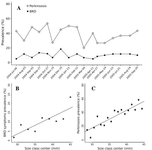
BRD prevalence was low (Fig. 4 A.; average prevalence: 9.7%) and showed no significant variation during the sampling period (χ2
= 12.96, df = 16, p-value =
0.675), disease intensity was also low: 90% of the symptomatic clams had a CDS lower than 4 on a scale going from 0 to 7.
Perkinsosis prevalence was moderated (Fig. 4 A.; 20–50% average prevalence: 38.2%). Although variations in prevalence during the sampling period were sig-nificant (χ2
= 37.79; df = 16; p–value = 2.10−3) no clear seasonal pattern appeared.
Perkinsus sp. infection intensity ranged between 0 and 1.63 106
cells/g gill WW, with a mean of 1.96 104. Perkinsus sp. prevalence and infection intensity were not correlated to temperature (linear models, p–value = 0.902 and 0.979 for prevalence and infection intensity, respectively).
There were significant positive correlations between size and both BRD (Fig. 4 B.) and perkinsosis prevalences (Fig. 4 C.)
A
B C
Fig. 4. A: Evolution of prevalence of perkinsosis and BRD during the sampling pe-riod; B: Relationship between center of the size classes and BRD symptom prevalence (y = 0.94 x − 0.25; r2 = 0.52; p-value = 0.005); C: Relationship between center of the
size classes and perkinsosis prevalence (y = 2.57 x −59; r2 = 0.77; p-value = 1.83 10−6)
3.5 Influence of diseases on hemocyte parameters
There were no difference in any measured hemocyte parameter residuals between BRD–asymptomatic clams and symptomatic clams (p–values > 0.05), neither be-tween BRD–asymptomatic clams and clams with CDS = 3 and higher (p–values > 0.05). No significant relationship between condition index residuals and BRD stage was found (linear model, p–value = 0.211).
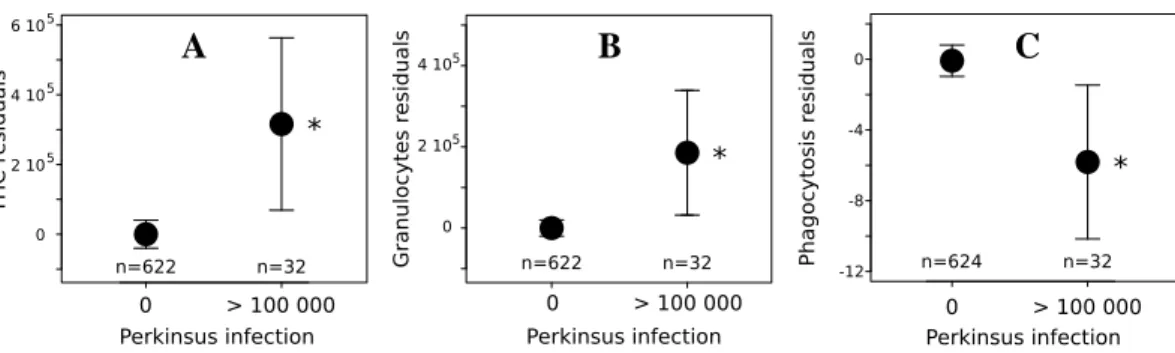
Effect of perkinsosis could only be detected (Fig. 5) when comparing uninfected clams with highly infected clams (>100 000 Perkinsus sp. cell/g gill WW).
Perkin-sus sp. significantly increased granulocyte concentration and THC and significantly
decreased the phagocytosis percentage (Fig. 5). No effect of Perkinsus sp. infection was observed on hyalinocyte concentration, number of aggregates (t–test, pvalue > 0.05), dead cells percentage (t–test, pvalue > 0.05) and PO activity (Wilcoxon Test,
A B C
Fig. 5. Comparisons of A: Total hemocyte count, B: Granulocyte count, C: Phagocyto-sis percentage between uninfected individuals (0 Perkinsus sp. cell/g gill WW; i.e. un-der the detection threshold) and highly infected individuals (> 100 000 Perkinsus sp. cell/g gill WW). Results are expressed as mean ±95% confidence interval. The stars (*) indicates significant differences (p–value < 0.05).
3.6 Overall variability of hemocyte parameters
Contributions of the environmental condition, internal factors and diseases to over-all variability of four hemocyte parameters (THC, granulocyte and hyalinocyte con-centration, phagocytosis) were investigated by the mean of a MANOVA (Tab. 3), only factors that had a significant effect on these hemocyte parameters were in-cluded. Results indicate that the most explained hemocyte parameter was the granu-locyte concentration (Tab. 3) for which only 16.4% of the variability was explained by temperature (11.3%), length (4.1%), Perkinsus sp. infection (0.6%) and condi-tion index residuals (0.4%). Only 10% of the THC variability was explained by these factors. Factors explaining most of the variance of the hemocyte parameters were temperature and length. Perkinsus sp. only explained very little of the overall variability of the hemocyte system : its effects are significant when comparing un-infected to heavily un-infected clams (Fig. 5) but no significant trend could be detected when using continuous linear model of THC or phagocytosis against concentration of Perkinsus sp.
4 Discussion
The aim of this study was to better understand the relative effect of environmental and internal factors and diseases on hemocyte parameters in the field. Independent effect of each factor on the hemocyte parameters will first be discussed. The com-bined effect of measured factors to the overall variability of hemocyte will finally be discussed.
Table 3. Summary of the weights of environmental factor (temperature) and internal factors (size/age and condition index residuals) and Perkinsus sp. infection on the total variability of THC, granulocyte concentration (cell mL−1), hyalinocyte concentration (cell mL−1) and phagocytosis percentage
computed from a MANOVA. Factors that had no significant effect were not included for computations. Partial r2 values indicate the weight of each factor on the total variability of each parameter, r2 indicate the weight of all included factor on the total variability of each parameter.
THC Granulocyte conc. Hyalinocyte conc. Phagocytosis Factor partial r2 p–value partial r2 p–value partial r2 p–value partial r2 p–value Temperature 0.033 ** 0.113 ** NI NI Length 0.055 ** 0.041 ** 0.030 ** 0.028 ** CI residuals 0.009 ** 0.004 * 0.009 ** NI Perkinsus sp. 0.003 NS 0.006 ** NI 2.4 10−5 NS r2 p–value r2 p–value r2 p–value r2 p–value All factors 0.100 ** 0.164 ** 0.039 ** 0.028 **
CI: condition index
NI: not included in MANOVA
NS: not significant (p–value > 0.05); *: p–value < 0.05; ** p–value < 0.01
1
4.1 Effect of environmental factors on hemocyte parameters
Temperature was the major environmental factor modulating hemocyte parameters. There were positive correlation between temperature and both granulocyte and total hemocyte counts. Since the slopes of the linear models relating temperature with granulocyte concentration and THC are equal (Tab. 1) it is possible to conclude that the increase in THC simply reflects the increase in granulocyte concentration. Several laboratory experiments emphasized the positive effect of temperature on hemocyte concentration in hemolymph in various bivalve and crustacean species (see e.g. Truscott and White, 1990; Chu and La Peyre, 1993; Fisher et al., 1996; Chu, 1998; Liu et al., 2004; Paillard et al., 2004; Monari et al., 2007), few studies however showed the occurrence of this pattern in the field (Fisher et al., 1996; Car-ballal et al., 1998; Soudant et al., 2004). The increase of circulating hemocyte count is generally considered as a consequence of proliferation or movement of cells from tissue into hemolymph (Pipe and Coles, 1995). Thus, an increase of the hemo-cyte proliferation with temperature could explain the observed pattern. Number of hemocyte aggregates was also positively correlated to temperature. Hemocyte ag-gregation is presumed to be involved in hemostasis and wound healing (Chen and Bayne, 1995) but also in defence mechanisms (Tiscar and Mosca, 2004). Formation of hemocyte aggregates is the result of the adhesion activity of hemocytes (Chen and Bayne, 1995; Auffret and Oubella, 1997). Since bivalves are poikilotherms, hemocyte activity may scale with temperature thus explaining the correlation be-tween aggregate concentration and temperature.
Extreme (high and low) salinities have been shown to induce variations of total hemocyte counts in several bivalve species (Chu and La Peyre, 1991; Reid et al., 2003; Matozzo et al., 2007), but generally variation of salinity within the life range of the species does not affect hemocyte counts (Matozzo et al., 2007). This tend to confirm that, in this study, the observed correlation between salinity and granulo-cyte concentration is only attributable to the correlation between temperature and salinity.
Although phagocytosis significantly varied during the studied period, this parame-ter does not seem to be affected by any of the measured environmental factors. This result appears contradictory with laboratory experiments which showed that phago-cytosis varied with temperature (Chu and La Peyre, 1993; Allam et al., 2002b; Monari et al., 2007) and salinity (Reid et al., 2003). The reasons for this difference remains unclear but this emphasizes the difficulty in extending laboratory results to the field.
Low food levels and starvation have been shown to reduce THC in oysters (Dela-porte et al., 2006; Butt et al., 2007). However, in an earlier study, Ashton-Alcox and Ford (1998) showed that a 4-week starvation did not affect hemocyte counts in oysters. This result suggest that this period is not long enough to induce a starvation
stress. In our study, indicators of food quantity (POM) and quality (POC, PON) did not appeared to affect any of the measured hemocyte parameters. This suggest that in such natural conditions food level and quality were not low enough to induce any modification of hemocyte parameters during the studied period.
The poor relationships between other environmental factors and hemocyte param-eters emphasize the difficulty to assess the environmental control of hemocyte sys-tem in the field.
4.2 Effect of endogenous variables
Size/age Variability of hemocyte parameters with size/age have been little stud-ied (Carballal et al., 1997; Lopez et al., 1997; Carballal et al., 1998; Barracco et al., 1999) and a clear effect of size on the hemocyte parameters was rarely found. Only Carballal et al. (1998) found a significant increase in number of circulating granu-locytes with age in Mytilus galloprovincialis. In most of the above studies, the size ranges were smaller than in our study (this study: 200 % of difference between the smallest and the largest individuals and 127 % to 150 % in Lopez et al., 1997; Carballal et al., 1998; Barracco et al., 1999, studies). Furthermore, the large num-ber of individuals sampled in our study allowed to distinguish the effect of size/age within the large variability of the hemocyte parameters. Our study emphasizes that, in the Manila clam, total hemocyte, granulocyte and hyalinocyte counts are signif-icantly positively correlated to size while phagocytosis and dead cells percentage are negatively correlated to size.
Sex and reproduction Links between gender and hemocyte system have poorly been studied (Barracco et al., 1999; Gagné et al., 2007). In both studies, as in the present one, results tend to show that hemocyte parameters are independent of gen-der. Such relationships may not be expected since hemocyte systems plays an im-portant role in maintaining organism homeostasis. Such requirement may not vary with gender.
However, gonadal cycle can influence the hemocyte system (see Oliver and Fisher, 1999, for a review), in a laboratory experiment Delaporte et al. (2006) observed that THC decreased during gametogenesis in the oyster Crassotrea gigas. Field studies showed that THC decrease during spawning period (in Mytilus
galloprovin-cialis and Crassostrea virginica, Pipe et al., 1995; Fisher et al., 1996) presumably
because of hemocyte infiltration in gonadal tissue. In our study, condition index profile indicated that clams had already spawned on the 2004-sep-01 and 2005-aug-24 ; these events could not be associated to a decrease in THC or any other of the measured parameters.
Condition index residuals Condition index residuals provide a rough informa-tion about the energetic status independently of seasonal variainforma-tion (i.e. is the in-dividual more or less fat for the season?). Our study shows that there is a little positive effect, but nonetheless significant, of condition index residuals on hemo-cyte counts. This tends to confirm the hypothesis of Ashton-Alcox and Ford (1998) who suggested that variability of molluscan hemocyte parameters could be linked to the amount of stored energy reserves to explain the high observed inter–individual variations. This relationship could be explained by the involvement of hemocytes in nutrient mobilisation (Cheng, 1996). Nevertheless, the relationship only explains little of the total variability of hemocyte parameters.
4.3 Diseases
Prevalence and intensity of perkinsosis and BRD Prevalence of Perkinsus sp. infection was moderated and ranged between 20 and 50% (average 38.2%) which is comparable to the values reported by by Ngo and Choi (2004) in R. philippinarum and R. decussatus beds of Jeju (Korea) and Portugal (Ngo and Choi, 2004; Leite et al., 2004). These prevalences are lower than those reported by Park and Choi (2001) in various places of Korea where prevalence reached 100% in some loca-tions and by Villalba et al. (2005) in natural R. decussatus beds of Galicia (Spain), where prevalence ranged between 45 and 100%. Although perkinsosis prevalence showed significant variation during the study period, no clear annual pattern ap-peared. Epizootiology studies showed that Perkinsus marinus infection in the oys-ter Crassostrea virginica prevalence is closely related to temperature and salinity (see review in Villalba et al., 2004) which results in an increased prevalence in sum-mer. Surveys of P. olseni infections in Ruditapes decussatus and R. philippinarum showed an annual pattern with an increase of prevalence in autumn and/or spring (Villalba et al., 2005; Ngo and Choi, 2004). These studies contrast with our results. Nevertheless, Leite et al. (2004) did not found any annual pattern of Perkinsus sp. in R. decussatus along the Portugese coasts during their 2–year survey which shows that seasonal pattern is not always observed in P. olseni infection prevalence. Estimation of Perkinsus sp. burden in gill (cells/g gill WW) provide an over–esti-mation of the total body burden (cells/g flesh WW) (Choi et al., 2002). In our study
Perkinsus sp. infection intensity was low (average: 2 104 cells/g gill WW) and our values are consistent with the values obtained by Lassalle et al. (2007) in Gulf of Morbihan. Higher values have been reported in location more southern along the French Atlantic coast : more than 8 104 cells/g gill WW in Arcachon bay (Lassalle et al., 2007). Higher values were also reported in Korea (up to 8.7 106
cells/g flesh WW; Park and Choi, 2001), in Japan (2.25 106
cells/g flesh WW; Ishaya bay; Choi et al., 2002) and in Spain (⋍ 5 104cells/g flesh WW in R. decussatus from Galicia; Villalba et al., 2005).
Average prevalence of BRD symptoms during the study period (9.7%) was low in comparison to the prevalences observed in aquaculture clams beds of North-ern Brittanny (Brouennou, Finistère, France) that ranged between 33 and 100% (Paillard, 1992; Paillard et al., 1997; Paillard, 2004a). Our results are in accor-dance with previous studies performed in natural populations of Gulf of Morbi-han (Paillard, 2004a; Lassalle et al., 2007) that found prevalences between 4 and 30%. However, no annual pattern in BRD prevalence was found and this contrasts with the field surveys of Paillard et al. (1997) and Paillard (2004a). These studies showed that symptomatic clams are found during all the year, but generally BRD symptoms prevalence increases toward the beginning of the spring and decreases towards the beginning of the summer. Low temperatures during the rising phase of prevalence have been invoked to explain this pattern (Paillard et al., 1997, 2004; Paillard, 2004a,b). The lack of pattern observed in our study may be explained by the low prevalence and intensity of BRD during the studied period : 80% of the symptomatic clams had a CDS of 3 or less, stage at which the extend of the brown deposit is localised to small area of the inner shell (Paillard and Maes, 1994))
Size-prevalence relationships The positive relationships between size and preva-lence for both diseases correspond to the general pattern observed for most para-sites in filter feeder bivalves (Guralnick et al., 2004). This pattern has already been shown for the P. marinus/C. virginica (Andrews and Hewatt, 1957) and P. olseni/R.
decussatus interactions (Villalba et al., 2005). The explanation of such a pattern lies
in both an increased filtration rate in biggest individuals and an accumulation of the parasite during life span, these processes leading to an increase of prevalence and infection intensity with size (Andrews and Hewatt, 1957; Guralnick et al., 2004; Villalba et al., 2005). Nevertheless, another interpretation for the size–BRD preva-lence relationship have been given in Flye-Sainte-Marie et al. (2008): increased body surface area with size could lead to higher probability of contact with big sediment grains that may induce disruptions and subsequently a potential entry for the pathogen V. tapetis.
Relations between hemocyte parameters and diseases High Perkinsus sp. in-fection significantly increased granulocyte concentration and consequently THC in highly infected individuals (> 100 000 Perkinsus sp. cells/g gill WW). This result is contradictory with those of Ordás et al. (2000) who found a decrease of THC in R.
decussatus infected by P. olseni. Nevertheless, augmentation of circulating
hemo-cytes have been widely documented in C. virginica heavily infected by P. marinus (see e.g. Anderson et al., 1992; Chu and La Peyre, 1993; Anderson et al., 1995; La Peyre et al., 1995; Anderson et al., 1996). These authors suggest that this in-crease reflects a mobilisation/production of hemocytes to counteract Perkinsus sp. development. In our study, high P. olseni infection also significantly reduced hemo-cyte phagocytosis percentage which is in accordance with previous studies who showed an inability of R. decussatus hemocyte to phagocyte P. olseni zoospores
(Lopez et al., 1997) and a decrease in percentage of phagocytosis in P. olseni-infected R. decussatus (Ordás et al., 2000). Although Muñoz et al. (2006) showed an increase in the PO activity in R. decussatus lowly and moderately infected by P.
olseni, no significant effect of Perkinsus sp. on this parameter could be found from
our data.
In this study, BRD had no significant influence on any of the hemocyte parame-ters. These results contrast with previous experimental studies which showed that (1) V. tapetis inoculation induces an increase in THC (Paillard et al., 1994; Allam et al., 2000, 2006; Paillard, 2004b), (2) a decrease in phagocytic activity (Allam et al., 2002b; Allam and Ford, 2006), and (3) an increase of dead cell percent-age (Allam et al., 2000; Allam and Ford, 2006; Allam et al., 2006). Symptoms of BRD results of the interaction between the bacteria and the clam over time. Exper-imental infection may uncouple bacterial burden and symptoms (brown deposit). In these conditions, effect on hemocyte system may be detected before apparition of symptoms. Thus, discrepancy between the above experimental observations and our results may be explained by the low natural infection intensity, presumably as-sociated to a low V. tapetis burden. Consistently with our study, Reid et al. (2003) experimental work failed to find any significant effect of BRD on PO activity. Allam et al. (2001) experimental study suggested that higher granulocyte concen-tration may be related to a higher resistance to BRD. Interestingly, in our study, granulocyte concentration significantly varied but BRD prevalence was not lower when granulocyte concentration was high. Granulocyte concentration significantly increased with size/age but prevalence and size were also correlated. Thus, it was not possible to link a high granulocyte concentration with any variation of BRD prevalence. This observation is consistent with Reid et al. (2003) who indicated that increased hemocyte population did not appear to have a direct role in reduc-ing BRD levels. This suggests that association of resistance to BRD and hemocyte parameters is not straightforward.
Diseases and condition index Low infection intensities of both diseases ob-served during the studied period explains that condition index was not affected in infected individuals. These results are consistent with previous studies indicating that both diseases are susceptible to interfere with energy balance and to affect con-dition index (Leite et al., 2004; Park et al., 2006; Flye-Sainte-Marie et al., 2007b) only at high infection intensities.
4.4 Variability of hemocyte parameters in the field
Consistently with previous studies, this multiparametric field study emphasizes that part of the variability of the hemocyte system of bivalves is attributable to
vironmental and internal factors as well as diseases. Temperature was the only environmental factor explaining seasonal variations of some of the hemocyte pa-rameters and contributed to 10% of the variability of granulocyte concentration. Effect of size/age on hemocyte parameters have been poorly described in literature (Carballal et al., 1997; Lopez et al., 1997; Carballal et al., 1998; Barracco et al., 1999). It was nevertheless the factor both affecting most of the measured hemo-cyte parameters and significantly contributing to the explanation of the variability of hemocyte system. Interestingly, although links between disease and hemocyte system has been extensively described in the literature these links were little con-firmed in our study. In accordance with Adamo (2004), this study emphasizes that the measures of hemocyte parameters cannot directly be interpreted as measure-ment of immunocompetence (i.e. capacity of defence against a pathogen).
Although this study identified factors that significantly affected the hemocyte sys-tem, these factors only explained little of the overall variability of the hemocyte parameters. For the mostly explained parameter (granulocyte concentration) only 16.4% of the variance could be explained from the measured factors. Part of the un-explained variability can be attributed to unun-explained month to month variations, suggesting that other environmental factors than temperature, salinity or trophic re-source may modulate the hemocyte system. Effect of contaminants and toxic algae on hemocyte system of bivalves have been described in literature (see e.g. Oliver et al., 2001; Auffret et al., 2004, 2006; Hégaret and Wikfors, 2005b,a; Hégaret et al., 2007) and could explain these month to month variations. Most of the vari-ability of the hemocyte system could be attributed to inter–individual differences and thus could not be associated with any parameter that we measured. Ashton-Alcox and Ford (1998) suggested that variability in molluscan hemocytes could be more immediately linked to individual metabolic condition than to an inabil-ity to buffer hemolymph against external ambient conditions. In the past 30 years, many authors have studied the link between hemocyte system and disease and thus tending to reduce the role of hemocyte system to immune functions. Nevertheless, although poorly documented, there are evidences of implication of hemocyte sys-tem in various other functions such as nutrition, wound repair, inflamation (Fisher, 1986; Cheng, 1996) and shell reparation and mineralization (Fisher, 1986, 2004; Mount et al., 2004). This study emphasizes the need for a better understanding of the various functions of the hemocyte system and how does the requirements of these functions control the hemocyte system. Such a knowledge is necessary to better understand the linkages between environment, bivalve metabolic status, hemocyte system and disease development, and to use of bivalve hemocyte param-eters as environmental biomarkers.
Acknowledgements
This work was founded by Région Bretagne within the MODELMAB regional re-search program. The authors greatly thank Lionel Allano for his technical help during field sampling. The authors thanks Jean–Francois Bouget from IFREMER la Trinité-sur-mer for fournishing salinity data. The authors thank Antoine Emery, Pierre Huonnic, Alain Lemercier, Morgane Lejart, Mirella da Silva, Sorcha Ni’Lon-gphuirt, Brivaela Moriceau, Pierre Fouillaron, Anne–Laure Cassonne, Angéline Fr-antz, Charlotte Dentan and Emmanuelle Ferret for their help for field sampling and analyses. The author also thank Annick Masson for CHN analysis.
References
Adamo, S. A., 2004. How sould behavioural ecologists interpret measurements of immunity? Animal behaviour 68, 1443–1449.
Allam, B., Ashon-Alcox, K. A., Ford, S. E., 2001. Haemocyte parameters associ-ated with resistance to brown ring disease in Ruditapes spp. clams. Developmen-tal and Comparative Immunology 25, 365–375.
Allam, B., Ashton-Alcox, K. A., Ford, S. E., 2002a. Flow cytometric comparison of haemocytes from three species of bivalve molluscs. Fish Shellfish Immunol. 13 (2), 141–158.
Allam, B., Ashton-Alcox, K. A., Ford, S. E., 2002b. Flow cytometric measure-ment of hemocyte viability and phagocytic activity in the clam, Ruditapes
philip-pinarum. J. Shellfish. Res. 21, 13–19.
Allam, B., Ford, S. E., 2006. Effects of the pathogenic Vibrio tapetis on defence factors of susceptible and non-susceptible bivalve species: I. Haemocyte changes following in vitro challenge. Fish Shellfish Immunol. 20, 374–383.
Allam, B., Paillard, C., Auffret, M., 2000. Alterations in haemolymph and extra-pallial fluid parameters in the Manila clam, Ruditapes philippinarum, challenged with the pathogen Vibrio tapetis. J. Invertebr. Pathol. 76, 63–69.
Allam, B., Paillard, C., Auffret, M., Ford, S. E., 2006. Effects of the pathogenic
Vib-rio tapetis on defence factors of susceptible and non-susceptible bivalve species:
II. Cellular and biochemical changes following in vivo challenge. Fish Shellfish Immunol. 20, 384–397.
Anderson, R. S., Burreson, E. M., Paynter, K. T., 1995. Defense responses of hemo-cytes withdrawn from Crassostrea virginica infected with Perkinsus marinus. J. Invertebr. Pathol. 66 (1), 82–89.
Anderson, R. S., Paynter, K. T., Burreson, E. M., 1992. Increased reactive oxygen intermediate production by hemocytes withdrawn from Crassostrea virginica in-fected with Perkinsus marinus. Biolgical Bull. 183 (3), 476–481.
Anderson, R. S., Unger, M. A., Burreson, E. M., 1996. Enhancement of
sus marinus disease progression in TBT-exposed oysters (Crassostrea virginica).
Mar. Envir. Res. 42 (1), 177–180.
Andrews, J. D., Hewatt, W. G., 1957. Oyster mortality studies in Virginia. II. the fungus disease caused by Dermocystidium marinum in oysters of Chesapeake Bay. Ecol. Monogr. 27 (1), 1.264.
Ashton-Alcox, K. A., Ford, S. E., 1998. Variability in molluscan hemocytes: a flow cytometric study. Tissue and Cell 30 (2), 195–204.
Auffret, M., Duchemin, M., Rousseau, S., Boutet, I., Tanguy, A., Moraga, D., Marhic, A., 2004. Monitoring of immunotoxic responses in oysters reared in areas contaminated by the" Erika" oil spill. Aquat. Living Resour. 17 (3), 297– 302.
Auffret, M., Oubella, R., 1994. Cytometric parameters of bivalve molluscs: effect of environmental factors. In: Stolen, J.S., Fletcher, T.C. (Eds.), Modulators of Fish Immune Response. SOS publication, Fair Haven, NJ, USA , 23–32.
Auffret, M., Oubella, R., 1997. Hemocyte aggregation in the oyster Crassostrea
gigas: In vitro measurement and experimental modulation by xenobiotics. Comp.
Biochem. Physiol. A 118 (3), 705–712.
Auffret, M., Rousseau, S., Boutet, I., Tanguy, A., Baron, J., Moraga, D., Duchemin, M., 2006. A multiparametric approach for monitoring immunotoxic responses in mussels from contaminated sites in Western Mediterranea. Ecotox. Env. Safty 63, 393–405.
Barracco, M. A., Medeiros, I. D., Moreira, F. L. M., 1999. Some haemato-immunological parameters in the mussel Perna perna. Fish Shellfish Immunol. 9 (5), 387–404.
Borrego, J. J., Castro, D., Luque, A., Paillard, C., Maes, P., Gracia, M., Ventosa, A., 1996. Vibrio tapetis sp. nov., the causative agent of the brown ring disease affecting cultured clams. Int. J. Syst. Bacteriol. B 46, 480–484.
Butt, D., Aladaileh, S., O’Connor, W. A., Ratios, D. A., 2007. Effect of starvation on biological factors related to immunological defence in the Sydney rock oyster (Saccostrea glomerata). Aquaculture 264 (1-4), 82–91.
Calvez, I., 2003. Approche de la variabilité spatiale d’une population de palour-des Ruditapes philippinarum (Adams et Reeve), aux stapalour-des larvaires et post-larvaires. Thèse de Doctorat, Université de Bretagne Occidentale, Brest.
Carballal, M., Villalba, A., López, C., 1998. Seasonal variation and effects of age, food availability, size, gonadal development, and parasitism on the hemogram of
Mytilus galloprovincialis. J. Invertebr. Pathol. 72 (3), 304–312.
Carballal, M. J., Lopez, C., Azevedo, C., Villalba, A., 1997. In vitro study of phago-cytic ability of Mytilus galloprovincialis Lmk. haemocytes. Fish Shellfish Im-munol. 7 (6), 403–416.
Castro, D., Martinez-Manzanares, E., Luque, A., Fouz, B., Morinigo, M., Borrego, J. J., Toranzo, A. E., 1992. Characterization of strains related to brown ring dis-ease outbreaks in southwestern spain. Dis. Aquat. Org. 14, 229–236.
Chen, J. H., Bayne, C. J., 1995. Bivalve mollusc hemocyte behaviors: Characteriza-tion of hemocyte aggregaCharacteriza-tion and adhesion and their inhibiCharacteriza-tion in the california mussel (Mytilus californianus). Biolgical Bull. 188 (3), 255–266.
Cheng, T. C., 1996. Hemocytes: forms and functions. In: Kennedy, V. S., Newell, R. I. E., Eble, F. (Eds.), The eastern oyster Crassostrea virginica. Maryland Sea Grant College, pp. 299–333.
Choi, K., Park, K., Lee, K., Matsuoka, K., 2002. Infection intensity, prevalence, and histopathology of Perkinsus sp. in the Manila clam, Ruditapes philippinarum, in Isahaya bay, Japan. J. Shellfish. Res. 21 (1), 119–126.
Choi, K.-S., Wilason, E. A., Lewis, D. H., Powell, E. N., Ray, S. M., 1989. The energetic cost of Perkinsus marinus parasitism in oysters: quantification of the thioglycollate method. J. Shellfish. Res. 8, 125–131.
Chu, F.-L. E., 1998. Host defenses against Perkinsus marinus: a review of recent findings in the eastern oyster, Crassostrea virginica. J. Shellfish. Res. 18 (1), 321–322.
Chu, F.-L. E., 2000. Defence mechanism of marine bivalves. In: Fingerman, M., Nagabhushanam, R. (Eds.), Recent advances in Biotechnology. Volume 5 Im-munobiology and pathology. Science publishers, Inc., pp. 1–42.
Chu, F.-L. E., La Peyre, J. F., 1991. Effect of salinity on Perkinsus marinus suscep-tibility and defense-related activities in eastern oysters, Crassostrea virginica. J. Shellfish. Res. 10, 294.
Chu, F.-L. E., La Peyre, J. F., 1993. Perkinsus marinus susceptibility and defense-related activities in eastern oysters Crassostrea virginica: temperature effects. Dis. Aquat. Org. 16 (3), 223–234.
Delaporte, M., Soudant, P., Lambert, C., Moal, J., Pouvreau, S., Samain, J.-F., 2006. Impact of food availability on the energy storage and defense related hemocyte of the Pacific oyster Crassostrea gigas during an experimental reproductive cycle. Aquaculture 254, 571–582.
Delaporte, M., Soudant, P., Moal, J., Lambert, C., Quéré, C., Miner, P., Choquet, G., Paillard, C., Samain, J.-F., 2003. Effect of a mono-specific algal diet on immune functions in two bivalves species, Crassostrea virginica and Ruditapes
philip-pinarum. J. Exp. Biol. 206 (17), 3053–3064.
Fisher, W. S., 1986. Structure and function of oyster hemocytes. In: Brehelin, M. (Ed.), Immunity in invertebrates, cells, molecules and defense reactions. Springer, Berlin, pp. 25–35.
Fisher, W. S., 2004. Relationship of amaebocytes and terrestrial elements to adult shell deposition in eastern oysters. J. Shellfish. Res. 23, 353–367.
Fisher, W. S., Oliver, L. M., Edwards, P., 1996. Hematologic and serologic vari-ability of eastern oysters from Apalachicola Bay, Florida. J. Shellfish. Res. 15, 555–564.
Flassch, J. P., Leborgne, Y., 1992. Introduction in Europe, from 1972 to 1980, of the Japanese Manila clam (Tapes philippinarum) and effects on aquaculture pro-duction and natural settlement. ICES Marine Symposium 194, 92–96.
Flye-Sainte-Marie, J., Jean, F., Ford, S. E., Paillard, C., 2008. Effect of sediment grain-size on development of brown ring disease in the Manila clam Ruditapes
philippinarum. Aquaculture 278, 184–187.
Flye-Sainte-Marie, J., Jean, F., Paillard, C., Ford, S., Powell, E., Hofmann, E., Klinck, J., 2007a. Ecophysiological dynamic model of individual growth of
ditapes philippinarum. Aquaculture 266, 130–143.
Flye-Sainte-Marie, J., Pouvreau, S., Paillard, C., Jean, F., 2007b. Impact of Brown Ring Disease on the energy budget of the Manila clam Ruditapes philippinarum. J. Exp. Mar. Biol. Ecol. 349 (2), 378–389.
Ford, S. E., Paillard, C., 2007. Repeated sampling of individual bivalve mollusks I: Intraindividual variability and consequences for haemolymph constituents of the Manila clam, Ruditapes philippinarum. Fish Shellfish Immunol. 23, 280–291. Gagné, F., Blaise, C., Pellerin, J., Fournier, M., Durand, M. J., Talbot, A., 2007.
Re-lationships between intertidal clam population and health status of the soft-shell clam Mya arenaria in the St. Lawrence Estuary and Saguenay Fjord (Québec, Canada). Environ. Int. , in press.
Guralnick, R., Hall, E., Perkins, S., 2004. A comparative approach to understanding causes and consequences of mollusc–digenean size relationships: A case study with allocreadiid trematodes and Cyclocalyx clams. J. Parasitol. 90 (6), 1253– 1262.
Hégaret, H., da Silva, P. M., Wikfors, G. H., Lambert, C., De Bettignies, T., Shumway, S. E., Soudant, P., 2007. Hemocyte responses of manila clams,
Ru-ditapes philippinarum, with varying parasite, Perkinsus olseni, severity to
toxic-algal exposures. Aquat. Toxicol. 84 (4), 469–479.
Hégaret, H., Wikfors, G. H., 2005a. Effects of natural and field-simulated blooms of the dinoflagellate Prorocentrum minimum upon hemocytes of eastern oysters,
Crassostrea virginica, from two different populations. Harmful Algae 4 (2), 201–
209.
Hégaret, H., Wikfors, G. H., 2005b. Time-dependent changes in hemocytes of east-ern oysters, Crassostrea virginica, and northeast-ern bay scallops, Argopecten
irradi-ans irradiirradi-ans, exposed to a cultured strain of Prorocentrum minimum. Harmful
Algae 4 (2), 187–199.
Humphreys, J., Caldow, R. W. G., McGrorty, S., West, A. D., Jensen, A. C., 2007. Population dynamics of naturalised Manila clams Ruditapes philippinarum in British coastal waters. Mar. Biol. 151, 2255–2270.
Jensen, A. C., Humphreys, J., Caldow, R. W. G., Grisley, C., Dyrynda, P. E. J., 2004. Naturalization of the Manila clam (Tapes philippinarum), an alien species, and establishment of a clam fishery within Poole Harbour, Dorset. J. Mar. Biol. Assoc. 84 (05), 1069–1073.
La Peyre, J. F., Chu, F.-L. E., Meyers, J. M., 1995. Haemocytic and humoral activi-ties of eastern and pacific oysters following challenge by the protozoan Perkinsus
marinus. Fish Shellfish Immunol. 5, 179–190.
Labreuche, Y., Soudant, P., Gonçalvez, M., Lambert, C., Nicolas, J.-L., 2006. Ef-fects of extracellular products from the pathogenic Vibrio aesturianus strain 01/32 on the lethality and cellular immune responses of the oyster Crassostrea
gigas. Developmental and Comparative Immunology 30, 367–379.
Laruelle, F., 1999. Phénologie et déterminisme de la reproduction chez Ruditapes
decussatus (L.) and Ruditapes philippinarum (Adams and Reeve) en Bretagne.
Thèse de Doctorat, Université de Bretagne Occidentale, Brest.
Rudi-tapes decussatus and RudiRudi-tapes philippinarum, on intertidal flats in Brittany. J.
Mar. Biol. Assoc. 172, 69–96.
Lassalle, G., de Montaudouin, X., Soudant, P., Paillard, C., 2007. Parasite co-infection of two sympatric bivalves, the Manila clam (Ruditapes philippinarum) and the cockle (Cerastoderma edule) along a latitudinal gradient. Aquat. Living Resour. 20, 33–42.
Leite, R. B., Afonso, R., Cancela, M. L., 2004. Perkinsus sp. infestation in carpet-shell clams, Ruditapes decussatus (L), along the Portuguese coast. Results from a 2-year survey. Aquaculture 240, 39–53.
Liu, S., Jiang, X., Hu, X., Gong, J., Hwang, H., Mai, K., 2004. Effects of temper-ature on non-specific immune parameters in two scallop species: Argopecten
ir-radians (Lamarck 1819) and Chlamys farreri (Jones & Preston 1904). Aquacult.
Res. 35 (7), 678–682.
Lopez, C., Carballal, M. J., Azevedo, C., Villalba, A., 1997. Differential phagocytic ability of the circulating haemocyte types of the carpet shell clam Ruditapes
decussatus (Mollusca: Bivalvia). Dis. Aquat. Org. 30 (3), 209–215.
Marie, D., Partensky, F., Vaulot, D., Brussaard, C., 1999. Enumeration of phyto-plankton, bacteria and viruses in marine samples. Curr. Protocols Cytom 11, 1– 15.
Marin, M. G., Moschino, V., Deppieri, M., Lucchetta, L., 2003. Variations in gross biochemical composition, energy value and condition index of T. philippinarum from the Lagoon of Venice. Aquaculture 219, 859–871.
Matozzo, V., Da Ros, L., Ballarin, L., Meneghetti, F., Marin, M. G., 2003. Func-tional responses of haemocytes in the clam Tapes philippinarum from the La-goon of Venice: fishing impact and seasonal variations. Can. J. Fish. Aquat. Sci. 60 (8), 949–958.
Matozzo, V., Monari, M., Foschi, J., Serrazanetti, G. P., Cattani, O., Marin, M. G., 2007. Effects of salinity on the clam Chamelea gallina. Part I: alterations in immune responses. Mar. Biol. 151 (3), 1051–1058.
Monari, M., Matozzo, V., Foschi, J., Cattani, O., Serrazanetti, G., Marin, M., 2007. Effects of high temperatures on functional responses of haemocytes in the clam
Chamelea gallina. Fish Shellfish Immunol. 22 (1-2), 98–114.
Mount, A. S., Wheeler, A. P., Paradkar, R. P., Snider, D., 2004. Hemocyte-mediated shell mineralization in the eastern oyster. Science 304, 297–300.
Muñoz, P., Meseguer, J., Ángeles Esteban, M., 2006. Phenoloxidase activity in three commercial bivalve species. changes due to natural infestation with
Perkin-sus atlanticus. Fish Shellfish Immunol. 20, 12–19.
Ngo, T. T. T., Choi, K.-S., 2004. Seasonal change of Perkinsus and Cercaria infec-tions in the Manila clam Ruditapes philippinarum from Jeju, Korea. Aquaculture 239, 57–68.
Oliver, L. M., Fisher, W. S., 1999. Appraisal of prospective bivalve immunomark-ers. Biomarkers 4, 71–82.
Oliver, L. M., Fisher, W. S., Winstead, J. T., Hemmer, B. L., Long, E. R., 2001. Relationships between tissue contaminants and defense-related characteristics of oysters (Crassostrea virginica) from five Florida bays. Aquat. Toxicol. 55 (3-4),
203–22.
Ordás, M. C., Ordas, A., Beloso, C., Figueras, A., 2000. Immune parameters in carpet shell clams naturally infected with Perkinsus atlanticus. Fish Shellfish Immunol. 10 (7), 597–609.
Paillard, C., 1992. Etiologie et caractérisation de la maladie de l’anneau brun chez la palourde d’élevage, Ruditapes philippinarum. Thèse de Doctorat, Université de Bretagne Occidentale, Brest.
Paillard, C., 2004a. Rôle de l’environnement dans les interactions hôtes-pathogènes; développement d’un modèle de vibriose chez les bivalves. Habilita-tion à diriger des recherches (HDR), Université de Bretagne Occidentale, Brest. Paillard, C., 2004b. A short-review of brown ring disease, a vibriosis affecting
clams, Ruditapes philippinarum and Ruditapes decussatus. Aquat. Living Re-sour. 17, 467–475.
Paillard, C., Allam, B., Oubella, R., 2004. Effect of temperature on defense param-eters in Manila clams Ruditapes philippinarum challenged with Vibrio tapetis. Dis. Aquat. Org. 59, 249–262.
Paillard, C., Maes, P., 1990. Etiologie de la maladie de l’anneau brun chez Tapes
philippinarum: pathogénicité d’un Vibrio sp. C. R. Acad. Sci. Paris 310, 15–20.
Paillard, C., Maes, P., 1994. Brown ring disease in the Manila clam Ruditapes
philippinarum: establishment of a classification system. Dis. Aquat. Org. 19,
137–146.
Paillard, C., Maes, P., 1995a. Brown ring disease in the Manila clam, Ruditapes
philippinarum. I. Ultrastructural alterations of the periostracal lamina. J.
Inver-tebr. Pathol. 65, 91–100.
Paillard, C., Maes, P., 1995b. Brown ring disease in the Manila clam, Ruditapes
philippinarum. II. Microscopy study of the brown ring syndrome. J. Invertebr.
Pathol. 65, 101–110.
Paillard, C., Maes, P., Mazurie, J., Claude, S., Marhic, A., Le Pennec, M., 1997. Epidemiological survey of the brown ring disease in clams of Atlantic coast : role of temperature in variation of prevalence. Proceedings of the VIIIe Symposium of the International Society for Veterinary Epidemiology and Economics, 31/32, 14.03.1–14.03.3.
Paillard, C., Maes, P., Oubella, R., 1994. Brown ring disease in clams. Ann. Rev. Fish Dis. 4, 219–240.
Paillard, C., Percelay, L., Le Pennec, M., Picard, D. L., 1989. Origine pathogène de l’"anneau brun" chez Tapes philippinarum (Mollusque, Bivalve). C. R. Acad. Sci. Paris 309, 235–241.
Park, K.-I., Choi, K.-S., 2001. Spatial distribution of the protozoan parasite
Perkin-sus sp. found in the manila clams, Ruditapes philippinarum, in Korea.
Aquacul-ture 203, 9–22.
Park, K.-I., Figueras, A., Choi, K.-S., 2006. Application of enzyme-linked im-munosorbent assay (ELISA) for the study of reproduction in the Manila clam
Ruditapes philippinarum (Mollusca: Bivalvia) II. Impacts of Perkinsus olseni on
clam reproduction. Aquaculture 251, 182–191.
function in marine bivalve molluscs. Fish Shellfish Immunol. 5 (8), 581–595. Pipe, R. K., Coles, J. A., Thomas, M. E., Fossato, V. U., Pulsford, A. L., 1995.
Evidence for environmentally derived immunomodulation in mussels from the Venice lagoon. Aquat. Toxicol. 32 (1), 59–73.
R Development Core Team, 2006. R: A Language and Environment for Statistical Computing. R Foundation for Statistical Computing, Vienna, Austria, ISBN 3-900051-07-0.
URLhttp://www.R-project.org
Ray, S. M., 1966. A review of the culture method for detecting Dermocystidium
marinum, with suggested modifications and precautions. Proc. Natl. Shellfish.
Assoc. 54, 55–69.
Reid, H. I., Soudant, P., Lambert, C., Paillard, C., Birkbeck, T. H., 2003. Salinity ef-fects on immune parameters of Ruditapes philippinarum challenged with Vibrio
tapetis. Dis. Aquat. Org. 56, 249–258.
Soudant, P., Paillard, C., Choquet, G., Lambert, C., Reid, H. I., Marhic, A., Don-aghy, L., Birkbeck, T., 2004. Impact of season and rearing site on the physio-logical and immunophysio-logical parameters of the Manila clam Venerupis (=Tapes,
=Ruditapes) philippinarum. Aquaculture 229, 401–418.
Tiscar, P. G., Mosca, F., 2004. Defense mechanisms in farmed marine molluscs. Vet. Res. Commun. 28, 57–62.
Truscott, R., White, K., 1990. The influence of metal and temperature stress on the immune system of crabs. Fisheries Research 4 (3), 455–461.
Villalba, A., Casas, S., Lopez, C., Carballal, M., 2005. Study of perkinsosis in the carpet shell clam Tapes decussatus in Galicia (NW Spain). II. Temporal pattern of disease dynamics and association with clam mortality. Dis. Aquat. Org. 65 (3), 257–267.
Villalba, A., Reece, K. S., Camino Ordás, M., Casas, S. M., Figueras, A., 2004. Perkinsosis in molluscs: A review. Aquat. Living Resour. 17, 411–432.