HAL Id: inserm-01933001
https://www.hal.inserm.fr/inserm-01933001
Submitted on 23 Nov 2018
HAL is a multi-disciplinary open access
archive for the deposit and dissemination of
sci-entific research documents, whether they are
pub-lished or not. The documents may come from
teaching and research institutions in France or
abroad, or from public or private research centers.
L’archive ouverte pluridisciplinaire HAL, est
destinée au dépôt et à la diffusion de documents
scientifiques de niveau recherche, publiés ou non,
émanant des établissements d’enseignement et de
recherche français ou étrangers, des laboratoires
publics ou privés.
Jacalin, the human IgA1 and IgD precipitating lectin,
also binds IgA2 of both allotypes.
Pierre Aucouturier, Françoise Duarte, Edith Mihaesco, Nathalie Pineau,
Jean-Louis Preud’Homme
To cite this version:
Pierre Aucouturier, Françoise Duarte, Edith Mihaesco, Nathalie Pineau, Jean-Louis Preud’Homme.
Jacalin, the human IgA1 and IgD precipitating lectin, also binds IgA2 of both allotypes.. Journal of
Immunological Methods, Elsevier, 1988, 113, pp.185-91. �inserm-01933001�
Journal oflmmunologicalMethods, 113 (1988) 185-191 185 Elsevier
JIM04896
Jacalin, the human IgA1 and IgD precipitating lectin,
also binds IgA2 of both allotypes
Pierre A u c o u t u r i e r a, Fran~oise D u a r t e 1, E d i t h M i h a e s c o 2, N a t h a l i e P i n e a u 1 a n d J e a n - L o u i s P r e u d ' h o m m e 1
1. Laboratory of Immunology and lmmunopathology (CNRS UA 1172), Poitiers University Hospital, BP 577, 86021 Poitiers Cedex, France, and 2 Laboratory of lmmunochemistry (INSERM U 108), Hbpital Saint Louis, 75475 Paris Cedex 10, France
(Received 8 March 1988, revised received 25 April 1988, accepted 3 May 1988)
The lectin jacalin from jackfruit seeds shows a human IgA-subclass specificity by gel precipitation and Western blotting. However, its reactivity with IgA2 is a matter of controversy. We further studied the immunoglobulin isotype specificity of jacalin by affinity chromatography with myeloma sera and by inhibition of jacalin binding to solid-phase IgA1 by purified monoclonal immunoglobulins. The lectin proved to bind IgA2 of both allotypes with a lower apparent affinity than for IgA1 and IgD.
Key words: Jacalin; Lectin; Chromatography, affinity; IgA subclass; IgA2 allotype
Introduction
The lectin jacalin from A r t o c a r p u s heterophyllus (or A . integrifolia L . ) seeds first came to the atten- tion of immunologists because of its mitogenic properties (Bunn-Moreno and Campos-Neto, 1981; Saxon et al., 1987). It was subsequently reported that jacalin could selectively bind human IgA (Roque-Barreira and Campos-Neto, 1985). This lectin thus seemed to be potentially useful for the purification of IgA from biological fluids. It soon became apparent that jacalin predominantly reacts with IgA1 and IgD (Kondoh et al., 1986; Aucoutufter et al., 1987; Zehr and Litwin, 1987) but the IgA2 reactivity of jacalin has been a
Correspondence to: P. Aucouturier, Laboratory of Im- munology and lmmunopathology (CNRS UA 1172), CHRU, BP 577, 86021 Poitiers Cedex, France.
Abbreviations: JCE, jackfruit seed crude extract; DEAE, diethylaminoethyl; JBP, jacalin-binding protein; PBS, phos- phate-buffered saline; OD, optical density; IEL, immunoe- lectrophoresis; HRP, horseradish peroxidase.
matter of controversy (Table I). We therefore un- dertook a re-evaluation of jacalin reactivity with purified monoclonal lgA2 of both allotypes. This study confirms the ability of jacalin to bind IgA2 weakly but significantly.
Material and methods
J a c a l i n
Jacalin crude extract (JCE) was prepared from jackfruit seeds collected in the island La Rrunion. Jacalin was purified by diethylaminoethyl (DEAE) chromatography as previously described (Aucou- tufter et al., 1987). Purification was confirmed by the presence of two single bands (the 15 kDa (glycosylated) and 12 kDa (unglycosylated) sub- units) after sodium d o d e c y l sulfate 12% poly- acrylamide gel electrophoresis and by thin layer agarose electrophoresis (Paragon, Beckman, Brea, CA). All the following experiments were per- formed with jacalin fractions with a 3 : 1 ratio of unglycosylated and glycosylated subunits (Aucou- 0022-1759/88/$03.50 O 1988 Elsevier Science Publishers B.V. (Biomedical Division)
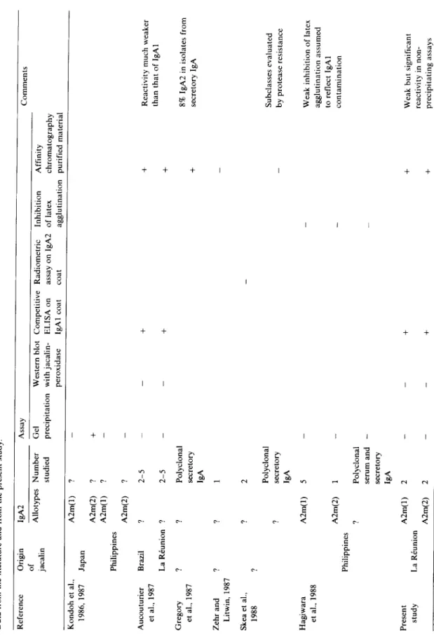
TABLE I IgA2 REACTIVITY OF JACAL1N Data from the literature and from the present study. Reference Origin IgA2 of Allotypes Number jacalin studied Kondoh et al., A2m(1) ? 1986, 1987 Japan Aucouturier et al., 1987 Gregory et al., 1987 Zehr and Litwin, 1987 Philippines Brazil La Rrunion ? A2m(2) ? A2m(1) ? A2m(2) ? ? 2-5 ? 2-5 ?
Polyclonal secretory lgA
? 1 Assay Gel Western blot Competitive Radiometric precipitation withjacalin- ELISA on assay on IgA2 peroxidase lgA1 coat coat + Inhibition of latex agglutination
Affinity chromatography purified
material m Comments Reactivity much weaker than that of IgA1 8% IgA2 in isolates from secretory IgA Skea et al., 1988 nagiwara et al., 1988 Present study Philippines La R6union ? 2 Polyclonal ? secretory IgA A2m(1) 5
A2m(2) ? A2m(1) A2m(2)
Polyclonal serum and secretory IgA m + + Subclasses evaluated by protease resistance Weak inhibition of latex agglutination assumed to reflect IgA1 contamination Weak but significant reactivity in non- precipitating assays
turier et al., 1987). More anodic fractions contain- ing a higher proportion of unglycosylated subunit were not used in this work.
Affinity chromatography purification of serum jaca-
fin binding proteins (JBP)
Sepharose 4B (Pharmacia, Uppsala, Sweden) was activated with cyanogen bromide according to March et al. (1974) and coupled to JCE or puri- fied jacalin (12 mg protein/ml of sedimented beads). In five separate experiments, the yields of coupling were 0.63, 0.53 and 0.54 with JCE and 0.87 on two occasions with purified jacalin. Seph- arose-coupled JCE or purified jacalin yielded identical results and hence were used inter- changeably. Ammonium sulfate precipitates from myeloma sera containing the proteins Mia (IgA2m(1)X), Gir (IgA2m(1)x), Felg (IgA2m(2)k) or Bel (IgA2m(2)r) (the allotypes were kindly determined by Dr. G. De Lange, Amsterdam), in addition to the controls, Ga (IgMr) and Ja (IgG2x), were adjusted to a concentration of 6.5 mg of protein/ml in 0.01 M phosphate-buffered saline pH 7.4 (PBS) and incubated for 20 rain at 4 ° C with jacalin-Sepharose (5 ml of settled beads for 10 ml of protein solution). After washing with cold PBS (at least 2 liters with numerous 2 or 3 min incubations) until the optical density (OD) of the effluent at 280 nm was below 0.002, JBP were eluted from the adsorbent by overnight incubation with 20 ml of 0.5 M D-galactose (Merck, Darm- stadt, F.R.G.) at 4°C, concentrated by vacuum dialysis against 0.15 M NaC1 and analyzed by standard immunoelectrophoresis (IEL) with poly- valent and anti-immunoglobulin light and heavy chain-specific antisera (Dako, Copenhagen, Den- mark) and with JCE (1.8 mg/ml). With the anti-a antiserum, IgA1 spurred over IgA2.
Purified monoclonal human Ig
Six monoclonal Ig were purified for this study; all steps were performed at 4 ° C and the purified concentrated solutions were stored in the presence of 50% glycerol at - 3 0 ° C . The IgAlr Min was isolated from serum by ammonium sulfate precipi- tation, gel filtration on Sepharose 6B (Pharmacia) followed by jacalin-Sepharose affinity chromatog- raphy of the 7-8 S fraction. It was pure by IEL at
187 27 mg/ml. The IgA2m(1)x Gir and IgA2m(2)~ Bel were purified by ammonium sulfate precipita- tion, gel filtration on Ultrogel ACA34 (IBF, Vil- leneuve-la-Garenne, France) and DEAE-Trisacryl (IBF) chromatography of the 7-8 S fractions in 0.01 M Tris/HC1 buffer pH 7.5 with a linear 0-0.3 M NaC1 gradient. Residual IgG of fast mobility and traces of polyclonal IgA1 were re- moved by adsorption on protein A-Sepharose (Pharmacia) and on a limited quantity of jacalin- Sepharose. After concentration by vacuum dialy- sis, the two IgA2 preparations were pure by IEL at 72 and 23 m g / m l respectively. The IgG3K Met has a very low electrophoretic mobility and was easily purified by DEAE-Trisacryl chromatogra- phy. The IgMx Mil was purified by ammonium sulfate precipitation, gel filtration on Sepharose 6B and euglobulin precipitation. It was then ad- sorbed on jacalin-Sepharose to remove small amounts of residual polyclonal IgA. The pure IgDX Car was prepared as previously described (Aucouturier et al., 1987) and stored at - 3 0 ° C with 5 mM c-aminocaproic acid and 1 mM phe- nylmethylsulfonyl fluoride in 50 % glycerol. Con- centrations of the purified Ig solutions were calculated from their absorbance at 280 nm using A~%cm values of 10.6 for IgA, 17.0 for IgD, 13.5 for IgG and 11.85 for IgM (Hudson and Hay, 1980; Johnstone and Thorpe, 1982). The mono- clonal IgA2m(2)h Felg and IgA2m(1)X Mia were purified as previously described (Aucouturier et al., 1987). Purity was controlled by IEL and by aminoterminal aminoacid sequencing.
Determination of isotype specificity of jacalin by
ELISA
Polystyrene microtitration plates (Nunc, Ros- kilde, Denmark) were coated with the IgA1 Min at 2 / t g / m l in 0.1 M carbonate buffer pH 9.6 (200 #l/well) overnight at 4°C, and saturated with PBS 0.05% Tween 20 (v/v) for 1 h at 37°C. After washing with PBS-Tween, 100 ttl/well of purified Ig at various dilutions were added in triplicate to 100 ttl of purified jacalin conjugated to horsera- dish peroxidase (HRP, Grade I, Boehringer; Manheim, F.R.G.) by the two-step glutaraldehyde method (Avrameas and Ternynck, 1971) at a final dilution of 1.04 ttg of jacalin/ml. The plates were incubated for 2 h at room temperature and washed
188
six times with P B S - T w e e n ; e n z y m e activity was
revealed with 0.4 m g / m l o - p h e n y l e n e d i a m i n e
0.012% H202 in 0.05 M s o d i u m citrate p H 5. A b s o r b a n c e values were read i n a Titertek (Flow, M c L e a n . VA) plate r e a d e r at a w a v e l e n g t h of 492 n m . As a c o n t r o l the same p u r i f i e d Ig were simi- larly tested o n identical plates with a m o n o c l o n a l a n t i - h u m a n IgA1 a n t i b o d y (clone M 4 D 8 . k i n d l y p r o v i d e d by Professor I.C.M. M a c L e n n a n . Bir- m i n g h a m , U . K . ) d i l u t e d 1 / 1 0 0 0 i n s t e a d of H R P - j a c a l i n . B i n d i n g of the a n t i b o d y to the i m m o b i - a n t i -r ~'c S 2 a n t i - ~( . . . $1 anti.~ " " ~ ' J' S1
anti-A
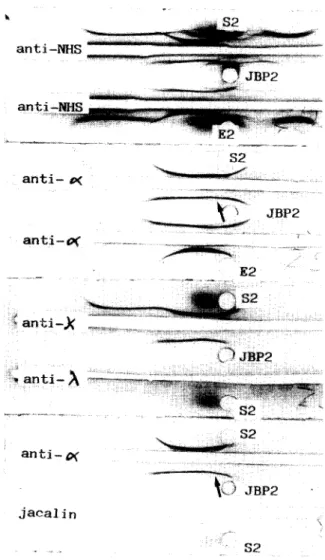
... S 1 , ... S I a n t i - ~ ... J a c a l i n ~ _ ~ $ 1Fig. 1. lmmunoelectrophoretic analysis of jacalin-binding pro- teins (JBP1) and effluent (El) obtained by affinity chromatog- raphy of an ammonium sulfate precipitate from the IgA2m(1)r myeloma serum Gir ($1). Anti-NHS: polyvalent anti-normal human serum antiserum. Note (1) that although the bulk of the IgA2 is in the effluent, it is also present in JBP, and (2) the
spur of polyclonal IgA1 over IgA2 (arrow).
a n t i - O f ... E 2 . . . :~ a n t a - . , a a n t i - O( . . . ~ ,,_,~
aBP2
j a e a l i nFig. 2. Immunoelectrophoretic analysis of jacalin-binding pro- teins (JBP2) and effluent (E2) obtained by affinity chromatog- raphy of an ammonium sulfate precipitate from the IgA2m(2)x
myeloma serum Bel ($2).
lized IgA1 was revealed b y i n c u b a t i o n for 1 h at r o o m t e m p e r a t u r e with 200 / ~ l / w e l l of H R P - c o n j u g a t e d r a b b i t a n t i - m o u s e I g G (2 / ~ g / m l , pre- p a r e d i n o u r l a b o r a t o r y ) .
Western blotting
Purified IgA1 M i n , I g A 2 m ( 1 ) G i r a n d I g A 2 m ( 2 ) Bel at 2.8 m g / m l were a n a l y z e d b y t h i n layer agarose electrophoresis ( P a r a g o n , B e c k m a n ) fol- lowed b y pressure b l o t t i n g o n n i t r o c e l l u l o s e ( H A H Y , Millipore, M o l s h e i m , F r a n c e ) a n d detec- t i o n with either H R P - a n t i - a a n t i s e r u m ( I n s t i t u t P a s t e u r P r o d u c t i o n , Paris, F r a n c e , 1 / 2 0 0 0 ) or
HRP-jacalin (0.52 #g jacalin/ml) for 90 min at room temperature. The enzymatic activity was de- veloped with benzidine 0.13% H202 0.03% in 0.1 M Tris/HC1 buffer pH 7.6.
Results
Study of proteins isolated by affinity chromatogra- phy on jacafin-Sepharose
IEL analysis of JBP from the four IgA2 myeloma sera (Figs. 1 and 2) revealed the presence of significant amounts of monoclonal IgA as shown in each case by a typical curvature of the IgA line with the anti-a and relevant anti-light chain antisera, and by a spur of polyclonal IgA1 with the anti-a serum. Indeed, as in our previous experiments (Aucouturier et al., 1987), residual polyclonal IgA1 was considerably enriched in jacalin affinity chromatography-purified material.
1
1 2 5 50 2(30 pg/ml
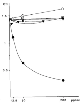
Fig. 3. Competition ELISA. Inhibition of HRP-jacalin binding to coated IgA1 by increasing concentration of purified human monoclonal IgA1 (@), IgA2m(1) (n), IgA2m(2) (o), IgD (v),
IgG3 (~) and IgM (*).
189 OD 1.5 (~""-" 1 O.S / o i i i 2.5 50 200 pg/ml
Fig. 4. Inhibition of binding of the anti-ed antibody M4D8 to coated IgA1 by increasing concentrations of purified human monoclonal IgA1 (O), IgA2m(1) (D), IgA2m(2) ((3), IgD (v),
IgG3 (or) and IgM (*).
Jacalin precipitated neither the IgA2m(1) nor the IgA2m(2) present in these JBP, but yielded a faint precipitating line (barely visible on the photo- graphs) corresponding to polyclonal IgA1. In con- trast, no traces of monoclonal IgG2r or IgMr could be found in JBP from the serum Ja and Ga respectively.
Competition ELISA
As previously observed, monoclonal IgA1 and IgD strongly inhibited the binding of HRP-jacalin to IgA1 coated plates while IgA2 of both allotypes yielding a weaker but significant inhibition (Fig. 3). The absence of contamination by IgA1 was confirmed by the control assay with the mono- clonal anti-al antibody instead of jacalin (Fig. 4), where only IgA1 was found to inhibit the binding of the antibody.
Gel precipitation and western blotting
As previously described and in striking contrast to IgA1, all IgA2 preparations tested failed to react with jacalin in Ouchterlony and IEL analysis or by Western blotting following agarose electro-
190
phoresis and detection with HRP-jacalin (not shown).
Discussion
We have previously reported that jacalin does not react with IgA2 by gel precipitation and west- ern blotting (which makes it very useful for the subclass typing of monoclonal IgA proteins) (Aucouturier and Preud'homme, 1987) but weakly binds to IgA2 (Aucouturier et al., 1987). This is confirmed in the present study of further IgA2 of known allotypes. Indeed, affinity chromatography prepared JBP from IgA2 myeloma sera contained the monoclonal IgA (in contrast to JBP from IgG or IgM myeloma sera which were free of myeloma proteins). Residual polyclonal IgA1 bound jacalin (with a considerable enrichment of the IgA1 com- pared to the starting sera) which clearly shows the much higher affinity of jacalin for the IgA1 sub- class. A large part of the myeloma IgA2 did not bind to jacalin-Sepharose and was recovered free of IgA1 in the effluent, thus permitting easy puri- fication of IgA2, in agreement with the findings of Gregory et al. (1987). Interestingly, even the IgA2 isolated in the JBP fraction did not precipitate jacalin in gel diffusion experiments, which strongly suggests that partial binding of IgA2 to jacalin columns results from low affinity or functional monovalency rather than from the presence of different populations of IgA2 molecules.
All of the IgA2 preparations tested weakly in- hibited the binding of jacalin (and not that of an anti-IgA1 monoclonal antibody) to insolubilized IgA1. In addition to the lack of reactivity with this monoclonal antibody, these IgA2 preparations were pure by electrophoretic, immunoelectro- phoretic and (in two cases) sequence criteria.
The strong reactivity of jacalin with human IgA1 and IgD is well explained by the presence of several o-linked Gal/31--* 3GalNAc in the hinge region of both heavy chains (Baenziger and Korn- feld 1974a,b; Mellis and Baenziger, 1983) since this disaccharide is the predominant reactive carbohydrate with jacalin (Sastry et al., 1986). Moreover, the location of the binding sites in the hinge region of IgA1 was confirmed by a study of proteolytic fragments (Skea et al., 1988). The
situation is not as clear for the other isotypes. Terminal galactose residues exist in all human Ig classes and subclasses, but their density and pre- sentation differ. /~ chains bear five kinds of asparagine-linked oligosaccharides, two of which are of the 'high mannose' type without galactose (Chapman and Kornfeld, 1979a,b), whilst the others have terminal N e u A c a 2 - - , 6Galfll 6GlcNAc or Galfll ~ 6GlcNAc (Hickman et al., 1972). The glycoside present on gamma chains is often terminated with Gal/31 ~ 4GlcNAc (Mizu- ochi et al., 1982; Takahashi et al., 1987), but its situation inside the pair of CH 2 domains in the entire molecule makes it almost inaccessible (Dei- senhofer, 1981). The location of IgA2 oligosac- charides has been studied by Toraho et al. (1977). However, their structures are not yet completely elucidated. Both allotypes seem to share two com- mon N-glycosides with IgA1, one located in the CH3 domain and the other, in the CH2 region, having exposed terminal Galfll ~ 4GlcNAc. IgA- 2m(1) and IgA2m(2) have two and three supple- mentary asparagine-linked oligosaccharides in contrast to IgA1, but their sequences have not been determined. Thus, the special feature of the carbohydrates found in IgA2 is the existence of at least one terminal Gal/31 --, 4GlcNAc exposed on the surface of the molecule. Sastry et al. (1986) showed that this disaccharide had some affinity for jacalin (although with a low association con- stant), and we previously showed that D-lactose (Gal/31 ~ 4Glc) inhibits HRP-jacalin binding to IgA1 at higher concentration than GalNAc and Gal (Aucouturier et al., 1987). Although the de- termination of the structure of the other oligosac- charides on the a2 chain may provide alternative explanations, the presence of Galfll--* 4GlcNAc in the CH2 domain is a possible explanation for IgA2 reactivity with jacalin.
The ability of jacalin to bind both IgA sub- classes can be used to remove IgA from biological samples and Ig preparations. However, the com- plete elimination of IgA2 would require a large excess of jacalin because of its low affinity of interaction. Preliminary experiments with com- mercially available intravenous IgG preparations showed that the IgA level sharply decreased after a first adsorption on jacalin-Sepharose but that several steps of adsorption were required to re-
m o v e all c o n t a m i n a t i n g I g A . N e v e r t h e l e s s , a m e t h o d b a s e d o n t h i s p r i n c i p l e s h o u l d b e o f g r e a t v a l u e f o r t h e p r e p a r a t i o n o f h u m a n I g G f o r t h e r - a p e u t i c use, i n w h i c h t h e p r e s e n c e o f I g A is a n i m p o r t a n t a n d d i f f i c u l t p r o b l e m . A c k n o w l e d g e m e n t s W e t h a n k D r . G . D e L a n g e f o r t y p i n g I g A 2 a l l o t y p e s , P r o f e s s o r I . C . M . M a c L e n n a n f o r t h e g i f t o f t h e m o n o c l o n a l a n t i b o d y t o I g A 1 , a n d D r . J. M e s t e c k y f o r p r o v i d i n g u s w i t h t h e I g A 2 F e l g . References
Aucouturier, P., Mihaesco, E., Mihaesco, C. and Preud'homme, J.L. (1987) Characterization of Jacalin, the human IgA and IgD binding lectin from jackfruit. Mol. Immunol. 24, 503. Aucouturier, P. and Preud'homme, J.L. (1987) Subclass distri- bution of human myeloma proteins as determined with monoclonal antibodies. Immunol. Lett. 16, 55.
Avrameas, S. and Ternynck, T. (1971) Peroxidase labeled anti- body and Fab conjugates with enhanced intracellular penetration. Immunochemistry 8, 1175.
Baenziger, J. and Kornfeld, S. (1974a) Structure of the carbo- hydrate units of IgA1 immunoglobuhn. I. Composition, glycopeptide isolation and structure of the asparagine-lin- ked ohgosaccharide units. J. Biol. Chem. 249, 7260. Baenziger, J. and Kornfeld, S. (1974b) Structure of the carbo-
hydrate units of the IgA1 immunoglobulin. II. Structure of the o-glycosidically linked ohgosaccharide units. J. Biol. Chem. 249, 7270.
Burm-Moreno, M.M. and Campos-Neto, A. (1981) Lectin(s) extracted from seeds of Artocarpus integrifolia (jackfruit): Potent and selective stimulator(s) of distinct human T and B cell functions. J. Immunol. 127, 427.
Chapman, A. and Kornfeld R. (1979a) Structure of the high mannose oligosaccharide of a human IgM myeloma pro- tein. I. The major oligosaccharides of the two high mannose glycopeptides. J. Biol. Chem. 254, 816.
Chapman, A. and Kornfeld, R. (1979b) Structure of the high mannose ohgosaccharides of a human IgM myeloma pro- tein. II. The minor ohgosaccharides of high mannose glyco- peptide I. J. Biol. Chem. 254, 824.
Deisenhofer, J. (1981) Cristallographic refinement and atomic models of a human Fc fragment and its complex with fragment B of protein A from Staphylococcus aureus at 2.9 and 2.8 A resolution. Biochemistry 20, 2361.
Gregory, R.L., Rundegren, J. and Arnold, R.R. (1987) Sep- aration of human IgA1 and IgA2 using jacalin-agarose chromatography. J. Immunol. Methods 99, 101.
191 Hagiwara, K., Collet-Cassart, D., Kobayashi, K. and Vaerman, J.P. (1988) Jacalin: isolation, characterization and influence of various factors on its interaction with human IgA1, as assessed by precipitation and latex agglutination. Mol. Im- munol. 25, 69.
Hickman, S., Kornfeld, R., Osterland, K. and Kornfeld, S. (1972) The structure of the glycopeptide of a human M-im- munoglobuhn. J. Biol. Chem. 247, 2156.
Hudson, L. and Hay, F.C. (1980): Practical Immunology. Blackwell, Oxford, p. 347.
Johnstone, A. and Thorpe, R. (1982) Immunochemistry in Practice. Blackwell, Oxford, p. 2.
Kondoh, H., Kobayashi, K., Hagiwara, K. and Kajii, T. (1986) Jacahn, a jackfruit lectin, precipitates IgA1 but not IgA2 subclass on gel diffusion reaction. J. Immunol. Methods 88, 171.
Kondoh, H., Kobayashi, K. and Hagiwara, K. (1987) A simple procedure for the isolation of human secretory IgA of IgA1 and IgA2 subclass by a jackfruit lectin, jacalin, affinity chromatography. Mol. Immunol. 24, 1219.
March, S.C., Parikh, I. and Cuatrecasas, P. (1974) A simplified method for cyanogen bromide activation of agarose for affinity chromatography. Anal. Biochem. 60, 149.
Melhs, S. and Baenziger, J. (1983) Structure of the o-glycosidi- cally hnked oligosaccharides of human IgD. J. Biol. Chem. 258, 11557.
Mizuochi, T., Taniguchi, T., Shimizu, A. and Kobata, A. (1982) Structural and numerical variation of the carbohydrate moiety of immunoglobulin G. J. Immunol. 129, 2016. Roque-Barreira, M.C. and Campos-Neto, A. (1985) Jacalin: an
IgA-binding lectin. J. Immunol. 134, 1740.
Sastry, M.V.K., Banarjee, P., Patanjali, S.R., Swamy, M.J., Swarnalatha, G.V. and Surolia, A. (1986) Analysis of sac- charide binding to Artocarpus integrifolia lectin reveals specific recognition of T-antigen (fl-D-Gal(1--* 3)o-Gal- NAc). J. Biol. Chem. 261, 11726.
Saxon, A., Tsui, F. and Martinez-Maza (1987) Jacalin, an IgA-binding lectin, inhibits differentiation of human B ceils by both a direct effect and by activating T-suppressor cells. Cell. Immunol. 104, 134.
Skea, D.L, Christopoulos, P., Plant, A.G. and Underdown, B.J. (1988) Studies on the specificity of the IgA-binding lectin, jacalin. Mol. Immunol. 25, 1.
Takahashi, N., Ishii, I., Ishihana, H., Mori, M., Tejima, S., Jefferis, R., Endo, S. and Arata, Y. (1987) Comparative structural study of the N-linked oligosaccharides of human normal and pathological immunoglobuhn G. Biochemistry 26, 1137.
Toraho, A., Tsuzukida, Y., Liu, Y.S.V. and Putnam, F.W. (1977) Location and structural significance of the oligosac- charides on human IgA1 and IgA2 immunoglobulins. Proc. Natl. Acad. Sci. U.S.A. 74, 2301.
Zehr, B.D. and Litwin, S.D. (1987) Human IgD and IgA1 compete for D-galactose-related binding sites on the lectin jacahn. Scand. J. Immunol. 26, 229.