RESEARCH OUTPUTS / RÉSULTATS DE RECHERCHE
Author(s) - Auteur(s) :
Publication date - Date de publication :
Permanent link - Permalien :
Rights / License - Licence de droit d’auteur :
Bibliothèque Universitaire Moretus Plantin
Institutional Repository - Research Portal
Dépôt Institutionnel - Portail de la Recherche
researchportal.unamur.be
University of Namur
Differential influence of anticancer treatments and angiogenesis on the seric titer of
autoantibody used as tumor and metastasis biomarker
Defresne, Florence; Bouzin, Caroline; Guilbaud, Céline; Dieu, Marc; Delaive, Edouard;
Michiels, Carine; Raes, Martine; Feron, Olivier
Published in: Neoplasia DOI: 10.1593/neo.10238 Publication date: 2010 Document Version
Publisher's PDF, also known as Version of record
Link to publication
Citation for pulished version (HARVARD):
Defresne, F, Bouzin, C, Guilbaud, C, Dieu, M, Delaive, E, Michiels, C, Raes, M & Feron, O 2010, 'Differential influence of anticancer treatments and angiogenesis on the seric titer of autoantibody used as tumor and metastasis biomarker', Neoplasia, vol. 12, no. 7, pp. 562-570. https://doi.org/10.1593/neo.10238
General rights
Copyright and moral rights for the publications made accessible in the public portal are retained by the authors and/or other copyright owners and it is a condition of accessing publications that users recognise and abide by the legal requirements associated with these rights. • Users may download and print one copy of any publication from the public portal for the purpose of private study or research. • You may not further distribute the material or use it for any profit-making activity or commercial gain
• You may freely distribute the URL identifying the publication in the public portal ? Take down policy
If you believe that this document breaches copyright please contact us providing details, and we will remove access to the work immediately and investigate your claim.
Differential Influence of
Anticancer Treatments and
Angiogenesis on the Seric Titer
of Autoantibody Used as Tumor
and Metastasis Biomarker
1,2Florence Defresne*, Caroline Bouzin*,
Céline Guilbaud*, Marc Dieu†, Edouard Delaive†, Carine Michiels†, Martine Raes†and Olivier Feron* *Angiogenesis and Cancer Research Laboratory, Pole of Pharmacology and Therapeutics, Université catholique de Louvain, Brussels, Belgium;†Laboratory of Biochemistry and Cellular Biology, University of Namur-FUNDP, Namur, Belgium
Abstract
Early detection of tumor-specific autoantibodies (auto-Abs) has the potential to be used for cancer screening and diagnosis. Whether auto-Ab may be useful to track metastatic progression or response to treatment is, however, largely unknown. To address these issues, the serological proteome was analyzed in an invasive but treatment-responsive mouse tumor model. Among 40 serum-reactive proteins identified by multiplex analysis, we chose to focus on glucose-regulated protein 78 (GRP78), a chaperone protein involved in the endoplasmic reticulum stress response. We first validated GRP78 as a protein overexpressed and mislocalized in tumor cells. We then docu-mented that an increase in GRP78 auto-Ab titer preceded the detection of a palpable tumor mass, correlated with metastatic progression, and was influenced by the onset of tumor neovascularization. We also found that chemo-therapy and radiochemo-therapy, both leading to inhibition of tumor growth, oppositely influenced the anti-GRP78 im-mune response. Whereas radiation increased the concentration of GRP78 auto-Ab by three-fold, the auto-Ab titer was reduced in response to bolus or metronomic administration of cyclophosphamide. Finally, we established a decrease in auto-Ab–producing B lymphocytes in response to chemotherapy and the overexpression of GRP78 together with a strong immunoglobulin response in irradiated tumors. In conclusion, we identified GRP78 auto-Ab as an early marker of tumor and metastatic progressions. However, the multiple influences of anticancer treatments on the humoral immune system calls for caution when exploiting such auto-Ab as markers of the tumor response.
Neoplasia (2010) 12, 562–570
Introduction
Autoantibodies (auto-Ab) are present in the blood of patients who are affected by different malignancies [1,2]. These antibodies are directed against a group of autologous cellular antigens generally known as tumor-associated antigens (TAAs) [3–5]. The expression by tumor cells of proteins, which are mutated, mislocalized, or pro-duced in abnormal quantities, is thought to mainly account for this humoral response. Auto-Abs circulate for a longer time than other polypeptides because they are very stable in the serum and often pro-duced in large amounts. Their biochemical properties are well under-stood, and many available reagents do exist for their detection. Serum profiling of circulating auto-Ab is therefore considered a very attrac-tive method to diagnose cancer at early stages.
Different proteomic techniques allow detecting auto-Ab and iden-tifying TAAs: serological expression cloning and serological proteome analysis (SERPA) are among them [6–9]. These methods use a patient’s sera to probe blotted phage expression libraries derived from tumor cells
or tumor cell lysates blotted onto a membrane after two-dimensional gel separation, respectively. Modification of the latter involves spotting
Abbreviations: auto-Ab, autoantibody; SERPA, serological proteome analysis; TAA, tumor-associated antigen
Address all correspondence to: Prof. Olivier Feron, Pole of Pharmacology and Therapeu-tics, Université catholique de Louvain– FATH5349, 52 Ave E. Mounier, B-1200 Brussels, Belgium. E-mail: olivier.feron@uclouvain.be
1
This work was supported by grants from the Fonds de la Recherche Scientifique Médicale, the Fonds National de la Recherche Scientifique (FNRS), the Télévie, the Belgian Federation Against Cancer, the J. Maisin Foundation, and the Région Bruxelles-Capitale and by an Action de Recherche Concertée (ARC 09/14-020) from the Communauté Française de Belgique. F.D. is FNRS Research Assistant and O.F. is FNRS Research Director.
2
This article refers to supplementary materials, which are designated by Tables W1 and W2 and Figures W1 and W2 and are available online at www.neoplasia.com. Received 3 February 2010; Revised 28 March 2010; Accepted 29 March 2010 Copyright © 2010 Neoplasia Press, Inc. All rights reserved 1522-8002/10/$25.00 DOI 10.1593/neo.10238
of fractionated tumor lysates onto microarrays [10], and for each of these techniques, final identification of the proteins of interest requires mass spectrometry. SERPA has the advantages to allow proteins with their posttranslational modifications to be analyzed for their immuno-genicity and to reveal, in a single experiment, the global reactivity of a given serum toward a tumor-derived proteome. Multiple studies have already used these techniques to identify auto-Abs in a variety of can-cers including hepatocellular carcinoma [3], colon cancer [11,12], lung cancer [13], and breast cancer [5,14].
Very little is known, however, about how the auto-Ab–based mark-ers of early cancer stages do evolve when the disease progresses to metastases or when patients undergo anticancer treatments. In the-ory, the ideal auto-Ab candidate would have to be upregulated when the tumor is growing or when metastases are developing and to fall down when the patients respond to the treatment. Collateral effects of treatments on the capacity of tumor or immune cells to contribute to the auto-Ab response, however, should not be underestimated. Chemotherapy may, for instance, lead to lymphodepletion and thereby interfere with the capacity of the humoral immune system to produce auto-Ab. Whether a reduction in auto-Ab reflects the effects of che-motherapy on tumor growth or instead acknowledges a systemic in-terference with the immune system needs to be addressed to fully exploit information derived from serological proteome analyses.
Here, we applied the SERPA technique to identify the fate of auto-Ab in tumor-bearing mice exposed to different treatments, in-cluding chemotherapy, radiotherapy, and surgery. Such an animal model allows to reduce interindividual serological variations under basal conditions as well as in response to treatments and to concen-trate in 2 to 3 weeks, the life of a tumor from the primary tumor emer-gence to the metastases development. Using SERPA technology, we identified glucose-regulated protein 78 (GRP78) as a reproducible immunogenic TAA in our mouse tumor model. A specific enzyme-linked immunosorbent assay (ELISA) was developed and confirmed that the increase in GRP78 auto-Ab titer was correlated with primary tumor and metastases development. Opposite variations in the GRP78 auto-Ab concentrations after chemotherapy and radiotherapy, how-ever, pointed out how treatment-driven modulation of the immune system may interfere with the auto-Ab production and detection. Materials and Methods
Cells and Mice
Lewis lung carcinoma (LLc) cells were routinely cultured in 175-mm flasks in serum containing Dulbecco modified Eagle me-dium (Invitrogen, Paisley, UK). Adult C57Bl/6J mice (Elevage Janvier, Le Genest Saint-Isle, France) received intramuscular injections of 106 syngeneic LLc cells in the posterior right leg. The tumor diameters were regularly tracked with an electronic caliper. Blood was collected for se-rological assays through retro-orbital or intracardiac routes according to the required amounts. Ten days after tumor cell injection, mice were exposed to radiotherapy or chemotherapy. Local irradiation was ad-ministered to mice using a RT-250 device (Philips Medical Systems, Brussels, Belgium) with a dose delivery of 1.2 Gy/min. The tumor was centered in a circular irradiation field, and healthy tissues were protected by a lead mask. Two different protocols were used for chemotherapy: cyclophosphamide (Bayer, Leverkusen, Germany) was either adminis-tered through intraperitoneal injection of a bolus dose (100 mg/kg) or added to the drinking water (renewed every 3 days) for a so-called metronomic administration [15] to reach a 20-mg/kg per day regimen.
Each procedure was approved by local authorities according to national animal care regulations.
SERPA Technique
Tumors from three different mice were pooled, lysed in difference in-gel electrophoresis labeling buffer (7 M urea, 2 M thiourea, 4% 3-[(3-cholamidopropyl)dimethylammonio]-1-propanesulfonate, and 30 mM Tris pH 8.5), homogenized using Ultra-Turrax T25 (IKA, Staufen, Germany), and clarified by centrifuging at 12,000g for 15 min-utes at 4°C. Protein concentration was determined by the Bradford method, and extracts were diluted to reach a final concentration of 5 to 10μg/μl. pH was adjusted to 8.5, and 25 μg of each sample was labeled with 200 pmol of amine-reactive cyanine Cy5 dye (Amersham GE Healthcare, Diegem, Belgium) for 30 minutes in the dark at 4°C, according to the manufacturer’s instructions. Labeling reaction was stopped by incubating the mixture for 10 minutes with 10 mM lysine (Sigma Aldrich, Bornem, Belgium), and nonlabeled proteins were added to reach 300μg. Samples were then diluted in immobilized pH gra-dient (IPG) buffer (4% 3-[(3-cholamidopropyl)dimethylammonio]-1-propanesulfonate, 7 M urea, 2 M thiourea, 30 mM Tris, 30 mM DTT, 1% IPG buffer 3-11 (vol./vol.) [Amersham GE Healthcare]) and in-cubated for 20 minutes in the dark at room temperature.
Proteins were loaded onto rehydrated 18-cm IPGstrips pH 3-11 NL for the isoelecteric focusing on the IPGphor; parameters were as fol-lows: 300 V for 3 hours, gradient steps of 1000 V for 8 hours, 8000 V for 3 hours, and 8000 V for 45 minutes at 20°C with a maximum cur-rent setting of 50μA per strip. IPGstrips were subsequently incubated for 15 minutes with equilibration solutions (6 M urea, 30% glycerol, 2% SDS, 1.5 M Tris pH 8.8) supplemented with 10 mg/ml DTT and 25 mg/ml iodoacetamide, respectively. One-dimensional strips were then washed and loaded onto the two-dimensional gels (10% acryl-amide). Electrophoresis was performed overnight at 15°C.
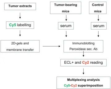
Separated proteins were finally transferred onto low-fluorescence polyvinylidene difluoride (PVDF) membrane (200 mA for 2 hours). All the materials and products were purchased from GE Health-care, except glycerol which was purchased from Sigma. Blotted two-dimensional membranes were incubated for 3 hours in 5% nonfat dry milk containing Tris–Tween buffered saline (TTBS) blocking buffer and were then exposed overnight to either control mouse serum or tumor-bearing mice (dilution, 1:100); a pool of sera collected from 15 different mice was used per condition. After several washes in TTBS containing 1% nonfat dry milk, membranes were incubated with horseradish peroxidase–conjugated goat antimouse immunoglobulin class G (IgG) antibody (1:5000; Jackson Immunoresearch, Sulfolk, UK) for 2 hours. Note that this secondary antibody (cat. no. 115-035) reacts with both heavy and light chains of IgG molecules and may thus also react with other immunoglobulin classes, including IgM. Immunodetection was performed using ECL Plus (GE Health-care) followed by scanning at the Cy2 wavelength on the Ettan Dige Imager (GE Healthcare; Figure W1).
Immunoblot Analysis and Immunocytochemistry
Collected tumors were homogenized with an Ultra-Turrax in RIPA lysis buffer containing 1% protease inhibitor cocktail. SDS-PAGE was performed as previously described on 10% acrylamide gels, and after transfer, PVDF membranes were probed overnight at 4°C with anti-GRP78 antibodies (BD Pharmingen [Erembodegem, Belgium] and Cell Signaling [Danvers, MA]). HRP-conjugated sec-ondary antibody was used for detection with ECL Plus.
For immunohistochemical analyses, frozen tumor sections were probed overnight at 4°C with anti-GRP78 antibody (dilution 1:50; Cell Signaling) or with anti-CD31 antibody (dilution, 1:50; Pharmingen) after a 30-minute blocking procedure in PBS containing 0.1% Tween and 5% BSA. Detection was performed with Alexa Fluor second-ary antibodies (dilution, 1:300); quantification was performed using ImageJ software (National Institutes of Health, Bethesda, MD). In some experiments, nonpermeabilized tumor sections were costained with rhodamine-labeled wheat germ agglutinin (Vector Laboratories, Burlingame, CA) to probe plasma membranes.
Laser Doppler Imaging
Local tumor blood flow was measured with a laser Doppler imager (Moor Instruments, Devon, UK). Mice were anesthetized, and fur was removed using a depilatory cream. The animals were placed on a heat-ing pad (37°C) to minimize variations in temperature. The perfusion of the tumor-bearing and control legs can be evaluated on the basis of colored histogram pixels.
In-gel Enzymatic Digestion and Mass Spectrometry
Preparative gels were performed with 300μg of unlabeled proteins according to the protocol described above for the analytical gels. Two-dimensional gels were krypton-stained (Pierce, Rockford, IL) after protein fixation. The proteins of interest were automatically picked from the gels with the Ettan Spot Picker (GE Healthcare). After rinsing, gel pieces were dehydrated in acetonitrile and further dried at 37°C for 20 minutes. Digestion was performed overnight with trypsin (12.5 ng/μl) in 50 mM ammonium bicarbonate. The extraction step was performed with formic acid 5% for 15 minutes at 37°C. Peptides were extracted with 5% formic acid at 37°C for 15 min-utes, and the collected supernatants were kept frozen at−20°C until mass spectrometry analysis.
Digested peptides were then processed for identification on the basis of their mass fingerprint obtained using a matrix-assisted laser desorption/ ionization–time of flight mass spectrometry (MALDI-TOF; Waters, Milford, MA) or using a nanoflow liquid chromatography coupled to tandem mass spectrometry with electrospray ionization (Waters) on a CapLC Q-TOF2 mass spectrometer (Waters; Supplementary Data for detailed information). Full-length proteins were identified with Mascot software (version 2.2; Matrix Sciences, London, UK) by sequence homology research against mouse protein databases.
GRP78 ELISA
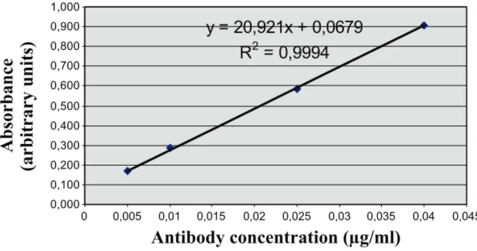
Amounts of circulating auto-Abs to GRP78 were determined by conventional ELISA. Ninety-six–well plates (Reacti-Bind; Thermo Scientific, Rockford, IL) were coated overnight at room temperature with 5μg/ml recombinant GRP78 protein (Stressgen, Ann Arbor, MI). Coating and blocking procedures were carried out using Ultra-block and Neptune buffers from AbD Serotec (Oxford, UK) according to the manufacturer’s instructions. Sera (dilution, 1:100) were incu-bated overnight at 4°C, and after washing, specific hybridization was measured with a peroxidase-conjugated antimouse IgG antibody (dilution, 1:10,000) and addition of 3,3′,5,5′-tetramethylbenzidine (Merck Chemicals, Nottingham, UK). Plates were read at 450 nm in a microplate reader (VictorX4; Perkin Elmer, Waltham, MA), and the calibration curve was performed for each single experiment using serial dilutions of GRP78 antibody (BD Pharmingen; Figure W2).
Immunoprecipitation and Immunodetection of Natural IgG
The same volumes of serum and protein G sepharose (slurry 1:1) were mixed together and incubated for 1 hour at 4°C under constant agitation. Pellets were collected by centrifuging at 10,000g at 4°C and were resuspended in 2× Laemmli buffer. Samples were boiled for 5 minutes and were centrifuged; supernatants were analyzed by im-munoblot analysis as described above. In some experiments, accumu-lation of natural IgG was evaluated in tumor sections using an Alexa Fluor 488 antimouse IgG.
Flow Cytometry Analysis
Blood samples were freshly collected, and red blood cells were elim-inated by centrifugation on Histopaque 1083 (Sigma). Cells were then labeled with a biotin-conjugated monoclonal antibody from BD Pharmingen (anti-B220, clone RA3-6B2) then with PE-conjugated streptavidin. Fluorescence signals were measured using a FACScan apparatus (BD Pharmingen) and analyzed by the FlowJo software (Tree Star, Inc, Olten, Switzerland).
Statistical Analysis
Data are expressed as means ± SEM or as scatter plots. Student’s t test and one-way analysis of variance were used where appropriate. Results
SERPA Identification of GRP78 Auto-Abs in
Tumor-Bearing Mice
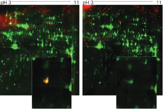
Total proteins extracted from LLc tumors were separated by two-dimensional PAGE and were transferred onto PVDF membranes. Pooled sera from tumor-bearing mice and control mice (n = 15 per condition) were probed separately for the presence of auto-Abs directed against tumor proteins. Multiplexing analysis was performed taking advantage of the Cy5 prelabeling of tumor proteins and the detection of the anti-IgG peroxidase-conjugated secondary antibody in the Cy2 wavelength (Figure W1). The long-lasting and stable chemiluminescence from the ECL Plus reagent used in our experi-ments gave a stable fluorescence signal. The low interindividual variability in this mouse tumor model led to the identification of 40 serum-labeled spots on the two-dimensional blots. The proteins of interest were excised from preparative gels and digested with tryp-sin, and the peptide mixtures were analyzed by mass spectrometry. This led to the identification of 24 proteins with satisfying scores (Table W1), among which 12 were tumor-bearing mouse serum– positive (Table 1). Only four antigens, however, were exclusively rec-ognized by the serum of tumor-bearing mice: GRP78, aldolase A1, vinculin, and heterogeneous nuclear ribonucleoprotein L. In the rest of the study, we chose to focus on GRP78 (also called BiP), which was the antigen giving the strongest immunoblot signal (see enlarged spot in Figure 1).
GRP78 Autoantigen Validation
The GRP78 identification was confirmed by probing two-dimensional membranes with commercially available anti-GRP78 antibody (Figure 2A). We then examined by Western blot analysis the GRP78 expression level in LLc tumor cells and found that it amounted to more than five-fold the abundance in the host tissue (Figure 2B). Furthermore, while in the host tissue, GRP78 was ex-clusively found intracellularly in agreement with its endoplasmic re-ticulum (ER) function; immunohistochemistry revealed that, in
tumor cells, GRP78 was aberrantly located at the plasma membrane (Figure 2C ).
GRP78 Auto-Abs as a Marker of Tumor Growth
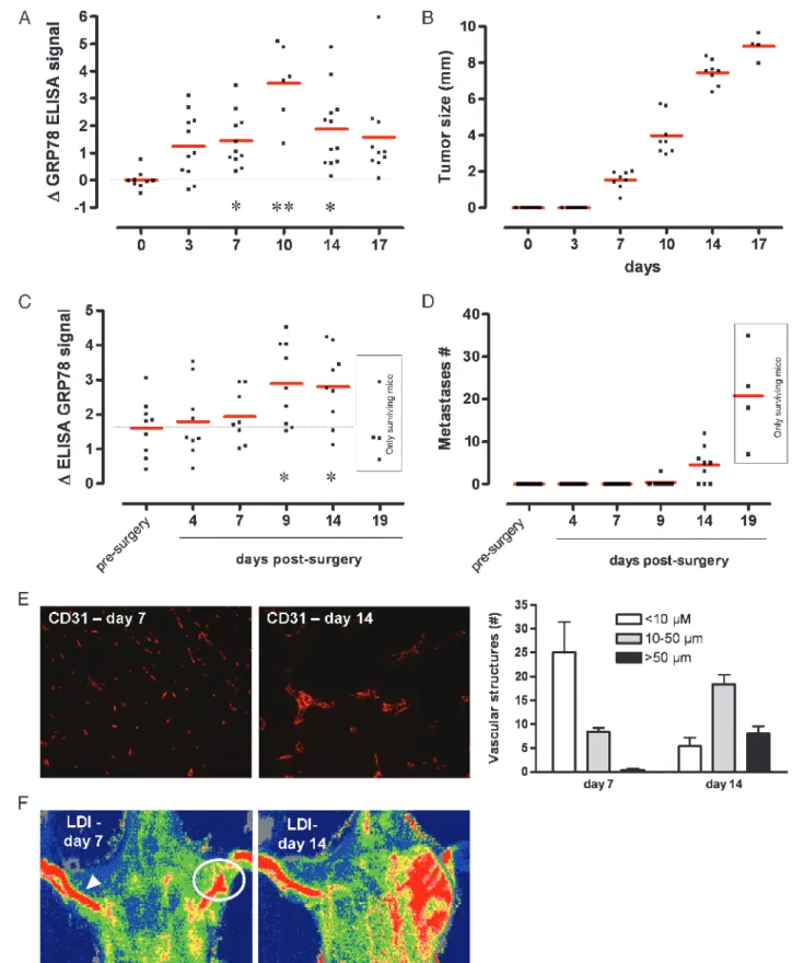
An ELISA was developed using recombinant GRP78 protein to confirm the presence of auto-Abs in the serum of tumor-bearing mice and to track titer changes in response to treatments. Calibration was carried out for each ELISA assay, and a linear relationship was consistently observed in the range of antibody concentration detected from the mouse serum (Figure W2). Blood was collected from tumor-bearing mice on days 0, 3, 7, 10, 14, and 17 after injection (intra-muscular) with 106tumor cells. ELISA revealed that a net increase in GRP78 auto-Abs was detectable in the tumor-bearing mouse serum as soon as 3 days after tumor cell injection. Interestingly, at that early time, tumor growth, as measured with an electronic caliper, was not yet detectable; tumors were actually palpable from day 7 (Figure 3B). GRP78 auto-Ab signal remained stable from day 3 to day 7 after
in-jection but peaked on day 10; values detected on days 14 and 17 were barely higher than those observed on day 7. Because day 10 is usually the time required for angiogenesis to become functional, we examined the difference in endothelial staining using CD31 anti-body and blood flow around this period. We confirmed a significant increase in large, mature blood vessels on day 14 versus a punctate, an-giogenic pattern on day 7 (Figure 3E ). A dramatic increase in tumor blood flow was also observed between day 7 and day 14, as determined using laser Doppler imaging (Figure 3F ).
In other series of animals aimed to examine the effect of metastases spreading on the anti-GRP78 titer, we stimulated the development of dormant lung metastases by surgically removing the primary tumor (when reaching 8 mm in diameter), as previously documented in this tumor model [16]. Interestingly, whereas a net increase in anti-GRP78 antibody was observed 9 days after surgery (Figure 3C ), the detection of metastases required 5 to 10 more days (see day 14 [6/9 mice] and day 19 in Figure 3D). Between day 14 and day 19,
Figure 1. SERPA of LLc tumor-bearing mice. Representative immunoblot analysis with pooled sera from tumor-bearing mice (left panel) or control mice (right panel) (n = 15) on Cy5-labeled LLc tumor extracts; yellow/red spots correspond to proteins recognized by auto-Ab present in the sera. This experiment was repeated four times; the enlarged spot corresponds to GRP78, as confirmed after picking, trypsin digestion, and consecutive MALDI identification.
Table 1. List of Identified Tumor Proteins Detected by Two-dimensional Immunoblot Analysis with the Sera of Tumor-Bearing Mice
Proteins pI MW (kDa) Mascot Score Uniprot no. Queries Matched Sequence Coverage (%) P (Expect)
MALDI identification
inner membrane protein mitochondrial cra 6.18 83 148 Q8CAQ8 34 52 1.81e−12
Villin 2 5.83 69 245 P26040 41 60 4.50e−20
Grp78 4.79 72 240 P20029 36 56 1.40e−19
Heat shock protein 1 4.93 84 125 P07901 29 51 1.80e−08
Heterogeneous nuclear ribonucleoprotein l 6.07 70 161 Q499X2 22 47 5.70e−13
Heterogeneous nuclear ribonucleoprotein k 5.39 51 119 P61979 18 41 1.80e−07
Vimentin 4.77 52 225 P20152 31 59 4.50e−18
Lap 3 7.61 56 153 Q9CPY7 27 56 1.10e−10
Lectin, galactose binding, soluble 3 8.50 30 122 P16110 9 80 9.10e−08
QTOF identification
Aldolase 1, A isoform 8.40 40 471 P05064 7
Procollagen lysine, 2-oxoglutarate 5-dioxygenase 3 5.81 85 389 Q9R0E1 4
more than 50% of the mice died because of lung metastases. Inter-estingly, among the mice alive on day 19, the anti-GRP78 levels were usually in the low percentile range (Figure 3C ).
Effect of Radiotherapy and Chemotherapy on GRP78
Auto-Ab Titer
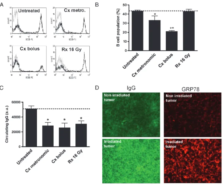
To study the effect of treatments on the expression of GRP78 auto-Abs, we chose radiotherapy and chemotherapy regimens. Accordingly, tumor-bearing mice were exposed to 16-Gy radiation, 100-mg/kg cyclo-phosphamide administered intraperitoneally (bolus, two injections at 5-day interval), or low-dose cyclophosphamide in the drinking water (so-called metronomic chemotherapy). Radiotherapy and bolus cyclophos-phamide are known to exert profound antitumor effects, as confirmed in Figure 4, A and C, respectively. Metronomic cyclophosphamide ther-apy is a therapeutic scheme proposed to have a better benefit/toxicity balance than the conventional maximal tolerated dose regimen [15]. In this study, we observed an initial response of the tumor to metro-nomic cyclophosphamide but a loss of efficacy on the longer term (Fig-ure 4E ). ELISA revealed that radiotherapy led to an increase in circulating anti-GRP78, peaking at five-fold the level observed 5 days before the radiation exposure (Figure 4B). Of note, local irradiation of control animals (ie, without tumor) failed to lead to any changes in anti-GRP78 concentrations (not shown). Chemotherapy gave rise to an opposite pattern, with a significant reduction of anti-GRP78 auto-Ab concentration (Figure 4, D and F ). Note the continuous decrease in anti-GRP78 auto-Ab with metronomic chemotherapy despite the lesser inhibitory effect of this drug regimen on tumor growth (Figure 4F ).
Immune Response Toward the Different Treatments
To understand the reasons of the decrease in anti-GRP78 auto-Ab after chemotherapy, we evaluated whether the systemic cytotoxic effects of cyclophosphamide could influence the humoral response by anti-body-producing lymphocytes. We therefore used flow cytometry to evaluate potential changes in the extent of B cells in treated versus un-treated tumor-bearing mice (Figure 5A). We observed that, whereas radiotherapy did not alter the amounts of B-220–positive B cells,
cy-clophosphamide administration led to a 20% to 50% reduction in B cells (Figure 5B). The reduction in B-cell numbers after chemotherapy did correlate with a reduction in the extent of circulating IgG as de-termined by immunoprecipitation from cyclophosphamide-exposed mouse sera (Figure 5C). Because radiotherapy led to a similar reduc-tion in circulating IgG despite a maintained amount of B cells, we fur-ther examined whefur-ther antibodies could be trapped into the irradiated tumor. Figure 5D shows that the overall detection of IgG from tumor sections was indeed significantly increased in response to radiotherapy (vs untreated tumors, n = 5). Immunohistochemical analysis of the same tumor sections further revealed that GRP78 expression was also dramatically increased in irradiated tumors (Figure 5D).
Discussion
The major findings of this study are that (i) increase in circulating anti-GRP78 auto-Ab is associated with the detection of primary tu-mor and metastases at earlier times than with palpation and micro-scopic analysis of the invaded tissues, respectively; (ii) chemotherapy and radiotherapy have opposite effects on the extent of circulating anti-GRP78 auto-Ab, prompting caution in interpreting changes in auto-Ab titers in response to anticancer treatments; and (iii) Cy-dyes multiplex analysis may optimize the SERPA workflow.
An increasing number of studies report the identification of auto-Ab in cancer patients as potential new biomarkers for cancer de-tection and prognosis [1–14]. Although several good candidates are making their route toward validation in larger studies, a variable pro-portion of patients with a given cancer type are usually positive for the considered Ab. Most of these studies aiming to identify auto-Ab as new cancer biomarkers are carried out by comparing the sera of cancer patients with the sera of healthy volunteers or patients with cancer-predisposing diseases (eg, polyps, cirrhosis, viral infection). The status of patients at the time of the serum collection is therefore vari-able, and the influence of treatments (targeting cancer or precancerous lesions) is rarely integrated in the interpretation of the results. Another related issue is the potential and reliability of auto-Ab–based markers to follow the response to treatments. From a theoretical point of view, Figure 2. Validation of GRP78 as a tumor autoantigen. (A) Immunoblot analysis with commercial anti-GRP78 of the putative GRP78 spot from LLc tumor extracts separated on two-dimensional membranes. (B) Representative Western blot analysis of the expression of GRP78 in host tissue and tumors (n = 4); simultaneous glyceraldehyde-3-phosphate dehydrogenase (GAPDH) immunoblot analysis was used as a control. (C) Representative GRP78 immunostaining on host tissue (top) and tumor (bottom) sections; immunostaining experiments were repeated several times with similar results.
Figure 3. Changes in the titer of anti-GRP78 auto-Ab as a marker of primary tumor growth and metastases. (A) GRP78-specific ELISA was performed from sera collected through retro-orbital bleeding of mice before (day 0) and after (days 3, 7, 10, 14, and 17) tumor cell injection. Results are presented as scatter plots; *P < .05, **P < .01. (B) Tumor diameters as measured with a caliper. (C) GRP78-specific ELISA was performed at the indicated time after primary tumor removal to activate lung metastases. Results are presented as scatter plots; *P < .05. (D) Number of lung metastases identified by histologic analysis of lungs collected from tumor-bearing mice at the indicated time before and after surgery. Note that on day 19 in panels C and D, only data arising from surviving animals are presented. (E) Representative CD31 immunostaining of tumor sections on days 7 and 14. Histogram represents the distribution of CD31-positive vascular structures according the indicated sizes. (F) Representative laser Doppler imaging (LDI) pictures from LLc tumor-bearing mice on days 7 and 14 after injection of tumor cells. The average perfusions of the tumor-bearing leg (white circle) and of the control leg (arrowhead) can be evaluated on the basis of the colored histogram pixels.
eradication of a tumor should lead to a decrease in TAAs and thus in the corresponding circulating auto-Ab.
Here, we used a mouse tumor model to work with a more con-densed process of tumor growth and consecutive metastatic spreading than what is observed in the course of the disease in humans. Such an animal model also allows to reduce the interindividual variability observed with patients. A multiplex analysis was further applied to optimize the detection of auto-Ab, through the coregistration of the Cy5 staining of tumor cell proteins separated on two-dimensional gels and the Cy2-channel detection of the ECL-driven chemilumi-nescence from the peroxidase-conjugated secondary antibody. This multiplex strategy, which advantageously replaced the conventional approach matching the immunoblot with Coomassie blue– or
silver-stained gels, led us to identify a rather limited number of spots. From the short list of proteins selectively recognized by the sera of tumor-bearing mice, we have selected GRP78 (ie, the highest immune signal after serum immunoblot analysis) to address the issues of changes in auto-Ab titer with metastatic spreading and treatments.
GRP78, a member of the heat shock protein 70 family, plays key roles in the ER stress response [17]. In tumor cells, ER stress is a common phenomenon observed in response to nutrient deprivation, acidosis, or hypoxia [18]. In these hostile conditions, GRP78 facili-tates proper protein folding and targets misfolded protein for protea-some degradation [18,19]. Shedding of GRP78 protein was reported to sign the presence of cancer [18] and a recent study identified crit-ical roles of GRP78 in tumor cell survival and angiogenesis [19]. Figure 4. Differential influence of radiotherapy and chemotherapy on GRP78 auto-Abs titer. Changes in tumor growth and seric GRP78 auto-Ab signal (detected by ELISA) observed after radiotherapy, or cyclophosphamide under bolus (100 mg/kg intraperitoneally) or metronomic (through the drinking water) administrations. Panels A, C, and E show the effects of either treatment on tumor growth; **P < .01 versus day 10. Bar graphs represent the effects of either treatment on GRP78 auto-Ab signal from tumor-bearing (B, D, and F). *P < .05, **P < .01 versus day 9 (before treatment administration).
Interestingly, anti-GRP78 auto-Abs were recently identified as bio-markers of the clinical progression of prostate [20,21] and ovarian cancers [22]. In these human studies, serum reactivity against GRP78 correlated positively with natural cancer progression, allowing to discriminate between organ-confined, locally advanced, and meta-static prostate cancers [20] and between early stages and stages III/IV of ovarian cancer [21]. The choice of anti-GRP78 as a generic auto-Ab detected in our mouse tumor model is therefore supported by clinical data validating this marker of cancer progression. In our study, the immunogenicity of GRP78 is likely to find its origin in the overexpres-sion and the mislocalization of GRP78 at the plasma membrane of tumor cells (Figure 2). Importantly, we showed that anti-GRP78 auto-Ab was detected in the serum of mice well before the detection of the tumor by palpation or caliper measurements (Figure 3, A and B). Taking advantage of the induction of dormant lung metastases after the removal of the primary tumor [16], we showed that an in-crease in anti-GRP78 auto-Ab concentrations was detectable at a time
when microscopic evaluation of the lungs failed to identify metastases (Figure 3, C and D). These results give credential to the use of anti-GRP78 antibody as early biomarkers of both primary tumor emer-gence and activation of dormant metastases after surgery. We found, however, that the titer of circulating anti-GRP78 auto-Ab did not in-crease linearly with the tumor burden (Figure 3, A and B). In par-ticular, the onset of a functional vascularization in the tumor (Figure 3, E and F ) was associated with an acute increase in auto-Ab detection (see day 10 in Figure 3A).
The data establishing GRP78 as a key immunogenic protein in our tumor model led us to evaluate whether anti-GRP78 auto-Ab could also be exploited as a marker of the response to radiotherapy and chemotherapy. Although we used a 16-Gy irradiation and bolus cyclophosphamide that inhibited tumor growth to the same extent (Figure 4, A and C ), we found that chemotherapy administration was associated with a reduction in the serum titer of anti-GRP78 antibodies, whereas radiotherapy increased it (Figure 4, B and D).
Figure 5. Humoral immune responses of tumor-bearing mice to radiotherapy or chemotherapy. B-cell number determination from the blood of control or treated tumor-bearing mice (see Figure 4 legend for treatment details): representative flow cytometry analyses of B220-expressing blood cells (A) and quantification (B) are presented; *P < .05, **P < .01 (n = 3). (C) Changes in the total amounts of circulating IgG were also determined after immunoprecipitation from the blood of tumor-bearing mice; *P < .05 (n = 3). (D) Represen-tative pictures of immunohistochemical analyses of tumor sections documenting the trapping of IgG (left) or the extent of GRP78 protein expression (right) in nonirradiated (top) or irradiated (bottom) tumors. These experiments were repeated on five different tumors per condition with similar results; staining and optic conditions were strictly identical between untreated and irradiated tumors.
We found that the decrease in anti-GRP78 auto-Ab (and circulat-ing Ig in general) after cyclophosphamide administration could be related to the observed net reduction in lymphocytes, in particular the antibody-producing B-cell population (Figure 5, A and B). More-over, the metronomic scheme of cyclophosphamide administration used in this study, although less efficient in inhibiting tumor growth, did not spare the anti-GRP78 titer, which also decreased with time (Figure 4F ). Altogether, these observations stress that myelotoxic treatments, as observed with most chemotherapeutic strategies [23– 25], could be misguiding by inadequately linking reductions in tumor biomarker concentrations and in tumor mass.
In contrast, as anticipated, local irradiation of the tumor did not influence the number of B cells in treated animals. The observed radiation-driven increase in anti-GRP78 auto-Ab may instead be related to an overall increased stress response favoring the expression of functional GRP78 (as shown in Figure 5D); additional mech-anisms could involve radiation-triggered degradation of GRP78 pro-tein, thereby increasing the pool of peptides presented by the MHC class I pathway. The observed local increase in IgG abundance in the tumor (Figure 5D) and the overall decrease in circulating IgG after radiation (Figure 5C ) support this hypothesis of an overall stimulated immune response. Also, the response was tumor-selective because we did not observe any increase in anti-GRP78 auto-Ab by irradiating the host tissue (in non–tumor-bearing mice; not shown). Together with the observation that, in response to radiotherapy, the circulating anti-GRP78 auto-Ab titer increased despite the overall decrease in circulating antibodies (Figures 4B and 5C ), our data confirm that GRP78 is one of the key protein against which an immune response is mounted in tumors.
In conclusion, we identified the anti-GRP78 auto-Ab as an early seric marker of tumor and metastatic progression and showed how its titer may be differently influenced by radiotherapy and chemotherapy. Although not excluding the use of auto-Ab as markers of response to conventional anticancer treatments, this study with an archetypical auto-Ab emphasizes the need to integrate factors such as B cells’ de-pletion or promotion of the antigen-presenting process to interpret the data. Our study therefore brings new insights in the process of identi-fying new biomarkers from the serological proteome by (i) drawing the attention to the therapeutic status of cancer patients recruited in stud-ies aiming to identify new auto-Abs as cancer biomarkers and (ii) re-porting the integration of CyDyes multiplex technology as a way to improve SERPA-based detection of tumor auto-Abs.
References
[1] Caron M, Choquet-Kastylevsky G, and Joubert-Caron R (2007). Cancer immu-nomics using autoantibody signatures for biomarker discovery. Mol Cell Proteomics 6, 1115–1122.
[2] Desmetz C, Cortijo C, Mange A, and Solassol J (2009). Humoral response to cancer as a tool for biomarker discovery. J Proteomics72, 982–988.
[3] Zhang JY, Megliorino R, Peng XX, Tan EM, Chen Y, and Chan EK (2007). Antibody detection using tumor-associated antigen mini-array in immunodiag-nosing human hepatocellular carcinoma. J Hepatol46, 107–114.
[4] Tan EM and Zhang J (2008). Autoantibodies to tumor-associated antigens: reporters from the immune system. Immunol Rev222, 328–340.
[5] Lu H, Goodell V, and Disis ML (2008). Humoral immunity directed against
tumor-associated antigens as potential biomarkers for the early diagnosis of cancer. J Proteome Res7, 1388–1394.
[6] Ludwig N, Keller A, Heisel S, Leidinger P, Klein V, Rheinheimer S, Andres CU, Stephan B, Steudel WI, Graf NM, et al. (2009). Improving seroreactivity-based detection of glioma. Neoplasia11, 1383–1389.
[7] Banks RE, Craven RA, Harnden P, Madaan S, Joyce A, and Selby PJ (2007). Key clinical issues in renal cancer: a challenge for proteomics. World J Urol25, 537–556. [8] Hardouin J, Lasserre JP, Sylvius L, Joubert-Caron R, and Caron M (2007). Cancer immunomics: from serological proteome analysis to multiple affinity protein profiling. Ann N Y Acad Sci1107, 223–230.
[9] Tan HT, Low J, Lim SG, and Chung MC (2009). Serum autoantibodies as biomarkers for early cancer detection. FEBS J276, 6880–6904.
[10] Kijanka G and Murphy D (2009). Protein arrays as tools for serum auto-antibody marker discovery in cancer. J Proteomics72, 936–944.
[11] De Monte L, Sanvito F, Olivieri S, Vigano F, Doglioni C, Frasson M, Braga M, Bachi A, Dellabona P, Protti MP, et al. (2008). Serological immunoreactivity against colon cancer proteome varies upon disease progression. J Proteome Res7, 504–514. [12] Ran Y, Hu H, Zhou Z, Yu L, Sun L, Pan J, Liu J, and Yang Z (2008). Profiling tumor-associated autoantibodies for the detection of colon cancer. Clin Cancer Res14, 2696–2700.
[13] Qiu J, Choi G, Li L, Wang H, Pitteri SJ, Pereira-Faca SR, Krasnoselsky AL, Randolph TW, Omenn GS, Edelstein C, et al. (2008). Occurrence of auto-antibodies to annexin I, 14-3-3 theta and LAMR1 in prediagnostic lung cancer sera. J Clin Oncol26, 5060–5066.
[14] Desmetz C, Bascoul-Mollevi C, Rochaix P, Lamy PJ, Kramar A, Rouanet P, Maudelonde T, Mange A, and Solassol J (2009). Identification of a new panel of serum autoantibodies associated with the presence of in situ carcinoma of the breast in younger women. Clin Cancer Res15, 4733–4741.
[15] Man S, Bocci G, Francia G, Green SK, Jothy S, Hanahan D, Bohlen P, Hicklin DJ, Bergers G, and Kerbel RS (2002). Antitumor effects in mice of low-dose (metronomic) cyclophosphamide administered continuously through the drink-ing water. Cancer Res62, 2731–2735.
[16] O’Reilly MS, Holmgren L, Shing Y, Chen C, Rosenthal RA, Moses M, Lane WS, Cao Y, Sage EH, and Folkman J (1994). Angiostatin: a novel angiogenesis inhibitor that mediates the suppression of metastases by a Lewis lung carcinoma. Cell79, 315–328.
[17] Melnick J and Argon Y (1995). Molecular chaperones and the biosynthesis of antigen receptors. Immunol Today16, 243–250.
[18] Lee AS (2007). GRP78 induction in cancer: therapeutic and prognostic impli-cations. Cancer Res67, 3496–3499.
[19] Dong D, Ni M, Li J, Xiong S, Ye W, Virrey JJ, Mao C, Ye R, Wang M, Pen L, et al. (2008). Critical role of the stress chaperone GRP78/BiP in tumor proliferation, survival, and tumor angiogenesis in transgene-induced mammary tumor devel-opment. Cancer Res68, 498–505.
[20] Mintz PJ, Kim J, Do KA, Wang X, Zinner RG, Cristofanilli M, Arap MA, Hong WK, Troncoso P, Logothetis CJ, et al. (2003). Fingerprinting the circu-lating repertoire of antibodies from cancer patients. Nat Biotechnol21, 57–63. [21] Gonzalez-Gronow M, Cuchacovich M, Llanos C, Urzua C, Gawdi G, and Pizzo SV (2006). Prostate cancer cell proliferation in vitro is modulated by antibodies against glucose-regulated protein 78 isolated from patient serum. Cancer Res 66, 11424–11431.
[22] Taylor DD, Gercel-Taylor C, and Parker LP (2009). Patient-derived tumor-reactive antibodies as diagnostic markers for ovarian cancer. Gynecol Oncol 115, 112–120.
[23] Machiels JP, Reilly RT, Emens LA, Ercolini AM, Lei RY, Weintraub D, Okoye FI, and Jaffee EM (2001). Cyclophosphamide, doxorubicin, and paclitaxel enhance the antitumor immune response of granulocyte/macrophage-colony stimulating factor–secreting whole-cell vaccines in HER-2/neu tolerized mice. Cancer Res61, 3689–3697.
[24] Zitvogel L, Apetoh L, Ghiringhelli F, and Kroemer G (2008). Immunological aspects of cancer chemotherapy. Nat Rev Immunol8, 59–73.
[25] Zitvogel L, Apetoh L, Ghiringhelli F, Andre F, Tesniere A, and Kroemer G (2008). The anticancer immune response: indispensable for therapeutic success? J Clin Invest118, 1991–2001.
Materials and Methods
MALDI identification.
Peptides digest were desalted on C18Geloader pipette Tips (Proxeon Biosystems, Odense, Denmark) and directly eluted on the target with a mix (1:1 vol./vol.) of α-cyano-4-hydroxyciannamic acid (in 7:3 vol./vol. acetonitrile/5% formic acid) and 2,5-dihydroxybenzoic acid (in 7:3 vol./vol. acetonitrile/0.1% tri-fluoracetic acid). Peptide mass fingerprints were obtained using a MALDI-MX mass spectrometer (Walters). ProteinLynx Global Server 2.2.5 (Waters) was used as a peak list–generating software. Two external calibrations were used: lock mass with ADH digest and lock mass with 1618.84 Da. The trypsin autodigestion peak at 2211.1046 Da was used for the internal calibration. In-house Mascot 2.2 server was used as database search engine; PMF search was performed on Mus musculus subset of the National Center for Biotechnology Information non-redundant database (NCBInr; 138,000 entries in 2008 sequences). Pa-rameters for peptide matching were a peptide tolerance of 100 ppm, a maximum of one missed cleavage, carbamidomethylation was allowed as a fixed modification, and oxidation of methionine was allowed as a variable modification. For all protein identifications, a minimal indi-vidual score of 64 (identity score) and expect value less than 1 were used for the identification criteria.
QTOF identification.
Peptides were analyzed by using a nanoflowliquid chromatography coupled to tandem mass spectrometry with electrospray ionization. (Waters) instrument on a CapLC Q-TOF2 mass spectrometer (Waters). The digests were separated by reverse-phase liquid chromatography using a 75-μm × 150-mm reverse-reverse-phase NanoEase column (Waters) in a CapLC (Waters) liquid chromatog-raphy system. Mobile phase A was 95% of 0.1% formic acid in water and 5% acetonitrile. Mobile phase B was 0.1% formic acid in aceto-nitrile. The digest (1μl) was injected, and the organic content of the mobile phase was increased linearly from 5% to 40% B in 40
min-side the Q-TOF source. Peptides were analyzed in data-dependent acquisition mode on a Q-TOF2 (Waters) instrument. In a survey scan, MS spectra were acquired for 1 second in the m/z range between 450 and 1500. For MS/MS raw data, peak lists were created using Distiller (Matrix Sciences), and in-house Mascot 2.2 (Matrix Sciences) server was used as database search engine. Enzyme specificity was set to tryp-sin, and the maximum number of missed cleavages per peptide was set at 1. Carbamidomethylation was allowed as a fixed modification, and oxidation of methionine was allowed as a variable modification. Mass tolerance for the monoisotopic precursor peptide window and MS/MS tolerance window were set to ±0.3 Da. We also specified ESI-Q-TOF as an instrument. The peak lists were searched against the Mus musculus subset of the National Center for Biotechnology Information non-redundant (NCBInr) database (138,000 entries in 2008). Control searches of all the files against the whole NCBInr database (5,454,477 entries in 2008) were used to confirm the identification. For all protein identifications in MS/MS, a minimal individual peptide score of 36 (if less than this score, no identity or homology was found for the ana-lyzed peptides) and expect value less than 1 were used for the initial iden-tification criteria. Moreover, the correlation between theoretical pI and molecular mass of the protein with the position of the corresponding spot in the two-dimensional gel was also taken into account.
Results
We identified 40 serum-labeled spots on the two-dimensional blots. The proteins of interest were excised from preparative gels and digested with trypsin, and the peptide mixtures were analyzed by MS. This led to the identification of 24 different proteins with satisfying scores (see criteria; Table W1), among which 12 were rec-ognized by pooled sera of tumor-bearing mice. The extensive data sets corresponding to the identification of the tumor serum–positive antigens are provided in Table W2.
Figure W1. Schematic overview of the multiplex-based detection of auto-Ab. Cy5-labeling of tumor extracts is coregistered with en-hanced chemiluminescence (ECL)–based detection of the peroxidase-conjugated secondary antibody using the Cy2 channel detection.
antimouse IgG antibody (dilution, 1:10,000) and addition of 3,3′,5,5′-tetramethylbenzidine (Calbiochem). Plates were read at 450 nm in a VictorX4 microplate reader.
Table W1. List of Identified Tumor Proteins Detected by Two-dimensional Immunoblot Analysis with the Sera of Control and/or Tumor-Bearing Mice.
Proteins Recognized by Control Mouse Serum Proteins Recognized by Tumor-Bearing Mouse Serum
Inner membrane protein mitochondrial, isoform CRA_a and b + +
Villin 2 + +
Glucose-regulated protein GRP78 +
Sorting-nexin 9 +
Heat shock protein 1 + +
Heterogeneous nuclear ribonucleoprotein L +
Heterogeneous nuclear ribonucleoprotein K +
Lamin A/C + Septin 11 + Vimentin + + Lap 3 + + Similar to actin + Pyrophosphatase +
Lectin, galactose binding, soluble 3 + +
Alanyl-tRNA synthetase +
Ubiquitin specific protease 5 (isopeptidase T) +
Aldolase 1, A isoform +
gamma-Actin +
Annexin A11 +
Heat shock protein HSP 90-beta Hsp 84 +
Procollagen-lysine, 2-oxoglutarate 5-dioxygenase 3 + +
Protein 40 kDa + +
Gelsolin-like capping protein +