Publisher’s version / Version de l'éditeur:
Proceedings of SPIE, 2009-02-01
READ THESE TERMS AND CONDITIONS CAREFULLY BEFORE USING THIS WEBSITE. https://nrc-publications.canada.ca/eng/copyright
Vous avez des questions? Nous pouvons vous aider. Pour communiquer directement avec un auteur, consultez la première page de la revue dans laquelle son article a été publié afin de trouver ses coordonnées. Si vous n’arrivez pas à les repérer, communiquez avec nous à PublicationsArchive-ArchivesPublications@nrc-cnrc.gc.ca.
Questions? Contact the NRC Publications Archive team at
PublicationsArchive-ArchivesPublications@nrc-cnrc.gc.ca. If you wish to email the authors directly, please see the first page of the publication for their contact information.
NRC Publications Archive
Archives des publications du CNRC
This publication could be one of several versions: author’s original, accepted manuscript or the publisher’s version. / La version de cette publication peut être l’une des suivantes : la version prépublication de l’auteur, la version acceptée du manuscrit ou la version de l’éditeur.
For the publisher’s version, please access the DOI link below./ Pour consulter la version de l’éditeur, utilisez le lien DOI ci-dessous.
https://doi.org/10.1117/12.809361
Access and use of this website and the material on it are subject to the Terms and Conditions set forth at
Label-free imaging of arterial tissues using photonic crystal fibre (PCF) based nonlinear optical microscopic system
Ko, Alex C. T.; Ridsdale, Andrew; Pegoraro, Adrian F.; Smith, Michael S. D.; Mostaço-Guidolin, Leila B.; Hewko, Mark D.; Kohlenberg, Elicia M.; Schattka, Bernie J.; Shiomi, Masashi; Stolow, Albert; Sowa, Michael G.
https://publications-cnrc.canada.ca/fra/droits
L’accès à ce site Web et l’utilisation de son contenu sont assujettis aux conditions présentées dans le site LISEZ CES CONDITIONS ATTENTIVEMENT AVANT D’UTILISER CE SITE WEB.
NRC Publications Record / Notice d'Archives des publications de CNRC: https://nrc-publications.canada.ca/eng/view/object/?id=bb462be8-6197-47c9-af3e-09cc07c8a5b3 https://publications-cnrc.canada.ca/fra/voir/objet/?id=bb462be8-6197-47c9-af3e-09cc07c8a5b3
Label-free imaging of arterial tissues using photonic crystal fibre (PCF)
based nonlinear optical microscopic system
Alex C.T. Ko1*, Andrew Ridsdale2, Adrian F. Pegoraro2, Michael. S.D. Smith1, Leila B. Mostaço-Guidolin 1, Mark D. Hewko1, Elicia K. Kohlenberg1 , Bernie Schattka1, Masashi
Shiomi3, Albert Stolow2, Michael. G. Sowa1 1
Institute for Biodiagnostics, National Research Council Canada, Winnipeg, Canada 2
Steacie Institute for Molecular Sciences, National Research Council Canada, Ottawa, Canada
3
Institute for Experimental Animals, Kobe University School of Medicine, Japan *Email: alex.ko@nrc-cnrc.gc.ca
Abstract
Nonlinear optical (NLO) microscopy provides a minimally invasive optical method for fast molecular imaging at subcellular resolution with 3D sectioning capability in thick, highly scattering biological tissues. In the current study, we demonstrate the imaging of arterial tissue using a nonlinear optical microscope based on photonic crystal fiber and a single femto-second oscillator operating at 800nm. This NLO microscope system is capable of simultaneous imaging extracellular elastin/collagen structures and lipid distribution within aortic tissue obtained from coronary atherosclerosis-prone WHHL-MI rabbits (Watanabe heritable hyperlipidemic rabbit-myocardial infarction) Clear pathological differences in arterial lumen surface were observed between healthy arterial tissue and atherosclerotic lesions through NLO imaging.
Keywords
Nonlinear optical microscopy; photonic crystal fibre; coherent anti-Stokes Raman scattering; two photon excited fluorescence; second harmonic generation; Atherosclerosis
1. Introduction
Atherosclerosis is a disease triggered by hypercholesterolemia and associated with progressive plaque accumulation in the arteries.[1-3] Morphological consequences of early disease often includes a thickening of the endothelium and the sequestering of lipids by macrophages. Advanced plaques show extensive migration of vascular smooth muscle cells to the luminal surface of the artery contributing to the formation of a fiborous cap overlying a lipid rich necrotic core. Changes in the extracellular matrix of the artery along with lipid accumulation are hallmarks of plaque development where elastin and collagen as the key structural protein constituents of the extracellular matrix and modified cholesterols as the primary lipid species are the dominant biomolecules associated with disease progresssion. A triad of nonlinear optical methods, two-photon excited fluorescence (TPEF), second harmonic generation
(SHG), and coherent anti-Stokes Raman scattering (CARS), is particularly suited to understanding the role and interplay between these biochemical species in plaque development.
TPEF and SHG are sensitive to elastin and type-I collagen fibrils, respectively, while CARS can be used to image the lipid-rich structures within tissue. The label-free nature of these techniques provides a means of in-vivo imaging potentially useful in medical diagnosis.
A few research groups have reported arterial tissue imaging using NLO in the past.[4-7] However, label-free NLO imaging of atherosclerotic lesions using three nonlinear optical methods (TPEF, SHG and CARS) on the same platform was only realized recently by Le et al [6] using a multi-modal CARS microscope based on two tightly synchronized Ti:sappire lasers and an Ossabaw swine animal model of metabolic syndrome-induced arterial disease. In our study, we demonstrate atherosclerotic plaques imaging using a photonic crystal fibre (PCF) based multi-modal nonlinear optical microscope equipped with a single femtosecond laser. Simultaneous elastin/type-1 collagen fibril /lipid imaging of arterial tissues harvested from Watanabe heritable hyperlipidemic (WHHL-MI) rabbit are achieved at micron resolution using an in-house laser-scanning nonlinear optical microscope. While the TPEF and the SHG images can be obtained using only the femtosecond pump beam, the CARS signal is generated by spatially and temporally overlapping the femtosecond pump pulse with the Stokes pulse produced in a photonic crystal fiber (PCF) pumped by the same femtosecond laser.
2. Experimental
2.1 Arterial tissue samples and sample preparation
All animal experiments conformed to the guidelines set out by the Canadian Council on Animal Care regarding the care and use of experimental animals and were approved by the local Animal Care Committee of the National Research Council of Canada. This study uses the myocardial infarction prone Watanabe heritable hyperlipidemic (WHHL-MI) rabbit model [8] which develops atherosclerotic plaques without the requirement of a diet modification due to a hereditary defect in LDL processing. Three older rabbits used in the study were born and raised in the Institute for Experimental Animals, Kobe University School of Medicine (Kobe, Japan) until they were at almost 12 months of age. These rabbits were later transported from Kobe University (Kobe, Japan) to the Institute for Biodiagnostics, National Research Council Canada (NRC-IBD). Three rabbits were sacrificed at the age of 16, 18 and 24 month-old, respectively. Another younger rabbit used in the study was born at NRC-IBD and was sacrificed at the age of 4 month-old.
The aorta was dissected from the ascending aorta to the external iliac artery and then rinsed in heparinized saline. The exterior aorta was delicately cleaned of connective tissue prior to being subdivided into ~20-30 mm sections resulting in 5-7 pieces. Additionally, some short segments were set aside for histology at this point. Each section was cut open longitudinally exposing the luminal surface. The samples were
placed in petri dishes with the luminal surface facing up on a moist surface and hydration was maintained throughout the measurements by applying phosphate buffered saline (PBS) solution periodically. Digital photos of the luminal surface were acquired and regions of interest were identified prior to measurements.
2.2 Nonlinear optical imaging
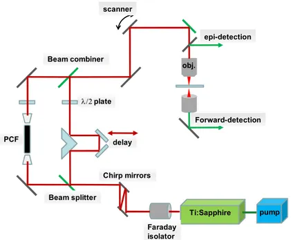
Samples were investigated using an in-house built multiphoton laser scanning microscopic system. Schematic of this microscope imaging system is illustrated in figure 1. The laser source is a Ti:Sapphire oscillator (Spectra-Physics, Tsunami) centered at 800nm with a pulse width of 100fs, average output power of 1W when pumped with 7.25 W green laser at 532nm (Spectra-Physics, Millennium Pro). The output femto-second pulses from the Ti:sapphire oscillator first pass through a Faraday isolator (Newport) to prevent back reflections from re-entering the laser cavity. The isolator contains highly dispersive material and requiring re-compression of the pulse. This is accomplished using a pair of GTI laser mirrors (Layertec GmbH, Germany). The fs pulse is then split into two beams, the reflected pulses (pump) and the transmitted pulses (Stokes), using a 50/50 beam splitter. The pump pulse was transmitted through a series of lenses and mirrors, including an optical delay stage before combining with the Stokes pulses at a beam combiner. Approximately 300mW of transmitted pulse is coupled into a two-zero-dispersion-point PCF ( Crystal Fibre, Denmark) through an objective lens (Newport) to generate a broadband emission. The NIR portion of the broadband emission is used as the Stokes pulses for generating the CARS signal. The pump and Stokes pulses are combined and sent into the microscope assembly. Three non-descanned modular type PMT detectors (Hamamatsu) are mounted on the microscope for simultaneous detection of TPEF, SHG and CARS signals through different combinations of dichroic and colour filters. TPEF, SHG and CARS signals are all collected in the backscattered (epi)-direction. The laser pulses are focused onto sample through a 20x, 0.75 NA infinity corrected air objective lens (Olympus) and the emitted TPEF and epi-SHG/CARS signals are collected through the same objective lens. For forward-SHG/CARS, the emitted signals are collected through an aspheric condenser lens (Comar Optics) placed beneath the translational stage. A 40x, 0.8 NA, water immersion objective lens (Olympus) was also used to collect tissue images at greater spatial and depth resolution on selected regions of interest. Typically 25mW of pump and 8mW of Stokes (measured after the objective) were used for imaging. For image acquisition and laser scanning control, we used ScanImage (ver. 3.5) software [9] developed at Cold Spring Harbour Laboratory (Cold Spring Harbour, NY). Pixel dwell time for an average of 4 scans for a signal collection is 21 µs. Post image processing and image viewing were carried out in ImageJ ver 1.42b, an open source image processing software initially developed at National Institute of Health.
Figure 1. Schematic of the in-house built nonlinear optical microscope with the Stokes
pulses being generated in PCF which is pumped by the same Ti:sapphire laser provides the pump pulse.
3. Results
From examining the digital photographs of the arterial intimal surfaces, it is clear that all the arterial samples harvested from the older rabbits (16, 18 and the 24 month-old rabbits) have a significant burden of plaque. Lipid-rich lesion is easily identifiable by human eyes and most of the intimal surfaces show large, confluent, whitish raised lesions. The arterial lumen from the 4 month-old rabbit, on the contrary, has a much larger area of smooth, flat surface with some slightly raised lesions interspersed with the occasional larger raised plaques.(Figure 2). This observation is consistent with the result reported earlier on progressive atherosclerosis in the WHHL rabbit [10].
Figure 2. An arterial lumen surface, obtained from a 4 month-old WHHL-MI rabbit. Arrow
marked: an early atherosclerotic lesion
λ/2 plate scanner obj. Ti:Sapphire pump Faraday isolator Chirp mirrors delay Beam combiner Beam splitter Forward-detection epi-detection PCF
Representative NLO images of smooth, healthy luminal surface of an arterial sample are shown in figure 3. This piece of thick arterial sample was harvested from the 4 month-old WHHL-MI rabbit and figure 3a and 3b were epi-images acquired using the 20x air lens and the 40x WI objective lens, respectively. Membrane-like structures giving rise to strong TPEF signal are evident whereas only minimum SHG and CARS signal are detected. When probing deeper into tissue, we observed strong TPEF signal from a fibre-structure showing very different morphology from those observed at shallower layer-see figure 3c.
Figure 3. Epi-NLO images of healthy arterial lumen collected using (A) 20x air lens (B) 40x
WI lens on the lunimal surface (C) 40x WI lens collected at deeper layer. Green:TPEF, Blue:SHG, Red:CARS
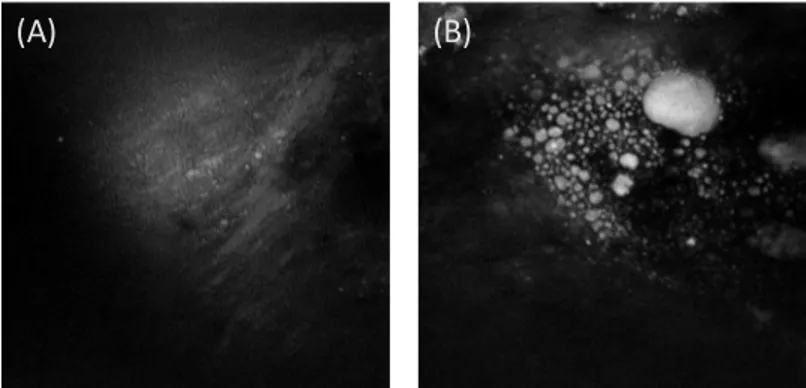
Representative NLO Image acquired from an early atherosclerotic lesion (arrow marked area in figure 2) is illustrated in figure 4a. In this image, the membrane-like structure shown in figure 3a is not visible. Instead, the image shows sparsely scattered collagen fibres, accumulated lipid-rich structures and dot-shaped strong fluorescent particles.
Images acquired from advanced plaque reveal similar luminal surface pathology to those detected on early lesions however they shows very different collagen structures. (see figure 4b) Older, more advance plaques show straight, highly directional collagen fibres whereas early lesions, show less directional collagen fibrils. Depth scan images of some advanced plaque show a thin collagen fibril layer overlaying a lipid rich structure. Figure 5a and 5b show images obtained at ~10µm and ~60µm depth in an advanced plaque, respectively. High collagen content is evident in the 10µm depth image while abundant lipid-rich structure without collagen fibrils is observed in the 60µm depth image. (A) 25 µm (B) 12.5 µm (C) 12.5 µm
Figure 4. Epi-NLO images of arterial lumen surface obtained from (A) an early lesion (B) an
advanced lesion using 20x air lens, showing different collagen morphology. Blue: SHG(collagen) , Red:CARS(lipid-rich structure) and Green:TPEF(elastin or other fluorescent particles)
Figure 5. Epi-NLO images of an advanced plaque obtained at (A) ~10 um depth (B) ~60um
depth. Blue:SHG(collagen), red/orange:CARS/TPEF(lipid-rich structure)
4. Discussion
The membrane-shaped structure detected on healthy luminal surface (shown in figure 3a and 3b) is consistent with internal elastic lamina in arterial intima and the fibril structure shown in figure 3c is the elastin in arterial media. Similar elastin membrane structures in porcine arterial tissue was also reported previously by Wang et al [7] using a multimodal CARS microscope based on a two 5-ps laser configuration. Compared to Wang’s work, our images show a slightly lower depth resolution as we were unable to visualize the endothelial cells on the lumen surface. No internal elastic lamina is visible on either early or advanced atherosclerotic lesions, which is expected as the lumen is thickened and the plaques overlay the arterial intima which contains the internal elastic lamina. The complex pathology detected for early lesions reveals early stage accumulation of lipid-rich materials and initial formation of collagen fibrils
(A) (B)
within the lesion. At this stage, no obvious collagen cap was observed in the lesion. On some advanced plaques, a thin collagen cap was observed overlaying a lipid-rich structure, which is in agreement with the pathology of a rupture-prone atherosclerotic plaque. The straight and highly directional collagen fibril structure observed in advanced plaques is notably different from the collagen structure observed in healthy or early stage lesions and is possibly caused by increased tissue tension experienced on the top of a raised plaque.
5. Conclusion
In this paper we demonstrated ex vivo nonlinear optical imaging of arterial tissue using a PCF based nonlinear optical imaging technique. Epi-NLO imaging of un-sectioned arteries from WHHL-MI rabbits allows label-free biochemical visualization of extra-cellular components relevant to the development of atherosclerosis. Clear differences in the surface morphology is observed between healthy artery wall and atherosclerotic lesions. Different collagen fibril structures and distributions are also noted between early lesions and advanced plaques.
6. Acknowledgments
The authors acknowledge Vijay Iyer of Cold Spring Harbour Laboratory (NY,USA) for the assistance provided in using ScanImage software. This work was supported by National Research Council Canada, Genomics and Health Initiative.
References
[1] Libby, P., “Atherosclerosis: Disease Biology Affecting the Coronary Vasculature,”
Am. J. Cardiol 98[suppl],3Q-9Q (2006).
[2] Hansoon, G.K.,“Inflammation, Atherosclerosis, and Coronary Artery Disease,” N.
Engl J Med 352, 1685-95 (2005).
[3] Lusis, A.J.,“Atherosclerosis,” Nature (London) 407, 233-241, (2000).
[4] Zoumi, A., Lu, X., Kassab, G.S., and Tromberg, B.J., “Imaging coronary artery microstructure using second-harmonic and two-photon fluorescence microscopy,”
Biophys. J. 87(4), 2778-2786 (2004).
[5] Lilledahl, M.B., Haugen, O.A., Davis, C.D.L., Svaasand, L.O., “Characterization of vulnerable plaques by multiphoton microscopy,” J. Biomed. Opt. 12(4), 044005 (2007). [6] Le, T.T., Langohr, I.M., Locker, M.J., Sturek, M., Cheng, J.-X., “Label-free molecular imaging of atherosclerotic lesions using multimodal nonlinear optical microscopy,” J.
Biomed. Opt. 12(5), 054007 (2007).
[7] Wang, H-W., Le, T.T., Cheng, J-X.,“Label-free imaging of arterial cells and extracellular matrix using a multimodal CARS microscope,” Opt. Comm. 281, 1813-1822 (2008).
[8] Shiomi, M., Ito, T., Yamada, S., Kawashima, S., Fan, J.,“Development of an Animal Model for Spontaneous Myocardial Infarction (WHHLMI Rabbit),” Arteriosclerosis,
Thrombosis, and Vascular Biology. 23, 1239 (2003).
[9] Pologruto, T.A., Sabatini, B.L., Svoboda, K.,“ScanImage: Flexible software for operating laser scanning microscopes,” BioMedical Engineering Online 2, 13 (2003). [10] Buja, L.M., Kita, T., Goldstein, J.L., Watanabe, Y., Brown, M.S.,“Cellular pathology of progressive atherosclerosis in the WHHL rabbit,” Atherosclerosis, 3, 87-101 (1983).