HAL Id: tel-01614913
https://tel.archives-ouvertes.fr/tel-01614913
Submitted on 11 Oct 2017HAL is a multi-disciplinary open access archive for the deposit and dissemination of sci-entific research documents, whether they are pub-lished or not. The documents may come from teaching and research institutions in France or abroad, or from public or private research centers.
L’archive ouverte pluridisciplinaire HAL, est destinée au dépôt et à la diffusion de documents scientifiques de niveau recherche, publiés ou non, émanant des établissements d’enseignement et de recherche français ou étrangers, des laboratoires publics ou privés.
Epidemiology and dynamic of hand, foot and mouth
disease in Vietnam
Nghia Ngu Duy
To cite this version:
Nghia Ngu Duy. Epidemiology and dynamic of hand, foot and mouth disease in Vietnam. Infectious diseases. Université Montpellier; National Institute of Hygiene and Epidemiology (Hanoi, Viet Nam), 2016. English. �NNT : 2016MONTT099�. �tel-01614913�
Délivré par Université de Montpellier
EtNational Institute of Hygiene and Epidemiology
Préparée au sein de l’école doctorale GAIA –ED 584
Spécialité : Biologie des Interactions
Présentée par Ngu Duy Nghia
Soutenue le 16 Décembre 2016 devant le jury composé de
M Le Tran Binh, Prof, USTH Rapporteur M Ngo Van Toan, Prof, Hanoi Medical University Rapporteur M Xavier de Lamballerie, Université Aix-Marseille Rapporteur M Christian Devaux Examinateur Mme Aneta Afelt, University of Warsaw Examinateur M Emmanuel Cornillot, University of Montpellier Examinateur M Nguyen Tran Hien, Prof, NIHE Directeur M Roger Frutos, Prof, CIRAD Directeur
! ! ! ! ! ! ! ! ! ! ! ! ! ! ! ! ! ! ! ! ! ! ! ! ! ! !
Epidemiology and dynamic of Hand, Foot
and Mouth Disease in Vietnam
SCIENTIFIC ADVISORS
VIETNAM
FRANCE
First advisor: Prof. Nguyen Tran Hien
First advisor: Prof. Roger Frutos
Second advisor: Prof. Nguyen .T.H. Thanh Second advisor: Dr. Laurent Gavotte
HƯỚNG DẪN KHOA HỌC
VIỆT NAM
PHÁP
Hướng dẫn 1: Giáo sư NguyễnTrần Hiển
Hướng dẫn 1: Giáo sư Roger Frutos
Hướng dẫn 2: Giáo sư Nguyễn .T.H.Thanh Hướng dẫn 2: Tiến sĩ Laurent Gavotte
TABLE OF CONTENTS
ACKNOWLEDGEMENTS! LIST OF ABBREVIATIONS! LIST OF FIGURES AND TABLES!
GENERAL INTRODUCTION ... 1!
PART I. LITERATURE REVIEW ... 6!
1. Picornaviridae ... 8!
1.1. Classification ... 8!
1.2. Virion ... 12!
1.3 Capsid ... 13!
1.4. Genome structure and features ... 14!
1.5. Replication ... 18!
2. Enterovirus ... 21!
2.1. Classification ... 21!
2.2. Human Enteroviruses cause Hand, Foot and Mouth Disease ... 22!
3. Pathogenesis... 26!
3.1. Virus entry and spread ... 26!
3.2. Pathological findings ... 26!
3.3. Virus virulence and host factors ... 27!
3.4. Protective immunity ... 29!
4. Diagnosis ... 29!
4.1. Case definition and classification ... 29!
4.2. Laboratory diagnosis ... 32!
5. Epidemiology of Hand, Foot and Mouth Disease ... 34!
5.1. History and epidemiological features of HFMD ... 34!
5.2. Sources, Transmission and Susceptibility of HFMD ... 37!
5.3. HFMD in Vietnam ... 38!
6. Prevention and control measures of HFMD ... 40!
6.1. Options for prevention and control ... 40!
PART II. EMPIDEMIOLOGY AND ETIOLOGY OF HAND, FOOT AND MOUTH
DIEASE IN VIETNAM ... 72!
CHAPTER 1. MONITORING INFLUENCE OF HFMD NEW GUIDELINES ON PATIENTS CARE DURING 2011-2012 OUTBREAK IN HAI PHONG CITY, VIETNAM ... 73!
1.1. Context of study ... 73!
1.2. Objective ... 74!
1.3. Discussion and conclusions ... 75!
CHAPTER 2. VALINE/ISOLEUCINE VARIANTS DRIVE SELECTIVE PRESSURE IN THE VP1 SEQUENCE OF EV-A71 ENTEROVIRUSES ... 111!
1.1. Context of study ... 111!
1.2. Objective ... 112!
1.3. Discussion and conclusions ... 112!
CHAPTER 3. MODELLING THE DYNAMIC OF A MULTIPHASE DISEASE/ THE EXAMPLE OF HFMD ... 143!
1.1. Context of study ... 143!
1.2. Objective ... 144!
1.3. Discussion and conclusion ... 144!
GENERATION CONCLUSSION AND PERSPECTIVES ... 165!
ACKNOWLEDGEMENTS
To write this thesis, I am greatly indebted to program, institutes and many people for their support and encouragements.
First of all, I would like to my thanks to the Erasmus Mundus program, granted me the scholarship to pursue this doctoral program; To Ecole doctorale SIBAGHE, University of Montpellier, Centre d’études d’agents Pathogènts et Biotechnologies pour la Santé (CPBS, CNRS), France, and National Institute of Hygiene and Epidemiology, Vietnam for giving me an opportunity to follow this academic program and for equipping me with such an updated knowledge to make this thesis possible.
My deep gratitude goes to Professor Roger Frutos, from French Agricultural Research center (CIRAD) and University of Montpellier, Montpellier, France for accepting to be my supervisor, for his great support, effort and help in articles and thesis writing, for making several round trips between France and Vietnam.
My special thanks goes to my supervisor, Professor Nguyen Tran Hien, former director of National Institute of Hygiene and Epidemiology, and Hanoi Medical Univerity, Ha noi, Viet Nam for his great supports, encouragement to complete the PhD program.
My huge thanks and appreciation to my cosupervisor, Dr Laurent Gavotte, Assistant Professor, Evolutionary Biology, Ecology Department, Univerity of Montpellier, France for the accomplishment of the work, for his help and support in supervision research and writing.
My special thanks to my cosupervisor, Prof. Nguyen Thi Hien Thanh, former deputy head of the Virology Department, Chief of Enterovirus Lab, National Institute of Hygiene and Epidemiology, Viet Nam for her supports and allowing me to work as part of her team and help me in her laboratory.
My special thanks goes to Professor Tran Nhu Duong, deputy director of National Institute of Hygiene and Epidemiology, Viet Nam for firstly allowing me to work on all the HFMD surveillance data in the Epidemiology Department, for his valuable supports, encouragement to complete the PhD program.
I would like to express my sincere gratitude and appreciation to Professor Emmanuel Cornillot, from University of Montpellier, France for his great support, effort and help in articles and thesis writing, encouragement to complete the PhD program.
My huge thanks and appreciation to my cosupervisor, Dr Patrice Ravel, Institut de Recherche en Cancérologie de Montpellier, Montpellier, France for the accomplishment of the work, for his help and support in an articles.
My special thanks goes to Professor Catherine Moulia, Montpellier 2 University, French for generously accepting me to his laboratory at Montpellier 2 University, for her valuable supports, encouragement to complete the PhD program.
I am also very grateful to Dr. Aneta Afelt, University of Warsaw, Poland, for her very important support in making all GIS analyses and maps and for having accepted to be a defense committee member.
I would like to thanks the medical staff of the Pediatric Hospital and Preventive Medicine Centers in Hai Phong city for their care of patients, supported, and their cooperation during conducted of this study.
My thanks to Enterovirus laboratory, National Institute of Hygiene and Epidemiology, Vietnam for allowed me to work as part of their team, and for their help.
I want to express my deep gratitude to Professor Xavier de Lamballerie, Aix-Marseille University, France, Professor Le Tran Binh, University of Science and Technology of Hanoi and Professor Ngo Van Toan, Hanoi Medical University, Viet Nam for having accepting and making me the honor to review this PhD work.
I would like to thank the following Professors, friends and colleagues:
To my colleagues at Epidemiology Department, National Institute of Hygiene and Epidemiology, Hanoi, Viet Nam thanks to Dr. Vu Dinh Thiem, Dr. Pham Quang Thai, Dr. Nguyen Cong Khanh, Dr. Ngo Huy Tu, Dr. Nguyen Quang Minh and Pham Thi Cam Ha for their help in all the works in order to generate data used in this thesis.
My thanks to Dr. Guilhem Kister in Faculté de Pharmacie, Univerity Montpellier, France and Dr. Ankit Dwivedi, Montpellier, France for their help in Montpellier, France.
All teachers, friends and colleagues for their help during the course.
Last but not a least, my heartfelt thanks and love to my parents, my wife, my children who have helped me in every possible way to my completion of this Doctor of philosophy study.
LIST OF ABBREVIATIONS
ANS Autonomic nervous system
cDNA Complementary Deoxyribonucleic acid
CPBS Center Agents Pathogens and Biotechnology for Health study CNRS The French National Centre for Scientific Research
CNS Central nervous system
CSF Cerebrospinal fluid
CODEHOP Consensus-degenerate hybrid oligonucleotide primer
CV-A Coxsackievirus A
DNA Deoxyribonucleic acid
EV-A Human Enterovirus A
EV-B Human Enterovirus B
EV-C Human Enterovirus C
EV-D Human Enterovirus D
HFMD Hand, Foot and Mouth Disease
HRV Human rhinovirus
HIV Human immunodeficiency virus
HLA Human leucocyte antigen
HA Herpangina
IL Interleukin
IP Interferon induced protein
IgG Immunoglobulin G
I/V Isoleucine/Valine
MoH Ministry of Health
MRI Magnetic resonance imaging
NIHE National Institute of Hygien and Epidemiology
ORF Open reading frame
PV Poliovirus
RD cell Human rhabdomyosarcoma cell
RNA Ribonucleic Acid
Real-time PCR Real-time Polymerase Chain Reaction
UTR Untranslate region
USA United state of America
US-CDC US - Centers for Disease Control and Prevention Vero cells African green monkey kidney cells
VLP Virus-like particle
VPg Virus protein, genome linked
VP 1 Capsid protein of Enterovirus 1
VP 2 Capsid protein of Enterovirus 2
VP 3 Capsid protein of Enterovirus 3
VP 4 Capsid protein of Enterovirus 4
WHO World Health Organizaion
LIST OF FIGURES AND TABLES
Figure 1: Phylogenetic tree of the different genera within Picornaviridae ... 9!
Figure 2: The picornavirus virion.. ... 13!
Figure 3: The capsid of human rhinovirus. ... 14!
Figure 4: Organization of a typical picornavirus RNA genome. ... 15!
Figure 5: Genome structure and gene organization of Picornaviridae ... 16!
Figure 6: Poliovirus translation and post-translational modifications. ... 19!
Figure 7: The picornavirus replication cycle.. ... 20!
Figure 8: Genogroupe B of Enterovirus 71(EV-A71) ... 24!
Figure 9: Genogroupe C of Enterovirus 71(EV-A71). ... 25!
Figure 10: The postulated pathogenesis of EV-A71. ... 28!
Figure 10: History of Hand, Foot and Mouth Disease ... 36!
Figure 11: Monthly distribution of HFMD in Vietnam ... 40!
Figure 12: Location of Hai Phong city, Vietnam. ... 73!
Table 1: Importance of Piconaviruses of Humans and Animals ... 10!
Table 2: Picornaviruses: Genera, Species and Serotypes ... 12!
Table 3: Major demonstrated and predicted functions of picornavirus proteins. ... 17!
Table 4: Enterovirus genus and species. ... 22!
Table 5: Proposed clinical case definitions for HFMD/HA and associated complications . 31! Table 6: Recent outbreaks of HFMD due to in the Asia Pacific region. ... 37!
1
GENERAL INTRODUCTION
In the recent decades, our world has been facing a series of communicable diseases threats such as avian influenza, SARS, MERS-CoV, Ebola, Zika… Beside of the that, the re-emergence of other communicable diseases still remains major burden for human. Hand, foot and mouth disease (HFMD) is the ones that reported for long time ago. This disease is an acute febrile illness in children with a papulovesicular skin rash at the palms or soles of the feet, or both. Presentation can be with or without inclusion of mouth ulcers. HFMD can result in severe complications such as encephalitis, aseptic meningitis, pulmonary edema, myocarditis, and death (1). HFMD is caused by types of Enterovirus A species which includes some Coxsackievirus A (CV-A) and Enterovirus A71 (EV-A71) (2), (3). The transmission can be faecal-oral and respiratory secretions route by direct person-to-person contact, droplets or fomites. Recent, the widespread community outbreaks of HFMD have occurred mostly in Asia Pacific region with notable amount of cases and deaths. The disease still has neither no specific treatment or vaccines so far.
It was thought that the disease attacks mostly in the poor hygiene areas because of transmission mode. However, HFMD has been hitting both developing and developed countries since reported. In 1969, in California, USA, EV-A71 strain firstly was isolated from a 9 months aged child with encephalitis diagnostic (1). There are then small and large outbreaks of HFMD have been reported throughout the world (2). In the early 1970s, several countries in different continent including Sweden, Australia, USA and Japan had reported small HFMD outbreaks with some tens of cases and almost of them are children whose clinical aspects are mostly typical of HFMD, sometimes aseptic meningitis (3), (4), (5), (6). After that, HFMD was only reported in Europe in late half of the 1970s with two large outbreaks in Bulgaria (year 1975, 451 cases and 44 deaths), Hungary (year 1978, 1550 cases and 47 deaths) and a small number of cases in France in 1979 (7), (8), (9). The 1980s there are also some small outbreaks in Hong Kong, Australia, USA (10), (11), (12) and no reported cases from other countries
In the late 1990s, many country members of Asia Pacific region have experienced large HFMD outbreaks. It began in 1997 with two large widespread community outbreaks
2
in Sarawak, Malaysia and Taiwan, with 2628 and 129,106 cases reported, respectively (13), (14). Following that, a series of small and large outbreaks happen throughout the region in which Japan, Australia, China, Malaysia, Singapore, Taiwan (China), Korea, Mongolia, Vietnam, Brunei has been the hotpots of epidemic with cycle of every 2–3 years period (15), (16), (17). So far, the latest large outbreak in the region was in one province named Anhui of China in 2008 with around 490,000 cases and 126 deaths in children were reported, the case-fatality rate is around 0.03% but in certain local outbreaks, such as in Fuyang City of Anhui Province, this rate can reach up to 0.3% (18), (19). During those outbreaks, almost of the cases are under 5 years old children and although clinical manifestations were mostly typical of HFMD, a cluster of deaths among young children was identified. Moreover, cases involving the central nervous system complication and/ or pulmonary oedema have also been observed for the first time (20), (21). There were several small outbreaks or sporadic HFMD cases outside the Asia-Pacific region.
Vietnam is located in South Eastern Asia and shares the border with China, Laos, and Cambodia. The climate is tropical in south; monsoonal in north with 4 seasons are spring, summer, autumn and winter. Although EV-A71 was isolated for the first time in Vietnam in 2003, the first outbreak of HFMD was not reported in the southern provinces until 2005. The 2005 outbreak was associated with EV-A71 C1, C4 and C5 subgenogroups and CV-A16 (22), (23). For the southern part of the country, in the periods of 2007 - 2009, the numbers of reported cases and deaths were 5,719 and 23; 10,958 and 25, and 10,632 and 23, respectively. In contrast, there were a few sporadic HFMD cases in the northern. In 2005 - 2007 periods, seven cases were identified. In 2008, 88 cases were reported from 13/28 provinces. No severe or fatal cases were reported. Since 2011, Vietnam have experienced continuously large outbreaks of HFMD and the disease became a notifiable one in the national communicable disease surveillance system. According to data of Viet Nam Ministry of Health (MoH) from 2011 to 2015, number of reported cases and deaths were 113,121 and 170 (2011), 157,391 and 45 (2012), 78,818 and 23 (2013), 77,296 and 9 (2014), 56,329 and 5 (2015), respectively which have been reported from across all 63/63 provinces. HFMD outbreaks have continuously occurred nationwide. Responding to HFMD outbreaks, MoH issued two specific guidelines applied nationwide. The first one published on the February 24, 2012 concerned surveillance, prevention and control of
3
HFMD. The second guideline was issued on March 30, 2012 were about diagnosis and treatment.
Indeed, in Viet Nam, HFMD is now important public health concern. Recent studies mainly observe for basic epidemiology and etiology characteristics (23), (24), (22), (25). Besides, the relationship between etiological agents and clinical epidemiological behavior, temporal and spatial analysis, pathogenicity-related mutations of virus, modeling approach for prediction, evaluation of countermeasures has not been reported. In order to more comprehensive understanding which contribute to prevention and control of HFMD in Vietnam, This work analyzed all HFMD cases reported in Hai Phong city by both the community and hospitals in 2011 and 2012. Since it was the very first outbreak to occur in Hai Phong city, it was a good model for investigating the dynamic of the disease without interference and potential remains from previous outbreaks or patient immunological adaptation and it provide findings related to influence of HFMD guidelines during the outbreak period too. This work is also an integrative analysis including spatial analysis and social evolution as well as genetic evolution to describe the dynamic of HFMD in a well delimited area. We will also analyze the VP1 gene of strains isolated throughout Northern Vietnam during the 2011-2012 outbreak and develop the modeling the dynamic of a multiphase disease. This part of the study is coordinated with Montpellier University 2, Montpellier, France.
The specific objective of the research aims:
1.! To describe the epidemiological and etiological characteristics during 2011-2012 outbreak in Hai Phong City, Vietnam.
2.! To monitor influence of HFMD new guidelines on patients care during 2011-2012 outbreak in Hai Phong City, Vietnam.
3.! To spatial analyze the HFMD dynamic during 2011-2012 outbreak in Hai Phong City, Vietnam
4.! To analyze pathogenicity-related mutations in the VP1 sequence of EV-A71 strains isolated throughout Northern Vietnam during the 2011-2012 outbreak.
4
5.! To better understand the monitor HFMD throughout modeling the HFMD dynamic during 2011-2012 outbreak in Hai Phong City, Vietnam.
The outline will be displayed in form of chapters with results presented as a form of in press, submitted or in preparation articles. The articles included in this thesis are listed below:
FIRST PART
Nghia Ngu Duy, Tran Nhu Duong, Christian Devaux, Roger Frutos. Emergence of EV-A71 infection in Asia Pacific region. Asia Pacific Jounal of Tropical Medicine
SECOND PART CHAPTER 1
Nghia Ngu Duy, Le Thi Thanh Huong, Patrice Ravel, Le Thi Song Huong, Ankit Dwivedi, October Michael Sessions, Yan’An Hou, Robert Chua, Guilhem Kister, Aneta Afelt, Laurent Gavotte, Catherine Moulia, Christian Devaux, Duane J Gubler, Vu Dinh Thiem, Nguyen Thi Hien Thanh, Tran Nhu Duong, Nguyen Tran Hien, Emmanuel Cornillot, Roger Frutos. Monitoring influence of Hand, Foot and Mouth Disease new guidelines on patients care during 2011-2012 outbreak in Hai Phong City, Vietnam. Asian Pacific Journal of Public Health.
CHAPTER 2
Nghia Ngu Duy, Le Thi Thanh Huong, Patrice Ravel, Le Thi Song Huong, Ankit Dwivedi, October Michael Sessions, Yan’An Hou, Robert Chua, Guilhem Kister, Aneta Afelt, Catherine Moulia, Duane J Gubler, Vu Dinh Thiem, Nguyen Thi Hien Thanh, Christian Devaux, Tran Nhu Duong, Nguyen Tran Hien, Emmanuel Cornillot, Laurent Gavotte, Roger Frutos*.!Valine/isoleucine variants drive selective
pressure in the VP1 sequence of EV-A71 enteroviruses. BMC Infectious Diseases
CHAPTER 3
Patrice Ravel, Nghia Ngu Duy, Aneta Afelt, Guilhem Kister, Le Thi Thanh Huong, Le Thi Song Huong, Ankit Dwivedi, October Michael Sessions, Yan’An Hou, Robert
5
Chua, Catherine Moulia, Duane J Gubler, Vu Dinh Thiem, Nguyen Thi Hien Thanh, Christian Devaux, Tran Nhu Duong, Nguyen Tran Hien, Laurent Gavotte, Emmanuel Cornillot, Roger Frutos.!Modelling the dynamic of a multiphase disease: the example
6
TÓM TẮT
Những thập kỷ gần đây, thế giới đang đối mặt với hàng loạt các bệnh truyền nhiễm mới nổi như cúm gia cầm, dịch SARS, MERS-CoV, Ebola, Zika…Bên cạnh đó, sự tái nổi trội của các bệnh truyền nhiễm khác cũng là gánh nặng bệnh tật rất lớn cho con người. Bệnh Tay chân miệng (TCM) là một trong số đó, TCM là bệnh sốt cấp tính, thường xảy ra ở trẻ em với biểu hiện chủ yếu là các ban dạng phỏng nước, thường xuất hiện ở gan bàn chân, bàn tay, mông, đầu gối và niêm mạc miệng. Bệnh đa số ở thể nhẹ, có thể tự khỏi trong vòng 1 tuần. Tuy nhiên có một số trường hợp có biến chứng thần kinh có thể dẫn tới tử vong. Căn nguyên gây bệnh là vi rút đường ruột, trong đó quan trọng nhất là EV-A71 và CA-16. Bệnh lây qua đường tiêu hoá, tiếp xúc với bề mặt bị ô nhiễm. Hiện nay chưa có vắc xin và thuốc điều trị đặc hiệu.
Bệnh TCM do EV-A71 lần đầu tiên ghi nhận tại California, Mỹ. Sau đó bệnh lưu hành và gây các vụ dịch nhỏ tại châu Mỹ, Châu Âu, Úc… Đến cuối những năm 1990s, TCM bùng phát và gây dịch lớn tại các quốc gia thuộc khu vực Châu á Thái bình dương, biến chứng thần kinh viêm thân não lần đầu tiên được ghi nhận. Tại Việt Nam, mặc dù EV-A71 được ghi nhận từ 2003, nhưng từ năm 2011, hàng năm, bệnh TCM gây dịch trên phạm vi cả nước với số mắc và tử vong cao. Thực vậy, TCM hiện nay là mối quan tâm về y tế công cộng. Một số nghiên cứu gần đây mới chỉ tập trung vào các đặc điểm dịch tễ, căn nguyên cơ bản nhất. Bên cạnh đó các mối liên hệ giữa tác nhân gây bệnh và đặc điểm dịch tễ học lâm sàng; các phân tích về phân bố bệnh theo không gian, thời gian; các biến đổi của vi rút liên quan đến độc lực; mô hình hoá sự biến động của bệnh dịch; đánh giá các biện pháp can thiệp kiểm soát bệnh dịch… vẫn chưa được nghiên cứu đầy đủ. Để cung cấp các kiến thức, thông tin toàn diện hơn về bệnh TCM góp phần phòng chống bệnh dịch này hiệu quả hơn, Nghiên cứa này phân tích toàn bộ các ca bệnh TCM được báo cáo trong vụ dịch đầu tiên tại Hải Phòng năm 2011-2012 tại cả cộng đồng và bệnh viện. Vụ dịch lớn đầu tiên này là một thực địa rất tốt cho điều tra , nghiên cứu bệnh dịch này. Nghiên cứu này tập trung phân tích các đặc điểm dịch tễ học, tác nhân gây bệnh, tác động của những hướng dân kiểm soát bệnh dich, biến động bệnh theo không gia, thời gian; phân tích các bieens đổi về di truyền liên quan đến độc lực; mô hình sự biến động bệnh dịch…
Một số khuyến cáo đối với lĩnh vực giám sát: Sự đa dạng về I/V variants là cách tốt để giám sát và xác định sự lưu hành của các chủng EV-A71; Chẩn đoán EV-A71 với bộ mồi MAS nên đuọc thực hiện một cách hệ thống từ sản phẩm của mồi SO. Cần chú ý tới
7
nhũng mẫu âm tính khi thực hiện giám sát do có thể mồi phát hiện không phù hợp. Bộ mồi AN89 nên được thiết kế lại để phát hiện EVs tốt hơn. Đối với kiểm soát bệnh: Tiếp tục áp dụng các hướng dẫn giám sát và kiểm soát của Bộ Y tế. Các biện pháp can thiệp nên tập trung vào nhóm nguy cơ cao (trẻ < 5 tuổi) và gia đình, người chăm sóc trẻ ở cả khu vực đô thị và nông thôn. Giai đoạn ưu tiên can thiệp là tháng 3 – 5 và tháng 9-10 hàng năm. Quan tâm đặc biệt đến trẻ dưới 1 tuổi do có nguy cơ diễn biến nặng. Mô hình biến động nên được áp dụng để quản lsy tốt các đợt dịch trong tương lai. Đối với nghiên cứu: Phân tích toàn bộ gen của tác nhân gây bệnh để thấy rõ hơn mối liên hệ giữa các biến đổi di truyền và độc lực, nghiên cứu nguyên nhân bùng phát dịch nên tập trung them vào các yếu tố khác ngoài tác nhân gây bệnh. Sau cùng, nghiên cứu này đã đóng góp những kết quả cần thiết và quan trọng giúp phần phát triển các công cụ giám sát và kiểm soát bệnh TCM tốt hơn.
8
PART I. LITERATURE REVIEW
1. Picornaviridae
1.1. Classification
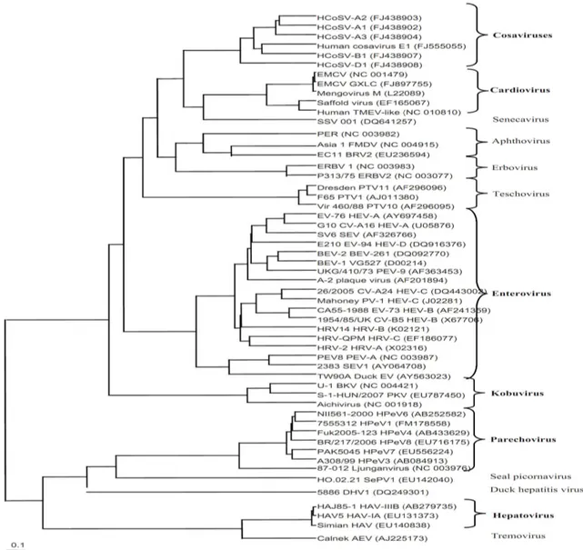
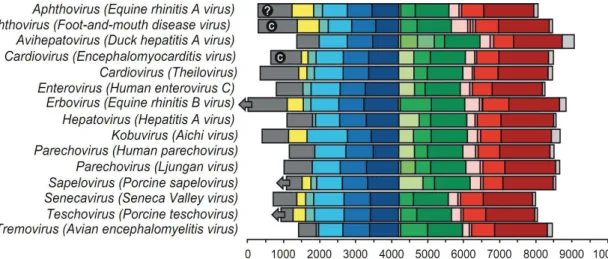
Picornaviruses are viruses which belonging to the family Picornaviridae. The name is derived from pico, meaning small, and RNA, referring to the ribonucleic acid genome, so "picornavirus" means small RNA virus (26). According to International committee on Taxonomy of Viruses (27), Picornaviruses are separated into 12 genera which consisting of
Enterovirus, Cardiovirus, Aphthovirus, Hepatovirus, Parechovirus, Erbovirus, Kobuvirus, Teschovirus, Sapelovirus, Senecavirus, Tremovirus, Avihepatovirus. There are many
9
Figure 1: Phylogenetic tree based on the deduced amino-acid sequence of the structural P1 region of representatives of the different genera within Picornaviridae. Within the genera, types infecting humans among other species are shown in bold, (28).
For the disease manifestation, Picornaviruses can cause diseases in both human and animal. They are grouped by genus based on their pathogenic properties and/or route of infection. For example, the enterovirus genus includes poliovirus and coxsackieviruses based upon the natural oral route of entry into the host and replication in gut tissue (29), (Table 1).
10
Genus Virus Principal Species
Affected Disease
Aphthovirus
Foot-and-mouth disease viruses
Equine rhinitis A virus Bovine rhinitis B virus
Cattle, sheep, swine, ruminant species Horses, camelids Cattle Foot-and-mouth disease Systemic infection Mild respiratory Cardiovirus Encephalomyocarditis virus Theiler’s mouse encephalomyelitis virus Rodents, swine, elephants, primates, mammals in contact with rodents Mice Encephalomyelitis and myocarditis in swine and elephants; rarely in other species Murine poliomyelitis Enterovirus Human enteroviruses A, B, C, and D Human rhinoviruses A, B, and C Bovine enteroviruses Simian enteroviruses Porcine enterovirus B Humans Humans Cattle Primates Swine Aseptic meningitis, poliomyelitis, myocarditis Respiratory disease Vesicular disease Mild enteric and respiratory disease Usually asymptomatic Erbovirus Equine rhinitis B virus Horses Mild rhinitis
Kobuvirus Bovine kobuvirus Cattle Possible enteritis
Teschovirus
Porcine teschovirus 1
Porcine teschoviruses Swine
Encephalomyelitis mild diarrhea, pericarditis Tremovirus Avian ncephalomyelitis
virus Chickens
Encephalomyelitis
Avihepatovirus Duck hepatitis A virus Ducks Hepatitis Table 1: Importance of Piconaviruses of Humans and Animals (29)
11
However, Base upon serology aspect (neutralized antibody against to capsid antigens), the Picornaviruses are further subclassified on the basis of serotype. This can range from a single serotype in the case of hepatitis A virus (HAV), or, in the case of rhinovirus, to about 100 identified serotypes (26) (Table 2).
Genus Species Serotypes
Enterovirus
Bovine enterovirus 2 types: bovine enterovirus 1-2
Human enterovirus A 21 types including some coxsackie A
viruses and enteroviruses
Human enterovirus B
59 types: enteroviruses, coxsackie B, echoviruses, swine vesicular disease virus
Human enterovirus C
19 types including poliovirus 1-3, some coxsackie A viruses,
enteroviruses
Human enterovirus D 3 types: EV-68, EV-70, EV-94
Porcine enterovirus B 2 types: porcine enterovirus 9-10
Simian enterovirus A 1 type: simian enterovirus A1
Human rhinovirus A 75 types
Human rhinovirus B 25 types
Human rhinovirus C 7+ types
Cardiovirus
Encephalomyocarditis virus 1 type: encephalomyocarditis virus.
Theilovirus
12 types: Theiler's murine
encephalomyelitis virus, Vilyuisk human encephalomyelitis virus, Thera virus, Saffold virus 1-9
Aphthovirus
Foot-and-mouth disease virus
7 types: O, A, C, Southern African Territories 1 - 3 and Asia 1
Equine rhinitis A virus 1 type: equine rhinitis A virus
Bovine rhinitis B virus 1 type: bovine rhinitis B virus
12
Parechovirus Human parechovirus 14 types: Human parechovirus 1-14
Ljungan virus 4 types: Ljungan virus 1-4
Erbovirus Equine rhinitis B virus 3 types: equine rhinitis B virus 1-3
Kobuvirus Aichi virus 1 type: Aichi virus
Bovine kobuvirus 1 type: bovine kobuvirus
Teschovirus Porcine teschovirus 11 serotypes: porcine teschovirus
Sapelovirus
Porcine sapelovirus 1 type: porcine sapelovirus
Simian sapelovirus 3 types: simian sapleovirus 1-3
Avian sapelovirus 1 type: avian sapelovirus
Senecavirus Seneca Valley virus 1 type: Seneca Valley virus
Tremovirus Avian encephalomyelitis virus
1 type: avian encephalomyelitis virus
Avihepatovirus Duck hepatitis A virus 3 types: duck hepatitis A virus 1-3
Table 2: Picornaviruses: Genera, Species and Serotypes
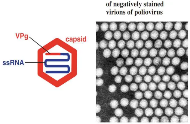
1.2. Virion
Picornaviruses have a simple structure with a small single-stranded RNA inside. The RNA is covered by a capsid, which is spherical shape with diameter of about 25–30 nm (30), (Figure 2).
13
Figure 2: The picornavirus virion.VPg: virus protein, genome linked. Electron micrograph courtesy of J. Esposito (US-CDC) and Professor Frederick A. Murphy (University of Texas Medical Branch) (30).
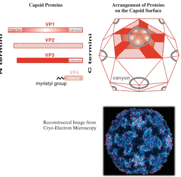
1.3 Capsid
According to (30), There are 4 capsid proteins named VP1-VP4 which numbered from the largest to smallest one. 60 copies each of these four proteins create the virus capsid with icosahedral symmetry. Each of the proteins VP1–3 contains an eight-stranded β-barrel like many virus capsid proteins. For human rhinovirus 14, the N termini of VP1–3 and all 60 VP4 molecules are completely internal while all the rest parts of VP1–3 are at the surface of the virion. Moreover, in many picornaviruses, there is an approximately 2 nm deep groove as canyon around each of the 12 vertices of the icosahedron (Figure 3). The canyons which contain the virus attachment sites are lined by the C termini of VP1 and VP3 molecules. Evolution of picornaviruses has generated a lot of variability in the capsid proteins, some of which is reflected in the existence of distinct serotypes such as poliovirus with 3 serotyps or rhinovirus with more than 100 serotypes.
14
Figure 3: The capsid of human rhinovirus 14. The canyons are visible in the reconstructed image, courtesy of J.Y. Sgro, University of Wisconsin – Madison (30).
1.4. Genome structure and features
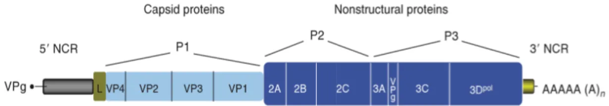
All picornaviruses have a similar genomic organization that is conserved in some but varies in other regions (31) (32). According to (26), picornaviruses have a quite short genome which containing a single molecule of positive-sense RNA within the capsid. The length of the RNA genome ranges from 6500 to 9500 nucleotides for whole. The genome consists of one single open reading frame (ORF) preceded by a long 5’-UTR and followed by a much smaller 3’UTR and a genetically encoded poly-(A) tail (Figure 4).
15
Figure 4: Organization of a typical picornavirus RNA genome. (26)
The 5’ end of the viral genome is covalently linked to the viral polypeptide VPg (virus protein, genome linked). The ORF which is divided into three consecutive regions, P1, P2 and P3 which translated into a single large polyprotein with approximately 2200 amino acid residues size. It is subsequently cleaved by viral protease(s) into mature proteins and their intermediates. For details, the P1 region encodes for the structural (capsid-forming) proteins 1A–1C which also known as VP4, VP2, VP3, and VP1, respectively. VP4 may not be formed in every picornavirus. The P2 and P3 region encodes for the nonstructural replication proteins 2A–2C and 3A–3D, respectively, as well as cleavage intermediates. In some picornaviruses, two or three unrelated 2A proteins may be formed while in others, two or three paralogous copies of 3B (also known as VPg) are (predicted to be) produced. The L region encodes for a leader protein in viruses of five genera. The L and 2A proteins may not be homologous, thus performing lineage-specific functions in different picornaviruses (33), (28), (27), (Figure 5), (Table 3).
16
Figure 5: Genome structure and gene organization of members of the family Picornaviridae. Each of the 12 genera is represented, as are species where there is a significant difference within a genus (27).
Genomic region
Protein
designation Genus Protein function
L Leader Aphthovirus,
Erbovirus
Papain-like cysteine protease implicated in virus–host interaction
L Leader Cardiovirus Involved in internal ribosome entry site-mediated translation of viral RNA
L Leader Kobuvirus No protease activity; involved in both viral RNA replication and encapsidation
P1 VP2, VP3,
VP1, VP4
All majority
Major capsid proteins
Small capsid protein implicated in virion uncoating that is present in viruses of most genera
P2 2A Enterovirus Chymotrypsin-like cysteine protease
17
nascent polypeptide; implicated in the control of RNA synthesis
2A Cardiovirus Aphthovirus Parechovirus Senecavirus Erbovirus Teschovirus
Small protein whose synthesis is accompanied by termination and
reinitiation of translation to separate 2A and 2B proteins
2A Hepatovirus Structural protein
2A Parechovirus
Tremovirus
Putative acyltransferase implicated in virus–host interaction
2B All Membrane–anchoring protein for the virus
replication complex
2C All
Multifunctional protein with ATPase and predicted helicase activity implicated in capsid assembly, virion uncoating, and RNA synthesis
P3
3A
Enterovirus and likely all
Membrane–anchoring protein for the virus replication complex; inhibits ER to Golgi membrane and secretory traffic
3B, VPg All Protein primer for the initiation of RNA-synthesis
3C All Chymotrypsin-like cysteine protease
mediating most cleavages in polyprotein
3D All RNA-dependent RNA polymerase
3CD Enterovirus
Responsible for processing capsid P1 precursor and regulation of RNA synthesis through binding to two RNA cis signals Table 3: Major demonstrated and predicted functions of picornavirus proteins (28).
18 1.5. Replication
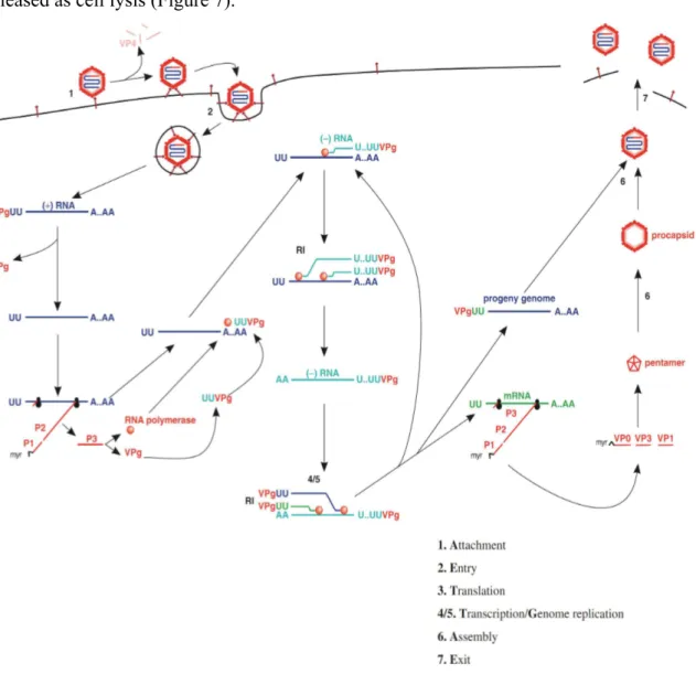
Attachment
The cell receptors for many picornaviruses have been characterized as the glycoprotein which is glycoprotein CD155 for poliovirus (30). It is the molecules with three immunoglobulin-like domains; the virus attachment site is located in the outermost domain. CD155 is found only in humans and some primate species. There are the major changes to capsid structure when virion binds to receptors which are the N termini of VP1 move to the exterior capsid surface and VP4 move out from virus capsid (Figure 7).
Entry
There are two modes of genome entry into the host cell have been proposed for picornaviruses. The first one is directly transfer of the RNA from the virion into the cytoplasm at the plasma membrane, leaving the capsid at the cell surface and the other is endocytosis. The VPg is removed from the 5’end by a cell enzyme as the virus genome is free in the cytoplasm (Figure 7).
Translation and post-translational modifications
The 40S ribosomal subunit binds internally at the IRES of the RNA. After a single polyprotein is created it is firstly cut to yield P1, P2 and P3. P1 becomes myristylated at the N terminus before being cleaved to VP0, VP3 and VP1, the proteins that will form procapsids; VP0 will later be cleaved to produce VP2 and VP4. Other cleavage products include 3B (VPg), 2C (an ATPase) and 3D (the RNA polymerase) (Figure 6).
Transcription/genome replication
RNA synthesis occurs in replication complexes are associated with membranous vesicles that created in the cytoplasm of infected cells and also contain cell proteins, as well as virus proteins and RNA. Multiple copies of (−) RNA are transcribed from infecting (+) RNA and then used as templates for (+) RNA synthesis. The small VPg is primer for both strands synthesis. Many RNA in the replication complexes play a role as replicative intermediates (RIs). Indeed, some RIs consist of a (+) RNA associated with growing (−) RNAs, while other RIs consist of a (−) RNA associated with growing (+) RNAs. Also
19
associated with RIs are copies of the RNA polymerase. The poly (A) sequence at the 3’end of the plus strand is transcribed and synthesized from a poly (U) sequence at the 5’end of the minus strand (Figure 7).
20
Assembly and exit
A procapsid is created by 12 pentamer which assembled from 5 copies each of VP0, VP3 and VP1. Each procapsid acquires a copy of the virus genome, with VPg still attached at the 5’end. Then the 60 copies of VP0 are cut into VP4 and VP2. The virions are released as cell lysis (Figure 7).
Figure 7: The picornavirus replication cycle. Note that translation gets under way before transcription, and that transcription and genome replication involve a single process (synthesis of (+) RNA). myr: myristyl group. RI: replicative intermediate (30).
21 2. Enterovirus
2.1. Classification
According to International Committee on Taxonomy of Viruses (27), enteroviruses are classified into 10 species and based on their sequence homologies, each species contains numerous viruses that are pathogenic to humans and animals (Table 4).
Species Subspecies
Bovine enterovirus Bovine enterovirus 1 Bovine enterovirus 2
Human enterovirus A
Coxsackievirus A2 [Fleetwood]
Other types: CV-A3 to A8, CV-A10, CV-A12, CV-A14, CV-A16, EV-A71, EV-A76, EV-A89 to EV-A92, EV-A114, SV19, SV43, SV46, baboon enterovirus A13
Human enterovirus B
Coxsackievirus B1
Other types: CV-B2 to CV-B6, CV-A9, EV-B1, EV-B2 to 7, EV-B9, EV-B11 to 21, EV-B24 to 27, EV-B29 to 33, EV-B69, EV-B73 to EV-B74, EV-B75, EV-B77 to EV-B88, EV-B93, EV-B97 to 101, EV-B106 to 107, EV-B110, SA5
Human enterovirus C
Poliovirus 1
Other types: PV-2, PV-3, CV-A1, CV-A11, CV-A13, CV-A17, CV-A19, CV-A20, CV-A21, CV-A22, CV-A24, EV-C95, EV-C96, EV-C99, EV-C102, EV-C104, EV-C105, EV-C109, EV-C113, EV-C116
Human enterovirus D Enterovirus D68
Other types: EV-D70, EV-D94, EV-D111
Porcine enterovirus B Porcine enterovirus 9 Porcine enterovirus 10
Simian enterovirus A Simian enterovirus A1 Other types: SV28, SA4 Human rhinovirus A Human rhinovirus A1
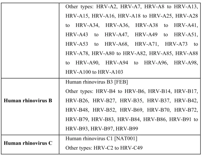
22
Other types: HRV-A2, HRV-A7, HRV-A8 to HRV-A13, HRV-A15, HRV-A16, HRV-A18 to HRV-A25, HRV-A28 to HRV-A34, HRV-A36, HRV-A38 to HRV-A41, HRV-A43 to HRV-A47, HRV-A49 to HRV-A51, HRV-A53 to HRV-A68, HRV-A71, HRV-A73 to HRV-A78, HRV-A80 to HRV-A82, HRV-A85, HRV-A88 to HRV-A90, HRV-A94 to HRV-A96, HRV-A98, HRV-A100 to HRV-A103
Human rhinovirus B
Human rhinovirus B3 [FEB]
Other types: HRV-B4 to HRV-B6, HRV-B14, HRV-B17, HRV-B26, HRV-B27, HRV-B35, HRV-B37, HRV-B42, HRV-B48, HRV-B52, HRV-B69, HRV-B70, HRV-B72, HRV-B79, HRV-B83, HRV-B84, HRV-B86, HRV-B91 to HRV-B93, HRV-B97, HRV-B99
Human rhinovirus C Human rhinovirus C1 [NAT001] Other types: HRV-C2 to HRV-C49
Table 4: Enterovirus genus and species, abbreviated as follows: bovine enterovirus (BEV); coxsackievirus (CV); echovirus (E); enterovirus (EV); human rhinovirus (HRV); poliovirus (PV); porcine enterovirus (PEV); simian agent (SA); simian virus (SV); simian enterovirus (SEV) (27).
2.2. Human Enteroviruses cause Hand, Foot and Mouth Disease
Origin, gene groups, evolution, and geographical distribution of EV-A71
Human enteroviruses have been classified into four species, HEV-A, HEV-B, HEV-C, and HEV-D. HFMD is caused by types of Enterovirus A species which includes some Coxsackievirus A (CV-A) and Enterovirus A71 (EV-A71) (34). The EV-A71 viruses are genetically related to CV-A; indeed, it has been suggested that these viruses may have diverged as recently as the 1920s. EV-A71 isolates are classified on the basis of VP1 gene into six independent genogroups: A, B, C, D, E and F. The EV-A71 B and C genogroups are each further subdivided into subgenogroups, B0 to B5 and C1 to C5 (Bessaud et al.,
23
2014). As are subdivided into serotypes A2-8, A10, A12, A14, CV-A16. Both EV-A71 and CV-A infection has been associated with severe HFMD in young children, sometimes resulting in death. But EV-A71 strain frequently causes severe HFMD as neurological forms and death in large epidemics while mild disease with other serotypes (35), (36), (37).
There is only one member in Group A, the prototype BrCr strain of EV-A71, which was first isolated in California, USA, in 1969. So far, the secondly isolates were reported in a HFMD epidemic among children in Anhui province of central China (38). Surveillance for HFMD by the Chinese Center for Disease Control and Prevention which do not seem to indicate any group A viruses (19).
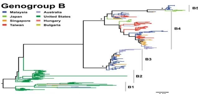
Group B viruses were firstly separated into B1 and B2 subgenogroups with 12% divergence at the nucleotide level. These subgenogroups were the predominant circulating strains in the 1970s and 1980s. In details, B1 was circulating in USA, Europe, Japan and Australia in 1970s and B2 mainly were isolated in USA in the 1980s. After that subgenogroup B3 was emerged from 1997 in Sarawak, Malaysia in several large outbreaks of Asia Pacific region. These viruses were also found in Singapore (39), (40), (41). Subgenogroup B4 were circulating and associated with the large outbreaks through Asia Pacific region in the 1990s but were also sampled by 2000. In 2003, subgenogroup B5 replaced subgenogroup B4 in Sarawak, Malaysia and circulate as the important viruses in Japan, Malaysia, Singapore and Taiwan (42), (43), (44), (15), (45), (Figure 6).
24
25
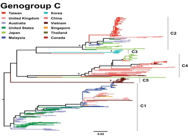
Group C viruses were initially separated into the C1 and C2 subgroups. Subgenogroup C1 viruses were first reported in USA and Australia in the 1970s, 1980s. From its initial detection, up to now these viruses have been identified continuously in many countries throughout Europe, Asia Pacific region. But they have not caused any large epidemics since the major community outbreak in Sydney in 1986 (46), (44). Subgenogroups C2, C4, and C5 is the novel subgenogroups which divers from genogroup C in the Asia Pacific region in the late 1990s. They cause fatal HFMD cases in Taiwan (14), China (47), and Vietnam (22), respectively. Subgenogroup C3 was isolated in Japan in 1994, and in Korea in 2000 (40), (48), (49). According to (45), subgenogroups C1, C2, and C4 globally distribute in many parts of the world while the C3 and C5 lineages are restricted to Korea and Vietnam, respectively (Figure 7).
26
Recombination and reservoir of EV-A71
The recombination of EV-A71 has been observed most frequently in the 5’UTR and 3’UTR regions. This rarely happens in the structural protein gene region (50), (51), (52). So far, the effect of EV71 recombination on the virulence or transmissibility is unknown. But some studies observe the replacement of the 3’ half of the genome of a non-recombinant EV-A71 field isolate with a EV-B species virus which improves growth in cell culture compared with a non-recombinant strain (53). Reservoir of EV-A71 is human and replication happens both in the intestinal tract and upper respiratory tract. The virion typically shed from 2 to 4 weeks and especially up to 12 weeks after infection. The transmission mode is through direct person-to-person contact, droplets or fomites by faecal-oral and respiratory secretions (54).
3. Pathogenesis
3.1. Virus entry and spread
The main transmission mode of HFMD is via the faces-oral route. It can also get infection through contact with virus contaminated surfaces, secretions, fluid or fomites. The virus can spread by respiratory droplets as well (55). According to (56), the enteroviruses initially replicate in the lymphoid tissues of the oropharyngeal cavity (tonsils) and small bowel resulting a mild viraemia. Most infections are asymptomatic at this time. The viruses follow bloodstream to other organs as the reticuloendothelial system (liver, spleen, bone marrow, and lymph nodes), heart, lung, pancreas, skin, mucous membranes, and center nervous system (CNS) at the onset time of disease with given clinical symptom. For virus shedding, it is up to 2 weeks from throat or 11 weeks from intestine after an acute EV71 infection (57).
3.2. Pathological findings
Clinical manifestations for HFMD widely vary from asymptomatic, mild to neurologic cases. But CNS infection is the features most frequently seen of EV-A71 infection (58).
27
For uncomplicated HFMD, regarding (59), the experiment on a mouse model shows the virus infect to skin which suggest the same results with human skin or oral mucosa lesions. But the information for skin or oral mucosa biopsies is still lacking.
For neurologic case, there is no inflammatory in cerebella cortex, basal ganglia, thalamus, peripheral nerve or autonomic ganglia. In contrast, the grey matter of the spinal cord and the whole medulla oblongata is affected predominantly and the hypothalamus, subthalamic, dentate nuclei as well. The motor cortex is also influenced but lesser. However, virus, viral antigen or RNA in the brainstem has not been seen or very rare for some cases in neuronal processes and phagocytic cells (20), (60).
For severe pulmonary oedema and cadiac dysfunction, It is supposed that explosive pulmonary oedema especially associated with neurogenic mechanisms secondary to brainstem inflammation, cardiac dysfunction, increased vascular permeability, and cytokine storm but its exact mechanism is still unclear and the pathogenesis is not completely understood (2), (Figure 8). In-vivo models, including those in mice and non-human primates, have replicated some of the features of severe EV-A71 disease, such as neuroinvasion with inflammatory changes, but none has yet been able to reproduce the severe systemic features, such as pulmonary oedema (59), (61), (62).
3.3. Virus virulence and host factors
So far, there is still not clear evidence from genetic analysis on virus-virulence factor. But data from some HFMD outbreaks show that the difference of genogroup may contribute to virulence aspect. Subgenogroup C2 from the epidemic in Taiwan (1998) was almost exclusively isolated from children with severe neurological disease. In contrast, subgroup B3 from the epidemic in Sarawak, Malysia (1997) was isolated mainly from children with mild HFMD (41), (44). As well, to date, still lack of information about infective virus doses related to the illness severity. It should be investigated further. In EV71 infection with explosive pulmonary oedema, there is a storm of cytokines and chemokines in cerebrospinal fluid and serum such as interleukin 6, 1beta, 10, IL-8, interferon induced protein (IP-10) and monokine induced by interferon gamma (MIG) (63), (64), (65). In contrast, number of T lymphocytes and NK cells reduce (66).
28
According to (67), HLA-A33, which is a common phenotype in Asian populations but is rare in white populations, was most significantly associated with EV-A71 infection and HLA-A2 was significantly related to cardiopulmonary failure. Receptors on the host target cell were identified as scavenger receptor B2 (68), and P-selectin glycoprotein ligand-1 (69), which expressed or limited to leukocytes, respectively.
! Figure 10: The postulated pathogenesis of enterovirus-71-associated acute pulmonary oedema (2). EV71=enterovirus 71. CNS=central nervous system. SVR=systemic vascular resistance. SBP=systemic blood pressure. HR=heart rate. LV=left ventricular.
29
Age and gender may play a role on susceptibility as a higher incidence of disease has been reported in male children. The relative immaturity of the innate and adaptive immune system in children aged between one and five years could contribute as well (70), (71), (25).
3.4. Protective immunity
According to (72), the cellular and humoral immune responses are both essential for decreasing the viral load and mortality in mice. In details, by mouse model, the disease severity, mortality, and tissue viral loads of mice lack of B, CD4 T, or CD8 T cells were significantly higher than those of wild-type mice after infection. Further, use a virus-specific antibody as therapy before or after infection significantly reduced the disease severity, mortality, and tissue viral loads of mice deficient in B cells. On the other hand, cellular immunity in human plays an important role of prevention of serious complications from HEV-71 infection (73), (74). Beside that there are some evidences of the protective immunity against infection by neutralizing antibodies from the humoral response (75), (76), (77). In humans, presence of maternal anti-EV-A71 antibodies has also been demonstrated in neonates, the prevalence and titer of which correlate with those levels in the mothers (78), (25). In mice, transplancental transfer of antibodies following maternal immunization against EV-A71 protects against lethal infection of newborn mice (79). Thus, it appears that the seroprevalence of neutralizing antibodies in women of childbearing age is important in protecting infants aged less than 6 months.
4. Diagnosis
4.1. Case definition and classification
For outbreaks, there are several kinds of clinical manifestation. It could be HFMD or Herpangina (HA) which usually is self-limiting and benign illness. But there is also a small number of cases can develop to neurological and systemic complication that may lead to death (Figure 9). According to (80), Case definition and classification is defined as below.
30
HFMD is a brief febrile illness in children with skin rash which typically is papulovesicular at the palms or soles of the feet, or both. This can be with or without mouth ulcers. Normally in younger children and infants the rash may be maculopapular without vesicles, and may also involve the buttocks, knees or elbows. HA is also fever and typically with multiple, painful mouth ulcers at mainly the posterior oral cavity (the anterior pharyngeal folds, uvula, tonsils and soft palate). In some cases, the mouth ulcers also affect other parts of the mouth such as the buccal mucosa and tongue.
For neurological and systemic complication, the enteroviruses, in general, can cause central nervous system complications as aseptic meningitis, acute flaccid paralysis, encephalitis. In particular, brainstem encephalitis is typically by EV-A71 infection and is frequently together with severe cardiorespiratory symptoms that have been attributed to neurogenic pulmonary oedema (21), (2). This typically clinical syndrome starts normally after 3–5 days of fever by a sudden deterioration as shock and pulmonary oedema or haemorrhages resulting from an acute and rapidly progressing cardiorespiratory failure. Finally, the children will die before reaching hospital or in the first day of admission, despite intensive care support (13), (20).
Fatal case firstly undergoes a short period of febrile illness with subtle neurological signs. Then dramatically suffer an acute refractory myocardial dysfunction and fulminant pulmonary oedema within hours of developing tachycardia, poor peripheral perfusion and tachypnea (80).
Disease Proposed Case Definition
HFMD
Febrile illness with papulovesicular rash on palms and soles, with or without mounth vesicles/ulcers. Rash may be maculopapular without vesicular lesion, and may also involve the buttocks, knees or elbows, particularly in younger children and infants.
Herpangina Febrile illness with multiple oral ulcers on the posterior parts of the oral cavity.
31 Aseptic meningitis
Febrile illness with headache, vomiting and meningism associated with presence of more than 5 – 10 white cells per cubic millimeter in cerebrospinal (CSF) fluid, and negative results on CSF bacterial culture.
Brainstem Encephalitis
Myoclonus, ataxia, nystagmus, oculomotor palsies, and bulbar palsy in various combinations, with or without MRI. In resource-limited settings, the diagnosis of brainstem encephalitis can be made in children with frequent myoclonic jerks and CSF pleocytosis.
Encephalitis Impaired consciousness, including lethargy, drowsiness or coma, or seizures or myoclonus.
Encephalomyelitis
Acute onset of hyporeflexic flaccid muscle weakness with myoclonus, ataxia, nystagmus, oculomotor palsies and bulbar palsy in various combinations.
Acute flaccid paralysis
Acute onset of flaccid muscle weakness and lack of reflexes.
ANS
dysregulation
Presence of cold sweating, mottled skin, tachycardia, tachypnea, and hypertension.
Pulmonary oedema/ haemorrhage
Respiratory distress with tachycardia, tachypnea, rales, and pink frothy secretion that develops after ANS dysregulation, together with a chest radiograph that shows bilateral pulmonary infiltrates without cardiomegaly.
Cardiorespiratory failure
Cardiorespiratory failure is defined by the presence of tachycardia, respiratory distress, pulmonary oedema, poor peripheral perfusion requiring inotropes, pulmonary congestion on chest radiography and reduced cardiac contractility on echocardiography.
Table 5: Proposed clinical case definitions for HFMD/HA and associated complications (80).
32 4.2. Laboratory diagnosis
Clinical samples
Throat swab, vesicle swab and stool, all can be used for virus detection. However, in the rate of detection point, throat and vesicle swab are considered to be the most useful specimens for both inpatients and outpatients. Especially, vesicle isolates always represent current systemic infection. It can also detect the virus from cerebrospinal fluid of central nervous system complication-patient, but the detection rate is very low (less than 5%). Serum sample from the patient is used in serological test as well (80) (81). The detection of the virus from the throat swabs was more frequent than from stool specimens; the time to positivity by viral culture was also shorter (82). In practice, it should be taken both kinds of sample as throat and vesicle swab or throat swab and stool in order to increase the possibility of virus detection.
Virus isolation
As usual for enterovirus isolation, some specimen pre-treatment such as complete mixing, filtration, chloroform treatment which needs to be implemented before inoculation. Two cell lines have been widely used for EV-A71 and CV-A16 virus detection are RD cells and Vero cells because of their relatively high sensitivity and the apparent cytopathic effect. For more, there are also some cell lines which are MRC-5, HEL, HeLa, L20B can be used as well (83). In addition, mouse cell lines are considered to give functional cellular receptors for enterovirus 71 which can be used for the selective isolation of EV-A71 or CV-A16 from clinical specimens (68), (69). Virus isolation and identification using suckling mice are still useful for some EV-A isolates, particularly for clinical samples from herpangina cases, but requires equipment, human resources and time (84).
Identification of virus isolates
Neutralization test recently gives confidence for virus identification. From virus isolation in cultured cells, conventional neutralization test which use a qualified type specific anti serum to identified the serotype of EV-A71 and CV-A16 virus, but these antisera are still not commercially available and it normally wait for a week to get the test result (80).
33
Reverse transcription - polymerase chain reaction (RT-PCR) and sequencing always has advantage of universal detection and amplification of gene targets, with any serotypes and genotypes, even though it is newly emerged variants or new serotypes of enteroviruses (85), (86), (39). With virus isolation from cultured cells, various gene targets which are 5’untranslated region, VP1 and VP4/VP2 genes have been widely used for molecular identification through by RT-PCR amplification of viral RNA and sequencing of the DNA amplicons (80). Indirect immunofluorescence assay (IFA) tests can provide quick, technically simple and reliable EV-A7 identification. It uses anti-EV-A71 monoclonal antibodies that are commercially available (80).
Rapid diagnosis directly from clinical samples
Nested RT-PCR: According to (87), use consensus-degenerate hybrid oligonucleotide primer (CODEHOP) on the VP1 region has enabled partial VP1 sequencing with a high sensitivity and broader specificity for all known enterovirus serotypes. Indeed, these viruses will be identified by VP1 sequences derived from the CODEHOP PCR products directly from clinical samples. It makes the advantage of quicker identification than virus culture. EV-A71-specific primers can be revised based on the sequences of newly emerging EV-A71 genogroups and variants for ensuring reliability of the test (88), (89). Multiplex RT-PCR methods are already developed for combined EV-A71and CV-A16 identification (90) and for the specific detection of EV-EV-A71and CV-A16 (91).
Real-time PCR can reduce the risk of cross-contamination when compared with conventional RT-PCR, particularly nested PCR systems. The reliability of recently EV-A71-specific real-time RT-PCR systems needs to be addressed by using different genogroups of EV-A71, CV-A16, EV-A strains, and clinical samples. In general, EV-A71-specific primers can be revised based on the sequences of newly emerging EV-A71 genogroups and variants for ensuring reliability of the test (80).
Molecular epidemiological analysis (genotyping) of EV-A71 strains
Determination of geographic and evolutional origin of a virus is normally today by genotyping technique (the molecular characterization genomes). For EV-A71 trains, based
34
upon a capsid VP1 sequence database, it can develop a quick way to tracking the regional transmission and current prevalence of EV-A71strains by comparing the extent of genetic changes that are observed between strains. For detailed molecular epidemiological analysis of EV-A71 strains, the entire VP1 sequence should be determined and analyzed, beside partial VP1 sequencing would be enough to genotype each EV71isolate (80).
Serological analysis
Testing for neutralizing antibodies against enteroviruses is not recommended for routine use in the diagnosis of enterovirus infections because of interpretation of serum antibody titers is sometimes difficult. In contrast, this test is useful for evaluating of immunity levels for EV-A71 infection within communities (54), also for monitoring the cross-reactivity of serum among different genogroups of EV71 (92) Serum samples from inpatients could be useful for the rapid immunoglobulin M (IgM) detection of EV71 (93), but the specificity and sensitivity of serological tests for EV-A71 infection remains difficulties in evaluation.
5. Epidemiology of Hand, Foot and Mouth Disease
5.1. History and epidemiological features of HFMD
In 1969, in California, USA, EV-A71 strain firstly was isolated from a 9 months aged child with encephalitis diagnostic (1). There are then small and large outbreaks of HFMD have been reported throughout the world (2), (Figure 10).
In the early 1970s, several countries in different continent including Sweden, Australia, USA and Japan had reported small HFMD outbreaks with some tens of cases and almost of them are children whose clinical aspects are mostly typical of HFMD, sometimes aseptic meningitis (3), (4), (5), (6). After that, HFMD was only reported in Europe in late half of the 1970s with two large outbreaks in Bulgaria (year 1975, 451 cases and 44 deaths), Hungary (year 1978, 1550 cases and 47 deaths) and a small number of cases in France in 1979 (7), (8), (9).
35
The 1980s there are also some small outbreaks in Hong Kong, Australia, USA (10), (11), (12) and no reported cases from other countries.
In the late 1990s, many country members of the Asia Pacific region have experienced large HFMD outbreaks. It began in 1997 with two large widespread community outbreaks in Sarawak, Malaysia and Taiwan, with 2628 and 129,106 cases reported, respectively (13), (14). Following that, a series of small and large outbreaks happen throughout the region in which Japan, Australia, China, Malaysia, Singapore, Taiwan (China), Korea, Mongolia, Vietnam, Brunei has been the hotpots of epidemic with cycle of every 2–3 years period (15), (16), (17). So far, the latest large outbreak in the region was in one province named Anhui of China in 2008 with around 490,000 cases and 126 deaths in children were reported, the case-fatality rate is around 0.03% but in certain local outbreaks, such as in Fuyang City of Anhui Province, this rate can reach up to 0.3% (18), (19). The explosive emergence of the disease in the region may be related to the association between HLA-A33 (which is higher prevalence in Asian populations than white population) and susceptibility to HEV-71 infection (67). During those outbreaks, almost of the cases are under 5 years old children and although clinical manifestations were mostly typical of HFMD, a cluster of deaths among young children was identified. Moreover, cases involving the central nervous system complication and/ or pulmonary oedema have also been observed for the first time (20), (21). The major pathogens of Hand, Foot and Mouth Disease (HFMD) are the EV-A in which some main serotypes are coxsackievirus A6, coxsackievirus A10, particularly coxsackievirus A16 (CV-A16) and enterovirus 71 (EV-A71). In the Asia Pacific region, EV-A71 strain frequently causes severe HFMD as neurological forms and death in large epidemics while mild disease with other serotypes (81), (36). Moreover, some EV71 subgenotypes can co-circulate in the same epidemic, as well as other non-EV-A71enteroviruses, such as Coxsackievirus-A. There are evidences show that the co-infection is possible in HFMD and it is not a primary cause leading to severe form (94), (21), (Table 10). Recurrent epidemics could be due to both of the accumulation of susceptible individuals in the community and introduction of new genotypes or strains in the Asia Pacific region (42). For instances, the outbreaks in Taiwan in 1998 and 2000 are caused by C2 and B4 strains, respectively (95), and by
36
sentinel surveillance in Sarawak, Malaysia, the emergence of a new subgenotype C1 is the cause of 2003 epidemic (15).
There were several small outbreaks or sporadic HFMD cases outside the Asia-Pacific region. In The Netherlands, there are only 58 cases of EV-A71 infection requiring hospitalization were reported in 2007 during 21 year period of very low endemicity by surveillance (96). In the United Kingdom, there were also only 32 patients, 01 death in 2003 with EV-A71 infection accompanied by neurological complications and/or cutaneous manifestations during that 1998 to 2006 period (97). 20 children with EV-A71 were admitted to a tertiary hospital in Canada in 1998, but no symptoms were severe and all improved rapidly (98). Similarly, in 2003 and 2005 in Denver, CO, USA, 16 children aged 4 weeks to 9 years get EV-A71 infection; one child died (99). A longitudinal study from Norway (2001- 2003), indicated asymptomatic circulation of EV71. Prevalence of EV-A71 in stool samples showed that EV-A71was circulating widely. But no clinical cases during this same period (100). In Nairobi, Kenya, two small institutional outbreaks of EV-A71 infection were reported in an HIV orphanage in 1999 and 2000 (101).
37
Year Location Genotype of EV-A71 and other co-circulating
viruses 1997 Peninsular and Sarawak,
Malaysia
EV-A71 (B3, B4, C1, C2); CV-A (16, 2, 4, 6, 9); CV-B5; EV (1,4,5,7)
1998 Taiwan EV-A71 (C2); CV-A16; CV-B (1, 2, 3, 5); EV(6,
7, 11, 22, 27).
1999 Australia EV-A71 (C2).
2000 Australia EV-A71 (B4).
2000 Singapore EV-A71 (B4); CV-A(16, 3, 4, 5, 6, 10, 23);
HEV-18.
2000 Taiwan EV-A71 (B4), CV(A16, A9, A24); CV-B (1, 3, 4);
EV (4, 9).
2001 Taiwan EV-A71 (B4); CV-A(16, 6, 9, 24); CV-B(4, 5); EV (4, 6).
2000 Sarawak, Malaysia EV-A71 (B4); CV-A16. 2000 Peninsular
Malaysia
EV-A71 (C1, B4)
2003 Sarawak, Malaysia EV-A71 (B4, B5, C1); CV-A16. 2005 Peninsular, Malaysia EV-A71 (B5, C1)
2006 Singapore EV-A71 (B5); CV-A16
2008, 2009
China EV-A71 (C4)
Table 6: Recent outbreaks of HFMD due to in the Asia Pacific region (102).
5.2. Sources, Transmission and Susceptibility of HFMD
Sources and transmission modes
Human beings now are the only known natural hosts of EV-A71. This pathogen can replicates in both the intestinal tract and the upper respiratory tract. It is typically shed for from 2 to 12 weeks and two weeks post-infection, respectively (57). So disease transmission can be faecal-oral and respiratory secretions route by direct person-to-person