EMT, the cytoskeleton, and cancer cell invasion
Mahmut Yilmaz
&Gerhard Christofori
Published online: 24 January 2009
# Springer Science + Business Media, LLC 2009
Abstract The metastatic process, i.e. the dissemination of
cancer cells throughout the body to seed secondary tumors
at distant sites, requires cancer cells to leave the primary
tumor and to acquire migratory and invasive capabilities. In
a process of epithelial-mesenchymal transition (EMT),
besides changing their adhesive repertoire, cancer cells
employ developmental processes to gain migratory and
invasive properties that involve a dramatic reorganization
of the actin cytoskeleton and the concomitant formation of
membrane protrusions required for invasive growth. The
molecular processes underlying such cellular changes are
still only poorly understood, and the various migratory
organelles, including lamellipodia, filopodia, invadopodia
and podosomes, still require a better functional and
molecular characterization. Notably, direct experimental
evidence linking the formation of migratory membrane
protrusions and the process of EMT and tumor metastasis is
still lacking. In this review, we have summarized recent
novel insights into the molecular processes and players
underlying EMT on one side and the formation of invasive
membrane protrusions on the other side.
Keywords Actin cytoskeleton . Cancer . Cell adhesion .
EMT . Metastasis . Tumorigenesis
1 Epithelial-mesenchymal transition (EMT)
and metastasis
Metastasis, the spread of tumor cells from a primary tumor
to a secondary site within the human body remains one of
the most life-threatening pathological events. In the last
years, major efforts have been taken to understand the
molecular mechanism underlying the distinct steps of
metastasis, which are (i) detachment of tumor cells from
the primary tumor, (ii) invasion into surrounding tissue, (iii)
intravasation into blood or lymphatic vessels, (iv)
dissem-ination in the blood stream or the lymphatic system and,
finally, (v) extravasation and outgrowth at a secondary site.
Each of these steps requires a distinct molecular program in
which the modulation of the adhesive and migratory and,
thus, the cytoskeletal properties of the disseminating tumor
cells play essential roles.
To detach from the primary tumor and to invade into the
surrounding tissue, tumor cells have to break down cell-cell
contacts, remodel cell-matrix adhesion sites, and follow a
chemoattractive path through the extracellular matrix, mined
by secreted proteinases. These processes are commonly
observed in various non-pathological conditions, such as in
developmental processes like gastrulation or neural crest cell
migration, where differentiated, epithelial cells dedifferentiate,
move to a distant site, and then re-differentiate to form a new
structure. This temporary and reversible phenomenon is
known as the epithelial-mesenchymal transition (EMT), a
process that is currently in the limelight of investigating the
onset of cancer cell migration, invasion and metastatic
dissemination [
1
,
2
]. During EMT, non-motile, polarized
epithelial cells, embedded via cell-cell junctions in a cell
collective, dissolve their cell-cell junctions and convert into
individual, non-polarized, motile and invasive mesenchymal
cells. Thereby, the molecular repertoire of a cell experiences
M. Yilmaz
:
G. Christofori (*) Institute of Biochemistry and Genetics,Department of Biomedicine, University of Basel, Basel, Switzerland
dramatic changes. For example, the function and expression
of the epithelial cell-cell adhesion molecule E-cadherin is
lost, whereas the expression of the mesenchymal cell-cell
adhesion molecule N-cadherin is induced, a process also
known as the cadherin switch. EMT can be prompted by
various intrinsic signals (e.g. gene mutations) as well
extrinsic signals (e.g. growth factor signaling). Among the
growth factors known to induce EMT are transforming
growth factor
β (TGFβ)[
3
], hepatocyte growth factor (HGF)
[
4
], members of the epidermal growth factor (EGF) family
[
5
], insulin-like growth factor (IGF)[
6
], and fibroblast
growth factor (FGF)[
7
,
8
]. Recently, also Notch signaling
has been implicated in EMT in human breast cancer cells by
activating the transcription factor Snail2 (Slug), a potent
repressor of E-cadherin gene expression [
9
]. Changes in the
composition of the extracellular matrix (ECM) are also able
to induce EMT, as shown for collagen I and hyaluronan [
10
,
11
].
With the diversity of signals inducing EMT the
complexity of the interactive downstream effector
path-ways increases. Among the candidates which are
engaged by TGF
β-induced EMT are the small GTPases
RhoA and Rac1 [
12
,
13
], Ras [
14
], phosphoinositol–3
kinase (PI3K) [
15
], Mitogen-activated protein kinase
(MAPK) [
13
], integrin-linked kinase (ILK) [
16
], and the
Jagged1/Notch signaling pathway [
17
]. With increasing
interest in microRNAs, miR-200 and miR-205 have been
recently shown to play an important role in TGF
β-induced
EMT by modulating the function of ZEB1 (δEF1) and
ZEB2 (Sip1), transcriptional repressors of E-cadherin gene
expression [
18
]. Such complexity of interactive signaling
upstream and downstream of the induction of EMT also
explains why EMT is not a simple matter of changes in a
cell
’s adhesive capabilities or its cytoskeletal organization,
it rather represents a fundamental reprogramming of
almost every aspect of a cell’s biology. Still, the different
signaling cascades underlying EMT can be grouped into
biological programs and, apparently, tumor cells
undergo-ing EMT hijack programs that are central for
develop-mental processes. The actual occurrence of EMT in
patients is still highly debated, yet with more detailed
molecular and histopathological analysis and the advent of
novel markers there is increasing evidence identifying
EMT in various human cancers [
19
,
20
]. Still, many
aggressive, invading tumors do not exhibit a molecular
signature of EMT, suggesting that EMT may not be
involved in every type of single cell invasion and that
some tumors may undergo a partial or incomplete EMT
[
21
]. In fact, cancer cells can invade in the absence of
EMT and have a broad repertoire for invasion, including
amoeboid or collective cell invasion [
22
,
23
].
This review highlights recent novel insights into EMT
research with a specific focus on the remodeling of the actin
cytoskeleton and the formation of invasive structures during
EMT and tumor cell invasion.
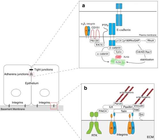
2 Epithelial cell-cell adhesion
The formation of a stable, polarized epithelium requires tight
cell-cell and cell-matrix connections. E-cadherin is the major
component of epithelial adherens junctions (AJ) which
mediate, along with tight junctions, intercellular adhesion.
AJ are located basal to the apical tight junctions (TJ) and form
a belt-like structure which tie neighboring cells together
(zonula adherens). E-cadherin is the prototype family member
of classical cadherins, single-span transmembrane
glycopro-teins that interact in a calcium-dependent, homophilic manner
with E-cadherins on neighboring cells. E-cadherin-mediated
cell-cell adhesion complexes are anchored to the actin
cytoskeleton via its cytoplasmic domain and
β-catenin and
α-catenin (Fig.
1
a). Thus, the formation of
E-cadherin-mediated cell-cell adhesion fundamentally modulates the
organization of cytoskeleton. This classical view of a direct
connection between the E-cadherin/
β-catenin/α-catenin
complex and the actin network has been challenged by
recent studies demonstrating that a reconstructed
cadherin-catenin complex fails to bind actin filaments in vitro [
24
],
and that E-cadherin, localized in electron-dense
micro-domains called spot adherens junctions (SAJs), binds to
actin in an
α-catenin-independent manner [
25
]. Here, the
authors describe a model in which two distinct actin
populations are involved in the zonula adherens architecture
(Fig.
1
a). One population represents stable, non-dynamic
patches of highly organized actin to which the
E-cadherin/β-catenin complex is attached in an
α-catenin-independent
manner (SAJs). The second population of actin is an
underlying, dynamic actin framework to which the SAJs
are linked and correctly positioned by
α-catenin. One protein
replacing
α-catenin in the E-cadherin/β-catenin complex to
SAJ could be eplin, a newly identified actin-binding protein
[
26
]. The juxtamembrane domain of E-cadherin binds to
p120-catenin which is important in surface tracking,
lyso-somal degradation and correct membrane localization of
E-cadherin [
27
–
30
]. Furthermore, p120-catenin plays an
elementary role in the stability of epithelial cell-cell adhesion
by repressing the activity of RhoA and activating Rac and
Cdc42 [
31
–
33
]. All three GTPases are key regulators of actin
assembly and play an essential role in the stability of cell-cell
adhesion by enforcing actin stress fibers (RhoA) and the
formation of migratory membrane protrusions, such as
lamellipodia and filopodia (Rac and Cdc42, respectively),
as discussed below.
Besides its adhesive function, E-cadherin also
encom-passes signaling capabilities, transduced predominantly by
proteins interacting with its intracellular domain, such as
β-catenin, or receptors that form multimeric complexes with
E-cadherin, such as c-Met, the cognate receptor for HGF, IGF1R
or integrins [
34
]. Notably, E-cadherin has been shown to
interact with a multimeric complex that consists of
α
3β
1-integrin, the tetraspanin CD151, which recruits protein
kinase C-βII (PKCβII), receptor of activated protein kinase
C-1 (RACK1), and the transmembrane protein tyrosine
phosphatase PTPµ [
35
] (Fig.
1
a). This multimeric complex
promotes association of the cadherin-catenin complex with
the actin cytoskeleton and supports cadherin mediated
cell-cell adhesion. CD151 appears to be important for the
filopodia-based
“adhesion zipper formation”, a process by
which initial filopodia-mediated contacts of epithelial cells
develop into mature cell-cell junctions [
36
,
37
]. Moreover,
CD151 expression accelerates E-cadherin-mediated
intercel-lular adhesion by inducing Cdc42-induced filopodia
exten-sions which form initial cell-cell contacts. Consistent with
these observations, E-cadherin colocalizes and interacts with
cortactin, a key regulator of actin-cytoskeleton assembly and
remodeling [
38
].
E-cadherin also associates with c-Met, IGF1R and
α
v-integrin at the plasma membrane [
39
,
40
]. Interestingly, in
the absence of
α-catenin the E-cadherin/IGF1R complex
does not form, suggesting that
α-catenin besides its
function as actin-anchoring protein also exerts a function
as an important scaffolding protein. Upon stimulation with
IGF-I,
α
v-integrin dissociates from the cell-cell adhesion
complex and translocates to focal contact sites of invasive
structures, such as invadopodia (see below). These findings
expand E-cadherin’s functional repertoire beyond its
adhe-ILK Talin Basement Membrane Tight junctions Integrins Integrins Epithelium Adherens junctions Src α β α β Paxillin Vinculin FAK p120 Actin Plasma membrane Actin RhoA α3β1-integrin PKCβII PTPµ CD151 E-cadherin RACK1 p190RhoGAP β−catenin Eplin α−catenin Cdc42 Rac1 stabilization ECM PINCH Parvin RTK
a
b
Integrins α βFig. 1 Differentiated, polarized epithelial cells are tightly attached to their neighboring cells by E-cadherin-mediated cell-cell adhesion complexes and to the extracellular matrix via integrins. a Epithelial cell-cell adhesion. E-cadherin is connected viaβ-catenin and Eplin to stable, electron-dense actin microdomains called spot adherens junctions (SAJ) in an α-catenin-independent manner. These E-Cadherin-SAJs complexes are attached and correctly positioned to an underlying dynamic actin framework via α-catenin. E-cadherin-mediated cell adhesion is stabilized by (i) p120-catenin recruited p190RhoGAP, which stabilizes the actin cytoskeleton underlying the adhesion complex by balancing the activities of RhoA and Rac1/ Cdc42, and by (ii) protein tyrosine phosphatase PTPµ, which keeps
β-catenin in an dephosphorylated state thereby preventing its degrada-tion. PTPµ is correctly positioned toβ-catenin by a multimeric protein complex consisting of receptor of activated protein kinase C-1 (RACK1), protein kinase C-βII (PKCβII), the tetraspanin CD151 andα3β1-integrin. b Epithelial cell-matrix adhesion. Integrin-mediat-ed cell-matrix adhesion and linkage to the actin cytoskeleton is accomplished by a multiprotein complex consisting of the adaptor proteins talin, paxillin, vinculin and the ternary complex of pinch, parvin and integrin-linked kinase (ILK), called tIPP complex. The interaction and phosphorylation status of focal adhesion kinase (FAK) and the non-receptor tyrosine kinase Src is critical for integrin complex assembly and turnover
sive functions and emphasize the critical role of E-cadherin
as a regulator of signaling complexes.
3 Loss of E-cadherin function and its consequences
Loss of E-cadherin gene expression or of E-cadherin
protein is frequently found during tumor progression in
most epithelial cancers. Hence, loss of E-cadherin function
is a clinical indicator for poor prognosis and metastasis
[
41
–
43
]. Since E-cadherin plays a key role in epithelial
structure and homeostasis its expression underlies a strict
control. As many other proteins, E-cadherin can be
regulated at the transcriptional as well as at the
post-translational level, yet both mechanisms usually cooperate
for an efficient repression of E-cadherin function.
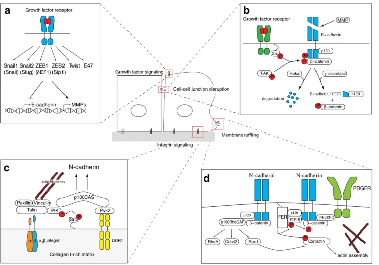
3.1 Transcriptional control of E-cadherin
Transcriptional repression of E-cadherin is mediated by a
list of transcription factors, among them intensely studied
factors like Snail1 (Snail), Snail2 (Slug), ZEB1 (
δEF1),
ZEB2 (Sip1), E47, and Twist [
44
] (Fig.
2
a). The
expression of these repressors can be induced by a variety
of stimuli, including activation of the TGF
β, HGF, EGF,
Wnt, and Notch signaling pathways. Moreover, they seem
to regulate each other’s expression in positive and negative
feedback loops. Engagement of these transcriptional
repressors at the E-cadherin gene promoter eventually leads
to epigenetic silencing of the gene by histone modifications
(acetylation, methylation, phosphorylation,
ubiquitinyla-tion, sumoylation) and subsequently by DNA
hypermethy-lation [
45
–
47
]. Such silencing of the E-cadherin promotor
is a complex process [
48
]. As a first step, Snail1 recruits the
histone deacetylase HDAC to the E-cadherin promotor
complex, thereby inducing histone deacetylation.
Subse-quently, the polycomb repressor complex 2 (PRC2) is
recruited to the site and methylates histones, thus
support-ing E-cadherin repression. Upon initial down-regulation of
E-cadherin gene expression, Snail1 induces ZEB1
expres-sion, which in turn engages a second, PRC2-independent
repressor complex that further inhibits E-cadherin
expres-sion. In addition, new interaction partners of Snail1 have
been identified, such as the LIM-domain protein Ajuba
which recruits the protein arginine methyltransferase 5
(PRMT5) to support Snail1-mediated transcriptional
repres-sion [
49
]. In a large variety of human cancers, the
E-cadherin gene is found to be highly hypermethylated, yet
how the initial silencing of the gene promoter converts into
a more long-term repression by DNA hypermethylation
remains to be resolved [
50
]. In the context of EMT and the
metastatic dissemination of tumor cells, the molecular basis
of the reversibility or irreversibility of E-cadherin
’s
epige-netic silencing is thus a future challenge. This complexity
and/or variability in E-cadherin regulation indicates that the
cell has a dynamic range of E-cadherin expression and
depending on the actual need can either totally suppress or
temporally attenuate its expression.
3.2 Post-translational control of E-cadherin
On the post-translational level, the transport of newly
synthesized E-cadherin to the cell membrane can be
inhibited via O-glycolysation [
51
] or mature,
membrane-bound E-cadherin can be degraded by proteolytic cleavage
or endocytosed from the plasma membrane [
52
–
55
]. The
proteolytic cleavage of cadherin can also produce
E-cadherin fragments which exert signaling functions. For
example,
γ-secretase-mediated cleavage of E-cadherin
produces a C-terminal, cytoplasmic fragment (CTF2) that
is transported into the nucleus in a p120-catenin-dependent
manner (Fig.
2
b). In the nucleus, CTF2 modulates the
interaction between p120-catenin and Kaiso, a
transcrip-tional repressor, thereby affecting for example cell survival
[
56
]. Destabilization of the E-cadherin adhesion complex is
also accomplished by receptor tyrosine kinase or
Src-mediated phosphorylation of E-cadherin, followed by its
ubiquitylation by the E3 ligase Hakai and subsequent
degradation [
57
,
58
] (Fig.
2
b). Moreover,
integrin-activated focal adhesion kinase (FAK) can phosphorylate
β-catenin and thus induce its ubiquitylation and
degrada-tion and the disassembly of the E-cadherin cell adhesion
complex [
59
]. Endocytosis of E-cadherin can occur via
clathrin or caveolin-dependent mechanisms [
60
–
62
]. A key
player in clathrin-mediated E-cadherin endocytosis is Arf6,
a Ras-related small GTPase. It promotes endocytosis via
recruitment of Nm23-H1, a nucleoside diphosphate kinase
(and the first metastasis suppressor gene ever identified
[
63
]), which in turn activates dynamin-dependent fission of
vesicles and destabilization of cortical actin by recruiting
the guanine nucleotide exchange factor (GEF) Tiam1, a
Rac1 inhibitor [
64
]. Recently, a new GTPase activating
protein (GAP) for Arf6, Smap1, has been identified, which
plays an essential role in E-cadherin endocytosis, revealing
a new player in post-translational E-cadherin control [
65
,
66
].
The downregulation of E-cadherin not only leads to a
mechanical disruption of AJ, it also liberates proteins from
the cytoplasmic cell adhesion complex which exert
ambiv-alent functions depending on their subcellular localization.
The probably most prominent cytoplasmic interaction
partner of E-cadherin is
β-catenin, well known for its dual
role in cell adhesion and Wnt signaling [
67
]. Stabilized by
active Wnt signaling or by mutations in the
β-catenin
phosphorylation/degradation pathway,
β-catenin
accumu-lates in the cytoplasm and enters the nucleus where it
interacts with members of the Tcf/Lef family of
transcrip-tion factors and modulates expression of a large number of
genes involved in cell proliferation, migration, invasion,
and morphogenesis, including cyclin D1, the cell adhesion
molecule L1-CAM, matrix metalloproteases (MMP) and
the metastasis gene S100A4 [
68
,
69
]. Another recently
discovered target of
β-catenin/Tcf signaling is Fascin, a
actin-bundling protein which is essential for filopodia
formation and cancer cell invasion [
70
,
71
].
Similar to
β-catenin, upon loss of E-cadherin function,
p120-catenin is also freed from the cytoplasmic cell
adhesion complex and accumulates in the cytoplasm. In
addition to its functions on Rho family GTPases and the
actin cytoskeleton (see above), p120-catenin can traffic to
the nucleus where it binds to the transcriptional repressor
Kaiso. In contrast to
β-catenin/Tcf-mediated transcription,
where
β-catenin acts as a transactivator, p120-catenin has
no transactivation domain and rather releases Kaiso from its
Paxillin Vinculin Talin Src α2β1integrin DDR1 α β p130CAS Pyk2 N-cadherin FAK P E-cadherin Snail1 (Snail) Twist MMPs
Collagen I-rich matrix
Cell-cell junction disruption Growth factor signaling
Integrin signaling p120 N-cadherin PDGFR FERPTP1B NHERF-2 p190RhoGAP RhoA P β−catenin N-cadherin β−catenin p120
b
c
d
p120 E-cadherin β−catenin Src FAK P P P E-cadherin / CTF2 p120 P β−catenin PGrowth factor receptor MMP
a
γ−secretase Hakai
+
Growth factor receptor
Snail2 (Slug)
ZEB1
(δEF1) (Sip1) E47
degradation ZEB2 Rac1 Cdc42 PCortactin P actin assembly P
Fig. 2 Induction of epithelial-mesenchymal transition (EMT). a Repression of E-cadherin gene expression. Growth factor stimulation induces various signaling cascades leading to the induction of EMT. Among the regulated genes are transcription factors that repress E-cadherin gene expression and induce expression of genes such as matrix metalloproteinases (MMP). b Loss of cadherin function. The E-cadherin cell-cell adhesion complex is destabilized by Src and/or FAK-mediated phosphorylation of either E-cadherin or β-catenin. The phosphorylation of the cytoplasmic E-cadherin domain induces its proteasomal degradation mediated by the E3-ubiquitin-ligase Hakai. Phosphorylation ofβ-catenin induces its detachment from E-cadherin and relocalization into the nucleus, where together with Tcf tran-scriptions factors modulates the expression of pro-invasive genes. Intracellular cleavage of E-cadherin byγ-secretase also results in a loss of E-cadherin function, yet its newly generated C-terminal fragment (CTF2) together with p120-catenin translocates into the nucleus, where it modifies the activity of the transcriptional repressor Kaiso. c
Integrin-mediated support of EMT. Enrichment of collagen I in the extracellular matrix supports EMT via the coordinated signaling ofβ1-integrins and the collagen receptor DDR1. These signals converge via FAK and Pyk2 kinases on the p130Crk-associated scaffold (CAS) protein, resulting in the induction of N-cadherin expression. d The formation of protrusive membrane structures. Upon PDGF stimulation, N-cadherin co-localizes with PDGFR in membrane ruffles. The interaction of both N-cadherin and PDGFR relies on Na+/H+exchanger regulatory factor-2 (NHERF-2) which is recruited to the complex by p120-catenin. Localized in membrane ruffles, N-cadherin contributes to actin cytoskeleton remod-eling (i) by recruiting p190RhoGAP and thereby favoring Rac1 and or Cdc42-mediated actin remodeling and (ii) by employing p120-catenin to recruit the non-receptor tyrosine kinase Fer. Fer in turn phosphorylates and activates cortactin which induces actin assembly. Morover, Fer stabilizes the N-cadherin complex by phosphorylating and activating protein tyrosine phosphatase PTP1B, thus preventingβ-catenin phos-phorylation and degradation
promoter binding sites and thus activates gene expression
by de-repression. However, the nature of p120/Kaiso target
genes is still poorly defined [
72
].
4 The cadherin switch and its consequences
The loss of epithelial E-cadherin and the gain of
mesen-chymal N-cadherin expression is a major hallmark of EMT.
This cadherin switch leads to a drastic change in the
adhesive properties of a cell, as it loses its affinity for
epithelial neighbors and gains affinity for mesenchymal
cells, such as fibroblasts or vascular endothelial cells.
Besides the change of the adhesive repertoire, the gain of
N-cadherin expression also provokes increased cell
migra-tion and invasion [
73
,
74
]. Like E-cadherin, N-cadherin
belongs to the family of classical cadherins and forms
homophilic cell-cell adhesion junctions. N-cadherin is
normally expressed in nervous tissue, in vascular
endothe-lial cells, and in skeletal and cardiac muscle cells. Its
expression is upregulated in the cells of the primitive streak
during mesoderm formation and during progression of a
variety of cancers, where its expression correlates with poor
prognosis [
74
–
76
]. Investigations into the molecular
mech-anisms underlying the induction of N-cadherin expression
during EMT have only recently begun. It has been shown
that N-cadherin can be upregulated by collagen I via the
coordinated engagement of the collagen receptor discoidin
domain receptor 1 (DDR1) and
α
2β
1-integrin based on a
p130
Crk-associated substrate (CAS) scaffold [
10
,
77
] (Fig.
2
c). On the transcriptional level, the transcriptional repressor
Twist appears to be involved in the induction of N-cadherin
gene expression in an
β
1-integrin-dependent manner [
78
–
80
].
Like its epithelial counterpart, N-cadherin is connected
via
α-catenin and β-catenin to the cytoskeleton and
functions as both a mechanical cell adhesion component
and a signaling molecule. Recently, it has been reported that
during neurite extension the traction forces generated by
retrograde actin flow are directly transmitted to N-cadherin
adhesions, thereby mechanically linking N-cadherin with
the formation of motile structures [
81
]. Interestingly, the
authors show that the mechanical engagement of
N-cadherin induces local actin polymerization and thereby
ensures the integrity of the adhesion complex. Engagement
of N-cadherin activates the RhoGTPase Rac1, which in turn
recruits the actin remodeling protein cortactin to the
N-cadherin adhesion complex [
82
] (Fig.
2
d). Within this
complex, the non-receptor tyrosine kinase Fer associates
with N-cadherin via p120-catenin [
83
]. Fer then
phosphor-ylates and activates cortactin, thereby inducing actin
remodeling, and increasing the mobility of N-cadherin
molecules to extend the adhesion zone and, finally, to
promote the formation of stable cell-cell adhesion. In line
with these observations, N-cadherin is found to be localized
in the lamellipodia of adjacent, contacting myoblasts [
84
].
Fer also phosphorylates the phosphatase protein tyrosine
phosphatase 1B (PTP1B) and promotes its binding to
N-cadherin. The PTP1B/N-cadherin interaction protects
β-catenin from degradation by keeping it in a dephosphorylated
state and thereby ensures the stability of the
N-cadherin-mediated cell-cell junctions [
85
,
86
] (Fig.
2
d).
Also similar to E-cadherin, N-cadherin interacts with a
number of signal transduction molecules and contributes to
various signaling pathways. For example, Na+/H+
ex-changer regulatory factor (NHERF)-2 has been shown to
physically link N-cadherin to PDGF receptor (PDGFR) by
binding both
β-catenin in the N-cadherin/β-catenin
com-plex and PDGFR [
87
] (Fig.
2
d). PDGFR activation is
known to induce actin reorganization and cell proliferation
and differentiation and to play an important role in EMT
[
88
,
89
]. Interestingly, PDGF stimulation of NIH3T3 cells
leads to colocalization of N-cadherin, p120-catenin and
p190RhoGAP in dorsal circular ruffles (DCRs), structures
known to depend on growth factor-induced Rac activity
and subsequent RhoA inhibition [
90
–
92
]. This is interesting
because p120-catenin is not only important for the
recruitment of Fer to the N-cadherin complex, but together
with p190RhoGAP also coordinates the antagonistic
func-tions between Rac and RhoA [
31
,
93
]. This antagonism
plays a critical role in defining the structure of the actin
cytoskeleton. The active form of RhoA stimulates
focal-adhesion (FA) formation and contractility via assembly of
predominantly radially-oriented actin stress fibers (ASF),
whereas Rac activation induces cell spreading, migration
and membrane ruffling via actin polymerization at the
cell periphery. Moreover, Rac activation inhibits Rho
activity, which can also be achieved by p120-catenin
over-expression [
94
] (Fig.
2
d).
Besides PDGFR, N-cadherin also interacts with FGF
receptors (FGFR) in a complex with neural cell adhesion
molecule (NCAM), a immunoglobulin domain cell
adhe-sion molecule [
42
,
95
]. The interaction between N-cadherin
and FGFR leads to stabilization of FGFR at the membrane
surface by preventing its internalization upon ligand
binding. As a result, sustained MAPK pathway activation
and increased cell motility and MMP secretion promote
invasiveness of N-cadherin-expressing cells [
96
–
98
].
NCAM accomplishes a similar induction of cell migration
and invasion by directly binding and stimulating FGFR via
its fibronectin type III domains [
95
]. Thereby,
NCAM-mediated stimulation of FGFR signaling differs substantially
from FGF-induced FGFR signal transduction resulting into
different cellular outcomes, such as increased cell-substrate
adhesion, migration and invasion by NCAM and cell
proliferation by FGF [
99
]. The interaction between
N-cadherin and NCAM with FGFR relies on a CAM-domain
proximally located to the acid box region of the FGFR,
which is not required for FGF ligand binding [
100
]. Such
complex interactions between different cell adhesion
mole-cules and tyrosine kinases raise another level of complexity
in the regulation of cell migration, invasion and metastasis
formation. For a more detailed insight into specific aspects of
cell adhesion and signaling complexes at the invasive cancer
front we refer the reader to recently published reviews [
42
,
101
].
Like E-cadherin, N-cadherin is also proteolytically
processed to generate shedded extracellular domain and
intracellular domain fragments with potential signaling
functions. In neurons, N-cadherin is cleaved by ADAM10
and by PS1/γ-secretase to produce a cytoplasmic fragment
of N-cadherin, N-Cad/CTF2, for example stimulated by
bone morphogenic protein-4 (BMP4) [
102
,
103
]. N-Cad/
CTF2 is able to interfere with the CPB/CREB transcription
complex, by binding the transcription factor CBP and
inducing its proteasomal degradation.
N-Cad/CTF2-induced repression of CPB/CREB-mediated transcriptional
control suppresses expression of genes important for
proliferation and differentiation, such as c-Fos. N-Cad/
CTF2 also promotes migration of neural crest cells by
increasing the expression of
β-catenin and therewith
inducing the expression of
β-catenin target genes like
cyclin D1. Interestingly, N-Cad/CTF2 facilitates
β-catenin-dependent signaling (i) by inhibiting
β-catenin
phosphor-ylation, (ii) by increasing
β-catenin transcription and (iii)
by reducing full-length N-cadherin protein levels to prevent
sequestration of
β-catenin to cell-cell junctions [
104
].
These data describe N-cadherin as a critical protein in
the regulation of EMT and cell invasiveness. Besides
simply exercising a cell adhesion function that changes a
cell’s affinity from epithelial cells to mesenchymal cells, it
is actively involved in delineating a cell’s migratory state
by modulating growth factor signaling and remodeling the
actin cytoskeleton. Such pleiotropic functions certainly
warrant detailed future experimental investigations.
5 Integrin-mediated cell-matrix adhesion and signaling
The extracellular matrix (ECM) is a constantly remodeled
3D structure consisting of a variety of specialized proteins
and proteoglycans which are able to regulate many cellular
processes, including cell proliferation, survival,
differenti-ation, and migration. This control is mainly based on a
constant communication between the adhesive adaptors of a
cell, integrins, and the ECM. Changes of a cell
’s integrin
repertoire or the composition of the ECM can have drastic
consequences for the cell and, in extremis, can lead to cell
death as well as transformation. Besides its scaffolding
function, the ECM is also able to bind and sequester diverse
growth factors and chemokines which can be retrieved by
local proteolysis and, dependent on activation or
inactiva-tion, affect cell behavior. Integrins are heterodimeric type-I
transmembrane proteins consisting of an
α-and a β-chain.
Mammals have 18
α and 8 β chains which are combined to
generate 24 different combinations which bind in a specific,
yet partially overlapping, manner various components of
the ECM.
Similar to cadherins, also integrins function as both
mechanical adhesion and signaling molecules. Importantly,
integrins switch between an inactive, low ligand-affinity
conformation and an active, high ligand-affinity
conforma-tion. Activation can be achieved by binding of intracellular
proteins to integrins, such as talin, or by MMP-mediated
proteolytic cleavage [
105
,
106
]. Integrins are linked to the
actin cytoskeleton and signal via the ternary complex of
integrin-linked kinase (ILK), pinch and parvin, also named
the tIPP complex [
107
] (Fig.
1
b). In the tIPP, integrins are
linked through ILK to the actin cytoskeleton, either via
parvin itself or via a paxillin/vinculin/parvin complex. ILK
plays a major role as a scaffold protein in assembling the
multimeric protein complex which is necessary for the
integrin-actin cytoskeleton linkage (via parvin, paxillin and
vinculin). Moreover, ILK is required for the formation of
signaling complexes with receptor tyrosine kinases via
pinch. The signaling cascades triggered by the activation of
integrins and its cytoplasmic partners are complex. Among
the direct targets downstream of integrins are FAK,
Src-family kinases, glycogen synthase-kinase-3
β (GSK3 β)
and protein kinase B (AKT/PKB). Effectors of these
signaling cascades include MAPK, NFκB, Jun, β-catenin
and others and together they modulate cell proliferation,
cell survival, cell migration and invasion. Among the
mitogenic integrins are
α
6β
4or
α
vβ
3-integrins which
cooperate with diverse growth factor receptors, including
EGFR, ErbB2, and c-Met [
108
–
111
]. On the other hand,
growth inhibitory signals can be transmitted, for example
via
α
2β
1-integrin, which in turn activates
p38-MAPK-mediated cell-cycle inhibition [
112
,
113
]. Integrin
α
vβ
6or
α
vβ
8engagement leads to the activation of latent TGFβ
thereby executing its cytostatic effect [
114
,
115
]. Faced by
this dichotomy of integrin signaling, cancer cells switch
their integrin expression to a pro-oncogenic repertoire in
order to invade and survive in the surrounding tissue.
6 Integrins in EMT and cell invasion
The function of integrins during EMT is diverse and
dynamic as they are able to initiate and enforce EMT and
invasion. For example, engagement of integrins
α
1β
1or
mediated cell-cell contacts, along with the activation of the
β-catenin/Tcf pathway in pancreatic cancer cells [
59
,
116
].
Furthermore, as described above, collagen type I is also
able to induce N-cadherin expression upon activation of the
integrin
α
2β
1together with the collagen receptor discoidin
domain receptor 1 (DDR1), a receptor tyrosine kinase [
77
].
Both, the downregulation of E-cadherin and upregulation of
N-cadherin play important roles in the initiation and
execution of EMT. Interestingly, Snail1, the transcriptional
repressor of E-cadherin expression and a potent inducer of
EMT, is able to induce the expression of
α
vβ
3-integrin
which is well known for its pro-invasive functions and its
localization in the invading front of cancers [
117
,
118
].
Along with their function as mechanical anchor proteins
for cell migration and invasion, integrins play an important
role in the correct localization of proteases. Several reports
demonstrate that the colocalization and cooperation of
β1-integrin and MT1-MMP1 is necessary for cancer cell
invasion into a collagen matrix and that both MT1-MMP
and
β
1-integrins have important roles in EMT [
119
–
123
].
The localization of MT1-MMP to
β
1-integrins is an
exocytic event dependent on the activity of the GTPase
Rab8 [
124
]. Integrin-mediated recruitment of ECM
remod-eling proteases is also responsible for the liberation and/or
activation of matrix bound growth factors and chemokines.
As described above, the cooperation of MT1-MMP and
α
vβ
8-integrin leads to the activation of latent TGFβ [
115
,
125
]. TGF
β usually acts as a cytostatic, tumor suppressing
factor, but it promotes tumor progression and invasion, if
the tumor cells overcome its cytostatic and apoptotic effects
[
126
]. In fact TGF
β is one of the most potent inducers of
EMT in cultured cells in vitro and in animal models in vivo
[
1
]. Other integrins which are upregulated during EMT,
such as
α
vβ
6-integrin, are also able to increase protease
expression and to liberate and activate TGFβ [
127
–
129
].
Also, the activities of the cytoplasmic interaction partners
of integrins, ILK and pinch, have been implicated in the
process of EMT [
130
–
132
].
7 EMT and the actin cytoskeleton
The actin cytoskeleton is a highly dynamic structure, which
is constantly remodeled in a living cell. This dynamics are
based on a well-balanced and highly controlled equilibrium
of local assembly and disassembly of actin filaments.
Obviously, such regulation is a prerequisite for processes
like endocytosis, cell motility, and cancer cell invasion.
7.1 RhoGTPases and EMT
The members of the Rho GTPase family are mainly
responsible for integrating and transmitting signals from
chemokine and growth factor receptors and from adhesion
receptors to effector proteins of actin remodeling.
RhoGT-Pases are activated upon GTP binding and inactive in their
GDP-bound form. RhoGTPase activation is tightly
con-trolled by three groups of regulatory proteins, guanine
nucleotide exchange factors (GEF), GTPase-activating
proteins (GAP), and guanine nucelotide dissociation
inhib-itors (GDI). GEF are responsible for the activation of
RhoGTPases by promoting the exchange of Rho-bound
GDP by GTP. This is counteracted by GAP which raise the
intrinsic GTPase activity of RhoGTPases and the
hydroly-sis of bound GTP to GDP. Finally, GDI bind inactive
Rho-GDP and prevent the interaction with RhoGEFs and thus its
activation. RhoA, Rac1 and Cdc42 are best studied among
the 23 family members of RhoGTPases. The complexity of
RhoGTPase signaling arises not only from the size of its
family members and number of effector proteins (~ 70
proteins), but also from the numbers of GEF (~ 70
members), GDI (~3 members) and GAP (~ 60 members)
which modulate their activity. Depending on which GEF,
GDI or GAP is interacting with the RhoGTPase the
biological response can be different. In the GTP-bound
form, RhoGTPases activate effector proteins, which are
often serine/threonine kinases, such as the p21-activated
kinases (PAK) for Rac1 and Cdc42 and the ROCK kinases
for RhoA. In general, RhoGTPases affect almost all cell
biological processes in a cell’s life [
133
–
135
]. With regard
to migration and invasion and in a simplified view, RhoA
induces actin stress fiber formation and regulates
cytoskel-etal changes affecting cell-cell or cell-matrix adhesion,
Rac1 is involved in lamellipodia and membrane ruffle
formation, and Cdc42 is involved in filopodia formation
[
136
,
137
]. Based on their central function in actin
remodeling and their ability to induce MMPs, Rho GTPases
play an important role in EMT as well [
138
].
During growth factor-induced EMT, tight control of the
activities of RhoGTPases is critical. As mentioned above,
depending on the presence of epithelial or mesenchymal
cadherins, the localization and function of p120-catenin and
thus the activity of RhoGTPases change dramatically [
139
].
In epithelial cells, p120-catenin localizes at the cell
membrane and associates with E-cadherin where it controls
the activity of RhoA and Rac1. RhoA activity, which is
required for the initial cell-cell contact formation, is
downregulated in established, mature cell adhesions. Both,
activation and inactivation of RhoA require the
p120-catenin-dependent recruitment of RhoGEFs, like Vav2, or
RhoGAPs, like p190-RhoGAP, respectively. The
recruit-ment of p190-RhoGAP results in the activation of Rac1
which leads to a stabilization of E-cadherin junctions by
inhibiting the activities of IQ-domain GTPase-activating
protein 1 (IQGAP1), a Rac1 effector protein and a mediator
of E-cadherin endocytosis (see also below). Moreover, the
actin cytoskeleton underlying cell contacts is reorganized
and stabilized [
140
].
During EMT, p120-catenin binds to mesenchymal
cadherins at the cell membrane but is also found localized
in the cytoplasm. Cytoplasmic p120-catenin functions as a
RhoA-GDI that binds and represses RhoA activity [
141
].
Simultaneously, p120-catenin bound to mesenchymal
cad-herins at the cell membrane promotes Rac1 activity and
induces the formation of motile, protrusive membrane
structures, such as lamellipodia. Thus, both cytoplasmic
and membrane-sequestered p120-catenin cooperate to
in-duce cell motility during EMT. Interestingly, Rac1 inhibits
RhoA activity by inducing the production of reactive
oxygen species (ROS), which in turn activate
p190Rho-GAP by inhibiting the low-molecular weight protein
tyrosine phosphatase (LMW-PTP) [
90
,
142
]. Moreover,
the expression of Snail1, the transcriptional repressor of
E-cadherin gene expression and potent inducer of EMT, is is
increased upon Rac1-mediated ROS production [
143
]. The
importance of RhoGTPases in EMT is also underscored by
the notions that Rac1b, a splice variant of Rac1, is highly
expressed in malignant breast tissue, that RhoA
down-regulation is required for EMT in colon carcinoma
progression, and that Rock activity is critical for cell
migration, invasion and metastasis after EMT [
141
,
144
,
145
].
The activity of RhoA during EMT not only effects
cell-cell adhesion but also microtubule-mediated cell-cell-matrix
adhesion and basement membrane integrity [
146
]. RhoA,
when localized at the cell-basement membrane (BM) gets in
contact with Net1, a RhoA-specific GEF, thereby exerting
an important function for the integrity of the BM in
epiblasts. During gastrulation, a process resembling EMT,
RhoA at the cell basis loses its activity, which leads to
microtubule destabilization, cell-BM contact disruption and
BM breakdown. Notably, destabilization of the cell-BM
contacts precedes breakdown of cell-cell adhesions.
Tiam1, a GEF for Rac1, also exerts a critical function in
both E-cadherin-mediated cell-cell junction stability and
during EMT. Loss of Tiam1 activity is required for the
induction of EMT; forced expression of constitutive-active
forms of Rac1 (RacV12) or Tiam-1 prevents HGF-induced
EMT in epithelial cells [
147
,
148
]. Interestingly, ablation of
Tiam-1 in a mouse model of chemical-induced skin
carcinogenesis reduces tumor incidence yet increases tumor
malignancy, demonstrating the ambivalent role of Rac1 in
tumor formation and tumor progression [
149
].
Despite major progress, the detailed role of Rho family
GTPases in EMT and tumor progression still remains
unresolved. The sophistication of tumor cell motility and
invasion on one side and the immense complexity of the
regulation of RhoGTPases on the other side obscure a
simple solution. The formation of membrane protrusions
and other RhoGTPase-dependent activities during EMT are
not linear processes, and rather depend on the integrated
activities of many members of the RhoGTPase family and
their interaction with various regulatory proteins which will
eventually determine the time and localization of their
specific activities.
7.2 Membrane ruffles
The onset of motility requires a relaxation of static actin
structures in order to form pliable membrane protrusions.
Rigid actin stress fibers are disassembled upon dorsal
circular ruffle (DCR) formation leaving a fine cortical actin
meshwork behind, from which cell membrane protrusions
like lamellipodia can emerge [
150
,
151
]. DCRs are
short-lived actin structures formed at the dorsal surface of growth
factor (PDGF, HGF, EGF)-stimulated cells. DCRs, formed
at the leading edge of growth factor-stimulated cells, are
also able to secrete MMP (e.g. MMP-2), revealing their
potential role for the onset of cell invasion [
152
]. Besides
relaxing static membrane structures, DCRs are also
impor-tant for macropinocytosis in growth factor-stimulated
epithelial cells [
153
,
154
]. Macropinocytosis plays an
important role in the modulation of cell signaling; it may
either inhibit signal transduction via degradation of growth
factor receptor complexes or it may potentiate signaling, as
shown in the case of EGF [
154
]. DCRs are enriched with
actin assembly proteins, such as Arp2/3, WASP and
cortactin, suggesting that DCR formation requires actin
assembly. As mediators of growth factor signaling,
RhoGTPases, like Rac1 or Cdc42, are required for DCRs
formation as well [
155
]. Other reports indicate the
involvement of protein kinase A (PKA) and the
cytoplas-mic tyrosine kinase Abl in DCR formation [
156
]. Both,
PKA and Abl, are known to be involved in EMT [
157
–
159
].
β-catenin in a complex with APC is also found to be
localized at the leading edge of migratory cells, implying an
important role for cell polarization and migration by linking
microtubules to the actin cytoskeleton [
160
–
162
]. Notably,
β-catenin seems to exert its function at the leading edge of
cells by co-localizing with N-cadherin and IQGAP1 in
membrane ruffles [
118
]. IQGAP1, an effector of Cdc42 and
Rac1, here acts as a key regulator of internalization of
N-cadherin and APC. The fact that PDGFR also co-localizes
with N-cadherin in membrane ruffles suggests an
interest-ing overlap of proteins known to be important in both EMT
and ruffle or podosome formation [
87
].
Other newly identified players in DCR formation and
important for motility are palladin and its interaction
partner Eps8 [
163
,
164
]. Palladin binds F-actin and
cross-links actin filaments into bundles. It also interacts with Src,
thereby affecting Src-mediated actin remodeling [
165
].
Both proteins are also involved in podosome formation
which highlights the kinship between DCRs and
podo-somes [
163
]. The cancer relevance of Eps8 and palladin is
underlined by their increased expression in various human
cancers, including breast and thyroid cancer [
166
–
170
].
Interestingly, palladin expression appears to be induced by
treatment of myofibroblasts with TGFβ1, suggesting a link
between TGFβ signaling, a major inducer of EMT, and the
formation of ruffles and podosomes [
171
].
7.3 Lamellipodia and filopodia
To be able to leave the primary tumor and to disseminate to
secondary sites, tumor cells have to break down cell-cell
and cell-matrix junctions, as described above, and they
have to invade into the surrounding tissue by gaining
motility and forming invasive, matrix degrading structures.
On planar two-dimensional culture substrates, cells utilize
two well-known organelles to explore and move into the
surrounding environment, filopodia and lamellipodia. Both
are actin-rich membrane protrusions that are formed upon
remodeling of the actin cytoskeleton beneath the plasma
membrane.
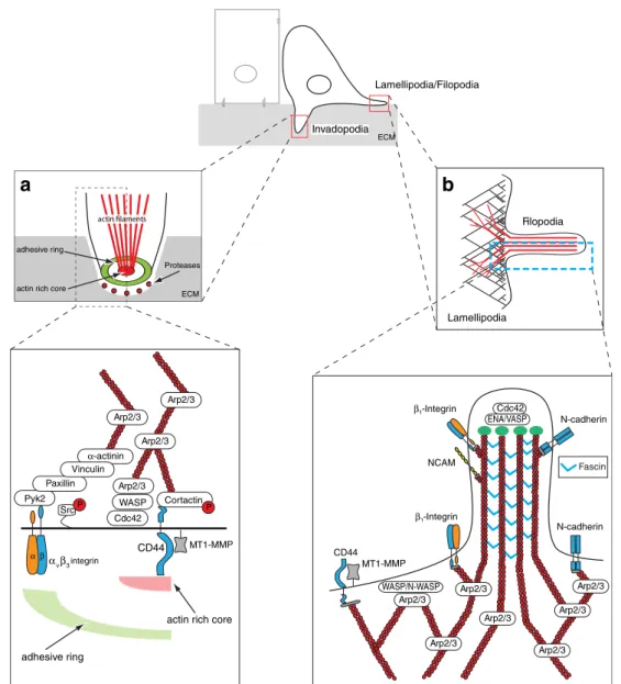
Lamellipodia are flat, sheet-like protrusion and they are
the main organelle for cell locomotion. The unbranched,
long actin filaments at the base of the lamellipodium are
progressing into a highly, lateral branched actin network at
the leading edge, giving lamellipodia their typical structure
(Fig.
3
b). Here, actin assembly and lateral branching
(dendritic nucleation) are mainly controlled by the Arp2/3
complex, a seven subunit protein and major initiator of
actin assembly. The activity of the Arp2/3 complex itself is
controlled by actin nucleation-promoting factors, such as
the N-WASP or the Scar/WAVE complexes which are
themselves recruited to and activated at the membrane by
Rac1 [
172
–
174
]. Increased expression of Arp2/3 and
WAVE2 has been shown to correlate with poor prognosis
in breast and liver carcinomas underlining the relevance of
lamellipodia-like structures in cancer progression [
175
,
176
]. Furthermore, the formation of lamellipodia is also
observed upon ErbB2-driven EMT in epithelial cells,
indicating that the formation of lamellipodia-like structures
underlies the increased invasiveness observed during EMT
[
177
].
Lamellipodia interact and attach to their environment via
different adhesion molecules, including integrins and
cadherins [
84
,
178
]. Also, CD44, the hyaluronan receptor
was identified to be in a complex with the protease
MT1-MMP in lamellipodial protrusions [
179
]. Here, MT1-MMP
binds directly to CD44 and mediates its proteolytic
cleavage, thereby stimulating migration (Fig.
3
b).
In contrast, filopodia are rod-like extension consisting of
tight bundled actin fibers which penetrate into the
surrounding environment originating from the basis of
lamellipodia. Filopodia can be considered as a sensory
organ of the cell that are used to detect and assimilate
signals like chemoattractants or nutrients released from e.g.
blood vessels. Interestingly, metastatic cells are rich in
filopodia-like structures, which correlates with their
inva-siveness [
180
,
181
]. Filopodia formation is based on
non-branched, processive actin assembly, controlled by fascin,
diaphanous and Ena/VASP [
173
,
182
] (Fig.
3
b). Actin
bundling via fascin, diaphanous and Ena/VASP, on the
other hand, is controlled by Rac and Cdc42. Fascin
upregulation correlates with poor prognosis in different
cancer types, including gastric cancer, lung cancer and
breast cancer [
183
–
185
]. Notably, fascin is a direct target
the
β-catenin/Tcf signaling pathway, which is active in the
process of EMT as well [
71
]. Recently, it has been reported
that filopodia can transform into lamellipodia by initiating
dendritic actin nucleation, characteristic for lamellipodia
formation, demonstrating that both filopodia and
lamelli-podia are highly interactive, inter-convertible structures
[
186
].
7.4 Podosomes and invadopodia
To move into or through a three-dimensional matrix, the
real life situation, cells have to gain the capability to
remodel the ECM via the expression of proteinases. In the
last few years, actin-rich protrusion with ECM proteolytic
activity, called invadopodia, have drawn particular attention
in the context of cancer cell invasion. Invadopodia are
considered the transformed counterparts of podosomes,
which are formed by non-transformed yet highly migratory
cells, such as macrophages, dendritic cells, osteoclasts, and
activated smooth muscle and endothelial cells. The exact
functions of podosomes are still rather elusive, yet it seems
that they are formed when cell adhesion junctions and
matrix needs to be concomitantly degraded. This may be
the case, when immune cells like monocytes or dendritic
cells have to cross tissue boundaries or during bone
resorption by osteoclasts for the control of bone
homeosta-sis [
187
]. The similarities or non-similarities of podosomes
and invadopodia are still under debate, yet the most striking
differences between both lies in the degree of ECM
degradation and their half-lives. Invadopodia are strong
degraders of ECM and persist for up to one h, whereas
podosomes exhibit less ECM degradation activity and are
stable only for minutes. Common for invadopodia and
podosomes is their architecture. They share an actin
filament-rich core containing the actin assembly machinery
(WASP, cortactin and Arp2/3) and a multimeric protein
complex surrounding the actin core, consisting of integrins
and integrin-associated proteins like vinculin, talin and
paxillin (Fig.
3
a). The multimeric adhesive ring complex is
connected to the actin core via radial actin filaments [
188
–
191
].
Invadopodia of invading tumors cells mediate
proteoly-sis of the ECM via the expression of different MMP, the
most prominent being MT1-MMP collagenase and one of
its direct targets, MMP2 gelatinase [
192
]. Invadopodia have
been identified in numerous cancer cell lines, including
malignant melanoma, breast cancer, glioma, and head and
neck cancer [
193
–
196
]. The formation of invadopodia can
be initiated by various signals, for example by EGF, HGF
or TGFβ-induced signal transduction [
193
,
197
,
198
] or by
α
6β
1-integrin engagement [
199
]. Invadopodia-inducing
Lamellipodia Filopodia Pyk2 α-actinin Arp2/3 Cdc42 Cortactin Paxillin Vinculin Src CD44 αvβ3integrin Arp2/3 ECM adhesive ring ENA/VASP ECM Invadopodia Lamellipodia/Filopodia adhesive ring
actin rich core
Proteases actin rich core
β1-Integrin NCAM WASP/N-WASP MT1-MMP CD44 Arp2/3 Arp2/3 Arp2/3 Arp2/3 Arp2/3 N-cadherin Arp2/3 Cdc42 Arp2/3 N-cadherin Fascin Arp2/3 Arp2/3 WASP β1-Integrin MT1-MMP P P
a
b
α βFig. 3 Upon completion of EMT, spindle-shaped, invasive cells migrate and invade into the surrounding environment by utilizing (i) membrane protrusion like lamellipodia and filopodia for horizontal cell movement and (ii) podosome-derived invasive structures, inva-dopodia, for ventral invasion. a Invadopodia-mediated invasion. Invadopodia are actin and phosphotyrosine-rich membrane protrusions with a high extracellular matrix degrading capacity. Invadopodia consist of an outer adhesive ring and a central actin-rich core. The non-receptor tyrosine kinase Src, the actin binding protein cortactin and the membrane bound matrix metalloproteinase MT1-MMP play an essential role in the formation and function of invadopodia. Integrins (likeαvβ3-integrin), along with their cytoplasmic interaction partners, are positioned in the adhesive ring where they mediate adhesion. The actin organization and nucleation is controlled by the Arp2/3 protein complex (adapted from Block et al. [191]). b
Lamellipodia/filopodia-mediated migration. Lamellipodia are the major organelles for cell movement and build upon highly branched dendritic actin networks, which are initiated by the nucleation promoting factors WASP/N-WASP and the Arp2/3 complex. Lamelli-podia interact with their environment via N-cadherin, β1-integrin or the hyaluronan receptor CD44, the latter also important for the correct positioning of the membrane-bound MT1-MMP. Filopodia are stiff, rod-like extension formed by tightly bundled actin filaments. They function as sensory organs of cells that penetrate into the surrounding microenvironment. They originate from the root of lamellipodia and are initiated and controlled by the concerted activity of the RhoGTPase Cdc42 and nucleation-promoting factor ENA/VASP. The major determinant for actin bundling and filopodia morphology is fascin. Filopodia interact with their environment via the cell adhesion molecules N-cadherin, NCAM, andβ1-integrins