RESEARCH OUTPUTS / RÉSULTATS DE RECHERCHE
Author(s) - Auteur(s) :
Publication date - Date de publication :
Permanent link - Permalien :
Rights / License - Licence de droit d’auteur :
Bibliothèque Universitaire Moretus Plantin
Institutional Repository - Research Portal
Dépôt Institutionnel - Portail de la Recherche
researchportal.unamur.be
University of Namur
Human epidermal keratinocytes upregulate expression of the prolactin receptor after
the onset of terminal differentiation, but do not respond to prolactin
Poumay, Yves; Jolivet, Geneviève; Pittelkow, Mark; Herphelin, Françoise; De Potter, Isabelle
Y.; Mitev, Vanio; Houdebine, Louis-Marie
Published in:
Archives of biochemistry and biophysics
Publication date: 1999
Document Version
Publisher's PDF, also known as Version of record
Link to publication
Citation for pulished version (HARVARD):
Poumay, Y, Jolivet, G, Pittelkow, M, Herphelin, F, De Potter, IY, Mitev, V & Houdebine, L-M 1999, 'Human epidermal keratinocytes upregulate expression of the prolactin receptor after the onset of terminal differentiation, but do not respond to prolactin', Archives of biochemistry and biophysics, vol. 364, no. 2, pp. 247-253.
General rights
Copyright and moral rights for the publications made accessible in the public portal are retained by the authors and/or other copyright owners and it is a condition of accessing publications that users recognise and abide by the legal requirements associated with these rights. • Users may download and print one copy of any publication from the public portal for the purpose of private study or research. • You may not further distribute the material or use it for any profit-making activity or commercial gain
• You may freely distribute the URL identifying the publication in the public portal ?
Take down policy
If you believe that this document breaches copyright please contact us providing details, and we will remove access to the work immediately and investigate your claim.
Human Epidermal Keratinocytes Upregulate Expression
of the Prolactin Receptor after the Onset of Terminal
Differentiation, but Do Not Respond to Prolactin
Yves Poumay,*,1
Genevie`ve Jolivet,† Mark R. Pittelkow,‡ Franc¸oise Herphelin,* Isabelle Y. De Potter,* Vanio Mitev,§ and Louis-Marie Houdebine†
*De´partement Histologie-Embryologie, Faculte´s Universitaires Notre-Dame de la Paix, B-5000 Namur, Belgium; †Unite´ de
Diffe´renciation Cellulaire, INRA, F-78352 Jouy-en-Josas, France; ‡Department of Dermatology, Mayo Clinic, Rochester, Minnesota 55905; and §Department of Biochemistry, University of Sofia, 1431 Sofia, Bulgaria
Received November 4, 1998, and in revised form January 22, 1999
Growing and differentiating keratinocytes maintain the epidermal barrier. This is partly controlled by growth factors and hormones. Prolactin (PRL) is named after its hormonal role in mammals during lac-tation, but is found in all vertebrates where PRL ex-erts various effects. In serum-free keratinocyte cul-tures, PRL was thought to be the factor responsible for the proliferative effect of bovine pituitary extract. Here, we evaluated PRL as a clonogenic factor for keratinocytes and found no mitogenic activity. Study-ing the expression of the PRL receptor by keratino-cytes, we found the receptor upregulated only after culture confluence, in differentiating keratinocytes, but we were unable to detect any cellular response to PRL. The hormone does not alter the gene expression of either early (suprabasal keratin) or late (involucrin) differentiation markers by keratinocytes. Accord-ingly, no activation of the transcription factor Stat5 by PRL can be detected in keratinocytes, Stat5 being nev-ertheless detected by Western blot. © 1999 Academic Press
Key Words: human epidermal keratinocyte;
prolac-tin receptor; growth and differentiation; Stat5.
The maintenance of the human epidermal barrier results from the growth and differentiation of keratin-ocytes and is under the control of multiple environmen-tal factors including growth factors and hormones (1). Beside the well-known mitogenic effects of the epider-mal growth factor (EGF) and insulin on keratinocytes,
the development of serum-free culture conditions has identified bovine pituitary extracts (BPE) as a source of factors stimulating the proliferation of these epidermal cells (2). Prolactin (PRL) has been suspected of being responsible for the in vitro stimulating effect of BPE because proliferation studies and binding studies using purified PRL have suggested responsiveness of the epi-dermal cells to this hormone (3).
Historically, PRL is a hormone named after its highly specialized role in mammals where PRL induces milk production by the mammary gland (4). However, PRL is systematically found in all vertebrates where the hormone produces various physiological responses, including growth-promoting activity for some organs, tissues, and cells (4 –7). Other observations have fur-ther suggested that PRL may be involved in coordina-tion of developmental processes (8).
In skin, epidermal keratinocytes, which are embry-ologically closely related to cells of the mammary gland, and cells of the cutaneous immune system have been proposed as potential targets for PRL (9). PRL in
vivo clearly induces the growth of the mammary gland
together with an abundant milk production (4). In cell culture though, PRL has only marginal effects on the proliferation of mammary epithelial cells (4, 10, 11), but is fully lactogenic, indicating that in vivo the hor-mone exerts its growth activity on the mammary epi-thelium through an as yet unidentified relay (4). PRL has been reported to induce the growth of nonmam-mary epithelial cells in culture (9, 12), and to stimulate specific peptide production by thymic epithelial cells. This cell type shares several features, including kera-tinization, with keratinocytes (9). Besides various ef-fects on epithelia, PRL is further involved in the
pro-1To whom correspondence and reprint requests should be
ad-dressed. Fax:132-81-724272. E-mail: [email protected].
0003-9861/99 $30.00 247
Copyright © 1999 by Academic Press
All rights of reproduction in any form reserved.
liferation of lymphocytes during an immune response (6, 7, 9).
These effects of PRL may be part of the systemic action of the circulating hormone, but may alterna-tively result from local production of PRL, particularly by mesenchymal cells found in the vicinity of respon-sive epithelia or immune cells (9). In skin for instance, the production of PRL by dermal fibroblasts, as it has been demonstrated recently in vitro (13), may result in the stimulation of neighbouring cells, including epider-mal keratinocytes.
When isolated and cultured on plastic, keratinocytes require stimulation by EGF or BPE to initiate clonal growth (14), but at higher cell density, keratinocytes can grow by autocrine stimulation (15). Then, when keratinocytes reach confluence under autocrine growth, their differentiation is triggered as revealed by the expression of the suprabasal keratins 1 and 10 (16). In the present study, we have reevaluated in serum-free culture conditions, whether PRL can stimulate the clonal growth of human keratinocytes. We further studied the expression of the PRL receptor by prolifer-ating and differentiprolifer-ating keratinocytes during auto-crine stimulation, and investigated the activation of the Stat5 transcription factor (17–19) and the expres-sion of differentiation markers in keratinocytes treated with PRL. To our surprise, cultured keratinocytes con-trol the expression of the PRL receptor, but do not respond to treatment with PRL.
MATERIALS AND METHODS
Cell culture and PRL treatment. Human adult normal skin and breast samples were obtained at plastic surgery (Dr. B. Bienfait, Clinique St. Luc, Namur-Bouge). Keratinocytes were isolated by the trypsin float technique as described (14), and primary cultures were initiated in KGM-2 medium (BioWhittaker). Keratinocytes har-vested from trypsinized primary proliferating subconfluent cultures were then plated into secondary cultures at 5–103 103cells/cm2in
the same medium. All experiments were performed on cells cultured in autonomous growth conditions (16), which means that, when approximately 40% of the culture substratum were covered by cells, the cultures were washed repeatedly with KBM-2 medium prepared by excluding bovine pituitary extract, insulin, transferrin, epineph-rine and EGF from the KGM-2 culture medium. Cultures were refed every other day with KBM-2 medium until treatment with PRL and analysis. The clonal growth assays were performed as described previously (14). HC11 mouse mammary epithelial cells were cul-tured in RPMI-1640 (BioWhittaker) with 10% fetal calf serum, 5 mg/ml insulin and 10 ng/ml EGF. MDA-453 human breast cancer cells were cultured in DMEM (BioWhittaker) with 10% fetal calf serum. Human PRL was obtained from Sigma (St. Louis, MO) or from the National Hormone and Pituitary Program (NIH, Bethesda, MD).
Poly(A)RNA isolation and Northern blot analysis. Poly(A)RNA was isolated and analyzed by Northern blotting as described previ-ously (16). The probes used to analyze the epidermal differentiation are cDNAs specific for the human basal keratin 14 (K14) or specific for the suprabasal keratins 1 (K1) or 10 (K10) (20), and cDNA specific for the human involucrin (21). The membrane was also hybridized with the 36B4 cDNA probe for the constitutively expressed human
acidic ribosomal phosphoprotein PO (22) to assure equivalent load-ing and transfer of RNA.
PRL receptor binding assay. Ovine PRL obtained from the Na-tional Hormone and Pituitary Program (NIH, Bethesda, MD) was used for iodination and competition in the binding assay. Radioiodin-ation with the lactoperoxidase method yielded specific activity of 53 106cpm/
mg. The binding assay was performed on subconfluent and confluent keratinocytes, MDA-453 cells and HC11 cells grown in 35-mm petri dishes. Briefly, the cells were washed twice in PBS then were incubated at room temperature for 4 h in 0.1% BSA–PBS containing PRL. For determination of total binding, 30 ng/ml of
125
I-PRL were prepared in each dish. Nonspecific binding was deter-mined by competition with 3 mg/ml of unlabeled PRL. After this incubation, the cells were washed twice in 0.1% BSA–PBS and then dissolved in 3% SDS before measurement of the cell-associated ra-dioactivity in a gamma counter. The results are presented as means6 SEM of replicates and analyzed using unpaired Student’s t test and P, 0.05.
Analysis by RT-PCR of the expression of the long form of the PRL receptor. Total RNA was isolated from superficial, mostly epider-mal, normal skin samples obtained with a keratome and from nor-mal breast tissue as described (23). Total RNA from breast tissue and poly(A)RNA from MDA-453 human breast cancer cells were used as positive controls for the mRNA expression of the long PRL receptor. Respectively, 1 and 0.1mg of total RNA and poly(A)RNA were used in the RT-PCR, and the reaction was performed using the GeneAmp RNA PCR Core kit (Perkin–Elmer), following instructions by the manufacturer. The cDNAs were amplified by 30 cycles and an am-plification product exhibiting the expected size of 889 bp was ob-tained with 59-ACTATGAGGACTTGCTGGTGGAGTATTT-39 as sense primer, and 59-CACTTGCTTGATGTTGCAGTGAAGTT-39 as antisense primer (5). Identity of the 889-bp cDNA fragment with the cDNA of the human PRL receptor was further assessed by automatic sequencing (24).
Preparation of cellular extracts, electrophoretic mobility shift assay and Western blot analysis of Stat5a and Stat5b expression. The methods used to detect Stat5 proteins and Stat5 activation have been described (18, 19). For the electrophoretic mobility shift assay, the probe used was a double-stranded oligonucleotide containing the sequence TGGACTTCTTGGAATTAAGG. HC11 mammary epithe-lial cells treated with PRL for 15 min were used as positive controls for the activation of Stat5. Identity of the DNA-binding transcription factor activated by PRL in HC11 was further established by super-shift experiment using two monoclonal antibodies to Stat5a and Stat5b (respectively from clone ST5a-2H2 and clone ST5b-10G1) purchased from Zymed Laboratories (San Francisco, CA).
For the Western blot analysis, the same monoclonal antibodies to Stat5a and Stat5b were utilized. Positive controls were provided by CHO cells expressing both Stat5a and Stat5b.
RESULTS
Clonal Growth of Human Epidermal Keratinocytes
No clonogenic potential of PRL on keratinocytes plated at clonal cell density (500 cells/dish) was de-tected when adding PRL alone at increasing concen-trations (0.1–50 ng/ml) to the keratinocyte growth medium containing insulin (5 mg/ml) but no EGF (Fig. 1, row I). Furthermore, no synergistic effect of PRL (0.1–10 ng/ml) with the clonogenic potential of EGF was either observed by addition of the hormone to the same culture medium containing suboptimal concentration (0.1 ng/ml) of EGF (Fig. 1, row II). These observations are not in accordance with
sults obtained by others with cultures plated at higher keratinocyte densities (3) and do not support the hypothesis proposing that PRL may be the com-ponent responsible for the previously reported clono-genic potential of BPE (14).
Expression of the PRL Receptor mRNA in Human Skin and Cultured Keratinocytes
By RT-PCR, RNA expression of the PRL receptor (24) in human breast cancer MDA-453 cells and in
FIG. 1. Clonal growth assay. Keratinocytes (500 cells/dish) were cultured in absence of BPE in medium containing insulin (5mg/ml) and either containing no EGF (I) or EGF at 0.1 ng/ml (II). No human PRL was added to the medium (A), or added at 0.1 ng/ml (B), 1 ng/ml (C), 10 ng/ml (D) and 50 ng/ml (E). After 10 days, culture plates were fixed with 4% formaldehyde and stained with crystal violet (0.2%).
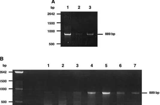
FIG. 2. RT-PCR analysis of the PRL receptor expression. (A) RT-PCR was performed on 0.1mg of poly(A)RNA from human breast cancer MDA-453 cells (lane 1) or on 1mg of total RNA from normal superficial human skin (lane 2) or from normal human breast tissue (lane 3) using specific primers (see Materials and Methods). (B) RT-PCR was identically performed on 0.1-mg samples of poly(A)RNA prepared from autonomous subconfluent keratinocyte cultures 4 days (lane 1) or 2 days (lane 2) before confluence of parallel cultures, prepared from cultures stopped on the first day of confluence (lane 3), or prepared from keratinocyte cultures 2 days (lane 4), 4 days (lane 5), and 6 days (lane 6) after confluence was reached. Amplification product produced by RT-PCR identically performed on RNA from human breast tissue was run on the same gel (lane 7) in order to identify unequivocally the fragment amplified from keratinocytes.
normal breast tissue was verified (Fig. 2A, lanes 1 and 3, respectively) and a lower but consistent expression was similarly detected in human skin biopsies ob-tained by the use of a keratome in order to mainly analyze expression in the epidermis (Fig. 2A, lane 2). Expression of the PRL receptor by cultured epidermal keratinocytes was also studied by RT-PCR. Keratino-cyte cultures (10 3 103
cells/cm2
) were initiated in parallel. When conditions of autonomous growth were reached, poly(A)RNA samples were extracted every other day, and the growth-state was determined by phase contrast microscopy in order to detect confluence of the cells. While poorly expressed by subconfluent keratinocytes (Fig. 2B, lanes 1 and 2) and by keratin-ocytes analyzed on the day they become confluent (Fig. 2B, lane 3), the PRL receptor was repeatedly upregu-lated during the 6 days following confluence of the culture, with a peak at 4 days (Fig. 2B, lanes 4 – 6). Since the confluence of cultured autonomously growing keratinocytes is concomitant with the induction of the suprabasal keratins 1 and 10 (16), our results suggest that the expression of the PRL receptor is upregulated at the onset of differentiation in keratinocytes, but
remains low in proliferating cells. Expression of the alternative short form of the PRL receptor (17) has been also investigated by RT-PCR using described primers (5), but was not found in cultured keratino-cytes (data not shown).
Measurement of125
I-PRL Binding on Cultured Cells
To assess whether the expression of the PRL recep-tor transcript at culture confluence regulates specific binding sites for PRL on human cultured keratino-cytes, we measured total and non specific binding of
125
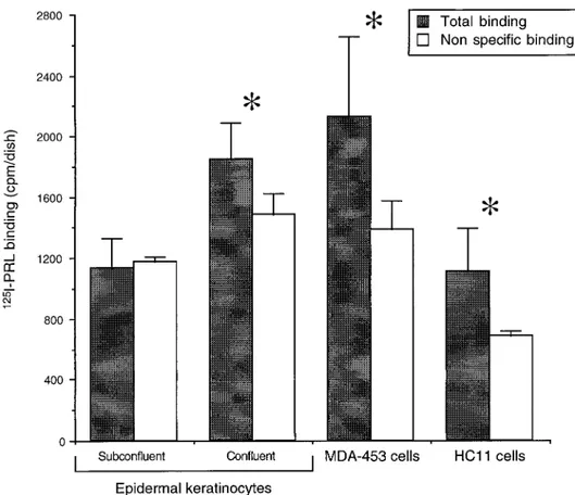
I-PRL on subconfluent epidermal keratinocytes and compared those results with the same binding on con-fluent differentiating keratinocytes, on MDA-453 breast cancer cells and on HC11 mammary cells (Fig. 3). The total binding of125
I-PRL in subconfluent kera-tinocytes does not differ from the non specific binding, suggesting absence of the PRL receptor in this epider-mal cell phenotype. On the other hand, when the total and non specific125
I-PRL bindings were measured and compared on differentiating confluent keratinocytes, a significant difference was seen, revealing that PRL
FIG. 3. PRL binding on cultured cells. Subconfluent (n5 3) and confluent (n 5 5) human epidermal keratinocytes, MDA-453 human breast
cancer cells (n5 3), and HC11 mouse mammary epithelial cells (n 5 3) were tested for total binding of125
I-PRL (30 ng/ml) in replicated
culture dishes. The data represent the mean6 SEM. Nonspecific binding was measured in the same number of dishes by including a
hundredfold excess of unlabeled PRL in the binding medium and compared with the measurement of total binding in identical cultures using unpaired Student’s t test. *P, 0.05.
receptors were present on these cells. Similarly study-ing MDA-453 and HC11 cells, we also found a signifi-cant difference between the total and non specific bind-ing, confirming presence of the PRL receptor on these cells (Fig. 3).
Treatment of Differentiating Keratinocytes with PRL
Because the PRL receptor is apparently expressed by keratinocytes undergoing epidermal differentiation at culture confluence, autonomous cultures were treated for 24 h with PRL (0.1–100 ng/ml), between 2 to 4 days after the confluence of keratinocytes had been de-tected. Unlike EGF and other factors of the same fam-ily activating receptors with tyrosine kinase activity (16, 17), PRL does not alter the mRNA expression of keratin 1 (Fig. 4) and keratin 10 (data not shown) in cultures of differentiating keratinocytes. PRL neither modifies the expression of involucrin, another marker of epidermal differentiation, or the expression of kera-tin 14 compared with the expression of 36B4 (Fig. 4).
Absence of Stat5 Activation by PRL in Epidermal Keratinocytes
The electrophoretic mobility shift and supershift as-says (Fig. 5) confirm that Sta5 can be activated in mammary HC11 cells by treatment with PRL for 15 min (Fig. 5A, lanes 1 and 2; Fig. 5B, lanes 1 and 2). The supershift experiment indicates that both forms Stat5a and Stat5b of the transcription factor are activated in HC11 cells by PRL. Conversely, treatment with PRL
(0.1–100 ng/ml) for 15 min of epidermal keratinocytes 2 days after confluence of the autonomous cultures does not activate Stat5 (Fig. 5, lanes 3–7), and prolongation of the treatment with PRL (10 ng/ml) up to 24 h does not activate Stat5 further (data not shown).
In order to assess whether epidermal keratinocytes express Stat5, nuclear and cytosolic protein extracts prepared from keratinocytes, either untreated or treated with PRL (10 ng/ml) for 15 min, were analyzed by Western blot using antibodies that specifically
rec-FIG. 5. Electrophoretic mobility shift assay of Stat5 activation. (A) HC11 cells were cultured for 24 h in absence of fetal calf serum and then treated (lane 1) or not (lane 2) with PRL for 15 min before preparation of nuclear extracts. Autonomous cultures of keratino-cytes were analyzed 2 days after confluence, when the PRL receptor is upregulated, and were kept untreated (lane 3) or treated for 15 min with 0.1 ng/ml (lane 4), 1 ng/ml (lane 5), 10 ng/ml (lane 6), and 100 ng/ml (lane 7) of human PRL. Then nuclear extracts were pre-pared and analyzed by electrophoretic mobility shift assay of the probe containing the consensus sequence recognized by Stat5 (see Materials and Methods). The arrow on the left side indicates the position of the probe recognized by Stat5. (B) To assess identity of the factor detected in the nuclear extracts only after incubation with PRL (A and lanes 8 and 9), a supershift experiment using monoclonal antibodies specific to Stat5a (lane 10) and Stat5b (lane 11) forms of the factor was performed on nuclear extracts (3mg/lane) from HC11 cells treated with PRL. Both antibodies induce a supershift of the factor detected in HC11 treated with PRL (lanes 10 and 11, right arrow), compared with the shift obtained in absence of antibodies (lane 8, left arrow). The asterisks label residual Stat5b (lane 10) and Stat5a (lane 11).
FIG. 4. Northern blot analysis of the mRNA expression of epider-mal differentiation markers by keratinocytes. Autonomous cultures of keratinocytes were used 2 to 4 days after the confluence was reached and either kept untreated (lane 1) or treated with human PRL at 0.1 ng/ml (lane 2), 1 ng/ml (lane 3), 10 ng/ml (lane 4), and 100 ng/ml (lane 5) for 24 h. Poly(A)RNA was then harvested from each culture and used for Northern blot hybridization with probes specific for keratin 1, keratin 14, involucrin, or 36B4 transcripts.
ognize the Stat5a and Stat5b forms of the transcription factor. Nuclear extracts of CHO cells were used as controls. Both forms of the transcription factor were found in autonomous confluent (Fig. 6), but also sub-confluent (data not shown) cultures of keratinocytes. Treatment of the keratinocytes with PRL (Fig. 6, lanes 2 and 5) did not induce any detectable relocalization of Stat5 between the cytosolic and nuclear cell compart-ments.
DISCUSSION
The involvement of PRL in skin physiology and pa-thology, more particularly in the control of the epider-mal keratinocyte, has been strongly suggested by both physiological and pathological observations (9), but so far whether PRL can induce signalling in keratinocytes has received only little attention. In the present work, we demonstrate for the first time that the PRL receptor is clearly expressed by cultured keratinocytes but only in confluent cultures undergoing differentiation. We also show that keratinocytes express the Stat5a and Stat5b proteins known as the main targets of the PRL-induced signalling pathway in mammary epithelial cells (4, 17).
On the other hand, we found no clonogenic effect of PRL on cultured keratinocytes, a result that differs from observations made previously by others (3) who found that PRL stimulates the proliferation of keratin-ocytes and who suggested that PRL could be the ele-ment of BPE responsible for its mitogenic effect on keratinocytes. However, our results excluding PRL as a clonogenic factor for keratinocyte concur with more
recent studies that identified a novel keratinocyte mi-togen of 95 kDa, unrelated to PRL, as the pituitary factor acting on keratinocyte growth (25). Then how could the results by Girolomoni et al. (3) be explained? In their proliferation assay, those authors used higher keratinocyte densities (5–103 103
cells/cm2
) than the density used in clonal assay (500 cells/60-mm diameter dish). Since keratinocytes at sufficient density produce growth factors with autocrine and paracrine effects (15), one may postulate synergistic effect of PRL with other factors in their results (although we found no synergy between PRL and EGF in our clonal assay). If PRL is really a growth factor for the epidermis as suspected from previous in vivo observations (9), it is surprising to detect no growth activity of PRL on cul-tured keratinocytes. Interestingly, the same discrep-ancy was noticed already between the effect of PRL in
vivo and in vitro on cells of the mammary gland (4).
Thus, we can speculate, by analogy with the closely related mammary tissue, that PRL exerts its in vivo epidermal action through an as yet unknown mecha-nism, but this mechanism is absent from cell culture conditions.
PRL was also proposed as a growth factor for kera-tinocytes because ligand-binding assays identified high affinity specific binding sites on those cells (3). By RT-PCR and binding measurements, we confirm ex-pression of the PRL receptor in differentiating keratin-ocytes but exclude its expression in subconfluent pro-liferating keratinocytes. Also in the poorly differenti-ated skin of 2-day-old mice, no RT-PCR signal for PRL receptor has either been reported (8). Again, this dis-crepancy with the results of Girolomoni et al. (3) can be explained. Since their binding assays were done only with keratinocytes freshly isolated from adult skin, they have consequently collected differentiating cells expressing the PRL receptor, whereas proliferating keratinocyte cultures might reveal negative in such binding assays in accordance with our observations.
In the mammary gland, activation of the PRL recep-tor by PRL results in the induction of expression of milk protein genes and this differentiating event re-sults from activation, through the JAK-Stat pathway, of mainly the Stat5 transcription factor (4, 17, 26). We show here that, even though the Stat5a and Stat5b proteins are expressed by epidermal keratinocytes, these proteins cannot be activated as a downstream cellular event following treatment of these cells with PRL. Of course, this result was expected in proliferat-ing keratinocytes where the expression of the PRL receptor is minimal, but the absence of Stat5 activation is more surprising in differentiating cultures express-ing the PRL receptor. One possible explanation for the absence, studied by electrophoretic mobility shift as-say, of Stat5 activation by PRL could probably be found as a defect in the signalling cascade downstream of the
FIG. 6. Western blot analysis of the protein expression of Stat5a and Stat5b. Nuclear (lanes 1 and 2) and cytosolic (lanes 4 and 5) extracts from confluent keratinocytes untreated (lanes 1 and 4) or treated with PRL (10 ng/ml) for 15 min (lanes 2 and 5), and nuclear extracts from CHO cells (lane 3) were analyzed by Western blot using antibodies specific to Stat5a (upper panels) or Stat5b (lower panels). The position of ladder proteins are given with respective molecular weight in kDa.
PRL receptor and involving the Jak2 kinase. However, the absence of the DNA-binding capacity of Stat5 after treatment with PRL does not exclude that the Stat1 or Stat3 transcription factors could be activated instead of Stat5 (17). Alternatively, recent results suggest that tyrosine phosphorylation of Stat5 through interaction with Jak2 and nuclear translocation of the factor are two independently regulated phenomena (26), but here, our data indicate no defect in the transfer of Stat5 between the cytoplasm and the nucleus.
Finally, no particular skin defect has been reported recently in mice with Stat5a and/or Stat5b gene dele-tion (27), suggesting that Stat5 and its activadele-tion may not play essential roles in cutaneous development and physiology. Nevertheless, the hypothetical involve-ment of PRL and PRL receptor in epidermal biology and pathology (9) remains intact. Indeed, the lack of effect of PRL in vitro on keratinocytes may simply reflect, like in the mammary epithelial cells (4), the absence in culture of a relay existing in vivo (4), maybe through interaction with surrounding mesenchymal cells (13). On the other hand, the expression of ele-ments of the PRL-dependent signal transduction path-way in keratinocytes could be too low in order to permit signalling by PRL. Thereby, the regulated expression of the PRL receptor in keratinocytes may simply indi-cate a lost function of PRL (28) in the epidermis of mammals.
ACKNOWLEDGMENTS
The authors thank Dr. B. Bienfait (Clinique Saint-Luc, Bouge) for providing the skin specimens, and Drs. D. R. Roop (Houston) and R. L. Eckert (Cleveland) for their gift of keratins and involucrin cDNA probes. Special thanks are addressed to F. Dubois (Namur) and N. Daniel (Jouy-en-Josas) for help, respectively, in the radiola-beling of PRL and the PRL binding procedure. This work was sup-ported by travel grant Tournesol No. 97.082 (Actions inte´gre´es franco-belges). I. Y. De Potter holds a fellowship from the Fonds pour la Formation a` la Recherche dans l’Industrie et l’Agriculture (FRIA).
REFERENCES
1. Pittelkow, M. R. (1992) in The Biology of the Epidermis (Oh-kawara, A., and McGuire, J., Eds.), pp. 109 –122, Elsevier Sci-ence, Amsterdam.
2. Boyce, S. T., and Ham, R. G. (1983) J. Invest. Dermatol. 81, 33S– 40S.
3. Girolomoni, G., Phillips, J. T., and Bergstresser, P. R. (1993) J. Invest. Dermatol. 101, 275–279.
4. Houdebine, L. M. (1994) in Dairy Products in Human Health and Nutrition (Serrano Rios, M., Sastre, A., Perez Juez, M. A., Es-trala, A., and De Sebastian, C., Eds.), pp. 3–12, Balkema, Rot-terdam.
5. Nevalainen, M. T., Valve, E. M., Ingleton, P. M., Nurmi, M., Martikainen, P. M., and Ha¨rko¨nen, P. L. (1997) J. Clin. Invest.
99, 618 – 627.
6. Matera, L. (1996) Life Sci. 59, 599 – 614.
7. Pellegrini, I., Lebrun, J.-J., Ali, S., and Kelly, P. A. (1992) Mol. Endocrinol. 6, 1023–1031.
8. Brown-Borg, H. M., Zhang, F.-P., Huhtaniemi, I., and Bartke, A. (1996) Eur. J. Endocrinol. 134, 751–757.
9. Paus, R. (1991) Med. Hypotheses 36, 33– 42.
10. Hammond, S. L., Ham, R. G., and Stampfer, M. R. (1984) Proc. Natl. Acad. Sci. USA 81, 5435–5439.
11. Edery, M., McGrath, M., Larson, L., and Nandi, S. (1984) Endo-crinology 115, 1691–1697.
12. Wilson, P. D., and Horster, M. F. (1983) Am. J. Physiol. 244, C166 –C174.
13. Richards, R. G., and Hartman, S. M. (1996) J. Invest. Dermatol.
106, 1250 –1255.
14. Wille, J. J., Jr., Pittelkow, M. R., Shipley, G. D., and Scott, R. E. (1984) J. Cell. Physiol. 121, 31– 44.
15. Cook, P. W., Pittelkow, M. R., and Shipley, G. D. (1991) J. Cell. Physiol. 146, 277–289.
16. Poumay, Y., and Pittelkow, M. R. (1995) J. Invest. Dermatol. 104, 271–276.
17. Bole-Feysot, C., Goffin, V., Edery, M., Binart, N., and Kelly, P. A. (1998) Endocrinol. Rev. 19, 225–268.
18. Tourkine, N., Schindler, C., Larose, M., and Houdebine, L.-M. (1995) J. Biol. Chem. 270, 20952–20961.
19. Goupille, O., Daniel, N., Bignon, C., Jolivet, G., and Djiane, J. (1997) Mol. Cell. Endocrinol. 127, 155–169.
20. Roop, D. R., Krieg, T. M., Mehrel, T., Cheng, C. K., and Yuspa, S. H. (1988) Cancer Res. 48, 3245–3252.
21. Eckert, R. L., and Green, H. (1986) Cell 46, 583–589. 22. Laborda, J. (1991) Nucleic Acids Res. 19, 3998.
23. Chomczynski, P., and Sacchi, N. (1987) Anal. Biochem. 162, 156 –159.
24. Boutin, J.-M., Edery, M., Shirota, M., Jolicoeur, C., Lesueur, L., Ali, S., Djiane, J., and Kelly, P. A. (1989) Mol. Endocrinol. 3, 1455–1461.
25. Gonzalez-Castro, U., Castells-Rodellas, A., and Krueger, J. G. (1997) Arch. Dermatol. Res. 289, 309 –316.
26. Ali, S., and Ali, S. (1998) J. Biol. Chem. 273, 7709 –7716. 27. Teglund, S., McKay, C., Schuetz, E., Van Deursen, J. M.,
Stra-vopodis, D., Wang, D., Brown, M., Bodner, S., Grosvold, G., and Ihle, J. N. (1998) Cell 93, 841– 850.
28. Takada, M., Yai, H., Komazaki, S., and Takayama-Arita, K. (1996) J. Exp. Biol. 199, 2573–2578.