Publisher’s version / Version de l'éditeur:
Infection and Immunity, 85, 11, 2017-09-05
READ THESE TERMS AND CONDITIONS CAREFULLY BEFORE USING THIS WEBSITE. https://nrc-publications.canada.ca/eng/copyright
Vous avez des questions? Nous pouvons vous aider. Pour communiquer directement avec un auteur, consultez la première page de la revue dans laquelle son article a été publié afin de trouver ses coordonnées. Si vous n’arrivez pas à les repérer, communiquez avec nous à PublicationsArchive-ArchivesPublications@nrc-cnrc.gc.ca.
Questions? Contact the NRC Publications Archive team at
PublicationsArchive-ArchivesPublications@nrc-cnrc.gc.ca. If you wish to email the authors directly, please see the first page of the publication for their contact information.
Archives des publications du CNRC
This publication could be one of several versions: author’s original, accepted manuscript or the publisher’s version. / La version de cette publication peut être l’une des suivantes : la version prépublication de l’auteur, la version acceptée du manuscrit ou la version de l’éditeur.
For the publisher’s version, please access the DOI link below./ Pour consulter la version de l’éditeur, utilisez le lien DOI ci-dessous.
https://doi.org/10.1128/IAI.00557-17
Access and use of this website and the material on it are subject to the Terms and Conditions set forth at
Characterization of two novel lipopolysaccharide
phosphoethanolamine transferases in Pasteurella multocida and their
role in resistance to cathelicidin-2
Harper, Marina; Wright, Amy; St. Michael, Frank; Li, Jianjun; Deveson Lucas,
Deanna; Ford, Mark; Adler, Ben; Cox, Andrew D.; Boyce, John D.
https://publications-cnrc.canada.ca/fra/droits
L’accès à ce site Web et l’utilisation de son contenu sont assujettis aux conditions présentées dans le site
LISEZ CES CONDITIONS ATTENTIVEMENT AVANT D’UTILISER CE SITE WEB.
NRC Publications Record / Notice d'Archives des publications de CNRC:
https://nrc-publications.canada.ca/eng/view/object/?id=f807b5fe-1a3d-4807-856b-3171be15d54f
https://publications-cnrc.canada.ca/fra/voir/objet/?id=f807b5fe-1a3d-4807-856b-3171be15d54f
Characterization of Two Novel
Lipopolysaccharide Phosphoethanolamine
Transferases in Pasteurella multocida and
Their Role in Resistance to Cathelicidin-2
Marina Harper,aAmy Wright,aFrank St. Michael,bJianjun Li,b
Deanna Deveson Lucas,aMark Ford,cBen Adler,aAndrew D. Cox,bJohn D. Boycea
Infection and Immunity Program, Monash Biomedicine Discovery Institute and Department of Microbiology, Monash University, VIC, Australiaa; Vaccine Program, Human Health Therapeutics Portfolio, National Research
Council, Ottawa, ON, Canadab; CSIRO Australian Animal Health Laboratory, Geelong, VIC, Australiac
ABSTRACT The lipopolysaccharide (LPS) produced by the Gram-negative bacterial
pathogen Pasteurella multocida has phosphoethanolamine (PEtn) residues attached to lipid A, 3-deoxy-D-manno-octulosonic acid (Kdo), heptose, and galactose. In this report, we show that PEtn is transferred to lipid A by the P. multocida EptA homo-logue, PetL, and is transferred to galactose by a novel PEtn transferase that is unique to P. multocida called PetG. Transcriptomic analyses indicated that petL ex-pression was positively regulated by the global regulator Fis and negatively regu-lated by an Hfq-dependent small RNA. Importantly, we have identified a novel PEtn transferase called PetK that is responsible for PEtn addition to the single Kdo mole-cule (Kdo1), directly linked to lipid A in the P. multocida glycoform A LPS. In vitro
as-says showed that the presence of a functional petL and petK, and therefore the pres-ence of PEtn on lipid A and Kdo1, was essential for resistance to the cationic,
antimicrobial peptide cathelicidin-2. The importance of PEtn on Kdo1and the
identi-fication of the transferase responsible for this addition have not previously been shown. Phylogenetic analysis revealed that PetK is the first representative of a new family of predicted PEtn transferases. The PetK family consists of uncharacterized proteins from a range of Gram-negative bacteria that produce LPS glycoforms with only one Kdo molecule, including pathogenic species within the genera Vibrio, Bor-detella, and Haemophilus. We predict that many of these bacteria will require the ad-dition of PEtn to Kdo for maximum protection against host antimicrobial peptides.
KEYWORDS lipopolysaccharide, phosphoethanolamine transferase, Pasteurella
multocida, 3-deoxy-D-manno-octulosonic acid, cathelicidin, cationic antimicrobial
P
asteurella multocida is a Gram-negative bacterium present in the nasopharynx of many healthy animals, including domestic cats and dogs. However, when trans-mitted to susceptible hosts, including birds, cattle, pigs, rabbits, and humans, the bacterium can cause serious joint, soft tissue, and respiratory infections as well as life-threatening septicemia (1, 2). P. multocida is the causative agent of acute fowl cholera, a systemic disease of birds that causes significant losses to poultry industries worldwide. Our structural and genetic analyses of the lipopolysaccharide (LPS) pro-duced by a number of P. multocida field isolates and historical type strains revealed that a wide range of outer core LPS structures were produced by P. multocida; accordingly, strains could be classified into eight different LPS genotypes, L1 to L8 (3–5). Many fowl cholera outbreaks are caused by P. multocida serogroup A:L1 strains (capsule type A, LPS genotype L1); these strains produce two major virulence factors that are essentialReceived 7 August 2017 Returned for modification 24 August 2017 Accepted 29
August 2017
Accepted manuscript posted online 5
September 2017
Citation Harper M, Wright A, St. Michael F, Li J,
Deveson Lucas D, Ford M, Adler B, Cox AD, Boyce JD. 2017. Characterization of two novel lipopolysaccharide phosphoethanolamine transferases in Pasteurella multocida and their role in resistance to cathelicidin-2. Infect Immun 85:e00557-17.https://doi.org/10.1128/ IAI.00557-17.
Editor Guy H. Palmer, Washington State
University
Copyright © 2017 American Society for
Microbiology.All Rights Reserved. Address correspondence to Marina Harper, marina.harper@monash.edu.
crossm
on December 8, 2017 by National Research Council Canada
http://iai.asm.org/
for their ability to cause disease in chickens, namely, a hyaluronic acid capsule which confers serum resistance and an L1 LPS (6–9).
The structure of the LPS inner core is conserved across all P. multocida strains and contains 3-deoxy-D-manno-oct-2-ulosonic acid (Kdo), heptose (Hep), and glucose (Glu). Unusually, most P. multocida strains, including those belonging to LPS genotype L1, simultaneously produce two major LPS glycoforms, A and B, that have identical outer core structures but differ in the inner core region with respect to the number of Kdo residues (one in glycoform A, two in glycoform B) and glucose residues (two in glycoform A, one in glycoform B) and with respect to the number and position of phosphoethanolamine (PEtn) residues (Fig. 1). PEtn is a positively charged moiety that can be added in a nonstoichiometric manner at up to three positions in the P. multocida LPS inner core (Fig. 1): to lipid A (at the 4= position), to the phosphate on the 4 position of Kdo in glycoform A, or to the 3 position of the second Hep (Hep II).
The addition of PEtn to the inner core of the P. multocida LPS is predicted to vary according to the strain and to the growth conditions. Capillary electrophoresis–mass spectrometry (CE-MS) analysis of O-deacylated LPS (LPS-OH) isolated from P. multocida strain VP161 grown in vitro in liquid broth revealed the presence of glycoform B and two glycoforms representing glycoform A, either with or without a PEtn residue attached to the phosphorylated Kdo residue. Analysis of LPS isolated from VP161 cells grown on chocolate blood agar revealed two different lipid A molecules (lipid A-OH species at 952 atomic mass units [amu] and 1,075.0 amu), the larger of which corre-sponds to lipid A with a PEtn residue attached (3). However, when P. multocida strain
FIG 1 Schematic representation of the two LPS glycoforms, A and B, produced by P. multocida strains X73
and VP161 that belong to LPS genotype 1 (Heddleston serotype 1). LPS glycoforms A and B have identical outer core sugars and linkages (not shown) but differ in the inner core (within square) with respect to the number of 3-deoxy-D-manno-oct-2-ulosonic acid (Kdo), glucose, and phosphoethanolamine (PEtn) residues. The addition of phosphoethanolamine (PEtn) is nonstoichiometric, but PEtn may be present at the 4= position of lipid A, on the phosphorylated Kdo in LPS glycoform A, at the 3 position on the second inner core heptose, and at the 6 position on both galactose residues located at the terminal end of the LPS molecule. Strain VP161 produces LPS glycoforms with PEtn only on lipid A and Kdo-P. Strain X73 produces LPS glycoforms with PEtn on the lipid A, Kdo-P, the second heptose, and the terminal galactose residues. Residues are indicated as follows: -D-Gal, -D-galactose; -D-Glc, -D-glucose; L-␣-D-Hep, L-␣-D-heptose; Kdo, 3-deoxy-D-manno-oct-2-ulosonic acid; P, phosphate; PCho, phosphocholine; PEtn, phosphoethanolamine.
on December 8, 2017 by National Research Council Canada
http://iai.asm.org/
VP161 or X73 was grown in liquid broth, no PEtn was detected on the lipid A moiety of the LPS produced by either strain (3, 10).
As P. multocida LPS does not contain a repeating O-antigen, the outer core sugars are the most distal part of the LPS molecule. The outer core region of the L1 LPS produced by highly virulent fowl cholera strains VP161 and X73 consists of a single heptose with two galactose (Gal) residues attached at the 4 and 6 positions on the heptose. Each of the two Gal residues in both VP161 and X73 is decorated with phosphocholine (PCho) at the 3 position (3), and the Gal residues are further decorated with PEtn in the LPS produced by strain X73 (Fig. 1) (10).
The transferases responsible for the addition of PEtn to lipid A (EptA) and to the 3 position or 6 position on Hep II in the LPS inner core (Lpt-3 or Lpt-6, respectively) have been identified in other bacteria, including Haemophilus influenzae and Neisseria men-ingitis (11, 12). We previously identified an lpt3 homologue in P. multocida and showed that Lpt-3 was responsible for addition of PEtn to the 3 position on Hep II of the P. multocida LPS (13). In many strains, however, lpt3 is present as a pseudogene and Hep II is not decorated with PEtn. Moreover, although strain X73 has a functional lpt3 gene, X73 cells grown in vitro do not elaborate PEtn on Hep II, suggesting that expression of lpt3 is regulated in response to the environment.
In this study, we used bioinformatic analyses combined with structural and func-tional assays to identify and characterize three PEtn transferases responsible for PEtn addition to the various positions on the genotype 1 LPS. We identified two novel LPS-associated PEtn transferases, PetG (which is required for the addition of PEtn to the terminal galactose residues in P. multocida X-73 LPS) and PetK (which transfers PEtn to Kdo-P), and showed that PetK represents a new family of bacterial PEtn transferases. We also proved that the presence of PEtn on both lipid A and Kdo-P is critical for P. multocida to resist killing by the antimicrobial peptide, cathelicidin-2.
RESULTS
Bioinformatic analysis identifies two novel P. multocida proteins that belong to the PEtn transferase family. In order to identify novel P. multocida PEtn transferases,
we used the amino acid sequences of three previously characterized PEtn transferases to query the P. multocida genomes. Each of the lpt3 genes present in N. meningitis (Lpt-3Nm/NMB2010) and P. multocida (Lpt-3Pm/A0R63_02570) encodes a PEtn
trans-ferase that is responsible for addition of PEtn onto the 3 position of Hep II in the LPS inner core (13, 14). N. meningitidis gene NMB1638 encodes EptANm, which transfers PEtn
to lipid A (11). These transferases all belong to the sulfatase family of proteins and contain five transmembrane helices, indicative of inner membrane-associated proteins. The amino acid sequences of these PEtn transferases were used to search the genomes of P. multocida strains VP161 and X73. In total, three genes were identified in X73 that encoded proteins with significant identity with Lpt-3Pm, Lpt-3Nm, and/or EptANm,
namely, A0R63_03465, A0R63_06305, and A0R63_09570. Each of the encoded products contained predicted sulfatase domains and at least four transmembrane helices. Intact homologs of X73 genes A0R63_06305 and A0R63_09570 were identified in strain VP161 (99% identity). A homologue of A0R63_03465 that shared 99% identity at the nucleo-tide level was also present in VP161 but was a pseudogene due to a single base deletion at nucleotide 1071.
The amino acid sequences of the characterized P. multocida PEtn transferase, Lpt-3Pm, and the newly identified putative P. multocida PEtn transferases (X73 locus tags
A0R63_03465, A0R63_06305, and A0R63_09570, respectively) were used in a multiple alignment together with all the functionally characterized bacterial PEtn transferases, as well as a number of uncharacterized proteins that shared a high level of identity with one or more of these proteins (see Fig. S1 in the supplemental material). The alignment data were then used to build a phylogenetic tree (Fig. 2). The functionally characterized transferases included three EptA homologs for which the catalytic domain structure has been resolved, namely, EptANm(LptA) (15) and the plasmid-mediated Mcr-1, both of
which confer resistance to polymyxins in certain Gram-negative bacteria via the
on December 8, 2017 by National Research Council Canada
http://iai.asm.org/
tion of PEtn to lipid A (16, 17), and Campylobacter jejuni protein EptC (18), a multifunc-tional transferase that can add PEtn to a range of cell components, including lipid A, heptose within the LPS inner core, and flagella (19–21). The amino acid sequences of these proteins, their active sites, and the associated metal binding sites are highly conserved. The presumed catalytic nucleophile in EptANmis Thr280, in EptC is Thr266,
and in Mcr-1 is likely to be Thr285, as an Escherichia coli strain harboring a Mcr-1 with an Ala substitution at Thr285 lost resistance to polymyxin (15, 16, 18). The zinc coordination sites are Glu240/227/246, Asp452/427/465, and His 453/428/466 in EptANm, EptC, and Mcr-1, respectively (15, 16, 18).
FIG 2 Phylogenetic analysis of a representative number of known and predicted phosphoethanolamine (PEtn) transferases. Shaded
clusters represent five different PEtn transferase families as follows; blue, lipid A-specific PEtn transferases, including EptA; green, PEtn transferases specific for the 6 position of the second heptose (Hep II) in the inner core region of lipooligosaccharide/lipopolysaccharide (LOS/LPS); gray, PEtn transferases specific for the 3 position of Hep II in the inner core of LOS/LPS; yellow, PEtn transferases, including PetG (this study), specific for the addition of PEtn to galactose residues in LPS; pink, putative PEtn transferases predicted to transfer PEtn to 3-deoxy-D-manno-oct-2-ulosonic acid (Kdo), including the PEtn transferase PetP (this study). Labels indicate species, strain, and gene/locus tag, separated by hyphens. Bacterial species are indicated as follows; Acti.p, Actinobacillus pleuropneumoniae; Aero.s, Aeromonas salmonicida; Aggr.a, Aggregatibacter actinomycetemcomitans; Bp, Bordetella pertussis; Cj, Campylobacter jejuni; Ec, Escherichia coli; Hd, Haemophilus ducreyi; Hf, Haemophilus felis; Hh, Haemophilus haemolyticus; Hi, Haemophilus influenzae; Hp, Helicobacter pylori; Mh, Mannheimia haemolytica; Ng, Neisseria gonorrhoeae; Nm, Neisseria meningitidis; Pm, Pasteurella multocida; Sf, Shigella flexneri; Shew.p, Shewanella putrefaciens; ST, Salmonella enterica subsp. enterica serovar Typhimurium; Vc, Vibrio cholerae; Vh, Vibrio harveyi; Vp, Vibrio parahaemolyticus.
on December 8, 2017 by National Research Council Canada
http://iai.asm.org/
The multiple alignment data revealed that only four amino acids were conserved across all analyzed proteins. These were the Glu, Asp, and His residues (at 240, 452, and 453, respectively, in EptANm) that form the zinc coordination site and a His at the
position equivalent to 384 in EptANm. (Fig. 2; see also Fig. S1). The presumed Thr
catalytic residue (Thr 280 in EptANm) was present in 34 of the 46 sequences. In the other
12 proteins, including P. multocida A0R63_09570, a serine residue was present at this position; these proteins formed a well-separated branch in the phylogenetic tree (Fig. 2, pink-shaded proteins).
The lipid A-specific PEtn transferases from a range of species formed a distinct branch on the phylogenetic tree and included the protein encoded by P. multocida X73 gene A0R63_06305 (Fig. 2, blue-shaded proteins). Accordingly, we predicted that the protein encoded by this gene would add PEtn to lipid A; thus, we named this gene petL (phosphoethanolamine onto lipid A). The transferases, which are specific for the addition of PEtn to Hep residues in the LPS inner core, formed three distinct branches (Fig. 2); the first contained Lpt-3 PEtn transferases specific for the addition of PEtn to the 3 position of Hep II, including the previously characterized proteins produced by P. multocida strain X73, N. meningitidis, and N. gonorrhoeae; the second contained Lpt-6 transferases from N. meningitidis, N. gonorrhoeae, and H. influenzae specific for the transfer of PEtn to the 6 position of Hep II; and the third contained CptA transferases from members of the Enterobacteriaceae family that are specific for the transfer of PEtn to Hep I.
P. multocida X73 protein A0R63_03465 grouped closely only with other P. multocida proteins from strains P1059, P2100, and HN06 (Fig. 2, yellow shading). This cluster was from a main branch that included two PEtn transferases with distinctly different acceptor molecules: glucose-specific OpgE from E. coli (which transfers PEtn to osmo-regulated periplasmic glucans [OPG] [22]) and PptA from N. gonorrhoeae (strain FA1090) (which is responsible for the transfer of PEtn to pilin [23]). Also in this group were the predicted PEtn transferases PtdA and PtdB from Haemophilus ducreyi. The acceptor molecule targeted by these H. ducreyi PEtn transferases has not been identi-fied, but these proteins were not required for the addition of PEtn to lipid A (24). There are no reports of PEtn decoration of cell components other than LPS in P. multocida, and this bacterium does not produce OPG. However, an alignment of the A0R63_03465 amino acid sequence with that of OpgE from E. coli revealed that they shared 31% amino acid identity (91% coverage), suggesting that X73 gene A0R63_03465 may encode a hexose-specific PEtn transferase. We therefore hypothesized that A0R63_03465 was a unique PEtn transferase required for the addition of PEtn onto the galactose residues at the distal end of the X73 LPS and named the gene petG (phosphoethanolamine onto galac-tose).
Importantly, the phylogenetic analysis revealed a well-separated and large group of proteins with unknown functions that included P. multicoda X73 protein A0R63_09570 (Fig. 2). A0R63_09570 shared amino acid identity with putative homologs (66% to 88% identity, 97% to 99% coverage) encoded on the genomes of a large number of other species belonging to the Pasteurellaceae family, including those within the genera Actinobacillus, Aggregatibacter, Haemophilus, and Mannheimia. More distant orthologs were also identified in species within the Aeromonas, Bordetella, Shewanella, and Vibrio genera (amino acid identity ranging from 50% to 55%, approximately 95% to 99% coverage). Surprisingly, a search of available protein structures revealed that a structure (resolved to 1.95 Å; PDB code 3LXQ) was available for the Vibrio parahaemolyticus protein within this group but the protein was identified only as a member of the alkaline phosphatase superfamily and no additional information was published. Previ-ous analyses of the LPS produced by strains representing many of these genera revealed the presence of a single, usually phosphorylated Kdo molecule, with or without substitution with PEtn (3, 25–28). We therefore hypothesized that this cluster of proteins represented a new family of PEtn transferases that are specific for the addition of PEtn onto Kdo, and we named the X73 gene petK (phosphoethanolamine onto Kdo).
on December 8, 2017 by National Research Council Canada
http://iai.asm.org/
Construction of P. multicoda PEtn transferase mutants. In order to confirm the
function of the predicted LPS PEtn transferases, directed mutants were generated in two fowl cholera isolates, VP161 and X73. Inactivation of petL and petK was achieved by single-crossover mutagenesis of strain AL435 (29), a tetracycline-resistant derivative of VP161 expressing wild-type LPS, to generate mutant strains AL486 and AL569, respec-tively (Table 1). In addition, petL and petK were inactivated in VP161 using Targetron mutagenesis to generate strains AL1354 and AL2577, respectively (Table 1). Finally, a petG mutant was generated using Targetron intron insertion into strain X73, as this gene is a pseudogene in VP161, generating mutant strain AL1911 (Table 1). All mutants showed normal in vitro growth rates equivalent to that of the wild type in heart infusion (HI) broth.
LPS analysis of P. multocida PEtn transferase mutants. Both of the P. multicoda
wild-type strains, VP161 and X73, produced LPS glycoforms consistent with those previously published (3, 10). Similarly, parent strain AL435, used in the construction of single-crossover mutants, produced all of the LPS glycoforms observed in wild-type VP161 (Table 2), including two glycoforms previously determined to contain different lipid A-OH species (952 and 1,075 amu) (3), with the smaller of the two corresponding to lipid A-OH without PEtn and the larger corresponding to lipid A-OH with PEtn. Examination of the LPS produced by petL mutant AL486, generated by single-crossover
TABLE 1 Bacterial strains and plasmids used in this study
Strain or plasmid Relevant descriptiona Source or reference
Strains
E. coli DH5␣ deoR endA1 gyrA96 hsdR17(rk⫺mk⫹) recA1 relA1 supE44 thi-1 (lacZYA-argFV169) ⌽80lacZ ΔM15 F⫺ Bethesda Research Laboratories E. coli SM10 pir E. coli strain for propagation of pUA826 and its derivatives 30
P. multocida AL435 VP161 carrying a Tn916 insertion in gene pm1417; fully virulent 29 P. multocida AL486 Single-crossover mutant of petL in AL435, constructed using pAL287 This study P. multocida AL569 Single-crossover mutant of petK in AL435, constructed using pAL302 This study P. multocida AL1354 petL TargeTron mutant of VP161, generated with pAL701 This study
P. multocida AL1405 fis TargeTron mutant of VP161 31
P. multocida AL1561 VP161 with expression vector pAL99 This study
P. multocida AL1563 VP161 with complementing plasmid pAL789 encoding petG This study P. multocida AL1911 petG TargeTron mutant of strain X73, generated with pAL910 This study P. multocida AL2577 petK TargeTron mutant of VP161, generated with pAL1124 This study P. multocida AL2581 AL1354 with complementing plasmid pAL1143 encoding petL This study
P. multocida AL2583 AL1354 with expression vector pAL99 This study
P. multocida AL2601 AL2577 with vector pAL99S This study
P. multocida AL2622 AL2577 with complementing plasmid pAL1151encoding petK This study P. multocida VP161 LPS genotype 1, capsule serogroup A, virulent poultry isolate 32 P. multocida X73 LPS genotype 1, capsule serogroup A, virulent poultry isolate 33 Plasmids
pAL99 P. multocida expression plasmid (Kanr) 8
pAL99S P. multocida expression plasmid (Specr) 5
pAL287 Internal 1-kb fragment of VP161 petL cloned into pUA826 using primers BAP2569 and BAP2570 This study pAL302 Internal fragment of VP161 petK cloned into pUA826 using primers BAP3425 and BAP3426 This study pAL692 P. multocida-specific TargeTron vector, markerless intron (plasmid Specr) 31 pAL701 TargeTron vector pAL692 retargeted to petL in VP161 using primers BAP5870, BAP5871, and
BAP5872 and EBS universal primer
This study pAL789 Complete X73 petG gene cloned into expression plasmid pAL99 using primers BAP6116
and BAP6117
This study pAL953 P. multocida-specific TargeTron vector (plasmid Specr, intron Kanr) 5 pAL910 pAL692 TargeTron vector retargeted to petG in X73 using primers BAP6617, BAP6618,
and BAP6619
This study pAL1124 pAL953 TargeTron vector retargeted to petK in VP161 using primers BAP6715, BAP6716,
and BAP6717
This study pAL1143 Complete VP161 petL gene cloned into expression plasmid pAL99 using primers BAP7478
and BAP7479
This study pAL1151 Complete petK gene cloned into expression plasmid pAL99S using primers BAP7509
and BAP7510
This study pUA826 Mob⫹, R6K replicon, single-crossover mutagenesis vector (AprSpecr) 34
aApr, ampicillin resistance; Specr, spectinomycin resistance.
on December 8, 2017 by National Research Council Canada
http://iai.asm.org/
mutagenesis in strain AL435, revealed the loss of all charged ions representing the LPS glycoforms corresponding to the larger lipid A-OH species with PEtn. To confirm the result, the petL TargeTron mutant (AL1354; Table 1) was also examined. LPS analysis of this second mutant also revealed charged ions corresponding to LPS glycoforms that contained only the smaller lipid A species (Table 2) (3). Together, these data confirm that petL encodes the PEtn transferase required for the addition of PEtn to lipid A in P. multocida.
Previous structural data revealed that the LPS produced by X73 contains a PEtn residue on the 6 position of both galactose residues located at the distal end of the LPS molecule (10). The LPS structure produced by VP161 is identical to the X73 structure except that the LPS structure does not have the PEtn residues (3). Consistent with this finding was the fact that the predicted galactose-specific PEtn transferase petG gene was intact in strain X73 but was present as a pseudogene in VP161. To prove that PetG was the PEtn transferase responsible for the addition of PEtn to each of the terminal galactose residues on the X73 LPS, we examined the LPS produced by X73 petG TargeTron mutant AL1911 (Table 1). LPS structural analysis of AL1911 revealed that the LPS glycoform profile was highly similar to the profile from strain VP161, with the
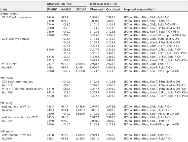
TABLE 2 Negative-ion CE-ES-MS data and proposed compositions of O-deacylated LPS and core oligosaccharide for P. multocida strainsa
Strain
Observed ion (m/z) Molecular mass (Da)
Proposed compositionb
(M-4H)4- (M-3H)3- (M-2H)2- Observed Calculated
Control strains
VP161* wild-type strain 744.0 992.4 2,980.1 2,978.8 2PCho, 3Hex, 4Hep, 2Kdo, lipid A-OH 749.0 999.6 3,000.9 3,000.8 2PCho, 4Hex, 4Hep, Kdo-P, lipid A-OH 774.0 1,033.0 3,101.0 3,101.9 2PCho, 3Hex, 4Hep, 2Kdo, lipid A-OH-PEtn 780.0 1,040.0 3,123.5 3,123.8 2PCho, 4Hex, 4Hep, Kdo-P, PEtn, lipid A-OH 780.0 1,040.0 3,123.5 3,123.8 2PCho, 4Hex, 4Hep, Kdo-P, lipid A-OH-PEtn 810.0 1,081.0 3,245.0 3,246.9 2PCho, 4Hex, 4Hep, Kdo-P-PEtn, lipid A-OH-PEtn X73* wild-type strain 1,033.8 3,104.4 3,101.9 2PCho, 3Hex, 4Hep, 2Kdo, PEtn, lipid A-OH
1,040.1 3,123.3 3,123.8 2PCho, 4Hex, 4Hep, Kdo-P, PEtn, lipid A-OH 1,073.1 3,222.3 3,224.9 2PCho, 3Hex, 4Hep, 2Kdo, 2PEtn, lipid A-OH 810.9 1,081.5 3,247.6 3,246.9 2PCho, 4Hex, 4Hep, Kdo-P, 2PEtn, lipid A-OH
1,116.2 3,351.6 3,348.0 2PCho, 3Hex, 4Hep, 2Kdo, 2PEtn, lipid A-OH-PEtn 841.6 1,122.4 3,370.3 3,369.9 2PCho, 4Hex, 4Hep, Kdo-P, 3PEtn, lipid A-OH 872.5 1,163.5 3,493.8 3,493.0 2PCho, 4Hex, 4Hep, Kdo-P, 3PEtn, lipid A-OH-PEtn VP161 Tetrd 743.7 991.8 1,488.5 2,978.7 2,978.8 2PCho, 3Hex, 4Hep, 2Kdo, lipid A-OH
(AL435) 749.3 999.4 1,499.1 3,000.9 3,000.8 2PCho, 4Hex, 4Hep, Kdo-P, lipid A-OH
780.0 1,040.2 1,560.8 3,123.7 3,123.8 2PCho, 4Hex, 4Hep, Kdo-P-PEtn, lipid A-OH PetG study
X73 petG intron mutant 1,040.1 3,123.3 3,123.8 2PCho, 4Hex, 4Hep, Kdo-P, PEtn, lipid A-OH
(AL1911) 1,081.0 3,246.0 3,246.9 2PCho, 4Hex, 4Hep, Kdo-P, PEtn, lipid A-OH-PEtn
VP161 ⫹ plasmid encoded petG 811.0 1,081.5 3,247.8 3,246.9 2PCho, 4Hex, 4Hep, Kdo-P, PEtn, lipid A-OH-PEtn
(AL1563) 841.5 1,122.0 3,369.5 3,369.9 2PCho, 4Hex, 4Hep, Kdo-P, 2PEtn, lipid A-OH-PEtn
872.0 1,163.0 3,492.0 3,493.0 2PCho, 4Hex, 4Hep, Kdo-P, 3PEtn, lipid A-OH-PEtn PetL study
petL mutantcin VP161 743.8 991.9 1,488.6 2,979.0 2,978.8 2PCho, 3Hex, 4Hep, 2Kdo, lipid A-OH
(AL486) 749.3 999.4 1,499.3 3,001.0 3,000.8 2PCho, 4Hex, 4Hep, Kdo-P, lipid A-OH
780.0 1,040.2 1,560.0 3,123.2 3,123.8 2PCho, 4Hex, 4Hep, Kdo-P-PEtn, lipid A-OH petL intron mutant in VP161 743.5 991.5 2,977.8 2,978.8 2PCho, 4Hex, 4Hep, 2Kdo, lipid A-OH
(AL1354) 749.0 999.0 3,000.0 3,000.8 2PCho, 4Hex, 4Hep, Kdo-P, lipid A-OH
780.0 1,040.0 3,123.5 3,123.8 2PCho, 4Hex, 4Hep, Kdo-P-PEtn, lipid A-OH PetK study
petK mutantcin VP161 743.8 992.2 1,488.5 2,979.3 2,978.8 2PCho, 3Hex, 4Hep, 2Kdo, lipid A-OH
(AL569) 749.3 999.3 1,499.5 3,001.0 3,000.8 2PCho, 4Hex, 4Hep, Kdo-P, lipid A-OH
aCE-ES-MS, capillary electrophoresis coupled to electrospray-mass spectrometry.
bProposed compositions and locations of PEtn residues are based on the full structural analysis of VP161 and X73 LPS (35). Average mass units were used for the
calculation of molecular masses based on the following proposed composition: lipid A, 953.01; Hex, 162.14; Hep, 192.17; Kdo, 220.18; Kdo-P, 300.16; PEtn, 123.05; PCho, 165.18.
cMutant generated by single-crossover mutagenesis in VP161 tetracycline-resistant strain AL435. dTetracycline-resistant (Tetr) strain AL435 was used to generate single-crossover mutants via conjugation.
on December 8, 2017 by National Research Council Canada
http://iai.asm.org/
number of PEtn residues detected in the oligosaccharide (LPS-OH) sample consistently lower than in the LPS-OH sample from the wild-type parent X73 strain (Table 2). Given that the only sugars in the oligosaccharide component of genotype 1 LPS known to be decorated with PEtn are the terminal galactose residues (10), we concluded that petG is the galactose-specific PEtn transferase. To provide further evidence, we provided strain VP161 with pAL789, containing the functional petG from X73 on expression plasmid pAL99, generating strain AL1563 (Table 1). Heterologous expression of X73 PetG in VP161 resulted in a strain producing LPS glycoforms containing additional PEtn residues relative to LPS glycoforms produced by VP161 and VP161 containing empty pAL99 (AL1561) (Table 2). Importantly, structural analysis of the LPS produced by VP161 containing a functional copy of petG (AL1563) identified new triply charged ions at m/z 1,122 and 1,163 consistent with one and two additional PEtn residues present in the AL1563 LPS core oligosaccharide component (Table 2).
To prove our hypothesis that PetK was the transferase required for the addition of PEtn to the phosphate on the Kdo residue, we examined the LPS produced by petK mutant AL569 (Table 1). LPS structural analysis of AL569 revealed the absence of the doubly, triply, and quadruply charged ions at m/z 1,560, 1,040, and 780, which corre-spond to an O-deacylated molecule containing one PEtn residue in parent strain AL435 (Table 2). Tandem MS (MS/MS) analysis of AL435 had established that this single PEtn residue was not located on the O-deacylated lipid A molecule (data not shown). Based on previous structural analysis of LPS produced by VP161 and AL435 (36), the absence of these ions in the spectra must correspond to the absence of PEtn on Kdo-P in LPS glycoform A. Thus, these data show that VP161 petK mutant AL569 did not produce any LPS glycoforms containing a PEtn molecule on phosphorylated Kdo. Therefore, these data are consistent with PetK being the PEtn transferase specific for the addition of PEtn to Kdo-P in P. multocida.
Susceptibility of the P. multocida X73 and VP161 PEtn transferase mutants to cathelicidin-2. The susceptibility of each of P. multocida petL, petK, and petG Targetron
mutants AL1354, AL2577, and AL1911, respectively (Table 1) to chicken antimicrobial peptide cathelicidin-2 was assessed alongside each of the wild-type parent strains, VP161 and X73 (Fig. 3). After 3 h of incubation in the presence of 2.5 M cathelicidin-2, the two wild-type parent strains, VP161 and X73, had survival rates of 34% and 27%, respectively, relative to the growth of each strain in the absence of cathelicidin-2. The X73 petG mutant (AL1911) did not display increased susceptibility to cathelicidin-2. In
FIG 3 Sensitivity of P. multocida to the chicken antimicrobial peptide cathelicidin-2. Percentages of
survival were determined by dividing the CFU count per milliliter following incubation in 2.5 M cathelicidin-2 at 37°C for 3 h by the CFU count per milliliter following incubation in vehicle only. Each data point shows the percent survival of each strain in the presence of cathelicidin-2 for a single assay. The average percent survival is indicated by the horizontal bars ⫾ standard error of the mean. For the VP161 study, the differences between the mean levels of survival of the parent and mutant (petL or petP) were significant, as were the differences between the data determined for the mutant (petL or petP) provided with vector and the data determined for the complemented mutant. In contrast, inactivation of petG had no effect on the ability of strain X73 to resist the action of cathelicidin-2. *, P ⬍ 0.05.
on December 8, 2017 by National Research Council Canada
http://iai.asm.org/
contrast, the survival rates of the VP161 petL mutant (AL1354) and the VP161 petK mutant (AL2577) were dramatically reduced to 0.3% and 9%, respectively, relative to growth of the same strain in the absence of cathelicidin-2 (Fig. 3). Complementation of the VP161 petL mutant with an intact copy of petL on expression vector pAL99 (AL2581) partially restored the survival rate in the presence of cathelicidin-2 (14.6%). Comple-mentation of the petK mutant with an intact copy of petK on expression vector pAL99 (AL2622) fully restored survival rates in the presence of cathelicidin-2 to wild-type levels (44%). In contrast, the petL and petK VP161 mutants containing the pAL99 vector only (AL2583 and AL2601, respectively) remained susceptible to the action of cathelicidin-2 (0.4% and 1.3%, respectively) (Fig. 3). The increased sensitivity of P. multocida following inactivation of either petL or petK was confirmed with cathelicidin-2 assays using the VP161 petL and petK mutants (AL486 and AL569, respectively; Table 1) that were generated using single-crossover mutagenesis (data not shown). Together, these data show that the addition of PEtn to lipid A by PetL, and the addition of PEtn to Kdo-P by PetK, is critical for strain VP161 to resist the action of cathelicidin-2.
Virulence of the petK and petL VP161 mutants. The petK and petL VP161
Target-ron mutants that showed increased susceptibility to the antimicrobial cathelicidin-2 (AL2577 and AL1354, respectively) were assessed for their ability to cause disease in the natural chicken host. P. multocida wild-type parent strain VP161, the petK mutant (AL2577), and the petL mutant (AL1354) were separately used to challenge two groups of 10 birds. The first group of birds received the designated P. multocida strain intramuscularly (i.m.) via injection into the pectoral muscle of each bird, while the second group received the strain via intratracheal (i.t.) inoculation, which mimics the natural route of infection. All birds that were challenged i.m. with parent strain VP161 showed late-stage signs of disease within 24 h and were euthanized (Table 3). Similarly, all birds injected i.m. with either the VP161 petK mutant or the VP161 petL mutant showed late-stage signs of disease at times similar to those seen with birds infected with the wild-type strain and were euthanized (Table 3). Moreover, birds that were challenged via the intratracheal route with parent strain VP161, petK mutant AL2577, or petL mutant AL1354 succumbed to disease within a similar time frame (Table 3). Muscle (i.m. groups only) and liver samples were recovered from a representative number of birds within each group immediately following euthanasia, and the samples were plated onto nonselective HI medium. All colonies that grew on the plates had typical P. multocida morphology, and PCR analysis of a representative sample of the recovered colonies revealed that the strain used to challenge each group was present in the corresponding samples. Together, these data demonstrate that the petK and petL VP161 mutants were capable of causing systemic disease in chickens. Thus, addition of PEtn to lipid A in glycoform A and B or to Kdo-P in glycoform A LPS is not required for P. multocida to cause fowl cholera disease in the natural host.
PEtn gene expression in VP161 and VP161 regulatory mutants. To identify if any
of the PEtn genes were differentially expressed during growth of VP161 in the early exponential, mid-exponential, and late exponential growth phases, available tomic data for each gene were examined. Also included in the analysis were
transcrip-TABLE 3 Virulence of the P. multocida VP161 petK and petL phosphoethanolamine transferase mutant strains, as determined by direct challenge in chickens Route of challenge Strain Dose (CFU) No. of deaths/total no. of chickensa Avg time to death (h) Intramuscular Wild-type P. multocida VP161 1.9 ⫻ 103 10/10 18.7 ⫾ 2.3
VP161 petL mutant 1.4 ⫻ 103 10/10 17.9 ⫾ 1.8
VP161 petK mutant 1.3 ⫻ 103 10/10 18.2 ⫾ 3.2
Intratracheal Wild-type P. multocida VP161 1.9 ⫻ 107 10/10 18.7 ⫾ 5.0
VP161 petL mutant 1.4 ⫻ 107 10/10 15.4 ⫾ 3.0
VP161 petK mutant 1.3 ⫻ 107 10/10 14.8 ⫾ 3.4
aAll birds showing terminal signs of fowl cholera were euthanized in accordance with animal ethic
requirements.
on December 8, 2017 by National Research Council Canada
http://iai.asm.org/
tomic data obtained from a VP161 strain with an inactivating mutation in fis (unpub-lished data), encoding the growth-phase-dependent transcriptional regulator Fis and data from a VP161 hfq mutant unable to produce Hfq, a protein essential for the activity of many small RNA (sRNA) regulatory molecules (37). The expression values for the four LPS PEtn genes, petL, petK, lpt3, and petG, and for the two regulatory genes fis and hfq during early exponential growth of the wild-type strain VP161 were compared to the expression values determined during late-exponential growth (Table 4). The expression levels of hfq and petK were unchanged in the wild-type VP161 strain during these two growth phases. However, expression of petL, encoding the lipid A-specific PEtn trans-ferase, was reduced more than 50-fold (log2 ratio, ⫺5.67; false-discovery rate [FDR],
4.5 ⫻ 10⫺7) during late exponential growth compared to early exponential growth. The
fis gene also showed a 7.1-fold reduction in expression during late exponential growth (log2ratio, ⫺2.84; FDR, 2.5 ⫻ 10⫺6; Table 4), confirming its growth-phase-dependent
expression reported previously for P. multocida (31) and other organisms. In contrast, transcript levels from lpt3 and petG (both intact in X73 but both present as pseudo-genes in VP161 due to single point mutations) showed significant 4.4-fold and 2.3-fold increases (FDR ⫽ 2.3 ⫻ 10⫺4and FDR ⫽ 4.7 ⫻ 10⫺3, respectively; Table 4) at the late
exponential growth phase. The expression of the same set of genes was then analyzed in the VP161 fis mutant (compared with expression in the wild-type strain) during the early exponential growth phase, when Fis is most active (31). There was no change in expression of petK, lpt3, or petG in the fis mutant. However, there was a major reduction in expression of petL in the fis mutant compared to expression in VP161 (⫺47.8-fold; FDR, 9.4 ⫻ 10⫺7). Thus, expression of petL is dependent on the presence of Fis. In
addition, as reported previously (37), fis inactivation in VP161 also resulted in reduced expression of hfq (2.2-fold reduction; log2 ratio, ⫺1.13; FDR, 9.7 ⫻ 10⫺4). A similar
comparison was performed using the gene expression values obtained from the published transcriptomic study on the VP161 hfq mutant (31) (Table 4). There was no change in expression of fis following inactivation of hfq in VP161, nor was there any change in expression of the PEtn genes, except for petL, whose expression increased 3.8-fold in the hfq mutant (log2ratio, 1.94; FDR, 1.3 ⫻ 10⫺3). Together, these data show
that expression of lpt3 and petG is greatest during late exponential growth but that petL expression is greatest during early exponential growth, when Fis is at its most active. Together with the fis and hfq mutant transcriptomic data, these data indicate that petL is positively regulated by Fis and negatively regulated by a Hfq-dependent sRNA molecule.
DISCUSSION
PEtn is a phosphomonoester that is used by eukaryotic cells during membrane biosynthesis and by bacteria for the decoration of multiple surface structures, including
TABLE 4 Relative expression levels of PEtn transferase genes petL, petK, lpt3, and petG and of regulatory genes hfq and fis in wild-type strain VP161 during the late exponential-growth phase (relative to expression during the early exponential-growth phase) and expression in the VP161 fis and hfq mutants relative to expression in WT strain VP161 in the same growth phasea
Gene
Expression in WT during late exponential growth compared with early exponential-growth phase
Expression in fis mutant relative to expression in WT during early exponential-growth phase
Expression in hfq mutant relative to expression in WT during mid-exponential-growth phasec Expression ratio (log2) Fold change FDR Expression ratio (log2) Fold change FDR Expression ratio (log2) Fold change FDR fis ⫺2.84 7.1 2 2.5e⫺6 ⫺0.84 NS 0.01 0.86 NS 0.03 hfq ⫺0.43 NS 0.05 ⫺1.13 2.2 2 9.7e⫺4 ⫺0.56 NS 0.07
petL ⫺5.67 50.9 2 4.5e⫺7 ⫺5.58 47.8 2 9.4e⫺7 1.94 3.8 1 1.3e⫺3
petK ⫺0.12 NS 0.60 ⫺0.84 NS 8.0e⫺3 ⫺0.12 NS 0.63
lpt3b 2.13 4.4 1 2.3e⫺4 ⫺0.44 NS 0.43 ⫺0.32 NS 0.56
petGb 1.18 2.3 1 4.7e⫺3 0.06 NS 0.90 0.14 NS 0.73
aGenes were identified as differentially expressed if they displayed a change in expression across the duplicate samples of ⱖ1.0 log
2(ⱖ2.0-fold) at a false-discovery
rate (FDR) of ⱕ0.05. NS, no significant change; WT, wild type.
bPseudogenes in VP161 due to single-point mutation but with both genes intact in strain X73. cData are from Mégroz et al. (37).
on December 8, 2017 by National Research Council Canada
http://iai.asm.org/
LPS, flagella, and type IV pili (20, 38, 39). PEtn is also incorporated into OPG, located in the periplasm of many bacteria, including E. coli (22). The addition of PEtn to key positions on bacterial surface structures confers resistance to polymyxins and host innate immune antimicrobials, including cathelicidins and alpha and beta defensins (21, 24, 40). The most common surface structure to be decorated with PEtn in Gram-negative bacteria is the LPS structure, where it can be attached to lipid A, to Kdo, to Hep within the inner core, or to the outer core galactose (10, 38, 41–43).
At least six PEtn transferase types, each required for the attachment of PEtn to specific residues on LPS, have been identified and characterized in other Gram-negative bacteria. The best-characterized LPS PEtn transferase is EptA, which adds PEtn to the 1 or 4= position of lipid A in N. meningitidis (11) and in many other bacteria. EptB is a PEtn transferase produced by E. coli that adds PEtn to the 7 position of the outer Kdo residue on LPS (44), and LptO is a plasmid-encoded transferase that adds PEtn to rhamnose in the O-antigen produced by Shigella flexneri (45). Finally, the heptose-specific trans-ferases, Lpt-3 and Lpt-6, transfer PEtn to the 3 position and 6 position of Hep II, respectively, in the LPS produced by P. multocida (Lpt-3 only), Neisseria spp., and Haemophilus spp. (12–14) and EptC/CptA adds PEtn to Hep I in the LPS produced by E. coli (46) and many other species belonging to the Enterobacteriaceae family.
In this study, we characterized the function of three LPS-associated PEtn transferases produced by P. multocida using a combination of bioinformatics, directed mutagenesis, and LPS structural analyses. Furthermore, we have shown that two of the PEtn transferases, PetL and PetK, are critical for mediating the resistance of P. multocida fowl cholera strain VP161 to the chicken antimicrobial peptide cathelicidin-2. P. multocida PEtn transferase PetL is an EptA homologue and is responsible for the addition of PEtn to the 4= position on lipid A. We predict that PEtn addition to lipid A will also be critical for conferring resistance to other antimicrobial agents whose action relies on a charged interaction with lipid A. In N. gonorrhoeae, the PEtn on lipid A mediates resistance to the classical complement pathway, is important for bacterial attachment to host cells, and also modulates the host immune response (40). Our direct virulence study indicates that the P. multocida petL mutant was still able to cause systemic disease when delivered intramuscularly or via the trachea. Moreover, inactivation of petL in VP161 had no effect on serum resistance (data not shown). Whether the loss of PEtn from P. multocida lipid A affects the binding of the bacterium to host cells or interaction with other components of the immune system will be the subject of future studies.
Importantly, we report for the first time the identification of two PEtn transferases with novel acceptor molecules. Phylogenetic analysis revealed that PetK represents the first member of a new family of PEtn transferases (Fig. 2). LPS structural analysis of the petK mutant, AL569, demonstrated that PetK is responsible for the addition of PEtn to the single, phosphorylated Kdo molecule on the glycoform A LPS in P. multocida. The addition of PEtn to this key position on the LPS molecule is also important for P. multocida strain VP161 resistance to the action of cathelicidin-2, although the petK mutant retained wild-type levels of virulence in chickens. The addition of PEtn to a single, phosphorylated Kdo molecule has been reported for other Gram-negative bacteria, including H. influenzae (substituting the phosphate group at the 4 position of the Kdo molecule) and Vibrio cholerae (27, 47). A search of numerous genomes representing H. influenzae strains revealed only one gene in each with significant identity to PetK at the amino acid level. For example, the product of the gene at locus tag HI_1246 in H. influenzae strain KW20 shares 76% identity with PetK. We suggest that this gene and homologs in other strains encode the PEtn transferase required for the addition of PEtn onto Kdo in those species that produce LPS containing a single Kdo residue. Strains belonging to the Vibrio genus also produce LPS with only one Kdo residue. Analysis of the LPS produced by V. cholerae serogroup O139 strain MO10-T4 showed that PEtn was on the 7 position of the single Kdo molecule whereas phosphate was retained at the 4 position (47). Although the genome sequence was not available for MO10-T4, the acapsular mutant strain used in the LPS study cited above, an analysis of the parent strain genome (MO10) revealed only one encoded protein (locus tag
on December 8, 2017 by National Research Council Canada
http://iai.asm.org/
VchoM_02105) that shared significant identity (53% identity, 97% coverage) with PetK; this protein also clustered within the same branch of the phylogenetic tree as PetK (Fig. 2). Many other Vibrio strains also encode a protein that shares significant identity with PetK (e.g., VCA0802 in O1 biovar El Tor strain N16961: 58% identity, 100% coverage). Interestingly, a search of the publicly available protein structures revealed that the structure of one protein within the PetK PEtn transferase family has been determined. This alkaline phosphatase family protein (PDB code3LXQ) from V. parahaemolyticus (strain not stated) has homologs in many strains belonging to the species and is of unknown function. The amino acid sequence of the crystalized protein is a 100% match to a sequence in the central region of a predicted protein within a number of V. parahae-molyticus strains, including VPUCM_ 1871 in strain UCMV493 (Fig. 2). It is possible, given that the protein clusters within the same branch as PetK, that VPUCM_ 1871 represents the PEtn transferase required for the addition of PEtn to Kdo in Vibrio spp.
The second novel PEtn transferase identified in this work was PetG from P. multocida strain X73. Inactivation of petG and subsequent LPS structural studies showed that petG was required for the unusual addition of PEtn to the 6 position on the Gal residues in the X73 LPS outer core. These Gal residues are also decorated with PCho at the 3 position (10), and this addition has been shown to be essential in strain VP161 for wild-type levels of resistance to cathelicidin-2 and for growth of VP161 in vivo in chickens (36). However, PEtn substitution of these Gal residues in the X73 LPS is not required for virulence, as mutation of petG in X73 did not affect virulence in chickens (data not shown). PEtn on Gal is also unlikely to play a role in resistance to cathelicidin antimicrobials as mutation of petG had no effect on the ability of strain X73 to resist the action of cathelicidin-2 (Fig. 3). Phylogenetic analysis showed that PetG clustered closely only with putative transferases from other P. multocida strains. However, its closest relatives outside the Pasteurella genus were PptA from N. gonorrhoeae, which adds PEtn to Ser within the type IV pilus major subunit; PilE (48); and OpgE from E. coli, which transfers PEtn to glucose in OPG.
The addition of PEtn to P. multocida LPS is nonstoichiometric in all strains examined to date. This may be due to the regulated expression of the PEtn transferase genes, availability of the enzyme acceptor molecules, and/or the activity of each PEtn trans-ferase. Some P. multocida strains belonging to LPS genotype L1 express LPS glycoforms similar to those expressed by VP161, with a complete absence of PEtn on the inner core Hep II and the terminal Gal residues (4). We predict that these strains, like VP161, also have inactivating mutations in lpt3 and petG. The presence or absence of PEtn on Hep II on the LPS is important for the serological differentiation of P. multocida strains belonging to LPS genotype 2 (13). In N. meningitis, Lpt-3 is important both for immunological recognition and for resistance to antimicrobial agents and expression of lpt3 is controlled by the two-component signal transduction system (TCSTS) MisR/MisS (49). There have been no reported studies on P. multocida TCSTS, but the genomes of strains X73 and VP161, which are very similar, each contain six predicted TCSTS as well as two genes encoding putative hybrid response regulator/sensor kinase proteins. It is likely that one or more regulators are involved in the regulation of expression of some of the P. multocida PEtn transferases. Whole-genome RNA expression analyses of P. multocida strain X73 revealed a significant increase in lpt3 expression during the late stages of P. multocida infection in chickens compared to growth in vitro (50), corre-sponding to the observation that PEtn is not present on Hep II in LPS isolated from X73 grown in vitro (10). In contrast, expression of petL was significantly decreased during late-stage in vivo growth (petL in vivo expression was reduced between 1.7-fold and 3.8-fold in bacteria isolated from blood and between 2.7-fold and 5.1-fold in bacteria isolated from liver), indicating that addition of PEtn to lipid A is reduced during late stages of systemic infection (our unpublished data). These data suggest that there is regulatory control of lpt3 and petL expression and that PEtn addition to Hep II and Kdo-P may not occur at the same time. Transcriptomic analyses of in vitro-grown P. multocida strain VP161 comparing gene expression during the early log phase with that seen during late log-phase growth also indicated an inverse relationship between the
on December 8, 2017 by National Research Council Canada
http://iai.asm.org/
levels of expression of petL and lpt3 (Table 4). Expression of petL was 50.9-fold higher in early exponential-phase cultures than in late exponential-phase cultures, whereas levels of transcripts from petG and lpt3 were greatest during late-exponential-phase growth (2.3-fold and 4.4-fold increases compared to early log expression, respectively) (Table 4). Moreover, growth-phase-dependent changes in petL expression correlate strongly with the abundance of the global regulatory protein Fis, expression of which is greatest during early log-phase growth (7.2-fold higher in early log phase compared to late log phase). Transcriptomic analyses from two P. multocida VP161 regulatory mutants revealed that petL expression was dramatically (48-fold) decreased following fis inactivation but increased when hfq was inactivated (37), suggesting that Fis and Hfq are involved in the control of PEtn decoration of the P. multocida lipid A molecule. Together, the data indicate that the relative positions of the PEtn residues on the P. multocida LPS molecule may change during different growth phases. A study of human and mouse urinary tract infections has shown that cathelicidins are produced very early in infection due to a rapid response from epithelial cells, followed shortly by the infiltration of cathelicidin-producing leukocytes (51). We have shown that petL, which adds PEtn to lipid A, is expressed most highly early in the growth of P. multocida and that its action provides protection against cathelicidin-2. Conversely, we have shown that expression of lpt3 is low early in infection and high late in infection. Thus, it is possible that the regulated addition of PEtn at different positions on the LPS may play important roles at some stage of P. multocida pathogenesis. Future studies will be focused on mapping the regulatory network that controls the expression of these important LPS PEtn transferase genes.
MATERIALS AND METHODS
Bacterial strains, plasmids, media, and growth conditions. The bacterial strains and plasmids
used in this study are listed in Table 1. E. coli and P. multocida were grown routinely in lysogeny broth (LB) and heart infusion (HI) broth, respectively (Oxoid, Australia), supplemented with kanamycin (Kan; 50 g/ml), or spectinomycin (Spec; 50 g/ml) when required. Solid media were obtained by the addition of 1.5% (wt/vol) agar. Isolation of LPS from P. multocida was performed as described previously (5).
Bioinformatic analyses. Putative PEtn transferase genes were identified by searching the available
genomes at the National Center for Biotechnology Information (NCBI) and Pathosystems Resource Integration Center (PATRIC) using one or more of the following bioinformatics tools: the protein-protein basic local alignment search tool (BLASTP), the conserved domain database (CDD), and the transmem-brane helices hidden Markov model (TMHMM). Multiple-sequence alignments were performed using the constraint-based multiple alignment tool COBALT available at NCBI. For phylogenetic analysis, the alignment data (output newick) were visualized with FigTree 1.4.3, developed by the Molecular Evolu-tion, Phylogenetics and Epidemiology Group at Biological Sciences, University of Edinburgh (http://tree .bio.ed.ac.uk/). The Vector Alignment Search Tool (VAST), available through NCBI, was used to identify proteins with structural similarity.
DNA manipulations. Restriction digests and ligations were performed using enzymes and buffers
obtained from NEB or Roche Diagnostics GmbH. Plasmid DNA and genomic DNA were prepared using a plasmid minikit (Qiagen) and a genomic DNA extraction kit (RBC), respectively. PCR amplification of DNA was performed using Taq DNA polymerase or an Expand High Fidelity PCR system (Roche Diagnostics), and amplified fragments were purified using a Qiagen PCR purification kit. The oligonu-cleotides used in this study were synthesized by Sigma (Australia) and are listed in Table S1 in the supplemental material. Transformation of P. multocida via electroporation was performed as described previously (31). DNA sequencing reactions were performed with BigDye Terminator version 3.1 (Applied Biosystems) using plasmid DNA. The use of PCR products or genomic DNA as the template and the amplification conditions were described previously (5, 52). All DNA sequences were determined on a capillary-platform genetic analyzer (Applied Biosystems 3730) and analyzed with Vector NTI Advance 11 (Invitrogen).
Construction of P. multocida mutants. Plasmids used for the mutagenesis of P. multocida genes are
listed in Table 1. The inactivation of genes in P. multocida strain VP161 using single-crossover mutagen-esis (strains AL569 and AL486) was performed as described previously (29). For the generation of the petL and petK single-crossover mutants (AL486 and AL569), conjugation was performed using a donor E. coli strain containing a gene-specific pUA826-derived plasmid together with a tetracycline-resistant P. multocida strain, AL435, as the recipient to allow for counterselection following conjugation. Transcon-jugants were selected on solid agar with the appropriate antibiotic selection. TransconTranscon-jugants with an insertion of a pUA826-derived plasmid were identified by PCR screening using primers flanking the target gene in combination with an outward firing, pUA826-specific primer located within the integrated plasmid (29). For the generation of group II intron insertional mutants in P. multocida strains VP161 and X73, we used the TargeTron mutagenesis method (Sigma-Aldrich) as described previously (5, 31) with oligonucleotides designed using the TargeTron design site (Sigma-Aldrich) (Table S1). Plasmids
on December 8, 2017 by National Research Council Canada
http://iai.asm.org/
ing a group II intron retargeted to the target gene (Table 1) were used to transform P. multocida wild-type strains VP161 and X73 via electroporation. For the identification of intron mutants, transfor-mants were screened for the correct intron insertion in the P. multocida genome as described previously (5). The presence of a single intron located in the target gene was determined using direct sequencing from genomic DNA as described previously (5) and the intron-specific EBS universal primer (Table S1).
In trans complementation of mutants. For complementation experiments, expression plasmid
pAL99 (kanamycin resistant [Kanr]) or expression plasmid pAL99S (spectinomycin resistant [Specr]) was
used as described previously (5). In brief, a complete copy of the PEtn transferase gene was amplified from the parent strain using an appropriately designed pair of oligonucleotide primers (Table S1). The PCR product was column purified and then ligated to the expression vector using BamHI and SalI restriction sites as described previously (5). The ligated products were then used to transform electro-competent P. multocida cells. Putative recombinant plasmids in the transformants were analyzed by colony PCR followed by DNA sequencing, as described previously (5).
Lipopolysaccharide compositional analyses. O-deacylated LPS (LPS-OH), core oligosaccharide (OS),
and completely deacylated LPS were all isolated and purified from LPS as described previously (3). To identify the locations of PEtn substitutions,31P-1H-labeled heteronuclear single quantum coherence
(HSQC) experiments were performed (3).
Antimicrobial peptide sensitivity assays. The MIC of the chicken antimicrobial peptide
cathelicidin-2 (RVKRVWPLVIRTVIAGYNLYAIKKK; a gift from H. Haagsman, Utrecht University) against P. multocida strains was determined using a modified antimicrobial peptide microdilution assay as de-scribed previously (35). Briefly, cell suspensions for each P. multocida strain were prepared by growing bacterial cultures (four replicates per strain) to mid-log phase (optical density at 600 nm [OD600],
approximately 0.4 to 0.45) in HI medium and then a 50-l aliquot from each culture was centrifuged for 2 min at 12,000 ⫻ g and the pelleted cells were resuspended in 1 ml solution A containing Na phosphate buffer (5 mM, pH 7.0) and dilute (20%) HI medium. A 25-l aliquot of 0 M or 5 M cathelicidin-2 was added to the appropriate wells of a 96-well polypropylene tray and mixed gently with 25 l of the cell suspension described above containing approximately 1 ⫻ 105 CFU of P. multocida cells and then
incubated at 37°C for 3 h. To determine the number of viable cells in the assay, a sample of each starting cell suspension used in the assays, and a sample from each assay well, obtained before and after incubation, was serially diluted in 0.9% NaCl and then 100 l of each dilution was plated onto HI agar and the plates were incubated overnight at 37°C. CFU were then enumerated, and the percentage of survival for each P. multocida strain was calculated by dividing the number of CFU per milliliter recovered following incubation with 2.5 M cathelicidin-2 at 37°C for 3 h by the number of CFU recovered from the control assay (0 M cathelicidin-2). Statistical differences between experiments were calculated using an unpaired t test with Welch’s correction (Graphpad Prism).
Chicken virulence trials. The virulence of the petL and petK VP161 Targetron mutants (AL1354 and
AL2577) relative to the virulence of wild-type parent strain VP161 was determined using groups of 10 Hy-Line Brown chickens (approximately 14 weeks of age). Two routes of infection were employed: an intramuscular (i.m.) route and an intratracheal (i.t.) route (to mimic the natural route of infection). For the i.m. route, approximately 1 ⫻ 103CFU of P. multocida in 100 l HI medium was injected into the left
pectoral muscle using a 23-gauge needle. For the i.t. route, birds were carefully restrained with the beak held open, allowing approximately 1 ⫻ 107CFU in 100 l HI medium of P. multocida to be delivered into
the open trachea using a silicone-sleeved, blunt-ended, 18-gauge needle. Viable counts of each culture used for challenge were determined by serial dilution followed by enumeration on HI agar. Following i.m. or i.t. challenge, all birds were observed hourly from 13 h postchallenge until the end of the experiment for signs of fowl cholera and euthanized when deemed incapable of survival. All animal work was performed with the approval of the relevant animal ethics committee.
High-throughput RNA sequencing. High-throughput RNA sequencing was performed on RNA
extracted and purified from P. multocida wild-type strain VP161 and the VP161 fis mutant (AL1405). Each strain was grown in HI broth (in duplicate), and samples were taken for RNA extraction during the early exponential growth phase (OD600⫽ 0.2), the mid-exponential growth phase (OD600 nm⫽ 0.4), and the
late-exponential growth phase (OD600⫽ 0.6). RNA was extracted and purified, rRNA was depleted, and
cDNA libraries were prepared, validated, and normalized as described previously (37). Libraries were sequenced and mapped to the draft VP161 genome as described previously (53). Genes examined in this study were identified as being differentially expressed if they displayed a ⱖ1.0 log2(ⱖ2.0-fold) change
in expression across the duplicate samples at a FDR of ⱕ0.05. SUPPLEMENTAL MATERIAL
Supplemental material for this article may be found at https://doi.org/10.1128/IAI .00557-17.
SUPPLEMENTAL FILE 1, PDF file, 0.4 MB.
ACKNOWLEDGMENTS
We thank Jacek Stupak at the National Research Council, Canada, for MS support. We also thank Jennifer Steen and Marietta John at Monash University for the construc-tion of strain AL1354 and for assistance during the chicken virulence trials, respectively. This study was funded in part by the Australian Research Council, Canberra, Aus-tralia.
on December 8, 2017 by National Research Council Canada
http://iai.asm.org/
REFERENCES
1. Wilkie IW, Harper M, Boyce JD, Adler B. 2012. Pasteurella multocida: diseases and pathogenesis. Curr Top Microbiol Immunol 361:1–22. 2. Wilson BA, Ho M. 2013. Pasteurella multocida: from zoonosis to cellular
microbiology. Clin Microbiol Rev 26:631– 655.https://doi.org/10.1128/ CMR.00024-13.
3. St Michael F, Li J, Vinogradov E, Larocque S, Harper M, Cox AD. 2005. Structural analysis of the lipopolysaccharide of Pasteurella multocida strain VP161: identification of both Kdo-P and Kdo-Kdo species in the lipopolysaccharide. Carbohydr Res 340:59 – 68.https://doi.org/10.1016/j .carres.2004.10.017.
4. Harper M, John M, Turni C, Edmunds M, St Michael F, Adler B, Blackall PJ, Cox AD, Boyce JD. 2015. Development of a rapid multiplex PCR assay to genotype Pasteurella multocida strains by use of the lipopolysaccharide outer core biosynthesis locus. J Clin Microbiol 53:477– 485.https://doi .org/10.1128/JCM.02824-14.
5. Harper M, St Michael F, John M, Vinogradov E, Steen JA, van Dorsten L, Steen JA, Turni C, Blackall PJ, Adler B, Cox AD, Boyce JD. 2013. Pasteurella multocida Heddleston serovar 3 and 4 strains share a common lipopoly-saccharide biosynthesis locus but display both inter- and intrastrain lipopolysaccharide heterogeneity. J Bacteriol 195:4854 – 4864.https:// doi.org/10.1128/JB.00779-13.
6. DeAngelis PL, Jing W, Drake RR, Achyuthan AM. 1998. Identification and molecular cloning of a unique hyaluronan synthase from Pasteurella multocida. J Biol Chem 273:8454 – 8458.https://doi.org/10.1074/jbc.273 .14.8454.
7. Harper M, Cox AD, Adler B, Boyce JD. 2011. Pasteurella multocida lipopolysaccharide: the long and the short of it. Vet Microbiol 153: 109 –115.https://doi.org/10.1016/j.vetmic.2011.05.022.
8. Harper M, Cox AD, St Michael F, Wilkie IW, Boyce JD, Adler B. 2004. A heptosyltransferase mutant of Pasteurella multocida produces a trun-cated lipopolysaccharide structure and is attenuated in virulence. Infect Immun 72:3436 –3443.https://doi.org/10.1128/IAI.72.6.3436-3443.2004. 9. Boyce JD, Harper M, Wilkie IW, Adler B. 2010. Pasteurella, p 325–346. In
Gyles CL, Prescott JF, Songer JG, Thoen CO (ed), Pathogenesis of bac-terial infections of animals, 4th ed. Blackwell Publishing, Ames, IA. 10. St Michael F, Li J, Cox AD. 2005. Structural analysis of the core
oligosac-charide from Pasteurella multocida strain X73. Carbohydr Res 340: 1253–1257.https://doi.org/10.1016/j.carres.2005.02.014.
11. Cox AD, Wright JC, Li J, Hood DW, Moxon ER, Richards JC. 2003. Phosphor-ylation of the lipid a region of meningococcal lipopolysaccharide: identifi-cation of a family of transferases that add phosphoethanolamine to lipo-polysaccharide. J Bacteriol 185:3270–3277.https://doi.org/10.1128/JB.185 .11.3270-3277.2003.
12. Wright JC, Hood DW, Randle GA, Makepeace K, Cox AD, Li J, Chalmers R, Richards JC, Moxon ER. 2004. lpt6, a gene required for addition of phosphoethanolamine to inner-core lipopolysaccharide of Neisseria meningitidis and Haemophilus influenzae. J Bacteriol 186:6970 – 6982.
https://doi.org/10.1128/JB.186.20.6970-6982.2004.
13. St Michael F, Harper M, Parnas H, John M, Stupak J, Vinogradov E, Adler B, Boyce JD, Cox AD. 2009. Structural and genetic basis for the serolog-ical differentiation of Pasteurella multocida Heddleston serotypes 2 and 5. J Bacteriol 191:6950 – 6959.https://doi.org/10.1128/JB.00787-09. 14. Mackinnon FG, Cox AD, Plested JS, Tang CM, Makepeace K, Coull PA,
Wright JC, Chalmers R, Hood DW, Richards JC, Moxon ER. 2002. Identi-fication of a gene (lpt-3) required for the addition of phosphoethano-lamine to the lipopolysaccharide inner core of Neisseria meningitidis and its role in mediating susceptibility to bactericidal killing and op-sonophagocytosis. Mol Microbiol 43:931–943.https://doi.org/10.1046/j .1365-2958.2002.02754.x.
15. Wanty C, Anandan A, Piek S, Walshe J, Ganguly J, Carlson RW, Stubbs KA, Kahler CM, Vrielink A. 2013. The structure of the neisserial lipooligosac-charide phosphoethanolamine transferase A (LptA) required for resis-tance to polymyxin. J Mol Biol 425:3389 –3402.https://doi.org/10.1016/ j.jmb.2013.06.029.
16. Stojanoski V, Sankaran B, Prasad BV, Poirel L, Nordmann P, Palzkill T. 2016. Structure of the catalytic domain of the colistin resistance enzyme MCR-1. BMC Biol 14:81.https://doi.org/10.1186/s12915-016-0303-0. 17. Hu M, Guo J, Cheng Q, Yang Z, Chan EW, Chen S, Hao Q. 2016. Crystal
structure of Escherichia coli originated Mcr-1, a phosphoethanolamine transferase for colistin resistance. Sci Rep 6:38793. https://doi.org/10 .1038/srep38793.
18. Fage CD, Brown DB, Boll JM, Keatinge-Clay AT, Trent MS. 2014. Crystal-lographic study of the phosphoethanolamine transferase EptC required for polymyxin resistance and motility in Campylobacter jejuni. Acta Crystallogr D Biol Crystallogr 70:2730 –2739. https://doi.org/10.1107/ S1399004714017623.
19. Cullen TW, Trent MS. 2010. A link between the assembly of flagella and lipooligosaccharide of the Gram-negative bacterium Campylobacter je-juni. Proc Natl Acad Sci U S A 107:5160 –5165.https://doi.org/10.1073/ pnas.0913451107.
20. Cullen TW, Madsen JA, Ivanov PL, Brodbelt JS, Trent MS. 2012. Charac-terization of unique modification of flagellar rod protein FlgG by Cam-pylobacter jejuni lipid A phosphoethanolamine transferase, linking bac-terial locomotion and antimicrobial peptide resistance. J Biol Chem 287:3326 –3336.https://doi.org/10.1074/jbc.M111.321737.
21. Cullen TW, O’Brien JP, Hendrixson DR, Giles DK, Hobb RI, Thompson SA, Brodbelt JS, Trent MS. 2013. EptC of Campylobacter jejuni mediates phenotypes involved in host interactions and virulence. Infect Immun 81:430 – 440.https://doi.org/10.1128/IAI.01046-12.
22. Bontemps-Gallo S, Cogez V, Robbe-Masselot C, Quintard K, Dondeyne J, Madec E, Lacroix JM. 2013. Biosynthesis of osmoregulated periplasmic glucans in Escherichia coli: the phosphoethanolamine transferase is en-coded by opgE. BioMed Res Int 2013:371429.https://doi.org/10.1155/ 2013/371429.
23. Naessan CL, Egge-Jacobsen W, Heiniger RW, Wolfgang MC, Aas FE, Rohr A, Winther-Larsen HC, Koomey M. 2008. Genetic and functional analyses of PptA, a phospho-form transferase targeting type IV pili in Neisseria gonor-rhoeae. J Bacteriol 190:387– 400.https://doi.org/10.1128/JB.00765-07. 24. Trombley MP, Post DM, Rinker SD, Reinders LM, Fortney KR, Zwickl BW,
Janowicz DM, Baye FM, Katz BP, Spinola SM, Bauer ME. 2015. Phosphoe-thanolamine transferase LptA in Haemophilus ducreyi modifies lipid A and contributes to human defensin resistance in vitro. PLoS One 10: e0124373.https://doi.org/10.1371/journal.pone.0124373.
25. Logan SM, Chen W, Aubry A, Gidney MA, Lacelle S, St Michael F, Kuolee R, Higgins M, Neufeld S, Cox AD. 2006. Production of aD-glycero-D -manno-heptosyltransferase mutant of Mannheimia haemolytica display-ing a veterinary pathogen specific conserved LPS structure; develop-ment and functionality of antibodies to this LPS structure. Vet Microbiol 116:175–186.https://doi.org/10.1016/j.vetmic.2006.04.024.
26. Anwar MA, Choi S. 2014. Gram-negative marine bacteria: structural features of lipopolysaccharides and their relevance for economically important diseases. Mar Drugs 12:2485–2514.https://doi.org/10.3390/ md12052485.
27. Månsson M, Hood DW, Li J, Richards JC, Moxon ER, Schweda EK. 2002. Structural analysis of the lipopolysaccharide from nontypeable Haemo-philus influenzae strain 1003. Eur J Biochem 269:808 – 818.https://doi .org/10.1046/j.0014-2956.2001.02707.x.
28. Chatterjee SN, Chaudhuri K. 2003. Lipopolysaccharides of Vibrio cholerae. I. Physical and chemical characterization. Biochim Biophys Acta 1639: 65–79.https://doi.org/10.1016/j.bbadis.2003.08.004.
29. Harper M, Boyce JD, Cox AD, St Michael F, Wilkie IW, Blackall PJ, Adler B. 2007. Pasteurella multocida expresses two lipopolysaccharide glyco-forms simultaneously, but only a single form is required for virulence: identification of two acceptor-specific heptosyl I transferases. Infect Immun 75:3885–3893.https://doi.org/10.1128/IAI.00212-07.
30. Miller VL, Mekalanos JJ. 1988. A novel suicide vector and its use in construction of insertion mutations: osmoregulation of outer membrane proteins and virulence determinants in Vibrio cholerae requires toxR. J Bacteriol 170:2575–2583. https://doi.org/10.1128/jb.170.6.2575-2583 .1988.
31. Steen JA, Steen JA, Harrison P, Seemann T, Wilkie I, Harper M, Adler B, Boyce JD. 2010. Fis is essential for capsule production in Pasteurella multocida and regulates expression of other important virulence factors. PLoS Pathog 6:e1000750.https://doi.org/10.1371/journal.ppat.1000750. 32. Wilkie IW, Grimes SE, O’Boyle D, Frost AJ. 2000. The virulence and protective efficacy for chickens of Pasteurella multocida administered by different routes. Vet Microbiol 72:57– 68.https://doi.org/10.1016/S0378 -1135(99)00187-X.
33. Heddleston KL. 1962. Studies on pasteurellosis. v. Two immunogenic types of Pasteurella multocida associated with fowl cholera. Avian Dis 6:315–321.
34. Cárdenas M, Fernández de Henestrosa AR, Campoy S, Perez de Rozas