HAL Id: inserm-00090010
https://www.hal.inserm.fr/inserm-00090010
Submitted on 25 Aug 2006
HAL is a multi-disciplinary open access
archive for the deposit and dissemination of
sci-entific research documents, whether they are
pub-lished or not. The documents may come from
teaching and research institutions in France or
abroad, or from public or private research centers.
L’archive ouverte pluridisciplinaire HAL, est
destinée au dépôt et à la diffusion de documents
scientifiques de niveau recherche, publiés ou non,
émanant des établissements d’enseignement et de
recherche français ou étrangers, des laboratoires
publics ou privés.
The epidermal growth factor receptor (EGFR) in head
and neck cancer: its role and treatment implications.
Michel Zimmermann, Abderrahim Zouhair, David Azria, Mahmut Ozsahin
To cite this version:
Michel Zimmermann, Abderrahim Zouhair, David Azria, Mahmut Ozsahin. The epidermal growth
factor receptor (EGFR) in head and neck cancer: its role and treatment implications.. Radiat Oncol,
2006, 1, pp.11. �10.1186/1748-717X-1-11�. �inserm-00090010�
Open Access
Review
The epidermal growth factor receptor (EGFR) in head and neck
cancer: its role and treatment implications
Michel Zimmermann
†1, Abderrahim Zouhair
†1, David Azria
2and
Mahmut Ozsahin*
1Address: 1Department of Radiation Oncology, Centre Hospitalier Universitaire Vaudois, Bugnon 46, 1011 Lausanne, Switzerland and
2Department of Radiation Oncology, INSERM Cancer Research Institute, CRLC Val d'Aurelle, Rue Croix-Verte, 34298 Montpellier cedex 05, France
Email: Michel Zimmermann - michel.zimmermann@chuv.ch; Abderrahim Zouhair - abderrahim.zouhair@chuv.ch; David Azria - azria@valdorel.fnclcc.fr; Mahmut Ozsahin* - mahmut.ozsahin@chuv.ch
* Corresponding author †Equal contributors
Abstract
Epidermal growth factor receptor (EGFR) is a member of the ErbB family of receptors. Its stimulation by endogenous ligands, EGF or transforming growth factor-alpha (TGF-α) results in activation of intracellular tyrosine kinase, therefore, cell cycle progression. High levels of EGFR expression are correlated with poor prognosis and resistance to radiation therapy in a variety of cancers, mostly in squamous-cell carcinoma of the head and neck (SCCHN). Blocking the EGFR by a monoclonal antibody results in inhibition of the stimulation of the receptor, therefore, in inhibition of cell proliferation, enhanced apoptosis, and reduced angiogenesis, invasiveness and metastases. The EGFR is a prime target for new anticancer therapy in SCCHN, and other agents in development include small molecular tyrosine kinase inhibitors and antisense therapies.
Review
Squamous-cell carcinoma of the head and neck (SCCHN) remains a challenging clinical problem, due to the persist-ing high rate of local and distant failure, as well as the occurrence of second primaries. Treatment for early stage disease involves usually surgery and radiation therapy (RT). Locally-advanced tumors are best treated with con-current chemotherapy to RT, either in the definitive set-ting or following surgery, according to each center's expertise.
Although altered radiation fractionation and chemoradi-otherapy had a favorable impact for advanced head and neck cancer patients, the outcome of patients presenting with stage III-IV SCCHN is still poor, with 5-year actuarial
survival rates fluctuating between 30% and 40% in most trials [1].
Recent research efforts have attempted to exploit biologic differences that may exist between normal and malignant cells, to develop tumor-specific therapies. The epidermal growth factor (EGF) and its receptor (EGFR, ErbB-1, or HER-1) were not only shown to play an influential role in cellular growth and differentiation in healthy tissues, but also in tumorigenesis and the progression of malignant disease [2].
As well as being expressed on the surface of healthy cells, the EGFR is commonly expressed at high levels in a variety of epithelial tumors, including SCCHN. The aberrant acti-vation of the EGFR leads to enhanced proliferation and
Published: 02 May 2006
Radiation Oncology 2006, 1:11 doi:10.1186/1748-717X-1-11
Received: 26 January 2006 Accepted: 02 May 2006
This article is available from: http://www.ro-journal.com/content/1/1/11 © 2006 Zimmermann et al; licensee BioMed Central Ltd.
This is an Open Access article distributed under the terms of the Creative Commons Attribution License (http://creativecommons.org/licenses/by/2.0), which permits unrestricted use, distribution, and reproduction in any medium, provided the original work is properly cited.
Radiation Oncology 2006, 1:11 http://www.ro-journal.com/content/1/1/11
other tumour-promoting activities, which provide a strong rationale to target this receptor.
During the past decade, intense research has initiated a new era of cancer treatment, that of molecular therapeu-tics. Today, the EGFR is a prime target for new anticancer therapy, with a broad range of inhibitors currently under investigation [3].
Promising preclinical studies have prompted the develop-ment of clinical trials testing EGFR inhibitors as single-agent therapy or in combination with conventional cyto-toxic therapy, with response rates lower than anticipated in the advanced disease setting. The clearest benefit of EGFR-inhibitor treatment to date is noted when it is com-bined with RT to treat locally advanced head and neck cancer [4].
The epidermal growth factor receptor
It was not until 1980 that Cohen et al managed to purify the EGFR [5], 15 years after the initial isolation of its lig-and, EGF [6]. EGFR is a glycoprotein of 170 kDa, encoded by a gene located on chromosome 7p12 [7]. It belongs to the ErbB receptor family (EGFR or Her-1, Her-2, Her-3, and Her-4). These receptors are composed of an extra-cel-lular ligand-binding domain, a hydrophobic transmem-brane segment, and an intracellular tyrosine kinase domain.
Binding to EGFR by its natural ligands, mainly EGF or transforming growth factor alpha (TGF-α) results in a con-formational change in the receptor, which promotes homodimerization with other EGFR molecules or het-erodimerization with other HER family members (espe-cially Her-2); dimerization results in subsequent autoactivation of the tyrosine kinase from the intracellular domain of the receptor. This process will activate an intra-cellular signalling pathway, leading to the inhibition of apoptosis, activation of cell proliferation and angiogen-esis, as well as an increase in metastatic spread potential [8].
The radiobiological rationale
The effect of radiation on tumor-cell proliferation has been extensively studied in the setting of RT of the head and neck. Accelerated repopulation, a condition of enhanced cellular proliferation after exposure to ionising radiation, appears to be responsible, at least in part, for radioresistance of head and neck cancers. Preclinical evi-dence suggests that EGFR has an important role in the pro-liferative response to ionizing radiation, counteracting the toxic effects of RT. Mechanisms of activation may be diverse, including increased EGFR expression [9] but one key mechanism involves probably ligand-stimulated acti-vation.
RT is able indeed to activate early the transduction signal-ling pathway of EGFR, through radiation-induced release of TGF-α, one of the EGFR ligands [10].
The inhibition of radiation-induced activation of the EGFR signalling pathway is one of the factors explaining the observed synergy between RT and EGFR inhibition; an increase in radiosensitivity through this pathway was demonstrated in vitro [11]. It has to be reminded that, at this point, no clear relationship has been demonstrated between EGFR expression (at least as measured by immu-nohistochemistry) and the level of radiation sensitization achieved with anti-EGFR. We are unable as well to identify tumors that are radioresistant by virtue of EGFR signal-ling, and are thus likely to become radiosensitized by EGFR inhibitors [12].
EGFR expression in head an neck cancer
In normal cells, the expression of EGFR ranges from 40,000 to 100,000 receptors per cell [13]. In SCCHN, EGFR and its ligand, TGF-α, are overexpressed in 80–90% of cases; the corresponding magnitudes of increase are 1.7-fold (P = 0.005) and 1.9-fold (P = 0.006) respectively, when compared to controls [14]. The nature of the pro-tein overexpression is thought to result from enhanced transcription, with no apparent change in mRNA stability; gene amplification has been observed less frequently. TGF-α is participating in an autocrine-signalling pathway in transformed, but not in normal mucosal epithelial cells. Targeting the translation start site of TGF-α mRNA with antisense oligonucleotides decreases TGF-α protein by up to 93% and reduces cell proliferation by a mean of 76% in human cell lines [15].
EGFR overexpression is an early event in SCCHN carcino-genesis; it is already present in "healthy" mucosa (field cancerization) from cancer patients, when compared to healthy controls; this overexpression will increase steadily in parallel to observed histological abnormalities, from hyperplasia to invasive carcinoma, through dysplasia and in situ carcinoma [16].
Prognostic value of EGFR expression
Most preclinical and clinical studies demonstrated a lower local control after radiation therapy in tumors overex-pressing EGFR [17]. A former study in 140 patients with primary laryngeal squamous-cell carcinoma showed that the 5-year survival rate was 81% for patients with EGFR non-expressing tumors, compared with 25% for patients with EGFR-expressing tumors (P < 0.0001) [18]. These results were also confirmed by others [19].
A recent retrospective study [20] evaluated the EGFR expression in 155 patients with stage III-IV SCCHN
accrued in the control arm of RTOG 9003 study, and received exclusive external beam RT (70 Gy in 7 weeks). A detectable expression of EGFR was found in 148/155 patients (95%), with a wide range in interindividual vari-ability. In this study, EGFR expression was found to be independent from tumor stage or initial nodal involve-ment; in multivariated analysis, it showed to be an inde-pendent pronostic factor of overall survival (P = 0.006), and of disease-free survival (P = 0.003), as well as a robust pronostic factor of locoregional relapse (P = 0.002) but not of distant relapse (P = 0.50).
If quantitative evaluation of EGFR by immunohistochem-istry has emerged so far as a convenient and promising marker for clinical outcome correlation, a more accurate reflection of the "activity state" of EGFR signalling status might be provided by the phosphorylated or "activated" forms of EGFR downstream signalling molecules like phosphorylated MAPK, phosphorylated AKT or Stat-3 [21,22]. They are currently actively evaluated as potential surrogate markers of EGFR signalling in clinical therapeu-tic trials.
Inhibition of EGFR activity
Two complementary therapeutic strategies have been developed. The first one targets the extracellular domain of the receptor with monoclonal antibodies (cetuximab, C225, or Erbitux®); binding of the antibody to the EGFR
prevents activation of the receptor by endogeneous lig-ands through competitive inhibition; it also results in internalization and degradation of the antibody-receptor complex, downregulating EGFR expression.
The second strategy targets the intracellular domain of the receptor with low-molecular-weight tyrosine kinase inhibitors (gefitinib, ZD 1839, Iressa®; erlotinib, OSI 774,
Tarceva®), competing with adenosine triphosphate (ATP)
for its binding site on the intracellular domain of EGFR. These two classes of anti-EGFR agents did not meet the expectations in clinical practice when used in mono-therapy, resulting more often in a cytostatic than a cyto-toxic effect [23].
Combining EGFR inhibitors with conventional chemo-therapy provided disappointing results so far. The Eastern Cooperative Oncology Group (ECOG) conducted a phase III study in 121 patients with relapsed or metastatic SCCHN; patients were randomized between a chemother-apy-only arm, and an arm combining cisplatin with cetux-imab. Survival differences were not significant [24]. Cetuximab and radiation therapy
In 2000, Bonner et al demonstrated in a pannel of SCCHN cell lines that the combination of cetuximab (5
µg/l) delivered simultaneously with radiation (3 Gy) resulted in a greater decrement in cellular proliferation than either treatment alone, regardless of the inherent EGFR expression of the cell line [25]. These promising results served as preclinical background for clinical inves-tigations involving C225/radiation in human SCCHN. The same author conducted a multinational phase III study involving 424 patients with locoregionally advanced SCCHN treated with curative intent [4]. Accrual took place between April 1999 and March 2002. Patients were randomized between definitive RT, versus the same RT regimen combined to weekly administration of cetux-imab. Median follow-up was 54 months. Radiotherapy plus cetuximab significantly prolonged progression-free survival (hazard ratio for disease progression or death: 0.70 (0.54–0.90); P = 0.006).
The median duration of overall survival in the definitive RT arm was 29.3 months, against 49.0 months in the arm combining radiation therapy plus cetuximab (P = 0.03). The 3-year survival rate was 45% for patients receiving RT alone, and 55% for those receving RT and cetuximab. Grade 3–5 acneiform rash was more common in the arm with cetuximab (17%) than in the RT alone arm (1%); this difference was statistically highly significant (P = < 0.001). Importantly, however, the use of cetuximab did not appear to exacerbate radiation-induced mucositis (grades 3–5: P = 0.44; all grades: P = 0.84) nor other tox-icities.
This is the first study to ever demonstrate a survival benefit related to the administration of an EGFR inhibitor (cetux-imab) when combined to RT in head and neck cancer, confirming the promising results provided by previous phase II clinical studies. Enthusiasm has still to be tem-pered, as the control arm was unfortunately RT alone and not concomitant chemoradiation.
It has to be reminded that this trial was designed in an era when radiation alone was still considered an acceptable standard in the treatment of advanced head and neck can-cer patients; in the mean time, concurrent radiochemo-therapy has assumed a preferred role for these patients. This trial enabled at least an unencumbered assessment regarding the capacity of cetuximab to augment radiation response and outcome without the confounding variable of chemotherapy [26].
The promising results from this phase III study will still require further cross-validation through additional trials to confirm outcome advantage for the combination of cetuximab with (chemo-) radiation therapy. A broad
Radiation Oncology 2006, 1:11 http://www.ro-journal.com/content/1/1/11
series of new clinical trials is currently under way [Table 1].
Tyrosine kinase inhibitors (TKI) and RT
In preclinical studies, when gefitinib was combined to RT, strikingly greater than additive effects were observed in
vivo [27].
At the ASCO 2005 meeting, Cohen et al. presented the results from a phase II study, integrating gefitinib into a concurrent chemoradiation regimen in patients with advanced SCCHN, followed by gefitinib alone in the adju-vant setting [28]. From the 69 patients accrued, only 42 subjects were evaluable for response, with a median fol-low-up of 10 months (16 patients had not yet been eval-uated, the others were not evaluable for various reasons). Grade III-IV toxicities were consistent with previous chemoradiotherapy trials. Complete response rate (CR) was 88% (37/42), suggesting that this regimen might be promising for patients with advanced SCCHN.
No mature data are available regarding erlotinib in com-bination with (chemo)radiotherapy in advanced SCCHN. Phase III studies will have to evaluate standard chemora-diotherapy in combination with TKIs or placebo for advanced HNSCC, as well as the potential role of these small molecules in the adjuvant setting.
Conclusion
Despite decades of intensive clinical investigations, the outcome of patients presenting with stage III-IV HNSCC is still poor, with 5-year actuarial survival rates fluctuating between 30% and 40% in most trials. These findings underscore the need to develop novel strategies in the management of patients with advanced HNSCC.
Accelerated radiation schemes lead to an enhanced 5-year local control from 60–70%, associated with an improved
disease-free survival, but with no benefit regarding overall survival [29].
Clinical trials testing combined modality therapy demon-strate that cytotoxic drugs given before (induction or neo-adjuvant chemotherapy) or after (neo-adjuvant chemotherapy) surgery or radiation do not improve sig-nificantly the local and distant control of the disease. In contrast, administering chemotherapy concurrently with radiation therapy has improved the 5-year overall survival rate by about 8%, but at the costs of increased local toxic-ity [30].
The magnitude of the survival benefit in favour of chemo-radiation is almost identical regardless whether mono-chemotherapy or poly-mono-chemotherapy is used. Cisplatin and 5-FU appear to be more effective than carboplatin or mitomycin C; no randomized data are available for newer cytostatic drugs like taxanes that have been shown to be effective in head and neck cancer [31].
Current chemo- and radiation therapies have reached their limits, with only minor improvements to be expected in the future; research is currently developing new treatment strategies, integrating novel targeted thera-pies in clinical practice.
In SCCHN, EGFR is not only an independent prognostic factor of outcome in multivariate analysis, but also a first-choice therapeutic target. The recent demonstration of a significant survival benefit when combining cetuximab with external RT is a major breakthrough in the manage-ment of SCCHN, establishing a new treatmanage-ment option for locally advanced SCCHN. This trial provided also an important proof of principle that targeting a pertinent sig-nalling pathway can enhance the radiation response of tumors. However, the improvement in the loco-regional control rate has been modest (within the range achieved with concurrent radiotherapy and chemotherapy) and
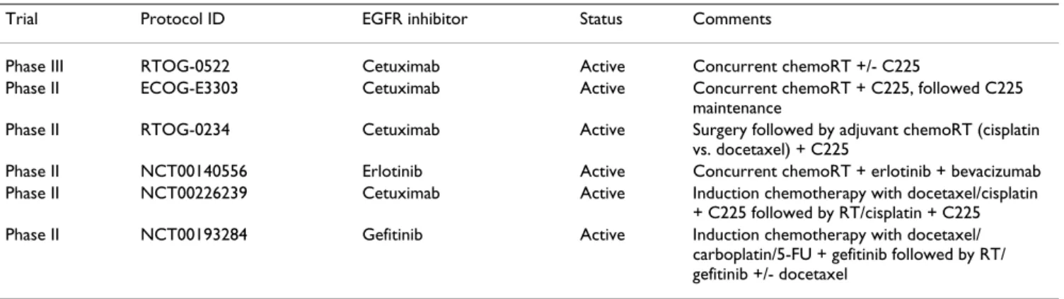
Table 1: Current phase II/III trials assessing EGFR inhibitors in combination with radiation (RT) locally advanced non-metastatic stage IV squamous-cell cancer of the head and neck
Trial Protocol ID EGFR inhibitor Status Comments
Phase III RTOG-0522 Cetuximab Active Concurrent chemoRT +/- C225
Phase II ECOG-E3303 Cetuximab Active Concurrent chemoRT + C225, followed C225
maintenance
Phase II RTOG-0234 Cetuximab Active Surgery followed by adjuvant chemoRT (cisplatin
vs. docetaxel) + C225
Phase II NCT00140556 Erlotinib Active Concurrent chemoRT + erlotinib + bevacizumab
Phase II NCT00226239 Cetuximab Active Induction chemotherapy with docetaxel/cisplatin
+ C225 followed by RT/cisplatin + C225
Phase II NCT00193284 Gefitinib Active Induction chemotherapy with docetaxel/
carboplatin/5-FU + gefitinib followed by RT/ gefitinib +/- docetaxel
more than half of patients receiving radiotherapy plus cetuximab still experienced local-regional relapse [4]. Therefore, there is a need to further improve outcome. Ongoing clinical efforts are devoted to address whether the addition of cetuximab to concurrent chemoradiation can yield a better outcome (i.e., RTOG study 0522). At this point, it has to be reminded that cancer cells rely on several, sometimes, redundant activation pathways; EGFR is only one of them. The risk of treatment failure is real, if only one receptor is targeted, hence the interest in combining broader range tyrosine kinase inhibitors such as CI-1033, which targets all four members of the Erb fam-ily (pan ErbB).
Finally, ionizing radiation stimulates the nitric oxyde (NO) pathway as well as the production of VEGF [32,33]. Angiogenesis inhibitors bear the potential to reinforce the cytotoxic action of RT. ZD6474, a small molecule tyrosine kinase inhibitor of EGFR and VEGF, looks very promising for the future.
Abbreviations
SCCHN: squamous-cell carcinoma of the head and neck RT: radiation therapy
EGF: epidermal growth factor
EGFR: epidermal growth factor receptor TGF-α: transforming growth factor alpha RTOG: Radiation Therapy Oncology Group ATP: adenosine triphosphate
ECOG: Eastern Cooperative Oncology Group TKI: tyrosine kinase inhibitor
NO: nitric oxyde
VEGF: vascular endothelial growth factor
Competing interests
The author(s) declare that they have no competing inter-ests.
Authors' contributions
MZ and AZ drafted the manuscript. DA helped the draft of the manuscript and participated in its design. MO con-ceived of the review, participated in its design, and helped the draft of the manuscript. All authors read and approved the final manuscript.
Acknowledgements
The authors thank Ms. Frances Godson for her excellent secretarial assist-ance.
References
1. Licitra L, Locati LD, Bossi P: Head and neck cancer. Ann Oncol 2004, 15(Suppl 4):iv267-iv273.
2. Arteaga C: Targeting HER1/EGFR: a molecular approach to
cancer therapy. Semin Oncol 2003, 30(Suppl 7):3-14.
3. Baselga J: Targeting the epidermal growth factor receptor: a
clinical reality. J Clin Oncol 2001, 19(Suppl):41S-44S.
4. Bonner JA, Harari PM, Giralt J, Azarnia N, Shin DM, Cohen RB, Jones CU, Sur R, Raben D, Jassem J, Ove R, Kies MS, Baselga J, Youssoufian H, Amellal N, Rowinsky EK, Ang KK: Radiotherapy plus
cetuxi-mab for squamous-cell carcinoma of the head and neck. N
Engl J Med 2006, 354:567-578.
5. Cohen S, Carpenter G, King L Jr: Epidermal growth
factor-receptor-protein kinase interactions: co-purification of receptor and epidermal growth factor-enhanced phosphor-ylation activity. J Biol Chem 1980, 255:4834-4842.
6. Cohen S: The stimulation of epidermal proliferation by a
spe-cific protein (EGF). Dev Biol 1965, 12:394-407.
7. Davies RL, Grosse VA, Kucherlapati R, Bothwell M: Genetic
analy-sis of epidermal growth factor action: assignment of human epidermal growth factor receptor gene to chromosome 7.
Proc Natl Acad Sci 1980, 77:4188-4192.
8. Roskoski R Jr: The ErbB/HER receptor protein-tyrosine
kinases and cancer. Biochem Biophys Res Commun 2004, 319:1-11.
9. Schmidt-Ullrich RK, Valerie KC, Chan W, McWilliams D: Altered
expression of epidermal growth factor receptor and extro-gen receptor in MCF-7 cells after single and repeated radia-tion exposures. Int J Radiat Oncol Biol Phys 1994, 29:813-819.
10. Dent P, Reardon DB, Park JS, Bowers G, Logsdon C, Valerie K, Schmidt-Ullrich R: Radiation-induced release of transforming
growth factor α activates the epidermal growth factor receptor and mitogen-activated protein kinase pathway in carcinoma cells, leading to increased proliferation and pro-tection from radiation-induced cell death. Mol Biol Cell 1999, 10:2493-2506.
11. Tofilon PJ, Saxman S, Coleman CN: Molecular targets for
radia-tion therapy: bringing preclinical data into clinical trials. Clin
Cancer Res 2003, 9:3518-3520.
12. Sartor C: Mechanisms of Disease: radiosensitization by
epi-dermal growth factor receptor inhibitors. Nat Clin Pract Oncol
2004, 1:80-87.
13. Carpenter G, Cohen S: Epidermal growth factor. Annu Rev
Bio-chem 1979, 48:193-216.
14. Grandis J, Tweardy D: Elevated levels of transforming growth
factor α and epidermal growth factor receptor messenger RNA are early markers of carcinogenesis in head and neck cancer. Cancer Res 1993, 53:3579-3584.
15. Grandis JR, Chakraborty A, Zeng Q, Melhem MF, Tweardy DJ:
Downregulation of TGF-alpha protein expression with anti-sense oligonucleotides inhibits proliferation of head and neck squamous carcinoma but not normal mucosal epithelial cells. J Cell Biochem 1998, 69:55-62.
16. Rubin Grandis J, Melhem MF, Barnes EL, Tweardy DJ: Quantitative
immunohistochemical analysis of transforming growth fac-tor α and epidermal growth facfac-tor recepfac-tor in patients with squamous cell carcinoma of the head and neck. Cancer 1996, 78:1284-1292.
17. Baumann M, Krause M: Targeting the epidermal growth factor
receptor in radiotherapy: radiobiological mechanisms, pre-clinical and pre-clinical results. Radiother Oncol 2004, 72:257-266.
18. Maurizi M, Almadori G, Ferrandina G, Ferrandina G, Distefano M, Romanini ME, Cadoni G, Benedetti-Panici P, Paludetti G, Scambia G, Mancuso S: Prognostic significance of epidermal growth factor
receptor in laryngeal squamous cell carcinoma. Br J Cancer
1996, 74:1253-1257.
19. Demiral AN, Sarioglu S, Birlik B, Sen M, Kinay M: Prognostic
signif-icance of EGF receptor expression in early glottic cancer.
Auris Nasus Larynx 31:417-424.
20. Ang KK, Berkey BA, Tu X, Zhang HZ, Katz R, Hammond EH, Fu KK, Milas L: Impact of epidermal growth factor receptor
Publish with BioMed Central and every scientist can read your work free of charge "BioMed Central will be the most significant development for disseminating the results of biomedical researc h in our lifetime."
Sir Paul Nurse, Cancer Research UK Your research papers will be:
available free of charge to the entire biomedical community peer reviewed and published immediately upon acceptance cited in PubMed and archived on PubMed Central yours — you keep the copyright
Submit your manuscript here:
http://www.biomedcentral.com/info/publishing_adv.asp
BioMedcentral
Radiation Oncology 2006, 1:11 http://www.ro-journal.com/content/1/1/11
advanced head and neck carcinoma. Cancer Res 2002, 62:7350-7356.
21. Albanell J, Codony-Servat J, Rojo F, Del Camp JM, Sauleda S, Anido J, Raspall G, Giralt J, Rosello J, Nicholson RI, Mendelsohn J, Baselga J:
Activated extracellular signal-regulated kinases: association with epidermal growth factor receptor/transforming growth factor alpha expression in head and neck squamous carci-noma and inhibition by anti-epidermal growth factor recep-tor treatments. Cancer Res 2001, 61:6500-6510.
22. Hambek M, Baghi M, Baumaun H, Strebhard K, Adunka O, Gstottner W, Knecht R: Iressa (ZD 1839) inhibits phosphorylation of
three different downstream signal transducers in head and neck cancer (SCCHN). Anticancer Res 2005, 25:1871-1875.
23. Raben D, Bianco C, Milas L, Ang KK: Targeted therapies and
radi-ation for the treatment of head and neck cancer: are we making progress? Semin Radiat Oncol 2004, 14:139-152.
24. Burtness BA, Li Y, Flood W, Mattar BI, Forastiere AA: Phase III trial
comparing cisplatin (C) + placebo (P) + anti-epidermal growth factor antibody (EGF-R) C225 in patients (pts) with metastatic/recurrent head and neck cancer (HNC). Proc Am
Soc Clin Oncol 2002, 21:226.
25. Bonner JA, Raisch KP, Trummell HQ, Robert F, Meredith RB, Spencer SA, Buschbaum DJ, Saleh MN, Stackhouse MA, LoBuglio AF, Peters GE, Carroll WR, Waksal HW: Enhanced apoptosis with
combi-nation C225/radiation treatment serves as the impetus for clinical investigation in head and neck cancers. J Clin Oncol
2000, 18(Suppl):47s-53s.
26. Harari PM, Huang S: Radiation combined with EGFR Signal
Inhibitors. Semin Radiat Oncol 2006, 16:38-44.
27. Solomon B, Hagekyriakou J, Trivett M, Stacker S, McArthur G, Cull-inane C: EGFR blockade with ZD1839 ("Iressa") potentiates
the antitumor effects of single and multiple fractions of ion-izing radiation in human A431 squamous cell carcinoma. Int
J Radiat Oncol Biol Phys 2003, 55:713-723.
28. Cohen EE, Haraf DJ, Stenson KM, Blair E, Brockstein BE, Mauer AM, Dekker A, Williams R, Lester E, Vokes EE: Integration of gefitinib
(G) into a chemoradiation (CRT) regimen, followed by G adjuvant therapy in patients with locally advanced head and neck cancer (HNC): a phase II trial. J Clin Oncol 2005, 23(Suppl):501s.
29. Overgaard J, Hansen HS, Specht L, Overgaard M, Grau C, Andersen E, Bentzen J, Basholt L, Hansen O, Johansen J, Andersen L, Evensen JF:
Five compared with six fractions per week of conventional radiotherapy of squmous-cell carcinoma of head and neck: DAHANCA 6 and 7 randomised controlled trial. Lancet 2003, 362:933-940.
30. Pignon JP, Bourhis J, Domenge C, Designe L: Chemotherapy
added to locoregional treatment for head and neck squa-mous-cell carcinoma: three meta-analyses of updated indi-vidual data. Lancet 2000, 355:949-955.
31. Budach W, Hehr T, Budach V, Belka C, Dietz K: A meta-analysis of
hyperfractionated and accelerated radiotherapy and com-bined therapy and radiotheapy regimens in unresected locally advanced squamous cell carcinoma of the head and neck. BMC Cancer 2006, 6:28.
32. Gorski DH, Beckett MA, Jasnowiak NT, Calvin DP, Mauceri HJ, Sal-loum RM, Seetharam S, Koons A, Hari DM, Kufe DW, Weichselbaum RR: Blockade of the vascular endothelial growth factor stress
response increases the the antitumor effects of ionizing radi-ation. Cancer Res 1999, 59:3374-3378.
33. Sonveaux P, Brouet A, Havaux X, Gregoire V, Dessy C, Balligand JL, Feron O: Irradiation-induced angiogenesis through the
up-regulation of the nitric oxide pathway: implications for tumor radiotherapy. Cancer Res 2003, 63:1012-1019.