IDENTIFICATION AND CHARACTERIZATION OF PUTATIVE
NUCLEOMODULINS IN MYCOBACTERIUM TUBERCULOSIS
Abstract
The nuclear targeting of bacterial proteins that modify host cell gene expression, the so-‐ called nucleomodulins, has emerged as a novel mechanism contributing to virulence of several intracellular pathogens. The goal of this study was to identify nucleomodulins produced by Mycobacterium tuberculosis (Mtb), the causative agent of tuberculosis (TB), and to investigate their role upon infection of the host. We first performed a screening of Mtb genome in search of genes encoding proteins with putative eukaryotic-‐like nuclear localization signals (NLS). We identified two genes of Mtb, Rv0229c and Rv3876, encoding proteins that are secreted in the medium by Mtb and are localized into the nucleus when expressed in epithelial cells or in human or murine macrophages. The NLSs of these two proteins were identified and found to be essential for their nuclear localization. The gene Rv0229c, a putative RNase, is present only in pathogen species of the Mtb complex and seems to have been recently acquired by horizontal gene transfer (HGT). Rv3876 appears more widely distributed in mycobacteria, and belongs to a chromosomal region encoding proteins of the type VII secretion system ESX1, essential for virulence. Ongoing studies are currently investigating the dynamics of these proteins upon infection of host cells, and their putative role in the modulation of host cell gene expression and Mtb virulence.
IDENTIFICATION ET CARACTERISATION DE NUCLEOMODULINES
PUTATIVES CHEZ LA BACTERIE MYCOBACTERIUM TUBERCULOSIS
Résumé
Les nucléomodulines sont des protéines produites par des bactéries parasites intracellulaires et qui sont importées dans le noyau des cellules infectées pour y moduler l’expression génique et contribuer ainsi à la virulence de la bactéries. L’identification de nucléomodulines chez plusieurs espèces de bactéries pathogènes a fait émerger ce concept comme une stratégie supplémentaire utilisée par les parasites intracellulaires pour contourner les moyens de défense de l’hôte. Le but de cette thèse était d’identifier et d’analyser d’éventuelles nucléomodulines produites par l’agent étiologique de la tuberculose, la bactérie Mycobacterium tuberculosis (Mtb). Nous avons tout d’abord analysé le génome de Mtb, à la recherche de gènes dont les produits portent des séquences analogues aux séquences de localisation nucléaire des eucaryotes (NLS). Nous avons pu identifier deux gènes de Mtb, Rv0229c et Rv3876, qui codent des protéines sécrétées dans le milieu de culture et qui se localisent dans le noyau lorsqu’elles sont exprimées dans des cellules épithéliales ou dans des macrophages murins ou humains. Les NLS de ces deux protéines ont été identifiées et leur modification abolit la localisation nucléaire dans les cellules eucaryotes. Le gène Rv0229c est trouvé spécifiquement dans le génome des espèces pathogènes du complexe Mtb. Ce gène semble avoir été acquis récemment par l’ancêtre de Mtb, via un transfert génétique horizontal. Rv3876 est plus généralement distribué chez les mycobactéries, et appartient à une région génomique codant un système de sécrétion type VII, ESX1, essentiel pour la virulence de Mtb. Les travaux en cours visent à analyser la dynamique de ces deux protéines au cours de l’infection de macrophages ou de modèles animaux, et leur rôle en tant que modulateurs de l’expression génique des cellules infectées et dans la virulence de Mtb.
ABSTRACT ... i
RÉSUMÉ ... ii
TABLE OF CONTENTS ... iii
LIST OF FIGURES ... vii
LIST OF TABLES ... viii
LIST OF ABBREVIATIONS ... viiii
ACKNOWLEDGEMENTS AND GRATITUDE ... xiii
ACKNOWLEDGEMENTS AND AUTHOR ATTRIBUTIONS ... xv
CHAPTER 1 INTRODUCTION ... 1
1.1 Tuberculosis ... 1
I. History of tuberculosis ... 1
II. Epidemiology of tuberculosis ... 1
III. Mycobacterium tuberculosis ... 2
A. Taxonomy and characteristics ... 2
B. Structure of the cell envelope of mycobacteria ... 3
IV. TB treatment and drug resistance ... 4
A. Drug resistance in TB today ... 5
B. Current and future treatments of MDR-‐ and XDR-‐TB ... 6
1.2 Host responses to mycobacterium tuberculosis ... 11
I. Immunity to Tuberculosis ... 12
A. Innate immune system during Mtb infection ... 12
B. Adaptive immune system during bacterial infection ... 15
i. Cell-‐mediated immunity ... 15
ii. Humoral immunity and B lymphocyte ... 17
II. Evasion of the immune response ... 18
A. Phagosome maturation ... 18
B. Resistance mechanisms ... 19
i. ROS and RNS ... 19
C. Nutritional mechanisms ... 21
D. Cell death: necrosis vs apoptosis ... 22
1.3 The concept of nucleomodulins ... 24
I. Nuclear transport ... 25
II. Nuclear Localization Signals (NLS) ... 26
III. Nucleomodulins ... 27
1.4 Statement of the problem ... 31
1.5 Purpose ... 32
CHAPTER 2 MATERIALS AND METHODS ... 33
2.1 In-‐silico prediction of Mtb proteins targeted to the host cell nucleus ... 33
2.2 Reagents and chemicals ... 33
I. Commercial Reagents ... 33
II. Enzymes and kits ... 34
III. Oligonucleotides and Sequencing ... 34
2.3 Cloning and expression ... 34
I. Amplification of DNA fragments by PCR ... 34
II. Plasmid constructions ... 35
A. GFP-‐Fusion plasmid clones ... 35
B. Mammalian expression vector constructs ... 35
C. Generation of plasmid constructs for overexpression ... 35
D. Plasmid constructs for overexpression under inducible promoter ... 36
E. Site directed mutagenesis ... 36
F. Construction of Knockout Mutant of Rv0229c and Rv3876 of Mtb ... 37
G. Recombineering experiments in Mtb ... 37
III. Ligation ... 38
IV. Media and culture conditions for bacteria ... 38
V. Preparation of chemically competent E. coli cells ... 38
VI. Transformation of bacteria ... 39
VII. Preparation of plasmids from small E. coli cultures ... 39
VIII. Digestion of DNA with restriction enzymes ... 39
X. Purification of DNA fragment by gel extraction ... 40
2.4 Cell culture and transfection ... 40
I. Primary cells and cell lines ... 40
A. Generation of human monocyte-‐derived macrophages ... 40
B. Murine cell lines: Mouse monocyte-‐macrophage BALB/c cells ... 41
C. Human embryonic kidney 293 (HEK 293) cells ... 41
D. Cell passaging ... 41
E. Assessing cell vitality ... 41
F. Freezing and thawing cells ... 42
II. Transfection of the constructed plasmids into host cells ... 42
A. Transient transfection of HMDM ... 42
B. Transient transfection of RAW 264.7 cells ... 42
C. Transient transfection of HEK 293 cells ... 43
III. Transfection efficiency analysis ... 43
A. Fluorescence microscopy and imaging ... 43
B. FACS analysis (Flow cytometry) ... 43
CHAPTER 3 RESULTS ... 44
3.1 Prediction of proteins with putative NLSs in Mtb ... 44
3.2 Screening of nuclear targeting proteins of Mtb in eukaryotic cells ... 46
3.3 Rv0229c encodes a protein of unknown function ... 47
3.4 Rv3876 (EspI) encodes a putative ATPase, involved in the regulation of the ESX-‐1 secretion system ... 48
3.5 Rv0229c was recently acquired by the ancestor of Mtb through horizontal genetic transfer as suggested by Comparative Genomics ... 48
3.6 Identification of the functional NLS of Rv0229c and Rv3876. ... 49
3.7 Rv0229c and Rv3876 are secreted proteins in Mtb ... 51
3.8 Construction of genetic tools for functional characterization of Rv0229c and Rv3876 during infection by Mtb. ... 52
A. Deletion of Rv0229c and Rv3876 ... 52
B. Over-‐expression of Rv0229c and Rv3876 ... 53
3.9 Transcriptome analysis ... 54 CHAPTER 4 DISCUSSION ... 56 REFERENCES ... 62 APPENDICES ... 79 APPENDIX A ... 79 APPENDIX B ... 80 1. Background ... 80
1.1 Toxin-‐antitoxin (TA) systems in bacteria ... 80
1.2 Gateway® cloning system ... 81
2. Material and methods ... 82
2.1 Generation of Rv1990c-‐1989c operon expression clones ... 82
2.2 Cloning and expression ... 83
2.3 Analysis of GFP expression ... 83
I. Fluorescence intensity ... 84
II. Fluorescence microscopy ... 84
3. Results and perspectives ... 84
3.1 Generation of expression clones by the Gateway®cloning system ... 84
3.2. Testing the Rv1990c-‐1989c operon expression ... 85
3.3. Dissection of the regulatory region of the Rv1990c promoter ... 86
3.4 Expression of the Rv1990c-‐1989c operon in a variety of stress conditions .... 87
Figure 1 Timeline showing historical highlights of scientific breakthroughs, and the
ongoing public health issues, in the fight against TB ... 1
Figure 2 Estimated TB incidence rates by country in 2013 ... 2
Figure 3 The cell envelope of Mtb ... 3
Figure 4 Current global pipeline of new tuberculosis drugs ... 6
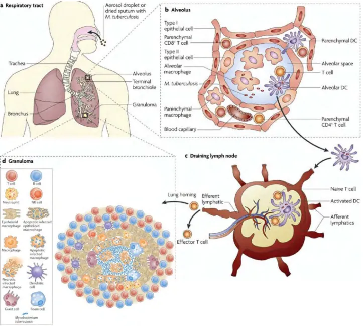
Figure 5 Mtb infection and granuloma formation ... 12
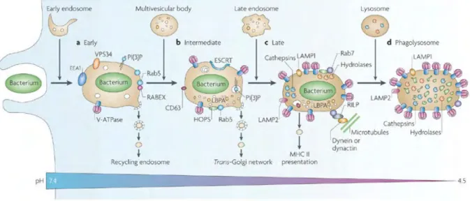
Figure 6 Stages of phagosomal maturation ... 18
Figure 7 Overview of nuclear protein import ... 25
Figure 8 Bacterial strategies to control gene expression in the nucleus ... 29
Figure 9 Mammalian expression vector ... 34
Figure 10 E. coli and mycobacterium shuttle plasmid ... 36
Figure 11 Genes associated with the Mtb ESAT-‐6 (esx) gene clusters ... 45
Figure 12 Nuclear localization of mycobacterial nucleomodulin candidates fused with GFP in eukaryotic cells ... 46
Figure 13 Nuclear localization of mycobacterial nucleomodulin candidates fused with GFP in RAW 264.7 macrophages ... 47
Figure 14 Comparative Genomics of Rv0229c ... 48
Figure 15 Rv0229c contains a functional nuclear localization signal ... 49
Figure 16 Rv3876 contains a functional nuclear localization signal ... 50
Figure 17 Rv0229c and Rv3876 are mycobacterial secretory proteins ... 51
Figure 18 Over-‐expression of Rv0229cv or Rv3876 has no effect on multiplication of Mtb inside human macrophages ... 53
Figure 19 Optimization of the GFP-‐reporter plasmid transfection ... 54
Figure 20 Analysis of the RNAs co-‐immunoprecipitated with Rv0229c ... 60
LIST OF TABLES
Table 1 Classification of drug used to treat TB ... 5
Table 2 Different classes of NLS allow nuclear localization of bacterial nucleomodulins 27 Table 3 Strategies exploited by pathogens to modulate the host signaling pathways .... 29
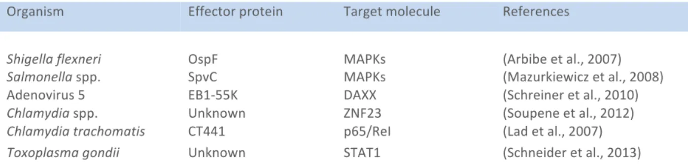
Table 4 Strategies exploited by pathogens to modulate the host nuclear proteins ... 29
Table 5 Strategies exploited by pathogens to modulate the host post-‐translation ... 30
Table 6 Construction of the different plasmids used in this study ... 35
Table 7 Construction of the different mutagenesis used in this study ... 35
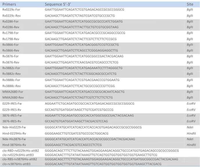
Table 8 Cloning primers used for plasmid constructions ... 36
Table 9 Cloning primers used for mutagenesis ... 37
Table 10 Primers used for Mtb mutant construction ... 37
Table 11 Sequencing primers used in this study ... 38
Table 12 Predicted nuclear targeting proteins with NLS sequences in Mtb H37Rv ... 45
AG AIDS AMs APC BAHD1 BCG BSA CIITA CD4 cDNA CLRs CMI DAMPs DC DC-‐SIGN DMSO DOTS EEA1 eis Emb ERK ESAT-‐6 FACS FAS-‐I FAS-‐II GalN HAUSP HGT HIV Arabinogalactan
Acquired Immune Deficiency Syndrome Alveolar macrophages
Antigen Presenting Cells
Bromo adjacent homology domain-‐containing protein 1 Bacillus Calmette Guerin
Bovine Serum Albumin MHC class II transactivator Cluster of Differentiation 4
Complementary Deoxy Ribonucleic Acid C-‐type lectin receptor
Cell Mediated Immune Response Damage-‐associated molecular patterns Dendritic Cells
Dendritic cell-‐specific intracellular-‐adhesion-‐molecule-‐ grabbing non-‐integrin Dimethyl sulfoxide
Directly Observed Treatment Short course Early endosome antigen 1
Enhanced intracellular survival Ethambutol
Extracellular signal-‐regulated kinase Early Signs of Antigenic Target 6 kDa Fluorescent Activated Cell Sorting Fatty-‐Acid-‐Synthase systems I Fatty-‐Acid-‐Synthase systems II N‑acetylated galactosamine
Herpes virus-‐associated ubiquitin-‐specific protease Horizontal gene transfer
ICL IFN-‐β IFN-‐γ Inh IL-‐2 IL-‐4 IL-‐5 IL-‐6 IL-‐10 IL-‐12 iNOS2 IRAK IRF JNK kDa LM LntA LAM LpqH LTBI MA MDR-‐TB ManLAM MAP MAPKs MAPc mDAP MLS MOMP MTBC MyD88 Isocitrate lyase Interferon-‐beta Interferon-‐gamma Isoniazid Interleukin-‐2 Interleukin-‐4 Interleukin-‐5 Interleukin-‐6 Interleukin-‐10 Interleukin 12
Inducible nitric oxide synthase IL-‐1 receptor-‐associated kinase Interferon-‐regulatory factor Jun N-‐terminal kinase Kilo Dalton
Lipomannan
Listeria nuclear targeted protein A Lipoarabinomannan 19-‐kDa lipoprotein Latent TB infection Mycolic acids Multidrug-‐resistant TB Mannosylated lipoarabinomannan Mitogen-‐activated protein
Mitogen-‐activated protein kinases
Mycolic acid-‐Arabinogalactan-‐Peptidoglycan complex meso-‐diaminopimelic acid
Malate synthase
Mitochondrial outer membrane permeabilization Mtb complex
NADPH NAGs NAM NAG-‐NAM NES NLRs NLS NF NOD NOS2 NPCs NTM PAMPs PG PGE2 PI PI3P PIMs PP2A PRRs PTMs Pza RLU RLRs Rif RNI ROI SRs SWI/SNF TAK1 TAL
Nicotinamide adenine dinucleotide phosphate N-‐acetylglucosamines
N-‐acetylmuramic acid
N‑acetyl glucosamine-‐N‑acetyl muramic acid Nuclear export signals
NOD-‐like receptors
Nuclear localization signals Nuclear transcription factor Nuclear oligomerization domain Nitric oxide synthases 2
Nuclear pore complexes Nontuberculous mycobacteria
Pathogen-‐Associated Molecular Patterns Peptidoglycan
Prostaglandin E2
Phosphatidyl-‐myo-‐inositol
Phosphatidylinositol 3-‐phosphate phosphatidyl-‐myo-‐inositol mannosides Protein phosphatase 2A
Pattern recognition receptors Post-‐translational modifications Pyrazinamide
Relative fluorescence units RIG-‐I like receptors
Rifampicin
Reactive nitrogen intermediates Reactive oxygen intermediates Scavenger receptors
SWItch/Sucrose NonFermentable TGFβ-‐activated protein kinase 1 Transcription activator-‐like
TB TCA TCR TDR-‐TB TDM TFs TLRs TMM TNF TRAF VPS34 XDR-‐TB Tuberculosis Tricarboxylic acid T cell receptors Totally drug-‐resistant TB Trehalose dimycolates Transcription factors Toll-‐like receptors Trehalose monomycolates Tumor necrosis factor
TNF receptor-‐associated factor Human vacuolar protein-‐sorting 34 Extensively drug-‐resistant TB
“Uncertainty is the only certainty”
-‐Buddha-‐
I would like to express my sincerest gratitude to Dr. Olivier Neyrolles, CNRS research director, for giving me the opportunity to carry out my research project. It also gives me immense pleasure to thank my mentor and immediate supervisor, Prof Claude Gutierrez, for his support and continuous guidance.
Also, I want to express my thanks to Dr. Geanncarlo Lugo who has always played a key role in encouraging and coordinating this project and keeping me abreast of scientific developments and for his inexhaustible patience during the correction phase of this dissertation.
I would also like to thank Prof. Denis Hudrisier for helping me with important comments, meticulous suggestions and astute criticism during the practical phase. And thanks to all other lab colleagues: Assoc. Prof Yannick Poquet, Florence Levillain, Ingrid Mercier, Dr. Carine Duval, Dr. Alan Benard, Dr. Alexandre Gouzy, Anthony Troegeler, Alexia Dumas and Lucie Bernard for their scientific help during the research program.
Sincerest thanks are also to:
My Dissertation Committee: Prof Matthieu Arlat and Assoc Prof Philippe Rousseau.
My Thesis Defense Committee: Prof Olivier Dussurget, Prof Carlos Martin and Prof Matthieu Arlat.
I also appreciate my pervious lab of Dr. Mamadou Daffé, CNRS research director, and his team. Special thanks to Assoc. Prof Nathalie Eynard for her precious support and encouragement. I am also thankful to my former advisor Dr. Didier Zerbib who supported me for the first 3 years of my research program.
I would also like to thank The Royal Thai Government Scholarships, offered by the Ministry of Science and Technology, for its financial support.
I am also thankful for my friends here in Toulouse, especially to Clément for the amazing support and the wonderful friendship when I was alone, and to his family. GC (Grumpy cat), Denis (senseï) and his family, Alan (the boy), Yanos and his family, Ingrid (la plus belle dans l’IPBS) and her family, Alexia (Madame Ouin Ouin), Alex (the best of the best), Stevie (beau gosse), Anthony (try to make it) and Carine (KK). They are lab colleagues who became my dear friends. I am also thankful my new friends from California: Alexandra and Natsinet and special thanks to Nat (that’s interesting) for her extensive knowledge of the English language, proofreading of my manuscript and also for the great times we spent together, and all others who helped make my stay in France a very pleasant one.
Last, but not the least, I would also like to thank to my teacher Prof Somsak Pantuwatana for constant support, encouragement and for never retiring from my life. I am also very thankful to my friends in Thailand, Sompong, Supirada, Sawanya, my lovely student Kanookporn and her family for unwavering support and lasting friendship.
Finally, I would like to express a deep sense of gratitude to my family, especially to my parents, who have always stood by me in all situations and I owe my life to them for the constant love, encouragement, moral support and blessings.
ACKNOWLEDGEMENTS AND AUTHOR ATTRIBUTIONS
The project was conceived by Dr. Olivier Neyrolles. Experiments were designed by Prof Claude Gutierrez. Experiments were performed in biosafety level 3 facility by Prof Claude Gutierrez and Dr. Anna Grabowska. Protein secretion test in Mtb using western blot analysis by Dr. Daria Bottai, Pisa University, Italy. Co-‐immunoprecipitation test by Dr. Anthony Henras and Dr. Yves Roméo, LBME -‐ UMR 5099 -‐ CNRS/UPS.
Figure 1. Timeline showing historical highlights of scientific breakthroughs, and the ongoing public health issues, in the fight against TB (Kaufmann, 2005).
1.1 TUBERCULOSIS
I. History of tuberculosis
Tuberculosis (TB), historically known as “phthisis” or “consumption,” due to the disturbing weight loss experienced by patients suffering from this disease, is one of the oldest recorded human afflictions as evidenced, for example, by Egyptian mummies dating as far back to 1550–1080 BC. They were found to have spinal columns with signs of tubercular decay and mycobacterial DNA still detectable in the bones (Cambau and Drancourt, 2014; Daniel, 2006; Nerlich et al., 1997). It was not until 1834 that the term “tuberculosis” was coined for this disease based on the original description (in 1650) of the apparent lung nodules (characteristic of patients succumbing to TB) as “tubercles” (Cambau and Drancourt, 2014). Another important historical event happened when Jean Antoine Villemin demonstrated (in 1865) that TB was an infectious disease; he showed transmissibility of TB from humans to rabbits, from cattle to rabbits, and from rabbits to rabbits (Cambau and Drancourt, 2014). The history of TB was changed dramatically in 1882, when Robert Koch identified the bacillus as the etiological agent causing TB, for which he was awarded the Nobel Prize in Physiology or Medicine in 1905. Four years later, the bacillus was named Mycobacterium tuberculosis (Mtb) (Grange, 1982). This revolutionary finding gave way to later discoveries such as that of tuberculin, which is an extract of Mtb used in skin testing in animals and humans to identify TB infection (in 1890), the Bacillus-‐Calmette Guérin (BCG) vaccine (in 1921), and the first anti-‐TB drug (i.e. streptomycin, in 1943) (Kaufmann, 2005). Together, these breakthroughs offered hope for the eradication of the disease (Fig. 1).
II. Epidemiology of tuberculosis
More than a century after the discovery of TB, despite advances in diagnostic, vaccination and treatment strategies, numerous challenges still remain. Today, TB ranks number eight on the list of the most common causes of death worldwide, and it is the second most common cause of death from an infectious disease after the human immunodeficiency virus (HIV /AIDS) (WHO, 2014). Mtb infection kills more people than any other bacterial pathogen. In 2013, there were more than 9 million TB cases accounting for over 1.5 million deaths. This
Figure 2. Estimated TB incidence rates by country in 2013 (WHO, 2014).
©2015 • KANJANA NUKDEE
is especially evident in the case of co-‐infection with HIV, TB is a leading killer of HIV-‐positive people causing one fourth of all HIV-‐related deaths (WHO, 2014). The overall TB burden continues to rise as a result of the rapid growth of the world population, specifically in Africa and Asia. The six countries with the largest number of incident cases in 2013 were India, China, Nigeria, Pakistan, Indonesia and South Africa; India (24%) and China (11%) alone accounted for 35% of the world’s TB cases. Figure 2 illustrates the TB incidence rates by country in 2013 (WHO, 2014).
III. Mycobacterium tuberculosis A. Taxonomy and characteristics
The bacillus causing TB in humans belongs to the genus Mycobacterium. Mycobacteria are classified as Gram-‐positive bacteria based on 16S ribosomal RNA sequence comparison, but are more closely related to Gram-‐negative bacteria when comparing the whole genome (Fu and Fu-‐Liu, 2002). Mtb is typically visualized by Ziehl-‐Neelsen (acidfast) staining and appears as a rod-‐shaped red bacillus. At 37°C and under optimal aerobic conditions and nutrients, Mtb has a generation time of 18–24 hours and forms a white to light yellow colony on agar within 3–4 weeks. Mtb does not form spores but has the capacity to become dormant, a non-‐replicating state characterized by low metabolic activity and phenotypic drug resistance (Gengenbacher and Kaufmann, 2012). The genome of the laboratory strain H37Rv of Mtb was the first completely sequenced in 1998 by Cole and coworker. The genome has a high guanine (G) and cytosine (C) content (65%), and comprises 4.4 mega base pairs (Mbp), encoding around 4,000 predicted proteins (Cole et al., 1998). Mycobacteria can be divided into three major groups for the purpose of diagnosis and treatment: i) the Mtb complex (MTBC) that cause TB in humans and animals, includes Mtb and the closely related species (i.e. M. bovis, M. bovis BCG, M. africanum, M. microti, and M. canetti (Richter et al., 2003), ii) M. leprae that causes leprosy, and iii) the nontuberculous mycobacteria (NTM) that groups all Mycobacterium species except the obligate pathogens MTBC and M. leprae, which are typically environmental organisms residing in soil and water and are in most cases nonpathogenic for humans or animals (van Ingen, 2013). Mycobacterium species that belong to NTM include human disease-‐associated bacteria: M. avium, M. kansasii, M. abscessus, M. chelonae, M. fortuitum, M. peregrinum and M. marinum. Other species are rarely the cause of disease, such as M. smegmatis and M. flavescens (Griffith et al., 2007). M. smegmatis and
Figure 3. The cell envelope of Mtb. The cell wall core is composed of three different covalently linked structures
peptidoglycan (PG ‐ grey), arabinogalactan (AG ‐ blue) and mycolic acids (MAs ‐ green), forming a complex known as MAPc (Mycolic acid‐Arabinogalactan‐Peptidoglycan complex). The covalent linkage of MAs results in a hydrophobic layer of extremely low fluidity. This layer is also referred to as the mycomembrane. The outer part of the mycomembrane contains various free lipids, such as PIMs, LM and LAM, which are intercalated with the MAs. The outer layer is known as the capsule that mainly contains polysaccharides (Abdallah et al., 2007).
©2015 • KANJANA NUKDEE
M. bovis BCG (an attenuated form of M. bovis) are often used as surrogate hosts to study Mtb gene expression (Gengenbacher and Kaufmann, 2012).
B. Structure of the cell envelope of mycobacteria
The mycobacterial cell wall consists of inner and outer layers that surround the plasma membrane. The inner compartment is composed of three distinct macromolecules: peptidoglycan (PG), arabinogalactan (AG) and mycolic acids (MA). Interestingly, the PG is covalently attached to AG, which in turn is attached to MA; these three components form the cell wall core otherwise known as the Mycolic acid-‐Arabinogalactan-‐Peptidoglycan complex (MAPc). This complex remains as the insoluble residue when cell walls are disrupted (e.g. with various solvents), whereas other free lipids and proteins are solubilized. The insoluble core is essential for the viability of the cell and many of the drugs used to combat Mtb specifically target these complexes (Brennan, 2003; Hett and Rubin, 2008). The outer layer, called capsule, contains only minor amounts of lipids and consist of a loosely-‐ bound structure of polysaccharides, proteins and noncovalently linked glycoconjugates, which is one of the unique features of the mycobacteria. Indeed, this last feature exhibits a high content of mannosylated molecules, including phosphatidyl-‐myo-‐inositol (PI) mannosides (PIMs), lipomannan (LM) and lipoarabinomannan (LAM) in particular (Kaur et al., 2009). Figure 3 illustrates the organization of the cell envelope of Mtb (Abdallah et al., 2007). The following is a brief description of some of the components of the inner and outer layers of the mycobacterial cell wall:
• The PG (or murein), which is surrounding the plasma membrane, is made of peptides and glycan strands. The glycan strand consists of repeating N-‐ acetylglucosamines (NAGs) linked to N-‐acetylmuramic acid (NAM) via peptide bridges to form N‑acetyl glucosamine-‐N‑acetyl muramic acid (NAG-‐NAM). The PG polymer can be also modified by glycolylation of NAM residues, or amidation of the d‑Glu and meso-‐diaminopimelic acid (mDAP) residues of the peptide side chain (Kieser and Rubin, 2014).
• AG is the major polysaccharide of the mycobacterial cell wall and is important for cell wall integrity, which is composed of arabinan and galactan. AG is covalently attached to the PG layer by a phosphoryl-‐N-‐acetylglucosaminosyl-‐rhamnosyl linkage and can also be modified by the addition of succinyl or unusual
©2015 • KANJANA NUKDEE
non‑N‑acetylated galactosamine (GalN) moieties. The GalN modifications are mostly present in pathogenic mycobacteria and are thought to promote infection efficiency (Hett and Rubin, 2008; Kieser and Rubin, 2014).
• MAs are long-‐chain, α-‐acylated, β-‐hydroxylated fatty acids, which are formed from the condensation of a medium chain fatty acid (C20-‐C26) produced by Fatty-‐ Acid-‐Synthase systems I (FAS-‐I) with a long meromycolic chain (C60-‐C90) produced by Fatty-‐Acid-‐Synthase systems II (FAS-‐II) (Cole et al., 1998). MAs are a major lipid component of the envelope and form the external mycomembrane (Hoffmann et al., 2008; Zuber et al., 2008). They are either inserted in the mycomembrane as trehalose dimycolates (TDM) and trehalose monomycolates (TMM) or linked covalently to the AG, itself bound to PG, to form the MAPc (Cole et al., 1998; Crick et al., 2001). The coordination of MAs and AG synthesis and insertion in the cell wall still unclear, recent report suggest that these macromolecules are produced and exported at the cell pole where new cell growth occurs (Carel et al., 2014; Meniche et al., 2014).
• PIMs contain up to six mannose residues, which are unique glycolipids anchored through a PI moiety to the inner and outer membranes of the cell envelope of all Mycobacterium species (Boldrin et al., 2014). PIMs play important roles in the permeability barrier of the cell envelope, cell membrane integrity, the regulation of septation and cell division (Morita et al., 2005; Patterson et al., 2003).
• LM and LAM are extensions of PIMs. LM is the mannan chain added to the PIM as a chain and a branch. Similarly, LAM is an additional arabinan group attached to the PIM (Chatterjee and Khoo, 1998). LAM from Mtb has short mannose containing oligosaccharide ‘‘caps’’ that are involved in mediating the binding of mycobacteria to, and subsequent entry into, macrophages (Brennan, 2003). Both LM and LAM show various immunomodulatory activities, and play critical roles in the integrity of cell wall and the pathogenesis of TB (Fukuda et al., 2013).
IV. TB treatment and drug resistance
Streptomycin, first purified from Streptomyces griseus in 1944 by Schatz and Waksman and then introduced as TB chemotherapy in 1946 (Zumla et al., 2013), was the first antibiotic with proven activity against Mtb opening the doors as a viable approach for the generation
Table 1. Classification of drug used to treat TB.
First‐line anti‐TB drugs Group 1. Oral: isoniazid (H/Inh), rifampicin/rifampin (R/Rif),
pyrazinamide (Z/Pza), ethambutol (E/Emb), rifapentine (P/Rpt) or rifabutin (Rfb)
Second‐line anti‐TB
drugs
Group 2. Injectable aminoglycosides: streptomycin (S/Stm), kanamycin (Km), amikacin (Amk). Injectable polypeptides: capreomycin (Cm), viomycin (Vim).
Group 3. Oral and injectable fluoroquinolones:
ciprofloxacin (Cfx), levofloxacin (Lfx), moxifloxacin (Mfx), ofloxacin (Ofx), gatifloxacin (Gfx).
Group 4. Oral: para‐aminosalicylic acid (Pas), cycloserine (Dcs), terizidone (Trd), ethionamide (Eto), prothionamide (Pto), thioacetazone (Thz), linezolid (Lzd).
Third‐line anti‐TB drugs Group 5. Clofazimine (Cfz), linezolid (Lzd), amoxicillin plus
clavulanate (Amx/Clv), imipenem plus cilastatin (Ipm/Cln), clarithromycin (Clr)
©2015 • KANJANA NUKDEE
of TB-‐specific therapy. The current chemotherapy of infectious TB is based on drugs classified into five groups based on evidence of efficacy, potency, drug class and experience of use (Table 1). The first-‐line anti-‐TB drugs (group 1) are currently recommended in a four drugs combination for the treatment of drug-‐susceptible TB; the second-‐line (groups 2-‐4) are reserved for drug-‐resistant TB; and the third-‐line (group 5) have unclear efficacy or undefined roles (Zumla et al., 2013).
The current recommended treatment of drug-‐susceptible TB is a minimum of six months and achieves cure rates of >95% when administered under directly observed therapy (DOT). This treatment is done in two phases. The first comprises a two-‐month regimen of first-‐line four drugs comprising isoniazid (Inh), rifampicin (Rif), pyrazinamide (Pza) and ethambutol (Emb). The second is characterized by administration of Inh plus Rif for the following four months (ISTC, 2014). Despite the success of this treatment, the emergence of drug resistant Mtb strains and co-‐infection incidence with HIV/AIDS make TB a difficult challenge to manage, as described below.
A. Drug resistance in TB today
The past 30 years have seen the worldwide appearance of multidrug-‐resistant TB (MDR-‐ TB), followed by extensively drug-‐resistant TB (XDR-‐TB), and, most recently, of totally drug-‐ resistant TB (TDR-‐TB).
MDR-‐TB is defined by resistance to at least Inh and Rif, or more of the first-‐line anti-‐TB drugs (Inh, Rif, Pza, Emb and Rfb). In 2013, the WHO estimated that there were 480,000 incidences of drug-‐resistant TB, and 210,000 drug-‐resistant TB-‐related deaths worldwide (WHO, 2014). There are two forms of drug resistance in Mtb: genetic drug resistance and phenotypic drug resistance. Genetic drug resistance refers to genetic alterations, such as mutations in target genes. In many bacteria, drug resistance is the result of horizontal gene transfer (HGT) by plasmids or transposons. In the case of Mtb, all strains with acquired resistance that are currently known appeared through chromosomal mutations fixed by the selective pressure of antibiotics (Almeida Da Silva and Palomino, 2011). Phenotypic drug resistance or drug tolerance arises when genetically susceptible cells become resistant to antibiotic treatment without genetic alterations (Dartois, 2014). Treatment for MDR-‐TB is then prolonged, less effective, costly and therapeutically poorly tolerated.
Figure 4. Current global pipeline of new tuberculosis drugs (WHO, 2014).
©2015 • KANJANA NUKDEE
XDR-‐TB is defined as resistant to at least Inh and Rif, plus any fluoroquinolone and at least one of the three injectable second-‐line drugs: Amk, Km and Cm. According to WHO in 2013, one hundred countries have reported XDR-‐TB, with an estimated 9% of patients with MDR-‐ TB that have XDR-‐TB (WHO, 2014). The principles used for MDR-‐TB and XDR-‐TB treatment are the same. The XDR‑TB takes longer to treat than MDR‑TB and requires the use of third-‐ line anti‑TB drugs, which are expensive and often have more side effects than first-‐line or second-‐line drugs. The XDR-‐TB is associated with a much higher mortality rate than MDR-‐TB due to the reduced number of effective treatment options (Velayati et al., 2009; Zumla et al., 2013).
TDR-‐TB is referred to the TB caused by the Mtb strains resistant to all available first-‐line, as well as second-‐line TB drugs (Udwadia et al., 2012).
B. Current and future treatments of MDR-‐ and XDR-‐TB
A growing number of MDR-‐TB and XDR-‐TB strains, and the full emergence of TDR-‐TB strains, has had a tremendous negative impact on the success of treatment programs against TB. Together with the toxicity reported for the new generation of drugs, the current drug therapy and the BCG vaccine are largely ineffective at least in preventing adult pulmonary disease. There is no doubt that improvement is largely needed for anti-‐TB treatments.
The last decade has seen the development of a promising TB drug pipeline (Fig. 4). Combining these new drugs with existing TB drugs offers hope for regimens that are better tolerated and are shorter in duration. A number of candidates in Phase II and Phase III clinical trials together with much activity in the TB therapeutic, such as Bedaquiline (Phase II) and delamanid (Phase III), represent promising treatment for the emerging MDR-‐TB (WHO, 2014). Nevertheless, new drug development and therapeutic combinations are still needed to eradicate various forms of TB and prevent the development of resistance. Several novel compounds are currently under investigation and new treatment regimens are in clinical trials including existing drugs that are redeveloped or repurposed for TB and new chemical compounds (Fig. 4).
©2015 • KANJANA NUKDEE • New anti-‐TB drug development
i. Repurposed compounds
FLUOROQUINOLONES
Several members of this class of compounds have been used as second-‐line drugs for the treatment of MDR-‐TB. Fluoroquinolones are broad-‐spectrum antimicrobial drugs that trap gyrase and topoisomerase IV on DNA as ternary complexes, thereby blocking the movement of replication forks and transcription complexes (Drlica et al., 2009). For instance, gatifloxacin and moxifloxacin (Fig. 4) are currently in Phase III clinical trials to establish whether treatment of drug-‐susceptible TB can be shortened to 4 months by substitution of gatifloxacin for ethambutol, or moxifloxacin for ethambutol or isoniazid (Ma et al., 2010).
RIFAMYCINS
Rifampicin has been the key component of the first-‐line treatment for TB chemotherapy for 40 years. This molecule prevents transcription by targeting the β subunit of RNA polymerase. Rifapentine (Fig. 4), another type of rifamycin, acts in the same way but has a much longer half-‐life than rifampicin with a better exposure and shorten treatment duration (Zumla et al., 2013).
OXAZOLIDINONES
Oxazolidinones are a new class of drugs that inhibit protein synthesis by binding to the 23S rRNA in the 50S ribosomal subunit of bacteria. These compounds have a broad spectrum of activity against anaerobic and gram-‐positive aerobic bacteria, and mycobacteria. Linezolid, a first-‐generation oxazolidinone (Fig. 4), has been used in combination regimens for the treatment of MDR-‐TB. Sutezolid (PNU‑100480; Fig. 4) is an analogue of linezolid that has stronger bactericidal activity in the murine model than linezolid, and it is currently in Phase II clinical trials. AZD5847 (Fig. 4) is a next-‐generation oxazolidinone that acts like linezolid; the drug has bactericidal activity against Mtb inside macrophages, as well as in murine models of acute and chronic TB infection (Zumla et al., 2013).
©2015 • KANJANA NUKDEE ii. New chemical compounds
DIARYLQUINOLINE
The newly approved drug bedaquiline (TMC-‐207; Fig. 4) is an ATP synthase inhibitor that decreases the intracellular ATP levels. It is highly potent against drug-‐susceptible Mtb and MDR-‐TB (Zumla et al., 2013).
BENZOTHIAZINONES
The 1,3-‐benzothiazin-‐4-‐ones (BTZs) (Fig. 4) are one of the most potent antimycobacterial agents that have activity against Mtb in vitro, ex vivo and murine TB models, by blocking enzymes important for the synthesis of the cell-‐wall arabinans (Makarov et al., 2009). Among the BTZ derived compounds is BTZ043 that is candidate for inclusion in combination therapies for both drug-‐sensitive and XDR-‐TB. Likewise, compared to BTZ043, the derivative PBTZ169 has improved potency, safety and efficacy in zebrafish and mouse models. When combined with other TB drugs, PBTZ169 showed additive activity against Mtb in vitro (Makarov et al., 2014).
NITROIMIDAZOLES
Two newer nitroimidazoles, PA‑824 and Delamanid (OPC67683) (Fig. 4), are prodrugs in clinical development. PA‑824 is activated intracellularly by active nitrogen species, including nitric oxide (NO), which are the major effectors of its aerobic activity. The drug component has progressed to Phase II trials and is being assessed as a component of novel regimens. Delamanid is a nitro-‐dihydro-‐imidazooxazole whose mechanism of action is probably through inhibiting mycolic acid biosynthesis; it also kills intracellular TB by producing NO. The action of Delamanid seems more potent than that of PA‑824, and it is currently in Phase III for the treatment of MDR‑TB (Zumla et al., 2013).
SQ109
SQ109 (Fig. 4), a 1,2-‐ethylenediamine, is a derivative of ethambutol. Its mode of action is to target the mycobacterial membrane protein Large 3 (MmpL3) (Tahlan et al., 2012), an essential membrane transport protein belonging to the resistance, nodulation and division (RND) family. SQ109 inhibits mycolic acid biogenesis by blocking the transport of trehalose monomycolates (TMM) into the cell envelope (Zumla et al., 2013).
©2015 • KANJANA NUKDEE • TB vaccine development
Achieving the goals to eliminate TB is requiring development of many aspects. Drug treatment is one strategy to control the disease; early and effective diagnosis is another critical aspect and development of an effective new vaccine is also of prime importance (Young et al., 2008). However, BCG vaccine is the only vaccine used in practice for the last 100 years, although it only protects younger children and exhibits poor efficacy in adults (WHO, 2014).The protection in children against pulmonary TB can reach up to 80%, whereas in adults only 50% are protected in the best scenario or no protection at all in the worst ones. In addition, BCG vaccination can lead to adverse effects (Principi and Esposito, 2015). Thus, the development of safe new vaccines is desired and considered as a priority to induce a long-‐lasting memory and promote better protection than the conventional BCG vaccine. There are three different approaches for new TB vaccines designers:
i. BCG replacements
The main strategies used to develop new vaccines are based on replacing the BCG antigen of the BCG vaccine to confer longer protection with increased safety in immuno-‐
compromised patients.
RECOMBINANT BCG (rBCG) STRAINS
Methods to construct rBCG include over expression of promising Mtb immunodominant antigens expressed by BCG, such as VPM 1002, which is a recombinant BCG strain based on live Mtb vaccine. The recombinant BCG strain has a gene of Listeria monocytogenes coding for the protein listeriolysin (Hly), which is integrated into the BCG genome with inactivated urease C gene (BCG ΔureC::hly). Hly is responsible for pore formation in the macrophage phagosome and BCG antigens trapped inside the phagosome, consequently enters the MHC II pathway of antigen presentation for a better activation of the immune response (Grode et al., 2013; Principi and Esposito, 2015).
ATTENUATED MTB STRAINS
The first live-‐attenuated Mtb-‐based vaccine is MTBVAC, which contains two independent deletions negatively affecting its virulence. The first one is in the gene encoding phoP, a key transcription factor for the regulation of Mtb virulence; this mutant strain showed a high