Functional characterization of the
Mycobacterium tuberculosis zinc metallopeptidase
Zmp1 and identifi cation of potential substrates
Agnese Petrera
1,a
, Beat Amstutz
1,a
, Magda Gioia
2,3,a
,
Janine H ä hnlein
1, Antonio Baici
4, Petra Selchow
1,
Davide M. Ferraris
5, Menico Rizzi
5, Diego Sbardella
2,3,
Stefano Marini
2,3, Massimo Coletta
2,3,*
and Peter Sander
1,6,*
1
Institute of Medical Microbiology , University of Zurich,
Gloriastrasse 30/32, 8006 Zurich , Switzerland
2
Department of Clinical Sciences and Translational
Medicine , University of Roma Tor Vergata,
Via Montpellier 1, I-00133 Roma , Italy
3
Interuniversity Consortium for Research on Chemistry of
Metals in Biological Systems , Via C. Ulpiani 27, I-70121
Bari , Italy
4
Department of Biochemistry , University of Zurich,
Winterthurerstrasse 190, 8057 Zurich , Switzerland
5
Department of Chemical , Food, Pharmaceutical and
Pharmacological Sciences, University of Piemonte Orientale
“Amedeo Avogadro”, Largo Donegani 2, I-28100 Novara ,
Italy
6
Nationales Zentrum f ü r Mykobakterien , Gloriastrasse
30/32, 8006 Zurich , Switzerland
* Corresponding authors
e-mail: [email protected]; [email protected]
Abstract
Zinc metallopeptidases of bacterial pathogens are widely
dis-tributed virulence factors and represent promising
pharma-cological targets. In this work, we have characterized Zmp1,
a zinc metallopeptidase identifi ed as a virulence factor of
Mycobacterium tuberculosis and belonging to the neprilysin
(NEP; M13) family, whose X-ray structure has been recently
solved. Interestingly, this enzyme shows an optimum activity
toward a fl uorogenic substrate at moderately acidic pH values
(i.e., 6.3), which corresponds to those reported for the Mtb
phagosome where this enzyme should exert its pathological
activity. Substrate specifi city of Zmp1 was investigated by
screening a peptide library. Several sequences derived from
biologically relevant proteins were identifi ed as possible
substrates, including the neuropeptides bradykinin,
neuro-tensin, and neuropeptide FF. Further, subsequences of other
small bioactive peptides were found among most frequently
cleaved sites, e.g., apelin-13 and substance P. We determined
the specifi c cleavage site within neuropeptides by mass
spec-trometry, observing that hydrophobic amino acids, mainly
phenylalanine and isoleucine, are overrepresented at position
P1
′ . In addition, the enzymatic mechanism of Zmp1 toward
these neuropeptides has been characterized, displaying some
differences with respect to the synthetic fl uorogenic substrate
and indicating that the enzyme adapts its enzymatic action to
different substrates.
Keywords: drug development; enzyme kinetics;
metallo-peptidase; Mycobacterium tuberculosis ; peptide hormones;
phagosome maturation.
Introduction
Tuberculosis (TB), the major bacterial cause of mortality
around the world, kills about 2 million people annually, and
approximately one third of the world ’ s population is
asymp-tomatically infected with Mycobacterium tuberculosis (Mtb),
the main causative agent of the disease (WHO Fact sheet;
http://www.who.int/mediacentre/factsheets/fs104/en/index.
html). The prolonged coevolution of Mtb with its human
hosts, and specifi cally with macrophages, has turned out in
a peculiar survival strategy. Unlike other bacteria, which
have developed different strategies, pathogenic Mtb has
evolved to block lysosomal delivery (Nguyen and Pieters ,
2005 ; Warner and Mizrahi , 2007 ). Hence, the pathogenic Mtb
strategy demands that it actively subverts normal cellular
mechanisms to avoid being killed (Nguyen and Pieters , 2005 ;
Masjedi et al. , 2006 ).
The Rv0198c gene of Mtb encodes for the zinc
metallo-peptidase 1 (Zmp1; 663 aa), which, although not required for
mycobacterial growth per se , turned out to be essential for
survival inside host macrophages (Master et al. , 2008 ). An
equivalent peptidase gene is also conserved in Mycobacterium
leprae (MLCL622.12c; 667 aa), the causative agent of
lep-rosy, whose genome exhibits massive gene decay (Cole et al. ,
2001 ). Master et al. (2008) have described Zmp1 as a secreted
virulence factor of Mtb, involved in phagosome maturation
arrest. Zmp1 manipulates host immune defence by
interfer-ing with the activation of a multiprotein complex termed
infl ammasome. Furthermore, Zmp1 deletion infl uences the
presentation of mycobacterial antigens, thus increasing the
immunogenicity of vaccine strain
Mycobacterium bovis
Bacille Calmette-Guerin (Johansen et al. , 2011 ). The
impor-tant role of Zmp1 in Mtb pathogenicity has been recently
con-fi rmed in a mouse model (Muttucumaru et al. , 2011 ), although
some questioning about the mechanism remains open.
Zmp1 belongs phylogenetically to the MEROPS peptidase
family M13 and displays the conserved HExxH active site
motif common to all Zn
2 +metallopeptidases. The targets of
M13 members usually are bioactive peptides with a key
func-tion in the regulafunc-tion of cardiovascular, nervous, and immune
systems (Turner et al. , 2001 ). Zmp1 shares particularly
pri-mary sequence similarity (48 % ) with human neprilysin (NEP;
749 aa), an extracellular peptidase that preferentially
hydro-lyzes extracellular oligopeptides (
< 5 kDa) on the amino side
of hydrophobic residues (also including amyloid
β peptides
implicated in Alzheimer disease) (Carson and Turner , 2002 ;
Madani et al. , 2006 ). We recently solved the crystal structure
of Zmp1, and our investigation demonstrated that Zmp1 and
NEP are also structurally conserved (Ferraris et al. , 2011 ). A
major aspect concerns the active site of Zmp1, which is
acces-sible only through a narrow channel, rendering diffi cult the
interaction with large macromolecular substrates; therefore,
the enzyme seems more tailored to process peptides. Here we
report the characterization of the proteolytic activity of Zmp1
metallopeptidase, describing its substrate preferences using a
microarray-based peptide library and analyzing its cleavage
specifi city by mass spectrometry (MS). In addition, we have
investigated the pH effect on Zmp1 catalysis, revealing an
optimum activity at moderately acidic pH values, which are
substrate-dependent.
Results
Zmp1 activity depends on Zn 2 + binding
Mtb Zmp1 was cloned into a prokaryotic expression
vec-tor carrying a His
6modifi cation at its N-terminus and heme
agglutinin (HA) tag at its C-terminus for purifi cation and
detection. In addition, glutathione-
S -transferase
(GST)-Zmp1-His/HA tagged protein was cloned. Both proteins were
expressed to high levels in Escherichia coli and purifi ed in a
two-step approach using HisTrap column and ion exchange/
GST-sepharose beads. Purity was
> 95 % according to a single
band at the expected molecular weight in the SDS-PAGE
(Figure 1 A). Two mutants in the HExxH signature motif were
produced by site-directed mutagenesis, namely H493A (His
493was altered to Ala) and E494A mutant (Glu
494was altered to
Ala). Both mutated proteins were produced in a stable and
soluble form and were purifi ed to a high degree (Figure 1A
and B).
To complement the crystallography data with the study of
the protein in solution, circular dichroism (CD) spectroscopy
was applied to Zmp1. In accord with the crystal structure
(Ferraris et al. , 2011 ), all purifi ed forms of Zmp1 [i.e., wild
type (wt) and the mutant] are properly folded, displaying
iden-tical CD spectra (Figure 1C). As predicted from the primary
amino acid sequence and in agreement with the crystal
struc-ture (Ferraris et al. , 2011 ), the secondary strucstruc-ture of Zmp1
displays over 70 % of an
α -helix structure, as estimated from
the molar ellipticity at 222 nm (Morrow et al. , 2000 ). Salts,
such as CaCl
2and NaCl, do not alter the thermal stability of
Zmp1 helical folding (data not shown).
Zmp1 is able to cleave a synthetic fl uorogenic substrate
(see Materials and methods), which is specifi c for a family
of zinc endoproteinases, called matrix metalloproteinases
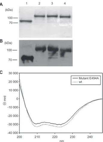
30 000 70 100 (kDa) 70 100 (kDa) 1
A
B
C
2 3 4 Mutant E494A wt 20 000 10 000 mrd 0 -10 000 -20 000 -30 000 -40 000 200 210 220 nm 230 240Figure 1 Purifi cation of Zmp1 and its structure in solution. (A) Coomassie stain of 12.5 % SDS-gel and (B) Western blotting of Zmp1 variants. Lanes: 1, Zmp1 wt; 2, GST-Zmp1; 3, GST-Zmp1 E494A; 4, GST-Zmp1 H493A. Zmp1 shows the predicted molecular mass of 72 kDa. The three GST-tagged variants have a molecular mass of approximately 100 kDa due to the fusion with a 26-kDa GST tag. (C) CD spectra of Zmp1. Dashed line corresponds to the CD spectrum of the Zmp1 wt; continuous line refers to the inactive E494A Zmp1.
(MMPs), sharing the catalytic motif (i.e., HExxH) with the
M13 family. GST-Zmp1 wt cleaved the fl uorogenic substrate
with high effi ciency and, as expected, mutating the catalytic
motif almost abolished the proteolytic activity. Likewise,
EDTA and the Zn
2 +chelator 1,10-phenanthroline reduced
Zmp1 activity (see Table 1
). Zmp1 activity was also strongly
inhibited by the potent M13 metallopeptidase inhibitor
phos-phoramidon with a
K
I= 3.5( ± 0.5) × 10
-8m
(Ferraris et al.
,
2011 ). The enzyme does not undergo autoproteolysis during
Table 1 Effect of peptidase inhibitors on the activity of Zmp1. Inhibitor Concentration (m m ) Zmp1 activity ( % of control) Phosphoramidon 0.1 5.1 ± 1.2 1,10-Phenanthroline 0.1 7.8 ± 3.2 EDTA 1 46.4 ± 8.3
Zmp1 (10 n m ) was preincubated with inhibitor for 10 or 60 min (in case of EDTA) at room temperature. Activity toward the synthetic fl uorogenic substrate was measured after 5 min at 37 ° C ( ± SD).
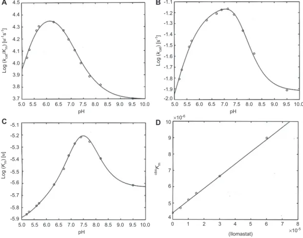
4.4 4.3 4.2 Log ( kcat /Km ) [ M -1s -1] Log ( Km ) [ M ] obs Km Log ( kcat ) [s -1] 4.1 4.0 3.9 -5.1 -5.2 -5.3 -5.4 -5.5 -5.6 -5.7 -5.8 -5.9 3.8 3.7 5.0 5.5 6.0 7.0 7.5 8.0 8.5 9.0 9.5 10.0 pH pH 6.5 5.0 5.5 6.0 7.0 7.5 8.0 8.5 9.0 9.5 10.0 pH 6.5 5.0 5.5 6.0 6.5 7.0 7.5 8.0 8.5 9.0 9.5 10.0 4.5
A
B
C
D
-1.1 -1.2 -1.3 -1.4 -1.5 -1.6 -1.7 -1.8 -1.9 10 9 8 7 6 5 4 0 1 2 3 4 5 6 7 8 (Ilomastat) ×10 -5 ×10⁻⁶ -2.0Figure 2 pH dependence of catalytic parameters for Zmp1 at 37 ° C against a synthetic fl uorogenic substrate.
(A) pH dependence of k cat / K m . Continuous line represents the non-linear least squares fi tting of data using Eq. (4a) with parameters reported in Table 2. (B) pH dependence of k cat . Continuous line represents the nonlinear least squares fi tting of data using Eq. (4b) with parameters reported in Table 2. (C) pH dependence of K m . Continuous line represents the non-linear least squares fi tting of data using Eq. (4c) with parameters
reported in Table 2. (D) Ilomastat concentration dependence of substrate affi nity constant K m for Zmp1 at 37 ° C. Values of obs K
m have been
obtained by Lineweaver-Burk plot of omni-MMPs fl uorogenic peptide cleavage in the presence of Zmp1 (0.4 n m ) in absence and presence of different Ilomastat concentrations (namely 5, 10, 15, 30, and 60 μ m ). The continuous line is the nonlinear least squares fi tting of data according to Eq. (5), from which a value of 0 K
m = 4.4( ± 0.6) × 10
-6 m and a value of K
I = 5.9( ± 0.7) × 10
-5 are obtained.
the activity assay, as proven by the Selwyn test (Selwyn ,
1965 ) (see Figure S1 ).
pH dependence of Zmp1 proteolytic activity
The pH dependence of Zmp1 activity was fi rst measured using
a synthetic fl uorogenic substrate. From the analysis of the
dependence on the substrate concentration at each pH value
according to Eqs. (2) and (3) (see Materials and methods),
we have obtained the catalytic parameters (i.e., k
cat/ K
m, k
cat,
and K
m) at each investigated pH. Their pH dependence is
reported in Figure 2 A – C for k
cat/ K
m(A), k
cat(B), and K
m(panel
C). obtaining the p K
avalues reported in Table 2
. Although
the nonlinear least squares fi tting of the pH dependence of
individual parameters might have been carried out with only
two protonatable groups, the global fi tting of the pH
depen-dence of all three catalytic parameters demanded three
proto-natable groups [i.e., n
= 3 in Eqs. (4a) – (4c); see Materials and
methods]. Evidently, the overall enzymatic activity of Zmp1
(corresponding to k
cat/ K
m) is highest at pH 6.3 (Figure 2A),
a pH value quite low compared with other metallopeptidases
(Fasciglione et al. , 2000 ). Further, the pH dependence of K
m(Figure 2C) indeed suggests that the substrate affi nity tends to
increase at pH lower than 7.5, as K
mdecreases. An
interest-ing pattern is observed for the pH dependence of k
cat(Figure
2B), where it appears evident that at least two groups with
markedly different p K
avalues are modulating this parameter.
In particular, it is worth underlining that the two-protonated
species appears to be the most active form of Zmp1 and that
this enhanced activity is connected to a residue characterized
by a p K
a= 6.73 ± 0.17 in the free enzyme, which then shifts to
p K
a= 7.72 ± 0.16 in the substrate-bound form (Table 2). As a
consequence, the pH-dependent profi le of k
catappears
some-what displaced toward higher pH values.
Zmp1 inhibition by Ilomastat
The inhibition constant by Ilomastat has been determined
for Zmp1, exploiting the inhibitor concentration
depen-dence of the substrate affi nity (Figure 2D). Application of
Table 2 Catalytic parameters for the different protonated species of Zmp1 toward the synthetic fl uorogenic substrate and p K a values for the protonating groups in the free enzyme (p K U ) and of the substrate-bound enzyme (p K L ). k cat / K m ( m -1 s -1) k cat (s -1) K m ( m ) 0-Protonated 5.0 ( ± 0.7) × 10 3 0.012 ± 0.004 2.4 ( ± 0.4) × 10 -6 1-Protonated 7.1 ( ± 0.9) × 10 3 0.131 ± 0.029 1.9 ( ± 0.3) × 10 -5 2-Protonated 3.0 ( ± 0.5) × 10 4 0.065 ± 0.017 2.2 ( ± 0.5) × 10 -6 3-Protonated 9.5 ( ± 1.6) × 10 2 0.008 ± 0.001 8.4 ( ± 1.6) × 10 -6 p K U1 8.17 ± 0.16 p K U2 6.73 ± 0.17 p K U3 5.43 ± 0.19 p K L1 7.22 ± 0.18 p K L2 7.72 ± 0.17 p K L3 5.69 ± 0.18
Eq. (5) (see Materials and methods) allows to determine the
K
I[
= 5.9( ± 0.7) × 10
-5m ], which is about 10
5-fold higher than
that observed for matrix metallopeptidases (Galardy et al. ,
1994 ), refl ecting a very low affi nity of Ilomastat for Zmp1.
This result indicates that Ilomastat is a very weak inhibitor for
Zmp1, confi rming the known difference between gluzincins
and metzincins, as indeed it turns out to be evident also from
the crystallographic structure, where the catalytic site results
different from that of other metallopeptidases (Ferraris et al. ,
2011 ).
Zmp1 hydrolyzes a variety of bioactive peptides
To possibly identify the native substrate(s) of Zmp1, we
car-ried out an extensive screening of a microarray-based
pep-tide library. The library consists of 1989 octameric peppep-tides
derived from annotated cleavage sites of peptidases or
ran-dom sequences and 1536 pentadecameric ranran-dom peptide
sequences. Peptides are C-terminally tagged with a
phospho-rylated tyrosine for detection. Peptide cleavage was
quanti-fi ed by incubation with antiphosphotyrosine and fl uorescent
secondary antibody labeling and following fl uorescence
measurement. Fluorescence intensity of 51 out of 3525
pep-tide spots was decreased by more than 90 % as compared
with the bovine serum albumin control. The 51 substrates
were categorized into three groups (Table S1
): (1)
neuropep-tides/substrates of NEP or endothelin-converting enzyme 1
(ECE-1); (2) coagulation factors; (3) others. Interestingly,
only short peptides (octameric) were represented among the
top substrates. The relative frequency of each amino acid in
the effi ciently cleaved peptides was calculated by dividing
the number of times this specifi c amino acid is represented
in the screened substrates over the number of times that the
amino acid is represented in the whole peptide microarray.
This statistical analysis provides information about the amino
acid preferences in the substrates, although it does not allow
prediction of actual cleavage sites. The most common motif
among the top substrates was the sequence SPFR, which is
present in the carboxy terminal part of the peptide hormone
bradykinin. In particular, the statistical analysis showed an
overrepresentation of arginine, phenylalanine, isoleucine,
leucine, and valine in the top substrate sequences, whereas
acidic amino acids such as aspartic or glutamic acid were
underrepresented, and no cysteine or tryptophan were present
in any of the 51 top substrates.
Zmp1 cleavage motif
Several cleavage motifs can be described from the
MALDI-TOF cleavage analysis of the investigated peptides (Figure
3
A). The phenylalanine was the most represented residue
at P1
′ position (Figure 3B), although isoleucine and leucine
were also abundant at this position. Proline was frequently
found in the P3 site, although other positions near the
cleav-age site showed little preference. We conclude that PxxF is a
preferred cleavage motif of Zmp1 and phenylalanine
occu-pies P1
′ position.
Identifi cation of substrate cleavage sites
First, we characterized the cleavage pattern of several
neuro-peptides identifi ed in the peptide array (i.e., bradykinin,
ape-lin-13, neurotensin, neuropeptide FF), including some NEP or
ECE-1 substrates [i.e., substance P, dynorphin A (1 – 13), and
1.0
B
A
0.5 0.0 P4 P3 P2 P1 P1′ P2′ P3′ P4′ ProbabilityFigure 3 Hydrolysis of hormone peptides by Zmp1.
(A) Cleavage sites detected in the Zmp1 hydrolyzed fl uorogenic pep-tides: 1, angiotensin I; 2, neurotensin; 3, apelin-13; 4, dynorphin A (1 – 13). Neuropeptide FF cleavage pattern was not studied because of poor detection signals in the MALDI-TOF analysis. (B) Amino acid preferences at various position of Zmp1 substrates calculated on the basis of the cleavage pattern of the studied hormone peptides. Size of amino acid letter is proportional to their occurrence in the recogni-tion/cleavage motif. Cleavage site (marked with red arrow) is loca-lized between position P1 and P1 ′ (drawn using Weblogo 3.0).
angiotensin I], and the reaction by-products were analyzed by
MALDI-TOF; to guarantee specifi c cleavage, a high substrate/
enzyme ratio was used (1000:1). Two cleavage sites were
detected in bradykinin (Figure 4 A), i.e., between Pro
7-Phe
8and
Gly
4-Phe
5, confi rming the results obtained with the extended
bradykinin, which is generated by the proteolytic cleavage of
the precursor kininogen-1 by the enzyme kallikrein (Kaplan
et al. , 2002 ). As a matter of fact, in rat, mouse, bovine, and
human, the SPFR motif of kininogen-1 is conserved, but it is
followed by a different carboxy terminal amino acid. Four zinc
metallopeptidases are mainly responsible for the metabolism
of bradykinin, namely angiotensin-converting enzyme (ACE),
NEP, aminopeptidase P, and carboxypeptidases M and N
(Moreau et al. , 2005 ). Both cleavages resulted in two respective
fragments that were detected by MALDI-TOF (Figure 4B and
C). We determined three cleavage sites in substance P: between
Gln
6-Phe
7, Phe
7-Phe
8, and Gly
9-Leu
10(Figure 4D; Figure S2).
Interestingly, all the cleavage sites of bradykinin and substance
P have a hydrophobic amino acid in the P1
′ site, mainly
phe-nylalanine, which is typical of NEP, ECE-1, and thermolysin;
20 000 Bradykinin Bradykinin+Zmp1 Mca-RPPGFSPFR-Dap(Dnp) Extended bradykinin
RPPGFSPFRSVTV
15 000 10 000 Intensity Intensity 5000 60 000 40 000 20 000 500 1000 1500Mca-RPKPQQFFGLM-Dap(Dnp)
Substance P m/z 1527.64 1511.65 972.41 641.27 574.21 558.22 889.37 905.38 2000 0 0 500 1000 1500 m/z 2000 List of mass peaksm/z=1527.64 MCA-RPPGFSPFR-Dap(Dnp) m/z=1511.65 MCA-RPPGFSPFR-Dap(Dnp)–(O) m/z=641.27 MCA-RPPG m/z=905.38 FSPFR-Dap(Dnp) m/z=889.37 FSPFR-Dap(Dnp)–(O) m/z=972.41 MCA-RPPGFSP m/z=574.21 FR-Dap(Dnp) m/z=558.22 FR-Dap(Dnp)–(O) 1511.65 1527.64
*
A
B
C
D
**
**
Figure 4 Cleavage sites of bradykinin and substance P by Zmp1.
(A) Determination of fragments of the ‘ extended ’ bradykinin (bradykinin sequence in bold) by Zmp1 through LC-MS analysis. Three different cleavage sites were detected in bradykinin. Primary site of hydrolysis is shown by the arrow with one asterisk, and secondary sites are shown by the arrows with two asterisks. (B) MALDI-TOF spectrum of fl uorogenic bradykinin. (C) MALDI-TOF spectrum of Zmp1 hydrolyzed fl uo-rogenic bradykinin. The C-terminal fragments showed an additional mass peak ( Δ 16 Da), due to a loss of oxygen (as described recently for nitro-phenyl-derivatives; see Petersson et al. , 2001 ). (D) Cleavage sites of fl uorogenic substance P by Zmp1. The arrows above the amino acid sequence of substance P indicate the cleavage sites.
further, this observation is in agreement with the
overrepre-sentation of this amino acid in the top substrates as compared
with the total peptide library. Specifi c sites of cleavage within
the other neuropeptides, such as angiotensin I (Figure S3),
apelin-13 (Figure S4), dynorphin A (1 – 13) (Figure S5), and
neurotensin (Figure S6) are summarized in Figure 3A.
Further, we synthesized a peptide fragment of mouse
kini-nogen with the sequence RPPGFSPFRSVTV to map the
cleavage site (bradykinin sequence in bold). The peptide was
incubated with Zmp1 for 60 and 120 min, respectively, and
products were analyzed by LC-MS. Cleavage sites were
deter-mined by calculating the molecular weight of putative
prod-ucts (data not shown). Primary cleavage occurred between
Pro
7and Phe
8(Figure 4A, arrow with one star). This cleavage
site lies within the bradykinin sequence and results in the
inac-tivation of bradykinin rather than releasing bradykinin from its
precursor. This peptide bond is also targeted by ACE, the main
enzyme responsible for the metabolism of bradykinin (Blais
et al.
, 2000
). Further cleavages were detected after longer
incubation of the peptide with Zmp1, e.g., between Gly
4-Phe
5and Ser
10-Val
11(Figure 4A, arrows with two stars).
Kinetic analysis of neuropeptides cleavage
Zmp1 hydrolysis of the neuropeptides was further studied
by the determination of kinetic constants K
mand k
cat. As the
kinetic course of peptide cleavage by Zmp1 was affected by
product inhibition and/or cleavage of multiple peptide bonds,
initial velocities were exploited for calculating the kinetic
parameters of the most susceptible peptide bond within a given
peptide. Results are summarized in Table 3
as best fi t values
with associated standard errors from nonlinear regression
of the Michaelis-Menten equation fi tted to data. The kinetic
parameters for angiotensin I and neurotensin could not be
cal-culated because even the initial velocity was strongly affected
by the concurrent multiple cleavage and multiple product
inhibition. Figure 5 A shows, as an example, the double
recip-rocal Lineweaver-Burk plot for substance P, apelin-13, and
bradykinin; it clearly emerges that substance P and apelin-13
are the best substrates among those investigated.
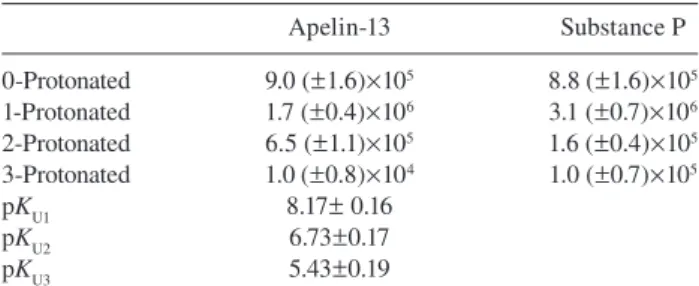
pH dependence of substance P and apelin-13 hydrolysis
In several studies, ECE-1 was found to cleave
neuropep-tides inside acidifi ed endosomes (i.e., at pH
≈ 5.5) (Padilla et
10
A
B
9 8 7 6 5 (E 0 )/v [s] Log ( kcat /K m ) [ M -1s -1] 4 3 2 1 0 6.5 6.4 6.3 6.2 6.1 6.0 5.9 5.8 5.7 6.0 7.0 8.0 pH 9.0 9.5 8.5 7.5 6.5 0 0.5 1.0 2.0 3.0 4.0 ×10⁶ 3.5 2.5 1/[Substrate] 1.5Figure 5 Kinetic analysis of Zmp1 hydrolysis of apelin-13, sub-stance P, and bradykinin.
(A) Double-reciprocal Lineweaver-Burk plot of the enzymatic pro-cessing by Zmp1 of apelin-13 (o), substance P (x), and bradykinin (*) at 37 ° C and pH 7.3. Solid lines represent the non-linear least squares fi tting of data according to Eq. (2). (B) pH dependence of the rate of cleavage ( k cat / K m ) of apelin-13 (o) and substance P (x) by Zmp1 at 37 ° C. Continuous lines represent the nonlinear least squares fi tting of data according to Eq. (4a).
Table 3 Kinetic parameters for the cleavage of the most susceptible peptide bond in fi ve peptides substrates of Zmp1.
Substrate k cat (s -1) K m ( μ m ) k cat / K m ( m -1 s -1 ) Apelin 1.54 ± 0.08 1.09 ± 0.23 1.41 ± 0.31 × 10 6 Substance P 4.78 ± 0.71 3.92 ± 1.37 1.22 ± 0.46 × 10 6 Bradykinin 2.76 ± 0.42 7.99 ± 1.45 3.46 ± 0.67 × 10 5 Dynorphin A 2.25 ± 0.22 5.72 ± 1.22 3.93 ± 0.92 × 10 5 Neuropeptide FF 1.07 ± 0.16 3.02 ± 1.15 3.56 ± 1.45 × 10 5
Values of k cat and K m , determined by non-linear regression from a fi t of the Michaelis-Menten equation to primary data, are shown together with the standard errors from regression.
al. , 2007 ; Roosterman et al. , 2007 ). We determined the pH
dependence of Zmp1 activity for additional substrates, i.e.,
apelin-13 and substance P to see the pH preference for these
substrates. Zmp1 cleaved the two neuropeptides preferentially
in the pH range between 6.5 and 8.0 rather than at more acidic
pH values, as observed for the fl uorogenic substrate (Figure 2
for the pH profi le of the fl uorogenic substrate, Figure 5B for
that of substance P and apelin-13). Data obtained from the
pH-dependence of apelin-13 and substance P degradation by
Zmp1 are reported in Table 4
.
Screening for macromolecular substrates (longer than 40 amino acids)
Using an in vitro proteolysis assay we have investigated the
enzymatic action of Zmp1 on four of the substrates reported
Table 4 Values of k cat / K m ( m -1 s -1 ) for the different protonated species
of Zmp1 toward apelin-13 and substance P and p K a for the protonating groups in the free enzyme (p K U ).
Apelin-13 Substance P 0-Protonated 9.0 ( ± 1.6) × 10 5 8.8 ( ± 1.6) × 10 5 1-Protonated 1.7 ( ± 0.4) × 10 6 3.1 ( ± 0.7) × 10 6 2-Protonated 6.5 ( ± 1.1) × 10 5 1.6 ( ± 0.4) × 10 5 3-Protonated 1.0 ( ± 0.8) × 10 4 1.0 ( ± 0.7) × 10 5 p K U1 8.17 ± 0.16 p K U2 6.73 ± 0.17 p K U3 5.43 ± 0.19
on the top substrates list of the peptide array:
β -amyloid,
α -fi brinogen chain, insulin, and denatured collagen. We have
further tested Zmp1 proteolytic activity on caspase 1, the
IL-1
β activating enzyme. However, even prolonged
exposi-tion to Zmp1 (up to 24 h) did not lead to any appreciable
cleavage of these potential substrates (data not shown). In
contrast, a control incubation with trypsin led to complete
digestion of the
α -, β -, and γ -fi brinogen chains already after
1 h of incubation.
Discussion
Zinc metallopeptidases represent promising
pharmacologi-cal targets and potential candidates for vaccine development
(Miyoshi and Shinoda , 2000 ; Aureli et al., 2008; Lopez -Otin
and Bond, 2008 ; Baillie et al. , 2010 ). We have investigated
the wt and the inactive mutants (H493A and E494A) of the
metallopeptidase Zmp1, which is an important virulence
fac-tor of Mtb (Master et al. , 2008 ).
Zmp1 belongs to the M13 family (MEROPS database:
http://merops.sanger.ac.uk/), such as NEP and ECE-1, whose
enzymatic action is restricted to peptides with a strong
prefer-ence for cleaving the amino terminal bond of hydrophobic
residues (Bland et al. , 2008 ). This preference for peptides is
also confi rmed by the recently solved X-ray crystallographic
structure of Zmp1, which reveals that the active site is located
at the bottom of a hydrophobic channel, likely impairing the
access of large macromolecular substrates in the proximity of
the reaction centre (Ferraris et al. , 2011 ).
To better characterize the specifi city of Zmp1 a peptide
microarray library was used (including octameric and
pen-tadecameric peptides); in particular, octameric peptides were
enzymatically cleaved with the highest effi ciency. Further,
a statistical analysis over the whole peptide array indicate a
marked tendency of Zmp1 to cleave bonds where
hydropho-bic amino acids are located at the P1
′ position, similarly to
NEP and ECE-1. In addition, as shown by the in vitro
clea-vage assay, sequences identifi ed by the peptide array could
not be cleaved when they were fl anked by the whole protein
sequences in a context of a structurally complex molecule
(e.g., fi brinogen,
β -amyloid).
Among the hits screened, several neuropeptides have been
identifi ed [i.e., substance P, neurotensin, bradykinin, apelin-13,
angiotensin I, neuropeptide FF, and dynorphin A (1 – 13)]. With
respect to these, it is interesting to remark that a member of M13
family (ECE-1) localizes into the luminal side of endosomal
membrane where it degrades interna lized neuropeptides (e.g.,
substance P, apelin-13) (El Messari et al. , 2004 ; Roosterman et
al. , 2007 ; Cottrell et al. , 2009 ), suggesting their possible
biolo-gical relevance as substrates of this type of enzymes. As a
mat-ter of fact, besides the classical involvement of neuropeptides
in neurotransmission and vasal smooth muscle cells
constric-tion, at least for some of them, a novel role in the modulation
of immune responses has been envisaged (Skidgel et al. , 1984 ;
Johnson et al. , 1999 ; Marriott and Bost , 2001 ; Yaraee et al. ,
2007
), affecting proinfl ammatory cytokine and chemokine
activation (in the case of apelin-13) and macrophage activation
(in the case of the substance P) in the early phases of immune
responses to Salmonella infection (Kincy -Cain and Bost, 1996 ;
O ’ Connor et al., 2004 ; Leeper et al. , 2009 ).
A comparison between the degradation profi le of these
substrates by Zmp1, NEP, and ECE-1 highlights overlapping
specifi cities in terms of cleavage sites (i.e., for bradykinin,
substance P, and dynorphin A) with minor differences (i.e.,
secondary cleavage sites in angiotensin I and neurotensin)
(Matsas et al. , 1984 ; Johnson et al. , 1999 ). However, a
sig-nifi cant quantitative difference in terms of catalytic effi ciency
with respect to ECE-1 is observed with k
cat/ K
mratios, which in
Zmp1 are 100- and 5-fold higher for substance P and
brady-kinin, respectively (Johnson et al. , 1999 ). On the whole,
ape-lin-13 turns out to be an excellent substrate for Zmp1, being
effi ciently cleaved with the highest specifi city constant (i.e.,
k
cat/ K
m) among all tested peptides, a feature mostly related to
the binding specifi city rather than the cleavage effi ciency.
Although we cannot state at this stage whether these
neuro-peptides represent the Zmp1 substrate in vivo , pH scanning of
Zmp1 proteolytic activity toward all investigated peptides
indi-cates that Zmp1 can modulate its activity through the
dynami-cal pH changes that occur in the maturating phagosome. In
this respect, it is noteworthy that degradation of neuropeptides
by ECE-1 in endosomes under acidic conditions (i.e., pH 5.5)
is documented (Fahnoe et al., 2000) and it is associated to
intracellular signaling and receptor turnover (El Messari et al. ,
2004 ; Roostermann et al., 2007; Cottrell et al. , 2009 );
analo-gously, we found that Zmp1 optimum activity toward the
syn-thetic substrate is at pH 6.3 (Figure 2A), a value corresponding
to that of the early endosome (Pethe et al. , 2004 ). Therefore,
it might be speculated that, similarly to ECE-1, Zmp1
oper-ates within the phagosome, arresting its maturation (Master
et al. , 2008 ; Johansen et al. , 2011 ). As a matter of fact, at the
acid pH of the maturating phagosome (i.e., pH 6.3), the
sub-strate affi nity of Zmp1 is fairly high (i.e., 2
μ m ), suggesting a
conformation of the active site particularly suitable to interact
with substrates under quite acidic pH conditions. In the case
of neuropeptides, such as apelin-13 and substance P, the
opti-mum pH is somewhat higher than that observed for the
syn-thetic substrate, although p K
avalues for the pH dependence of
k
cat/ K
mare fully comparable. This strongly supports the idea
that these p K
avalues refer only to the protonation of Zmp1
residues with no contribution from residues of the substrate(s).
Therefore, as the whole enzymatic mechanism of Zmp1 seems
to be modulated by the protonation of three classes of residues
(characterized by p K
a1, p K
a2, and p K
a3, respectively), the
dif-ferent pH profi les between these substrates can be simply
explained by the involvement of different protonated species
(the 2-protonated for the synthetic substrate and the
1-proto-nated for neuropeptides). This feature is clearly refl ecting a
relevant difference between the two classes of substrates in
their ionic interactions with the enzyme.
These investigations demonstrate the importance of Zmp1
for mycobacteria host interaction and highlight its potential
as a pharmacological target. Two major conclusions can be
drawn: Zmp1 preferentially cleaves small peptides, among
which neuropeptides appear particularly interesting; Zmp1
adapts its activity to different microenvironmental conditions.
The present biochemical characterization of the enzymatic
properties of Zmp1 can be merged with the structural
infor-mation on Zmp1 (Ferraris et al. , 2011 ), allowing a
structural-functional correlation, which will be of the utmost importance
for a rational drug design.
Materials and methods
Molecular cloning and protein expression
Mtb Zmp1 gene was subcloned into pET100/D-TOPO ® vector
(Invitrogen) using a PCR-amplifi ed product from pMV361-Zmp1 vector, carrying Zmp1 gene, as previously reported (Master et al. , 2008 ).
For all constructs, 1-L overnight culture of E. coli BL21 (Invitrogen) in L-broth medium (induced at OD 600 = 0.8 with 0.5 m m IPTG and grown at 18 ° C) was centrifuged at 5000 g for 20 min, followed by washing with PBS and additional centrifugation at 5000 g for 20 min. Cells were then lysed by three French press cy-cles (Thermo Electron) at 1.4 × 10 8 Pa in the presence of peptidase
inhibitors, followed by ultracentrifugation at 30 000 g for 60 min. Supernatants were loaded on HisTrap ™ HP column (GE Healthcare) equilibrated with buffer containing 20 m m NaH 2 PO 4 , pH 7.4, 0.5 m NaCl. Proteins were eluted with a concentration gradient of imida-zole. Both Zmp1 and GST-Zmp1 were eluted at 250 m m imidazole (protein concentration was 0.5 – 1 mg/ml). After overnight dialysis against PBS, Zmp1 wt was further purifi ed by ion exchange using a Q Column (GE Healthcare). GST-Zmp1 purifi cation was performed using glutathione-sepharose beads (Glutathione Sepharose 4B; GE Healthcare). Proteins were dialyzed against PBS, and fi nally, glycer-ol at a fi nal concentration of 10 % was added to store them in aliquots at -20 ° C. Proteins were analyzed by 12.5 % SDS-gel and subsequent Coomassie stain or Western blot against HA. All chemicals used were from Acros, Applichem, Fluka or Sigma-Aldrich. For detection of recombinant Zmp1 the following antibodies are used: mouse anti-HA (Roche) and goat anti-mouse-HRP (Molecular Probes).
CD experiments
CD spectra for the validation of the correct folding of the protein were recorded on a Jasco J-710 spectropolarimeter equipped with a thermostated cell holder and connected to a data station for signal averaging and processing. All spectra were averages of four scans and were recorded using quartz cells of 2-mm path length. Zmp1 was dissolved in CD buffer (20 m m Tris-HCl, 1 m m CaCl 2 , 100 m m NaCl) at about 2 μ m concentration. The α -helix content of Zmp1 was calculated from the molar ellipticity of circular dichroic spectrum at 222 nm according to the following equation
% α -helix = (-[ θ ] 222 nm + 3000)/39 000 (1) where [ θ ] 222 nm is the molar ellipticity at 222 nm, as previously described (Morrow et al. , 2000 ).
Kinetic analysis and pH dependence of Zmp1 catalysis
Internally quenched fl uorogenic substrates with 7-methoxycoumarin (MCA) as fl uorophore and 2,4-dinitrophenol (DNP) as quencher were either a synthetic fl uorogenic substrate, displaying the follow-ing sequence:
[(MCA-yl)acetyl]-Pro-Leu-Gly-Leu-DNP[ N l -2,3-diaminopropionyl]-Ala-Arg-NH 2 )
or neuropeptides from EMC microcollections (T ü bingen, Germany). Neurotensin was synthesized with glutamine as N-terminal amino acid instead of pyroglutamic acid for ease of synthesis. The substrate peptides were prepared as stock solutions in 100 % dimethyl sulfoxide (DMSO) at a fi nal concentration of 1.0 m m and further diluted into the univer-sal buffer (25 m m bis-Tris-HCl, 25 m m Tris-HCl, 100 m m NaCl, and 10 m m CaCl 2 prepared at 25 ° C) to maintain an unaltered composition at different pH values over the range investigated, keeping the DMSO concentration constant at 1 % (v/v) for all dilutions. Substrate hydrolysis was measured fl uorometrically with an Eclypse fl uorimeter (Varian) or with a Flx800 spectrofl uorometer (BIOTEK) equipped with KC Junior Software (BIOTEK) ( λ exc = 327 nm, λ em = 393 nm); reactions were start-ed by adding Zmp1 (at a fi nal concentration of 10 n m ) to a solu tion con-taining different substrates concentrations, spanning between 0.3 μ m and 5 μ m . For the pH dependence, Zmp1 was incubated in the universal buffer between pH 5.2 and 9.5 for 5 min at 37 ° C, then substrate was added, and fl uorescence signal was followed. Background fl uorescence of all buffers was similar. The initial velocities were measured at 37 ° C within a time interval during which the rate was constant and <10 % of the substrate has been degraded. It ensured a steady-state condition, and it was a prerequisite for the subsequent analysis step, which was based on the observation of an inverse linear correlation between velocity and substrate concentration according to Lineweaver-Burk equation [Eq. (2)] and Eadie-Hofstee linear regression [Eq. (3)]
[ ]
0 m cat cat1
1
SE
K
v
=
k
⋅ +
k
(2)MAX m m m m
-v V
v
K
S
S
K
K
K S
ε
+
=
+
(3)where E 0 is the total enzyme concentration, v is the actual rate (expressed as mol/s), K m is the Michaelis-Menten equilibrium con-stant (expressed as mol), k cat is the rate-limiting step kinetic constant
(expressed as s -1 ), [ S ] is the substrate concentration, and V MAX is the
ratio between E 0 and v . Linear regression plots were constructed from the velocity data, and the catalytic parameters k cat and K m were extracted. The pH dependence of the catalytic parameters has been fi tted according to the following equations
cat m ua obs 0 0 cat ua 0 0
/
/
i n r i r i r r i m i n r i r r r ik
K
K
H
k
K
K
H
= = + = = = = + = =⎡
⎤
⋅
⋅⎣ ⎦
=
⎡
⎤
⋅⎣ ⎦
∑
∏
∑∏
(4a)cat La obs 0 0 cat La 0 0 i n r i r i r r i i n r i r r r i
k
K
H
k
K
H
= = + = = = = + = =⎡
⎤
⋅
⋅⎣ ⎦
=
⎡
⎤
⋅⎣ ⎦
∑ ∏
∑∏
(4b)ua obs 0 0 0 m m La 0 0 i n r i r r r i i n r i r r r i
K
H
K
K
K
H
= = + = = = = + = =⎡
⎤
⋅⎣ ⎦
=
⋅
⎡
⎤
⋅⎣ ⎦
∑∏
∑∏
(4c) where obs k cat / K m , obs k cat, and obs Km are the observed parameters as
a function of pH; i k cat / K m ,
i k cat , and
i K
m are the parameters for the
i th-protonated forms; i K
ua are the i th protonation constants for the
free enzyme E; and i K
La are the i th protonation constant(s) for the
substrate-bound enzyme ES when it is involved in the cleavage event at the rate-limiting step. The global fi tting procedure, simultaneously using Eqs. (4a) – (4c) to describe the pH dependence of all three cata-lytic parameters of Zmp1 (i.e., k cat / K m , k cat , and K m ), allows to deter-mine the p K a of protonating groups, which modulate the enzymatic activity of Zmp1.
Inhibition assay
For inhibition experiments, Zmp1 (10 n m ) was preincubated with EDTA, phosphoramidon, and 1,10-phenanthroline (Sigma) in the uni-versal buffer (see above) at room temperature before addition of the synthetic fl uorogenic substrate. Fluorescence was measured after 5 min of incubation with the fl uorogenic substrate (5 μ m ) at 37 ° C. In the case of Ilomastat (British Biotech Pharmaceutical, Cowley, Oxford, UK), the inhibition constant K I was measured obtaining the various
K m from the substrate concentration dependence at various inhibitor concentration, using Eqs. (2) and (3) and then plotting data of K m as a function of inhibitor concentration, as from the following equation
[ ]
0 m m I1
I
K
K
K
⎛
⎞
=
⋅ +
⎜
⎟
⎝
⎠
(5)where K m is the Michaelis constant at different inhibitor concentra-tions, 0 K
m is the Michaelis constant in the absence of the inhibitor, [ I ]
is the inhibitor concentration, and K I is the inhibition constant.
Peptide array
Peptide array was performed at JPT Berlin using a commercially available microarray (batch 1562, both for peptidase incubation and control incubation in triplicates; JPT Peptide Technologies GmbH, Berlin, Germany). The library consists of 1989 octameric peptides derived from annotated cleavage sites of peptidases or random sequences and 1536 pentadecameric random peptide sequences. Peptides contain a C-terminal phosphorylated tyrosine for detec-tion with an antiphosphotyrosine antibody. The peptide microarray was incubated with 10 μ g/ml Zmp1 (total reaction volume 400 μ l) for 4 h at 37 ° C, using a microarray sandwich-like construction. As a control sample, an additional incubation was carried out in the absence of the target peptidase, which was followed by washing steps with 50 m m TBS buffer, pH 7.2, and ddH 2 O. Subsequent dry-ing with a microarray centrifuge was performed. Microarrays were fi rst incubated with anti-pTyr-antibody (for about 45 min) and then for 30 min with Dylight 649-labeled antimouse-antibody. For detec-tion, a Tecan HS4800 microarray processing station was used. Total incubation time was 45 min for anti-pTyr-100-antibody and 30 min for antimouse-antibody, followed by washing steps with 50 m m TBS, pH 7.2, and SSC buffer, pH 7.0 (JPT). Finally, the microarray plates were dried using a nitrogen stream. Analysis was performed with high-resolution fl uorescence scanner (Axon Genepix 4200 AL Scanner), and Spot-recognition software Genepix 6.0 and Microsoft
Excel were used for data analysis. A reduction by more than 90 % of signal intensity was considered as effective cleavage.
Cleavage site determination by MALDI-TOF
Peptide cleavage assay was performed as following: 100 μ m neuropep-tides were incubated with or without Zmp1 (20 n m ) for 5 min at 37 ° C in a total volume of 200 μ l. Then, 1 μ l of a 1:100 dilution with MALDI Matrix solution (consisting of 60 % acetonitrile, 0.1 % trifl uoroacetic acid in ddH 2 O, and 4 mg/ml α -cyano-4-hydroxy cinnamic acid) was spotted on the MALDI plate. After evaporation of solutes, MALDI spectra were recorded with a 4800 plus MALDI TOF/TOF Analyzer (Applied Biosystems, Foster City, CA, USA). MALDI was equipped with a 355-nm Nd:YAG laser that operates at a repetition rate of 200 Hz. MS spectra were acquired in the refl ectron mode, typically in the mass range from 600 Da to 4000 Da. The MS spectra were externally calibrated using a standard peptide mixture (Applied Biosystems). Recording software was 4000 Series Explorer, and for the fi nal prepa-ration of the fi gures, Data Explorer, Microsoft Excel, and Prism Graph were used.
LC-MS of bradykinin sequence
Bradykinin-elongated peptide with the sequence H 2
N-RPPGFSPFRSVTV-NH 2 (C-terminally tagged with an amide group to
avoid negative charge effects) was synthesized by JPT and dissolved in the assay buffer (100 m m Tris, pH 7.3, 100 m m NaCl, 10 m m CaCl 2 ) at a fi nal concentration of 1 mg/ml. Zmp1 was added to individual vials at concentrations of 10, 40, and 200 n m ; a vial was kept in the absence of peptidase as a control. At different time intervals, 8 μ l were injected into LC-MS system, and UV traces were recorded with Agilent LC-1200 VWD Detector at 220 nm; MS traces were recorded using Agilent Single-Quad G6130 mass spectrometer. All evaluations were performed with Agilent Chemstation B0301 data analysis software.
Acknowledgments
We acknowledge the support from the Functional Genomics Center Zurich (FGCZ). This work was also supported in part by the Swiss National Science Foundation (3100A0-135705 to P.S.), the Italian Ministry for University and Research (PRIN 200993WWF9 to M.C.), and the European Union (EU-PF7 New TB Vac, Project No. 241745 and EU-PF7 SysteMtb Collaborative Project no. 241587). B.A. was supported by the Forschungskredit 2010 from University of Zurich (54232101).
References
Aureli, L., Gioia, M., Cerbara, I., Monaco, S., Fasciglione, G.F., Marini, S., Ascenzi, P., Topai, A., and Coletta, M. (2008). Structural bases for substrate and inhibitor recognition by matrix metalloproteinases. Curr. Med. Chem. 15 , 2192 – 2222.
Baillie, L.W., Huwar, T.B., Moore, S., Mellado-Sanchez, G., Rodriguez, L., Neeson, B.N., Flick-Smith, H.C., Jenner, D.C., Atkins, H.S., Ingram, R.J., et al. (2010). An anthrax subunit vac-cine candidate based on protective regions of Bacillus anthracis protective antigen and lethal factor. Vaccine 28 , 6740 – 6748. Blais, C., Jr., Marceau, F., Rouleau, J.L., and Adam, A. (2000). The
kallikrein-kininogen-kinin system: lessons from the quantifi ca-tion of endogenous kinins. Peptides 21 , 1903 – 1940.
Bland, N.D., Pinney, J.W., Thomas, J.E., Turner, A.J., and Isaac, R.E. (2008). Bioinformatic analysis of the neprilysin (M13) family of peptidases reveals complex evolutionary and functional relation-ships. BMC Evol. Biol. 8 , 16.
Carson, J.A. and Turner, A.J. (2002). Beta-amyloid catabolism: roles for neprilysin (NEP) and other metallopeptidases ? J. Neurochem. 81 , 1 – 8.
Cole, S.T., Eiglmeier, K., Parkhill, J., James, K.D., Thomson, N.R., Wheeler, P.R., Honore, N., Garnier, T., Churcher, C., Harris, D., et al. (2001). Massive gene decay in the leprosy bacillus. Nature 409 , 1007 – 1011.
Cottrell, G.S., Padilla, B.E., Amadesi, S., Poole, D.P., Murphy, J.E., Hardt, M., Roosterman, D., Steinhoff, M., and Bunnett, N.W. (2009). Endosomal endothelin-converting enzyme-1: a regulator of beta-arrestin-dependent ERK signaling. J. Biol. Chem. 284 , 22411 – 22425. El Messari, S., Iturrioz, X., Fassot, C., De Mota, N., Roesch, D., and Llorens-Cortes, C. (2004). Functional dissociation of apelin receptor signaling and endocytosis: implications for the effects of apelin on arterial blood pressure. J. Neurochem. 90 , 1290 – 1301. Fahnoe, D.C., Knapp, J., Johnson, G.D., and Ahn, K. (2000).
Inhibitor potencies and substrate preference for endothelin-con-verting enzyme-1 are dramatically affected by pH. J. Cardiovasc. Pharmacol. 36 , S22 – S25.
Fasciglione, G.F., Marini, S., D ’ Alessio, S., Politi, V., and Coletta, M. (2000). pH- and temperature-dependence of functional modula-tion in metalloproteinases. A comparison between neutrophil col-lagenase and gelatinases A and B. Biophys. J. 79 , 2138 – 2149. Ferraris, D.M., Sbardella, D., Petrera, A., Marini, S., Amstutz, B.,
Coletta, M., Sander, P., and Rizzi, M. (2011). Crystal structure of Mycobacterium tuberculosis zinc-dependent metalloprotease-1 (Zmp1), a metalloprotease involved in pathogenicity. J. Biol. Chem. 286 , 32475 – 32482.
Galardy, R.E., Cassabonne, M.E., Giese, C., Gilbert, J.H., Lapierre, F., Lopez, H., Schaefer, M.E., Stack, R., Sullivan, M., Summers, B., et al. (1994). Low molecular weight inhibitors in corneal ulceration. Ann. NY Acad. Sci. 732 , 315 – 323.
Johansen, P., Fettelschoss, A., Amstutz, B., Selchow, P., Waeckerle-Men, Y., Keller, P., Deretic, V., Held, L., Kundig, T.M., Bottger, E.C., et al. (2011). Relief from Zmp1-mediated arrest of pha-gosome maturation is associated with facilitated presentation and enhanced immunogenicity of mycobacterial antigens. Clin. Vaccine Immunol. 18 , 907 – 913.
Johnson, G.D., Stevenson, T., and Ahn, K. (1999). Hydrolysis of peptide hormones by endothelin-converting enzyme-1. A com-parison with neprilysin. J. Biol. Chem. 274 , 4053 – 4058. Kaplan, A.P., Joseph, K., and Silverberg, M. (2002). Pathways for
bradykinin formation and infl ammatory disease. J. Allergy Clin. Immunol. 109 , 195 – 209.
Kincy-Cain, T. and Bost, K.L. (1996). Increased susceptibility of mice to Salmonella infection following in vivo treatment with the substance P antagonist, spantide II. J. Immunol. 157 , 255 – 264. Leeper, N.J., Tedesco, M.M., Kojima, Y., Schultz, G.M., Kundu,
R.K., Ashley, E.A., Tsao, P.S., Dalman, R.L., and Quertermous, T. (2009). Apelin prevents aortic aneurysm formation by inhib-iting macrophage infl ammation. Am. J. Physiol. Heart Circ. Physiol. 296 , H1329 – H1335.
Lopez-Otin, C. and Bond, J.S. (2008). Proteases: multifunctional enzymes in life and disease. J. Biol. Chem. 283 , 30433 – 30437. Madani, R., Poirier, R., Wolfer, D.P., Welzl, H., Groscurth, P., Lipp,
H.P., Lu, B., El Mouedden, M., Mercken, M., Nitsch, R.M., et al. (2006). Lack of neprilysin suffi ces to generate murine amy-loid-like deposits in the brain and behavioral defi cit in vivo. J. Neurosci. Res. 84 , 1871 – 1878.
Marriott, I. and Bost, K.L. (2001). Substance P receptor mediated macrophage responses. Adv. Exp. Med. Biol. 493 , 247 – 254. Masjedi, M.R., Farnia, P., Sorooch, S., Pooramiri, M.V., Mansoori,
S.D., Zarifi , A.Z., Akbarvelayati, A., and Hoffner, S. (2006). Extensively drug-resistant tuberculosis: 2 years of surveillance in Iran. Clin. Infect. Dis. 43 , 841 – 847.
Master, S.S., Rampini, S.K., Davis, A.S., Keller, C., Ehlers, S., Springer, B., Timmins, G.S., Sander, P., and Deretic, V. (2008). Mycobacterium tuberculosis prevents infl ammasome activation. Cell Host Microbe 3 , 224 – 232.
Matsas, R., Kenny, A.J., and Turner, A.J. (1984). The metabolism of neuropeptides. The hydrolysis of peptides, including enkepha-lins, tachykinins and their analogues, by endopeptidase-24.11. Biochem. J. 223 , 433 – 440.
Miyoshi, S. and Shinoda, S. (2000). Microbial metalloproteases and pathogenesis. Microbes Infect. 2 , 91 – 98.
Moreau, M.E., Garbacki, N., Molinaro, G., Brown, N.J., Marceau, F., and Adam, A. (2005). The kallikrein-kinin system: current and future pharmacological targets. J. Pharmacol. Sci. 99 , 6 – 38. Morrow, J.A., Segall, M.L., Lund-Katz, S., Phillips, M.C., Knapp,
M., Rupp, B., and Weisgraber, K.H. (2000). Differences in stabil-ity among the human apolipoprotein E isoforms determined by the amino-terminal domain. Biochemistry 39 , 11657 – 11666. Muttucumaru, D.G., Smith, D.A., McMinn, E.J., Reese, V., Coler,
R.N., and Parish, T. (2011). Mycobacterium tunerculosis Rv0198c, a putative matrix metalloprotease, is involved in patho-genicity. Tuberculosis (Edinb) 91 , 111 – 116.
Nguyen, L. and Pieters, J. (2005). The Trojan horse: survival tactics of pathogenic mycobacteria in macrophages. Trends Cell Biol. 15 , 269 – 276.
O ’ Connor, T.M., O ’ Connell, J., O ’ Brien, D.I., Goode, T., Bredin, C.P., and Shanahan, F. (2004). The role of substance P in infl am-matory disease. J. Cell Physiol. 201 , 167 – 180.
Padilla, B.E., Cottrell, G.S., Roosterman, D., Pikios, S., Muller, L., Steinhoff, M., and Bunnett, N.W. (2007). Endothelin-converting enzyme-1 regulates endosomal sorting of calcitonin receptor-like receptor and beta-arrestins. J. Cell Biol. 179 , 981 – 997.
Petersson, A.S., Steen, H., Kalume, D.E., Caidahl, K., and Roepstorff, P. (2001). Investigation of tyrosine nitration in proteins by mass spectrometry. J. Mass Spectrom. 36 , 616 – 625.
Pethe, K., Swenson, D.L., Alonso, S., Anderson, J., Wang, C., and Russell, D.G. (2004). Isolation of Mycobacterium tuberculosis mutants defective in the arrest of phagosome maturation. Proc. Natl. Acad. Sci. USA 101 , 13642 – 13647.
Roosterman, D., Cottrell, G.S., Padilla, B.E., Muller, L., Eckman, C.B., Bunnett, N.W., and Steinhoff, M. (2007). Endothelin-converting enzyme 1 degrades neuropeptides in endosomes to control recep-tor recycling. Proc. Natl. Acad. Sci. USA 104 , 11838 – 11843. Selwyn, M.J. (1965). A simple test for inactivation of an enzyme
dur-ing assay. Biochim. Biophys. Acta 105 , 193 – 195.
Skidgel, R.A., Engelbrecht, S., Johnson, A.R., and Erdos, E.G. (1984). Hydrolysis of substance p and neurotensin by converting enzyme and neutral endopeptidase. Peptides 5 , 769 – 776. Turner, A.J., Isaac, R.E., and Coates, D. (2001). The neprilysin (NEP)
family of zinc metalloendopeptidases: genomics and function. Bioessays 23 , 261 – 269.
Warner, D.F. and Mizrahi, V. (2007). The survival kit of Mycobacterium tuberculosis. Nat. Med. 13 , 282 – 284.
Yaraee, R., Ebtekar, M., Ahmadiani, A., Sabahi, F., and Ghazanfari, T. (2007). The effect of substance P on nitric oxide production by HSV-1 infected macrophages. Int. Immunopharmacol. 7 , 135 – 139. Received January 20, 2012; accepted March 29, 2012