Département de Biologie Unité de Biologie Végétale Université de Fribourg (Suisse)
Arabidopsis-Phytophthora, un pathosystème modèle pour la caractérisation
d’une interaction entre une plante et un pathogène oomycète
THESE
présentée à la Faculté des Sciences de l’Université de Fribourg (Suisse) pour l’obtention du grade de Doctor rerum naturalium
Présentée par Alexandra Roetschi de
Soleure, Oensingen et Bolken (SO) Diss. No 1345
Acceptée par la faculté des Sciences de l’Université de Fribourg (Suisse) sur la proposition du
Professeur Dr. Felix Mauch, du Professeur Dr. Ulrich Gisi et du Professeur Dr. Jean-Pierre
Métraux.
SUMMARY
Oomycetes are pathogens responsible for many plant diseases over the world and the economical impact of their damage is quite important. Although these organisms show a mycelar growth, their biology is quite different from that of fungi. This make them not easy to fight against and up to now no fungicide is able to stop an epidemy due to Oomycetes in a durable way. The particular biology of these organisms, which is still not well known, make them very interesting to study together with the study of the mechanisms of the interactions with their hosts.A novel pathosystem using Phytophthora porri as pathogen, an Oomycete infecting, among other hosts, cabbage (Brassica sp.) and Arabidopsis thaliana, a little weed from the family of the Brassicacea, was established. The choice of both protagonists was justified by the feature of plant model owned by A. thaliana and because P. porri is a facultative biotroph, an advantage not shared by every Oomycete. With this pathosystem, it will be possible, for the first time, to study not only the plant-Oomycete interaction but also both organisms separately because they are amenable to molecular and genetical studies.
This thesis is the next step after my diploma work, which aim was to set up the novel pathosystem. For this, 15 accessions of A. thaliana and 7 isolates of P. porri were screened in order to find accessions that could be hosts for P. porri and to establish a reproducible and reliable inoculation method. The cytological characterisation of the interaction enabled to distinguish between two distinct interactions. First, an incompatible interaction in which the growth of P. porri is rapidly stopped by the various hindrances built up by A. thaliana, among them the hypersensitive reaction which represents the death of one or several cells around the location where the pathogen attempted to penetrate. Secondly, a compatible interaction, in which the colonisation of the tissue by the pathogen takes places without any visible reaction of the plant. P. porri is able to complete its whole life cycle by producing its vegetative and sexual structures in the plant tissue of the compatible host, which confirms that this Oomycete is a true pathogen of A. thaliana.
In this thesis, the implication of different biosynthetic pathways leading to resistance in A. thaliana, as observed in other plant-pathogen interactions, was investigated during the interaction between this plant and P. porri. Biochemical and molecular analysis established that the main defense pathways of A. thaliana, namely the salicylic acid-, the jasmonic acid- and the ethylene pathways as well as the one leading to the biosynthesis of the phytoalexins are induced during the interaction but are not the principal components of the resistance phenotype. The study of mutants impaired in these four different biosynthetic pathways resulted in the discovery that a mutant originally found in a screen for deficiency in phytoalexin accumulation after inoculation with a virulent bacteria and named pad 2-1, showed a hypersusceptible phenotype towards P. porri when all the other mutants (npr 1-1, jar 1-1, etr 1-1, ein 2.1 and pad 3-1) and the transformant nahG showed, like the wild type, resistance. The induction of systemic acquired resistance, with a biotic and an abiotic inducer, helped to show that the salicylic acid pathway is not involved because there is no switch from the susceptible phenotype towards a resistant phenotype when this pathway is turned on. The biochemical and molecular analysis also showed that pad2 is impaired in the salicylic acid pathway. So, the resistance towards P. porri seems to be under the control of a different mechanism than the one known so far for plant-Oomycete interactions and requires a functionnal PAD2 gene. A genetical analysis showed that the resistance is under the control of at least one resistance gene and this was confirmed by the susceptible phenotype of two mutants (ndr 1-1 and eds 1.2) which are impaired in the signalling pathways lying downstream of the gene-for-gene recognition events.
Another aspect investigated was the role of a small protein during the infection process. This protein is abundantly secreted by P. porri in liquid culture and the infiltration of a culture filtrate in leaves of Nicotiana benthamiana, a Soleanaceous species very sensitive towards different elicitors, caused necrosis in the infiltrated area. When A. thaliana leaves were infiltrated with the same culture filtrate, a differential reaction was observed which indicates a recognition mechanism depending on the accession tested. The protein responsible for these reactions was identified as an elicitin and was
RESUME
Les Oomycètes sont de redoutables pathogènes pour les végétaux, particulièrement pour les plantes de culture et les pertes annuelles occasionnées par ces organismes sont considérables. Leur biologie est très différente de celle des champignons, même s’ils partagent avec ces derniers un mode de croissance mycélaire. Ainsi, il n’est pas aisé de les combattre et il n’existe que peu de fongicides capables de stopper une épidémie de manière durable. Leur biologie particulière, encore peu connue, rend ces organismes très intéressants à étudier de même que les mécanismes régissant les interactions avec leurs hôtes respectifs.Un nouveau pathosystème a été élaboré en utilisant Phytophthora porri, un Oomycète infectant, entre autres, les choux (Brassica sp.) comme pathogène et Arabidopsis thaliana, une petite Brassicacée, comme hôte. Ce choix a été motivé par le caractère de plante modèle possédé par A. thaliana ainsi que par la qualité de biotrophe facultatif de P. porri, avantage que l’on ne retrouve pas chez tous les Oomycètes. Ainsi, pour la première fois lors d’une interaction entre un Oomycète et une plante, il est possible d’étudier non seulement l’interaction mais aussi les deux partenaires aux niveaux moléculaires et génétiques.
Cette thèse a fait suite à mon travail de diplôme, dans lequel il s’est agi, à partir d’une quinzaine d’écotypes d’A. thaliana et de sept isolats de P. porri, d’une part de déterminer si cette plante pouvait servir d’hôte pour ce pathogène et d’autre part, d’établir une méthode d’inoculation fiable et reproductible. Ainsi, lorsque A. thaliana est infectée par P. porri, deux types d’interactions ont pu être observées lors de la caractérisation cytologique. On trouve une interaction incompatible, dans laquelle P. porri est très rapidement stoppé dans sa progression par les différentes barrières mises en place par A. thaliana, dont par exemple la réaction hypersensible qui représente la mort cellulaire d’une ou de plusieurs cellules végétales à l’endroit où le pathogène a tenté de s’introduire. On observe aussi une interaction compatible, dans laquelle la colonisation du tissu par le pathogène a lieu sans qu’il y ait de réaction visible de la part de la plante. P. porri est ainsi capable d’effectuer son cycle de vie en élaborant ses structures de reproduction végétative et sexuée dans le tissu végétal de l’hôte compatible, ce qui confirme que cet Oomycète est un véritable pathogène pour A. thaliana.
Dans cette thèse, l’implication de différentes voies de biosynthèse présentes chez A. thaliana et conduisant à un état résistant dans d’autres pathosystèmes a été étudié dans le cadre de l’interaction entre cette Brassicacée et P. porri. Il a pu être établi par une analyse biochimique et moléculaire, que les principales voies de défense, à savoir celles de l’acide salicylique, de l’acide jasmonique, de l’éthylène ainsi que celle conduisant à la synthèse des phytoalexines sont mises à contribution lors du processus d’infection mais qu’elles ne sont toutefois pas les principales responsables de l’état résistant. Par ailleurs, de l’étude de mutants de ces quatre différentes voies de biosynthèse, un mutant, originellement isolé lors d’un criblage pour la déficience dans l’accumulation de phytoalexines après inoculation avec une bactérie virulente et nommé de ce fait pad 2-1, a été mis en évidence car ce dernier a présenté un phénotype d’extrême susceptibilité envers P. porri alors que les autres mutants (npr 1-1, jar 1-1, etr 1-1, ein 2.1, pad 3-1) et le transformant nahG, de même que l’écotype sauvage sont eux restés résistants suite à leur inoculation avec cet Oomycète. Des expériences d’induction d’une réaction systémique acquise, effectuées à l’aide d’un inducteur biotique et abiotique, ont donné une indication supplémentaire que la voie de l’acide salicylique, qu’elle soit enclenchée ou non, n’a pas le pouvoir d’inverser un phénotype susceptible vers un phénotype résistant. Ceci a aussi permis de confirmer les résulats des analyses biochimiques et moléculaires, desquelles découlent que pad2 est aussi empêché dans la voie de l’acide salicylique. Ainsi, la résistance envers P. porri semble être régie par un mécanisme différent de ce que l’on connaissait jusqu’à présent pour d’autres interactions impliquant une plante et un Oomycètre et nécessite un gène PAD2 fonctionnel. Par ailleurs, une analyse génétique a permis d’établir que la résistance envers P. porri est régie par au moins un gène de résistance et ceci s’est vu confirmé par le phénotype susceptible de deux mutants (ndr 1-1 et eds 1.1) empêchés dans les voies de biosynthèse situées en aval de la reconnaissance gène pour gène.
Un autre aspect étudié a été le rôle d’une petite protéine lors du processus d’infection. Cette dernière est abondamment sécrétée par P. porri lorsqu’il est placé en culture liquide. L’infiltration d’un filtrat de culture dans des feuilles de Nicotiana benthamiana, une Solanacée très sensible envers divers éliciteurs, a causé des nécroses sur la surface infiltrée alors que chez A. thaliana une réaction différentielle a pu être mise en évidence, ce qui indique un processus de reconnaissance selon l’écotype testé. La protéine responsable de ces réactions a été identifiée comme étant une élicitine et a été nommée Porrine I. Le gène codant pour cette dernière a été cloné est s’est révélé faire partie d’une famille multigénique à l’instar des autres élicitines isolées chez la plupart des Phytophthora. L'ARN de Porrine I a pu être mis en évidence lors de l’interaction compatible toutefois il n’a pas été possible, dans le cadre de cette thèse, de clarifier la fonction biologique de cette élicitine.
TABLE DES MATIERES
Chapitre 1: Introduction générale 1
L’interaction entre Arabidopsis et Phytophthora
Chapitre 2: Characterization of an Arabidopsis-Phytophthora pathosystem: 10 Resistance requires a functionnal PAD2 gene and is independent
of salicylic acid-, ethylene- and jasmonic acid-signaling A. Roetschi, A. Si-Ammour, B. Mauch-Mani, and F. Mauch Submitted for publication
Chapitre 3: Les mécanismes de défense impliqués dans l’interaction A. thaliana- 27 P. porri nécessitent des gènes NDR1 et EDS1 fonctionnels et l’état
résistant ne peut être induit ni par un traitement au BTH ni par une
bactérie avirulente
Chapitre 4: Analyse de Porrine I, une élicitine produite in vitro et in planta 44 par Phytophthora porri
Chapitre 5: Conclusion générale 65
Chapitre 1
INTRODUCTION GENERALE
L’interaction entre Arabidopsis et Phytophthora
Dans leur environnement naturel, les plantes sont constamment confrontées à des microorganismes d’origines diverses (bactériennes, virales ou fongiques) dont certains se révèleront être des pathogènes. Ceci implique, du côté des plantes, le développement de stratégies fort élaborées qui leur donneront la possibilité de se défendre envers ces différents envahisseurs. De même, ces derniers vont aussi développer des stratégies leur permettant de contourner les défenses mises en place par les plantes. Un aspect intéressant à étudier est comment les différents mécanismes issus des plantes et des pathogènes interagissent entre eux et quelle est leur régulation. Pour cela, il est important de se munir de bons outils d’étude, tant du côté de la plante que du côté du pathogène. Dans cette optique, un nouveau pathosystème a été mis au point, ouvrant ainsi la porte à de nouvelles investigations.
L’hôte: Arabidopsis thaliana
A. thaliana (fausse arabette), considérée comme “mauvaise herbe“ lorsqu’elle apparaît dans les jardins, est en fait un organisme particulièrement utile pour des études en laboratoire. En effet, appartenant à la vaste famille des Brassicacées (env. 340 genres et 3350 familles), elle est apparentée à des plantes de culture (par ex. le colza) ou à certains légumes tels les choux ou les radis. Sa petite taille, les commodités relatives à sa culture ainsi que son temps de génération (de graine à graine) rapide (env. 2 mois) rendent A. thaliana pratique à utiliser en chambre de culture. C’est aussi un bon outil pour effectuer des études génétiques car il est relativement aisé de procéder à croisements manuels sur ses fleurs, permettant ainsi de suivre un trait de caractère donné sur plusieurs générations. Par ailleurs, son génome diploïde est l’un des plus petits parmi les plantes à fleurs (123 Mbp pour 5 chromosomes) et il est totalement séquencé. C’est aussi un organisme facilement mutagénisable (par agents chimiques) et transformable (par la bactérie Agrobacterium tumefaciens), ce qui a pour conséquence de pouvoir engendrer de nombreux mutants et transformants intéressants à étudier.
Face à certains phénomènes, il est commun d'utiliser des modèles pour tenter de les expliquer. La biologie n'échappe pas à cette règle et elle recherche toujours des organismes pouvant servir de modèles pour d'autres organismes que l'on aimerait bien modéliser. Ainsi, A. thaliana diffère des autres organismes utilisés comme modèles par le fait que contrairement à Saccharomyces cerevisiae (modèle pour les eucaryotes), Caenorhabditis elegans ou Drosophila melanogaster (modèles pour le développement animal) elle est très apparentée aux espèces pour lesquelles elle peut servir de modèle. En effet, toutes les angiospermes partagent un cycle de vie, un mode de reproduction et un plan corporel plus ou moins similaire. Selon toute vraisemblance, ces dernières ont dû évoluer à partir d'un ancêtre commun, ce qui donne à penser que les gènes d'A. thaliana présentent une certaine homologie avec ceux de beaucoup d'autres angiospermes.
Le pathogène: Phytophthora
Les Oomycètes, classe à laquelle appartient le genre Phytophthora, sont des organismes à croissance mycélaire. Les similitudes avec les champignons s’arrêtent toutefois là; en effet, les Oomycètes, ne sont pas apparentés à ces derniers mais plutôt aux algues hétérokontes photosynthétiques (Dick, 1990; Förster et al., 1990) comme l’indique la figure 1. Ils ont été classifiés à part parmi les Stramenopila (Barr, 1992), malgré le fait que l’on parle d’eux encore sous le qualificatif de champignons.
Figure 1 : Illustration de la position des Oomycètes par rapport
aux champignons. Ce schéma a été réalisé sur la base d'une étude de la petite sous-unité de l'ARN ribosomal. Tiré de Alexopoulos et al., 1996.
La plupart des Oomycètes sont des pathogènes et sont responsables de nombreuses maladies touchant les plantes, principalement les plantes de culture. L’organisme probablement le plus connu est Phytophthora infestans qui s’attaque à la pomme de terre et qui a été responsable d’une famine et d’un exode sans précédents en Irlande au 19 ième siècle. Mais P. infestans n’est pas le seul à
créer des problèmes, d’autres membres du genre Phytophthora ainsi que les espèces appartenant au genre Pythium sont de redoutables pathogènes lorsque les conditions climatiques sont appropriées, c’est à dire par temps humide et par une température externe oscillant entre 15 et 18°C. Les deux genres cités sont des organismes que l’on qualifie de biotrophes facultatifs car ils sont capables de survivre indépendamment de l’hôte. Parmi les Oomycètes, il y a encore quatre autres genres
cette raison qu’ils sont considérés comme étant plus évolués par rapport à Pythium et Phytophthora. Toutefois, ils restent encore dépendants des conditions atmosphériques appropriées pour effectuer leurs infections. La figure 2 illustre la phylogénie des différents genres appartenant à la classe des Oomycètes.
Phytophthora Algues hétérokontes Pythium Peronospora Plasmopara Peronosclerospora Sclerospora
PATHOGENES DES GRAMINEES PATHOGENES DES ANGIOSPERMES
biotrophes facultatifs biotrophes obligatoires
Figure 2 : Illustration de la phylogénie des Oomycètes. Les algues hétérokontes sont
considérées comme étant l'ancêtre commun de tous les genres. Adapté d'après Shaw, 1981.
Comme la biologie des Oomycètes est très différente de celle des champignons et qu’elle n’est pas encore très connue, il n’est pas aisé de combattre ces organismes, les fongicides classiques restant inefficaces. Ceci constitue, entre autre, une des raisons pour lesquelles il est important d’apprendre à mieux connaître ces organismes.
L’utilité d’un tel pathosytème
Plusieurs pathosystèmes impliquant un Oomycète et une plante sont étudiés à l’heure actuelle mais si l’interaction peut être étudiée, il n’en va pas de même pour l’un des deux partenaires. En effet, dans les interactions impliquant d’une part la laitue (Lactuca sativa L.) et Bremia lactucae Regel (MacLean et al., 1979) et d’autre part A. thaliana et Peronospora parasitica (Koch and Slusarenko, 1990), les pathogènes sont des biotrophes obligatoires, ce qui rend leur étude en dehors de leur hôte quasiment impossible. En ce qui concerne les deux interactions suivantes, celle entre la pomme de terre et P. infestans (Fry et al., 1997) et celle entre le soja et Phytophthora sojae (Schmitthenner, 1985), quelques problèmes peuvent survenir du côté des plantes. En effet, la pomme de terre étant un organisme tetraploïde, cela complique les études génétiques alors que le soja, lui, ne se laisse pas transformer ce qui peut aussi constituer un sérieux obstacle.
Ainsi l’interaction entre A. thaliana et Phytophthora, outre sa nouveauté, présente le grand avantage de pouvoir étudier ce qui se passe lors l’interaction mais aussi de procéder à des expériences sur les deux partenaires de manière séparée. En effet, tant A. thaliana que Phytophthora peuvent être étudiés au niveau génétique et moléculaire et du fait de sa qualité de biotrophe facultatif, Phytophthora est cultivable in vitro sur un milieu approprié.
Les objectifs
Il s’est agit tout d’abord de trouver un représentant des Phytophthora qui puisse infecter A. thaliana, P. infestans ayant été écarté vu qu’il ne déclenche qu’une réaction non-hôte chez A. thaliana (Kamoun et al. 1999). Le choix s’est donc porté sur P. porri qui est un organisme, initialement isolé du poireau (Allium porrum L.) (Foister, 1931) mais qui n’est pas un pathogène restreint aux seules Liliacées, tels les oignons (Allium cepa L.) ou les échalottes (Allium bakeri Regel) (Hickman, 1943; Katsura et al., 1969). D’autres légumes, tels les choux (Brassica sp.) ou la carotte (Daucus carota) sont aussi concernés par P. porri (Semb, 1971; Geeson, 1976; Stelfox and Henry, 1978; Ho, 1983). Des cas de pathogénicité de la campanule (Campanula persicifolia), de l’oeillet (Dianthus caryophylus) ou des marguerites (Chrysanthemum sp.) ont également été signalés (Legge, 1951; Kouyeas, 1977). P. porri présente une carte de distribution assez vaste car des infections, dont il a été identifié comme étant l’agent causal, ont été signalées en Europe occidentale (Italie, Belgique, Angleterre, Pays-Bas, Grèce), au Canada, en Afrique du Sud, en Australie, au Japon ainsi qu’aux Etats-Unis.
Selon une étude moléculaire effectuée sur 13 isolats de P. porri provenant de 5 hôtes différents, il a été observé que les isolats pathogènes des choux et de la carotte ne présentent qu’une similarité limitée avec les isolats pathogènes des Liliacées (De Cock et al., 1992). Cette distinction est aussi appuyée par d’autres facteurs, dont des tests de pathogénicité. En effet, les isolats pathogènes pour le poireau ne le sont pas pour le chou et vice-versa. Il a, par conséquent, été suggéré que les isolats pathogènes des Brassicacées et de la carotte pourraient être des espèces différentes, voire des sous-espèces, malgré leur ressemblance morphologique avec les isolats pathogènes des Liliacées.
Ainsi, comme A. thaliana et les choux font partie de la même famille, c’est-à-dire des Brassicacées, il y avait quelque chance que les isolats de P. porri spécifiques aux choux soient capables d’infecter A. thaliana. A partir d’une quinzaine d’écotypes d’A. thaliana et de 7 isolats de P. porri, il s’est agit tout d’abord de trouver une méthode d’infection fiable et facilement reproductible. Puis des combinaisons entre les différents écotypes et isolats ont été effectués de manière à trouver des réactions différentes de la part d’A. thaliana. De cette première étape, deux interactions ont pu être mises en évidence, la première impliquant l’écotype Columbia et l’isolat HH et qui se solde par une réaction d’incompatibilité, c’est-à-dire que cet écotype est capable de se défendre et de stopper ainsi la progression de P. porri. La deuxième implique l’écotype Landsberg erecta et l’isolat HH et elle se solde par une réaction de compatibilité car P. porri est capable de coloniser le tissu végétal et d’effectuer son cycle de vie sur cet hôte. Avec ces deux types de réactions, l’établissement de ce pathosystème a pu être légitimé et cela a permis d’ouvrir la voie à de nouvelles perspectives.
Les différentes expériences effectuées au cours de ce travail de recherche ont été regroupées en trois chapitres. Chaque chapitre fesant l’objet d’une introduction propre, les courtes introductions présentées ci-après ont pour but de servir de préambule de manière à pouvoir situer le contexte de ces expériences.
Etude de l’implication des principaux mécanismes de résistance présents chez A.
thaliana lors de l’interaction avec P. porri
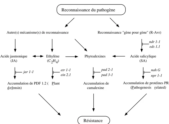
A. thaliana possède plusieurs mécanismes de défense qu’elle peut enclencher lorsqu’il y a menace de la part d’un pathogène, qu’il soit d’origine fongique, bactérienne ou virale. Ces différents mécanismes sont extrêmement complexes et semblent être d’une certaine manière reliés entre eux. L’implication de cinq mécanismes, à priori différents mais peut-être bien plus liés qu’on ne le pense à l’heure actuelle, a été étudiée lors de l’interaction entre A. thaliana et P. porri. La figure 3 en illustre une version schématisée.
Reconnaissance du pathogène Résistance Acide jasmonique (JA) Ethylène (C2H4) Accumulation de PDF 1.2 ( Plant d efensin) jar 1-1 etr 1-1 ein 2.1 Acide salicylique (SA) Accumulation de protéines PR (Pathogenesis related) nah G npr 1-1 Phytoalexines Accumulation de camalexine pad 2-1 pad 3-1
Reconnaissance "gène pour gène" (R-Avr) Autre(s) mécanisme(s) de reconnaissance
ndr 1-1 eds 1.1
Figure 3 : Schéma, simplifié, des différentes voies de biosynthèse conduisant à un état résistant et pouvant être mises à contribution chez A. thaliana après que cette dernière ait perçu la présence d'un pathogène.
Les mutants empêchés dans les différentes voies de biosynthèse sont indiqués en italique et seront décrits dans les chapitres traitant de leur analyse.
Dans un premier temps les analyses se sont effectuées pour les interactions entre les écotypes sauvages d’A. thaliana et l’isolat HH de P. porri puis ont été élargies à un certain nombre de mutants, préalablement séléctionnés et indiqués dans la figure 3, de ces différentes voies de défense. Une caractérisation biochimique et moléculaire a permis de déterminer quel est le rôle de ces différentes voies de biosynthèse lors de cette interaction. Une brève présentation de ces cinq voies est donnée ci-dessous.
La voie de reconnaissance R-Avr (gène pour gène)
La résistance des plantes envers un pathogène donné est souvent déterminée par l’arrière-fond génétique de ces dernières. En effet, lors des interactions impliquant les Oomycètes et leurs hôtes, la résistance est souvent dûe à une interaction dite “gène pour gène“ (Flor, 1971). Ce mécanisme implique la présence d’un gène de résistance (R) chez l’hôte et d’un facteur d’avirulence (Avr) chez le pathogène, si ces deux composantes sont présentes, il y aura reconnaissance de la part de l’hôte et ce dernier pourra activer ses mécanismes de défense. Si toutefois l’un des deux partenaires ne possède pas de gène de résistance resp. de facteur d’avirulence, il n’y aura pas de reconnaissance et l’hôte ne pourra pas réagir assez rapidement pour éviter la colonisation de ses tissus. Ce mécanisme de reconnaissance rend évidemment l’identification des gènes R importante. Plusieurs de ces gènes présents chez les plantes dicotylédones et monocotylédones ont été clonés, ce qui a permis de fournir des indices quant à leur fonction et leur localisation dans la cellule végétale (Staskawicz et al., 1995). Ces gènes présentent des caractéristiques différentes et ont été regroupés en 5 classes. La classe prédominante des produits de ces gènes, spécifiant la résistance envers des virus, des bactéries et des champignons, est cytoplasmique et possède des séquences composées d’un site de liaison nucléotidique (nucleotide binding site, NBS) ainsi que des répétitions riches en leucine (leucine rich repeats, LRR) (Hammond-Kosack and Jones, 1996). Les protéines R du type NBS-LRR ont été classifiées selon les différences dans leur séquence amino-terminale. Une classe comprenant le gène RPP5 (un gène de résistance envers l’Oomycète Peronospora parasitica) isolé chez A. thaliana présente des similarités avec la partie cytoplasmique du récepteur Toll de la drosophile ainsi qu’avec des récepteurs transmembranaires interleukin-1 présents chez les mammifères (TIR), ce qui suggère une conservation fonctionnelle avec le système immunitaire des mammifères (Baker et al., 1997; Medzhitov et al., 1997). Une seconde classe englobe les gènes R, toujours chez A. thaliana, dirigés envers les bactéries Pseudomonas syringae pv. maculicola et Pseudomonas syringae pv. syringae (RPM1, RPS5 et RPS2). Ces derniers possèdent un motif leucine zipper (LZ) à la place de TIR ce qui implique un mécanisme de signalisation différent (Bent et al., 1994; Mindrinos et al., 1994 ; Grant et al., 1995).
La voie de l’acide salicylique (SA)
De nombreuses espèces végétales, dont A. thaliana, sont capables de faire face à leurs pathogènes en induisant une résistance de longue durée, la résistance systémique acquise (SAR) (Ross, 1961). Cette réaction est déclenchée lorsqu’une lésion nécrotique est formée suite à l’infection par un pathogène avirulent, donc incompatible. Dès lors, un certain nombre de gènes vont être induits de
d’une dizaine liées à la pathogenèse, les protéines PR (pathogenesis related) qui possèdent des propriétés antimicrobiennes (Van Loon and Van Kammen, 1970; Gianinazzi and Ahl, 1983). Ces types de gènes sont qualifiés de gènes SAR car l’accumulation de leur ARN messager corrèle avec la SAR (Ward et al., 1991). Par ailleurs, le rôle de ces protéines dans la résistance a été confirmé par l’analyse de tabacs transgéniques surexprimant PR-1a, un des produits de gènes liés à la SAR, qui présentent une tolérance augmentée vis-à-vis de Peronospora tabacina et Phytophthora parasitica (Alexander et al., 1993), deux Oomycètes pathogènes du tabac. L’acide salicylique (SA), un composé synthétisé via la voie phénylpropanoide (Lee et al., 1995), est impliqué dans la voie de transduction conduisant à la SAR car des tabacs transgéniques exprimant l’enzyme bactérienne salicylate hydroxylase (SAH), qui dégrade l’acide salicylique en catechol (Gaffney et al., 1993), sont incapables de réaliser une SAR. Cependant, des greffes entre des tabacs transgéniques exprimant la SAH et des tabacs sauvages ont pu démontrer que le SA n’est pas transporté vers les parties systémiques de la plante (Vernooij et al., 1994). Par conséquent, le SA n’est pas le signal systémique mais il est toutefois nécessaire pour l’induction de la SAR.
La voie de l’acide jasmonique (JA)
L’acide jasmonique (JA) est un composé ubiquiste chez les plantes qui dérive de l’acide linolénique lors d’un processus d’oxygénation, dirigé par une lipoxygénase (LOX) (Creelman and Mullet, 1995). Ce composé affecte de nombreux processus physiologiques incluant la croissance des racines, l’inhibition de la germination des graines, la sénescence des feuilles ainsi que l’ouverture des stomates (Staswick et al., 1992; Sembdner and Parthier, 1993). Un rôle supplémentaire est celui de la réponse envers un stress occasionné par des pathogènes ou des insectes. Des inhibiteurs de protéinases (Farmer and Ryan, 1990), une thionine (Andresen et al., 1992), une osmotine (Xu et al., 1994), des protéines de la paroi cellulaire riches en prolines (Creelman et al., 1992), ainsi que différentes enzymes impliquées dans les réactions de défense telles la chalcone synthase (Creelman et al., 1992), la PAL (Gundlach et al., 1992) ou encore la LOX (Bell and Mullet, 1995) sont induites par de faibles concentrations de JA, ce qui suggère que ce composé est actif lors de la réaction de défense. Une indication supplémentaire provient du fait que des blessures ainsi que le traitement avec des éliciteurs fongiques conduit à une augmentation de la biosynthèse du JA (Gundlach et al., 1992). Par ailleurs, il a été démontré que A. thaliana, après avoir été infectée avec une race avirulente d’Alternaria brassicicola, qui est un pathogpne fongique, exprime une protéine qui possède des propriétés anti-fongiques, la PDF 1.2 (plant defensin) et que cette dernière est sous le contrôle de la voie du JA (Penninckx et al., 1996).
La voie de l’éthylène (C2H4)
L’éthylène est une hormone végétale affectant différentes étapes du développement et de la croissance d’une plante. Une augmentation de ce gaz va pouvoir influencer la germination des graines, la croissance des plantules, l’abscission des feuilles, la sénescence ainsi que la maturation
des fruits (Abeles et al., 1992). La biosythèse de ce composé est étroitement régulée et sa production est induite par certains facteurs ou stress environnementaux tels l’absence d’oxygène, par des blessures d’ordre mécanique ou dûes au contact avec des pathogènes. D’autres hormones comme l’auxine ou les cytokinines, ainsi que l’éthylène lui-même, vont pouvoir influencer la production de ce gaz (Yang and Hoffmann 1984; Mattoo and Suttle, 1991). La voie de la biosynthèse est sous le contrôle de l’ACC synthase qui en est l’enzyme clef (Kende, 1993). Chez A. thaliana, il existe au moins 5 gènes codant pour l’ACC synthase qui présentent des expressions distinctes selon le tissu concerné et les inducteurs employés (Liang et al., 1992; Van der Straeten et al., 1992; Rodrigues-Pousada et al., 1993; Abel et al., 1995).
Il a aussi été postulé que l’éthylène est impliqué dans la voie de l’acide jasmonique car chez des plantes exposées à ce gaz, une expression de PDF 1.2 a été détectée (Penninckx et al., 1996). Toutefois, il ne semble pas que cette hormone soit intégrée dans la voie de transduction du JA, elle serait plutôt induite de manière coordonnée avec cette dernière (Penninckx et al., 1998), en tout cas pour ce qui concerne l’interaction entre A. thaliana et Alternaria brassicicola. Les voies de l’acide jasmonique et de l’éthylène constituent ainsi des voies de défenses alternatives à celle de l’acide salicylique.
Les phytoalexines
Ce sont des composés antimicrobiens inductibles chez les plantes lors d’une infection ou d’un traitement avec des éliciteurs abiotiques. L’activation des gènes codant pour les enzymes impliquées dans la biosynthèse des phytoalexines est la plus rapide des réactions observée chez les plantes. La plupart des phytoalexines possèdent une activité antimicrobienne in vitro, alors que leur rôle précis in vivo n’a pas encore été élucidé (Kuc et al., 1995). Certaines évidences laissent à supposer un rôle dans la défense; en effet, une corrélation positive entre les quantités de phytoalexines produites et le degré de résistance envers certains pathogènes a pu être établie (Long et al., 1985, Conn et al., 1988). Lors de plusieurs interactions plante-pathogène, il a pu être observé que des phytoalexines s’accumulent autour du site d’infection au cours d’une infection avec un isolat avirulent mais pas lors d’une infection avec un isolat virulent (Darvill and Albersheim, 1984; Essenberg et al., 1992). Ainsi, la détoxification des phytoalexines pourrait être une façon pour certains pathogènes de contourner la résistance de l’hôte.
Chez A. thaliana, la camalexine (3-thiazol-2’-yl-indole) est la phytoalexine prédominante qui apparaît après inoculation avec Pseudomonas syringae pv. syringae (Tsuji et al., 1992). La biosynthèse de ce composé passe par la voie du tryptophane et il a été observé que les enzymes de cette voie sont régulées de manière coordonnée avec la production de la camalexine (Zhao and Last, 1996). In vitro, cette phytoalexine montre un effet inhibiteur sur la croissance de P. syringae pv. syringae et Cladosporium cucumerinum , un pathogène du concombre (Tsuji et al., 1992). La camalexine est aussi un inhibiteur de la germination des conidies d’Alternaria brassicacea, un autre pathogène fongique (Browne et al., 1991). Ces constations suggèrent évidemment un rôle de cette phytoalexine dans les
La caractérisation cytologique de l’interaction
Lors de la mise en place du pathosystème, les observations microscopiques ont revêtu une importance particulière. En effet, après avoir observé les divers symptômes macroscopiques, il a fallu passer au niveau microscopique afin d’être capable d’illustrer les divers types de réactions survenant lors des différentes interactions. Pour cela deux colorations ont été effectuées, la première étant le bleu de Trypan qui est un colorant qui réagit avec certains éléments du cytoplasme et le colore en bleu (Keogh et al., 1980). Cela donne la possibilité de visualiser, en champ clair, toutes les structures, pour autant qu’elles soient restées intactes, du pathogène ainsi que les cellules végétales mortes, que la cause de la mortalité soit P. porri ou non. La seconde coloration est une coloration au bleu d’aniline, qui est observable en fluorescence. Il s’agit d’un colorant qui met en évidence les β-glucanes, dont la callose est constituée (Smith and McCully, 1978). Ce composé est, entre autres, déposé contre les parois des cellules végétales, ceci afin de les renforcer et d’empêcher l’invasion du pathogène.
Ces colorations ont permis de mieux cerner les manifestations de résistance, resp. de susceptibilité au niveau cytologique chez A. thaliana. Elles ont aussi permis d’illustrer le cycle de vie de P. porri que ce dernier est capable d’effectuer lors de l’interaction compatible. Par ailleurs, l’analyse de différents mutants dans leurs réponses envers P. porri a aussi été effectuée par des observations microscopiques.
La caractérisation d’un facteur potentiel de reconnaissance
Lors de l’interaction entre un pathogène et son hôte, certains métabolites se retrouvent présents dans le tissu végétal alors qu’on ne les trouve pas lorsque la plante se trouve dans son état normal. Ces métabolites peuvent être des produits issus de la plante, ou bien provenir du pathogène lorsque ce dernier est en contact avec son hôte. Du côté du pathogène, on distingue deux types de facteurs, les facteurs d’avirulence et les facteurs de pathogénicité. Dans le cas des facteurs d’avirulence, comme expliqué précédemment, l’hôte pour autant qu’il possède le gène de résistance correspondant va être ainsi capable reconnaître son pathogène et d’agir en conséquence. En ce qui concerne les facteurs de pathogénicité, ils peuvent agir, par exemple, tels des toxines et car peuvent être responsables de dommages survenant dans le tissu végétal, ce qui va évidemment favoriser le processus de colonisation.
Dans le cas de l’interaction qui nous intéresse, P. porri doit selon toute vraisemblance sécréter des composés lorsqu’il se trouve dans le tissu. Cette hypothèse est soutenue par les manifestations morphologiques que l’on peut observer lors des interactions, qu’elles soient compatibles ou incompatibles. La dernière partie de cette thèse concerne donc la recherche d’un facteur issu de P. porri qui pourrait jouer un tel rôle lors de l’interaction.
Chapitre 2
CHARACTERIZATION OF AN ARABIDOPSIS-PHYTOPHTHORA
PATHOSYSTEM: RESISTANCE REQUIRES A FUNCTIONAL PAD2 GENE
AND IS INDEPENDENT OF SALICYLIC ACID-, ETHYLENE- AND
JASMONIC ACID-SIGNALING
Arabidopsis accessions were screened with isolates of Phytophthora porri originally isolated from Brassicas. The described Arabidopsis-Phytophthora pathosystem shows the characteristics of a facultative biotrophic interaction similar to agronomically important diseases caused by Phytophthora species. In susceptible accessions, extensive colonization of the host tissue occured and sexual and asexual spores were formed. In incompatible combinations the plants reacted with a hypersensitive response (HR) and the formation of papillae at the sites of attempted penetration. Defense pathway mutants such as jar1 (jasmonic acid insensitive), etr1 (ethylene receptor mutant) and ein2 (ethylene insensitive) remained resistant towards P. porri. However, pad2, a mutant with reduced production of the phytoalexin camalexin, was hypersusceptible. The accumulation of salicylic acid (SA) and PR1-protein was strongly reduced in pad2. Surprisingly, this lack of SA accumulation does not appear to be the cause of the hypersusceptibility because interference with SA-signaling in nahG plants or npr1 mutants had only a minor effect on resistance. Similarly, the complete blockage of camalexin biosynthesis in pad3 did not cause susceptibility. Resistance of Arabidopsis against P. porri appears to depend on unknown defense mechanisms that are under the control of PAD2.
INTRODUCTION
Plant diseases caused by oomycetes are known for their importanteconomical and social impact, the most prominent example being the late blight disease caused by Phytophthora infestans (Gregory, 1983; Bourke, 1991). Because of their fungus-like life style, the oomycetes have long been classified as fungi. However, based on their biology and phylogeny they belong to the separate kingdom Stramenopila and are believed to form a monophyletic group with the Hyphochytriomycota and Labyrinthulomycota (Barr, 1992; Dick, 1995). The nearest relatives of the oomycetes are not fungi but heterokont algae (Patterson, 1989). As in other plant diseases, race-cultivar specificity in diseases caused by oomycetes seems to be determined by gene-for-gene interactions (De Wit, 1997; Hammond-Kosack and Jones, 1997). In most cases though, evidence is based on genetic studies of the host alone. The most thoroughly investigated systems are the interactions between Bremia lactucae and lettuce, Phytophthora infestans and potato and Phytophthora sojae and soybean (Judelson, 1996). Many resistance genes have been genetically identified in these pathosystems (Illot et al., 1989; Spielman et al., 1989; Anderson and Buzzell, 1992; Buzzell and Anderson, 1992; Al-Kherb et al., 1995; Crute and Pink, 1996) but none, nor any of the corresponding
11
attributes of the systems at hand: Bremia being an obligate pathogen has made the molecular analysis of this organism difficult; in the potato-Phytophthora system the usually tetraploid host plant hinders rapid progress; the major drawback of the soybean system are the problems encountered in soybean transformability. From a practical point of view, there is no oomycete-plant interaction available that would allow the molecular and genetic analysis of both host plant and pathogen. The interaction between Peronospora parasitica and Arabidopsis is the most thoroughly characterized (Holub, 1994) but it has a major drawback: P. parasitica is an obligate biotrophic pathogen and thus not easily amenable to molecular analysis. To overcome these limitations, we have developped an Arabidopsis-Phytophthora pathosystem in which both organisms are accessible to genetic analysis and transformation, thus, allowing in the future the genetic and molecular analysis of the host plant and the oomycete pathogen.
The genus Phytophthora consists of over 62 different species, all but 3 species are plant-pathogens. Phytophthora has an intermediate position within the oomycetes between the nutritionally less demanding facultative saprophytes such as Pythium, and the downy mildews, which are obligate biotrophic plant parasites with a strong host specialization. Since no natural infections of Arabidopsis with Phytophthora have been reported in the literature, we decided to test a species, Phytophthora porri, which is able to infect plants of the family Brassicaceae. P. porri is mainly known as a pathogen of the family of the Amarillidaceae (Foister, 1931). Later reports describe infections on carrots (Semb, 1971; Stelfox and Henry, 1978; Ho, 1983), cabbage (Semb, 1971; Geeson, 1976) and different ornamentals (Legge, 1951, Kouyeas, 1977). Based on the limited size of its host range, P. porri is placed into the group of the more highly evolved Phytophthora species such as P. infestans and P. megasperma. Differences in mtDNA as well as in morphology and physiology suggested that P. porri forms a heterogeneous group containing different species (De Cock et al., 1992). Isolates capable of infecting members of the Brassicaceae were not infectious on members of the Amarillidaceae and vice versa (De Cock et al., 1992). The isolates infectious on Brassicaceae appear to represent a different species from P. porri and were proposed to be renamed as P. brassicae (De Cock et al., 1992).
In the present publication, we report on the initial chararacterization of a novel Arabidopsis- Phytophthora pathosystem. It is shown that Arabidopsis is a true host of P. porri isolates. Susceptible accessions are extensively colonized and the pathogen produces asexual as well as sexual spores while resistant accessions react with a hypersensitive response and the rapid halt of pathogen ingress. The disease phenotype of different Arabidopsis defense response mutants in the resistant Col background suggests that the establishment of resistance against Phytophthora is not based on SA-, ethylene- or jasmonic acid-dependent mechanisms. Thus, the resistance mechanisms effective against Phytophthora appear to be different from the ones effective against Peronospora parasitica and many other pathogens (Mauch-Mani and Métraux, 1998). Interestingly, resistance against Phytophthora was completely abolished in the previously described pad2 mutant (Glazebrook and Ausubel, 1994) indicating that PAD2 plays an important role in controlling the expression of resistance responses of Arabidopsis against P. porri.
RESULTS
P. porri has long been considered a pathogen with a narrow host range infecting plants mainly from the family Amarillidaceae, the best known example being leek, after which it has been named (Foister, 1931). P. porri had later also been described to be infectious on cabbage causing root rot (Heimann, 1994). Seven isolates of P. porri were tested on 15 Arabidopsis accessions to determine whether these plants could serve as a host. This screening resulted in the identification of susceptible and resistant hosts and some combinations with intermediate phenotype. The resistant accessions Columbia (Col-0) and Wassilewskija (Ws-0) and the susceptible accession Landsberg erecta (Ler) and Mt-0 were chosen for further analysis.
Incompatible Interaction between Arabidopsis and P. porri
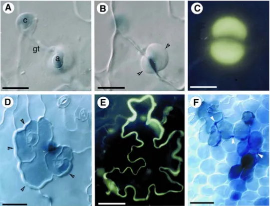
P. porri can penetrate Arabidopsis plants over the roots (data not shown) as well as over above-ground parts. The mode of penetration is independent of the initial propagule used for infection and the initial steps are the same in resistant and susceptible plants. With both zoospores and mycelium, penetration occured preferentially over anticlinal walls of epidermal cells (Figure 1A, B), occasionally via the stomatal opening (data not shown). Zoospores applied on leaves of Arabidopsis encysted, developed a germ tube reaching up to several spore diameters in length before forming an appressorium over the point of penetration (Figure 1A). A penetration hyphae then started to grow between the anticlinal walls of two epidermis cells. At this point, differences between compatible and incompatible interaction became apparent. In resistant plants, the earliest microscopically visible response was observed starting 6 hours after inoculation and consisted of the deposition of dense material, presumably of host origin, around the site of penetration as visualized for an attempted infection of Ws-0 by P. porri isolate HH (Figure 1B). Staining of the tissue with aniline blue revealed that these depositions contained callose, which is specifically stained by this dye (Figure 1C). Another resistance phenotype frequently encountered was the hypersensitive reaction (HR). One or several epidermal cells in the case of direct penetration through the epidermis (Figure 1D), or one or several mesophyll cells in the case of indirect penetration through a stomatal opening (data not shown), underwent a rapid cell death visualized microscopically by the retention of trypan blue in their cytoplasm. In cells adjacent to the dead ones a dense deposition of material was observed at the wall directly in contact with the dead cell (Figure 1D). Aniline blue staining revealed that the material encasing the HR cells consisted of callose (Figure 1E). Occasionaly, the hyphae were able to penetrate further into the plant tissue but were soon surrounded by necrotic cells (Figure 1F). This trailing necrosis response succesfully stopped further infection and became macroscopically visible as small necrotic regions on the leaves (data not shown).
Figure 1. Cytological characterization of the incompatible interaction of Arabidopsis with P. porri. Differential
interference contrast (DIC) (A, B, D, F) and fluorescence (C, E) micrographs.
A, B, D and F show lactophenol trypan-blue stained preparations, C and E were stained with decolorizcd aniline blue as described under Methods.
(A) A cyst (c) of P. porri isolate HH has formed a germ tube (gt) and an appressorium (a) on the upper epidermis of
a leaf of A. thaliana accession Ws-0 6 hours after inoculation. The faint blue staining inside the cyst and the appressorium visualizes the cytoplasm. (bar = 10 µm).
(B) Same as (A) focussed on the layer right below the appressorium. Arrowheads point to a heavy deposit of
material called a papilla surrounding the attempted penefration site at the border of two anticlinal walls of epidermal cells. (bar = 10 µm).
(C) Fluorescence of callose in an anime blue-stained papilla in a leaf of A. thaliana accession Col-0 24 hours after
infection with mycelium of P. porri isolate HH. (bar = 15 µm).
(D) Hypersensitive reaction of A. thaliana accession Col-0 after infection with P. porri isolate HH 24 hours after
inoculation with mycelium, The cells which have undergone a HR are stained a darker blue due to retention of trypan-blue. The penetrating hypha is out of the focal plane and only the actual point of penetration can be seen as a dark blue area between the two stomata in the HR region. In the adjacent cells, deposits of material (arrowheads) can be seen on the side were their cell walls are in contact with the HR cells. (bar =50 µm).
(E) Fluorescence of callose visualizing the limits of an epidermal cell of A. thaliana accession Col-0 which has
undergone a HR after an attempted penetration by P. porri isolate HH . The picture was taken 24 hours after inoculation. (bar =25 µm).
(F) Trailing necrosis in a leaf of A. thaliana accession Col-0 48 hours after inoculation with mycelium of P. porri
isolate HH. The hypersensitive cells are stained a darker blue, arrowheads point to places where the hypha is visible. (bar = 120 µm).
Compatible Interaction between Arabidopsis and P. porri
In susceptible Arabidopsis accessions, penetration also occurs preferentially at the border of adjacent epidermal cells. In an initial phase, during up to 3 days depending on the Arabidopsis accession, the mycelium grew exclusively in the intercellular spaces spreading in all directions away from the penetration site (Figure 2A, B). The hyphae were fairly regular in diameter and often in close contact with the plant cells (Figure 2B). Haustoria-like protuberances into the plant cells were only rarely observed (data not shown). During this first biotrophic phase, no reactions of plant cells were visible microscopically (Figure 2B) and macroscopically (data not shown). In a later phase, the tissue was colonized by a dense network of intra- and extracellular hyphae and plant cells started retaining the trypan blue stain (Figure 2C). Macroscopically, this phase was characterized by the watersoaked and wilted appearance of the infected tissue. Under conditions of high air humidity, P. porri started to grow out of the stomata (Figure 2D) and the emerging sporangiophores gave rise to mostly obpyriform zoosporangia (Figure 2E). Seven days after inoculation, sexual spores, the oospores, started to appear (Figure 2F). Antheridia were either amphigynous as shown in Figure 2F or paragynous (data not shown). In the latter case, one to three antheridia per oogonium were observed. The results show that P. porri can extensively colonize and reproduce in susceptible accessions of Arabidopsis.
Figure 2. Cytological characterization of the compatible interaction of Arabidopsis with P. porri.
Bright field (A, C, D, F) and differential interference contrast (B, E) micrographs of the compatible interaction. All the preparations were stained with lactophenol trypan-blue as described under Methods.
(A) Young colony of P. porri isolate HH in A. thaliana accession Mt-0 3 days after inoculation with
zoospores. The mycelium is visible as a dark blue network ramifying inside the leaf. (bar = 150 mm).
(B) Hyphae (arrowheads) of P. porri isolate HH growing intercellularly in the mesophyll of a leaf
of A. thaliana accession Ler 4 days after inoculation with mycelium. Note the absence of any necrosis in the plant cells. (bar = 60 mm).
(C) Heavy colonization as seen in a leaf of A. thaliana accession Mt-0 one week after inoculation
with zoospores of P. porri isolate HH. The hyphae grow inter- and intracellularly and the plant tissue shows macroscopical symptoms of wilting. (bar = 150 mm).
(D) Sporangiogenous hyphae of P. porri isolate II emerging through the stomatal opening in a leaf
of A. thaliana accession Mt-0 5 days after inoculation with zoospores. (bar = 40 mm).
(E) Tear-shaped zoosporangium on the surface of a leaf of A. thaliana accession Ler 4 days after
inoculation with mycelium of P. porri isolate HH. (bar = 50 mm).
(F) Oogonium and amphigynous antheridium of P. porri isolate D in a leaf of A. thaliana accession
Ws-0. (bar = 25 mm).
Inheritance of Resistance
Crosses between Col-0 and Ler were performed and the progeny was tested in the F2 generation.
Resistance in the F2 population segregated in a 3:1 ratio resistant:susceptible as tested for two
independent crosses (representative data for the first F2 population was 38 resistant and 10 susceptible, χ2
for a 3:1 ratio = 0.235, P > 0.05; the second population consisted of 36 resistant and 12 susceptible plants, perfectly fitting the 3:1 segregation). Backcrosses of F1 individuals with the susceptible parent Ler
segregated in a 1:1 ratio for resistant to susceptible (13 resistant and 7 susceptible, χ2 for a 1:1 ratio = 0.920, P > 0.5; 12 resistant and 8 susceptible, χ2 for a 1:1 ratio = 0.404, P > 0.05). Both segregation patterns in the crosses and the backcrosses point to a single dominant gene responsible for resistance in Col-0 against isolate II of P. porri.
Interaction between P. porri and Selected Arabidopsis Defense Pathway Mutants
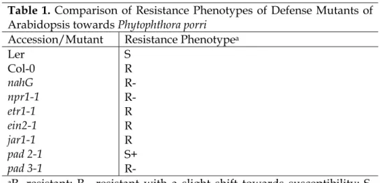
In order to learn more about the basis of resistance towards Phytophthora, several Arabidopsis mutants or transgenics with defects in defense signaling were tested for their reaction towards an attempted infection with P. porri isolate HH. The tested Arabidopsis mutants included: nahG and npr1-1 with defects in SA signaling (Cao et al., 1994; Delaney et al., 1995; Gaffney et al., 1993), the ethylene receptor mutant etr1-1 (Bleecker et al., 1988), the ethylene-insensitive mutant ein2-1 (Guzmann and Ecker, 1990), the jasmonate insensitive mutant jar1-1 (Staswick et al., 1992) and two mutants with reduced camalexin levels: pad2-1 and pad3-1 (Glazebrook et al., 1994; 1997). All the mutants were in the background of the resistant accession Col-0. The results of the phenotypical analysis of the mutant collection are summarized in Table 1.
Table 1. Comparison of Resistance Phenotypes of Defense Mutants of Arabidopsis towards Phytophthora porri
Accession/Mutant Resistance Phenotypea
Ler S Col-0 R nahG R- npr1-1 R- etr1-1 R ein2-1 R jar1-1 R pad 2-1 S+ pad 3-1 R-
aR, resistant; R-, resistant with a slight shift towards susceptibility; S,
17
Interference with ethylene or jasmonic acid signaling in etr1, ein2 and jar1, respectively, had no effect on the resistant phenotype. Interestingly, the jar1 mutant showed a much higher incidence of callose containing papillae (Figure 3E). This, however, had no effect on the already resistant phenotype. Prevention of SA accumulation in nahG or SA signaling in npr1 had only a minor effect on the resistance towards P. porri. The resistance was slightly shifted towards susceptibility: P porri could occasionally colonize small parts of the tissue but was soon stopped by host cell necrosis with the effect that zoosporangia and oospores were never observed in nahG or npr1 plants. A similar shift towards susceptibility was observed in pad3 which has a defect in camalexin biosynthesis and as a result is unable to synthesize camalexin (Zhou et al., 1999). Thus, SA-signaling and camalexin production appear to contribute to resistance but do not seem to be part of the main defense mechanism. However, the pad2 mutation appeared to knock out all mechanisms that are relevant for the establishment of resistance: pad2 plants proved to be hypersusceptible towards P. porri. Pad2 was even more susceptible than the susceptible accession Ler. Figures 3A-D show the results of an inoculation of pad2 with P. porri isolate HH. The pathogen rapidly colonized the leaf tissue. The hyphae ramifiyed in the intercellular spaces, and often the density of colonization was such, that several hyphae grew side by side filling the entire space between two cells (Figure 3A). Characteristic for infections in pad2 was, that P. porri was able to colonize host cells intracellularly. Some host cells appeared completely filled with hyphae but there was no apparent reaction of the plant cell to this invasion (Figure 3B). Furthermore, the formation of haustoria happened more frequently compared to a normal compatible infection (Figure. 3C). The ring of cells surrounding the base of trichomes seemed especially attractive to P. porri. In colonized areas of leaves of pad2 these cells were all extensively colonized (Figure. 3D). Colonization of pad2 by P. porri was not apparent macroscopically until 3 days after inoculation, when the colonized tissue started to get a watersoaked appearance followed by a total collapse without visible necrosis (data not shown).
Figure 3: Cytological characterization of the interaction of P. porri with the hypersusceptible pad2-1 mutant and with
the jasmonate insensitive mutant jar1-1.
Differential interference contrast (DIC) (A, B, C), bright field (D) and fluorescence (E) micrographs. A, B, C, D show lactophenol trypan-blue stained preparations and (E) was stained with decolorized aniline blue as described under Materials.
(A) Intercellularly growing mycelium of P. porri isolate HH in the mesophyll of the pad2 mutant.
Note the locally high concentration of hyphae (arrowhead) without visible reaction of the host cells. (bar = 60 µm).
(B) Extremely dense intracellular colonization of mesophyll cells of the A. thaliana pad2 mutant with hyphae of P.
porri isolate HH. Note that no visible reaction of the host cell can be detected. (bar = 40 µm).
(C) Intracellular fingershaped haustoria (arrowheads) of P. porri isolate HH in mesophyll cells of the pad2 mutant.
(bar = 20 µm).
(D) Preferential colonization of the cells surrounding the base of trichomes (tr) by P. porri isolate HH in the pad2
mutant. (bar = 40 µm).
(E) Low magnification picture of part of a leaf of jar1 after infection with mycelium of P. porri isolate HH. All the
bright green spots are papillae stained for callose at attempted penetration points of hyphae in the leaf. (bar = 150 µm).
19
Analysis of Marker Gene Expression in Different Defense Mutants
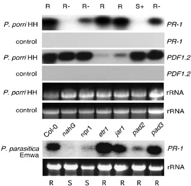
The expression of PR-protein 1 (PR-1) was used as a marker of SA-dependent defense responses (Ward et al. 1991) and the expression of a plant defensin PDF1.2 served as a marker of ethylene and jasmonic acid dependent defense gene induction (Penninckx et al., 1998). As shown in Figure 4, the inoculation of the resistant accession Col-0 with P. porri isolate HH lead within 24h to an increased expression of PR-1 and PDF1.2. PR-1 gene expression was completely blocked in nahG plants and partially blocked in the npr1 mutant, while the etr1 and jar1 mutations showed no effect on PR-1-expression compared to wild type. PR-1 PR-1-expression was only slightly down-regulated in the pad3 mutant but was completely blocked in the pad2 mutant. PDF1.2 expression was strongly down regulated in inoculated etr1 and jar1 mutants but remained unaffected in the SA signaling mutants and the two tested pad mutants. Despite the lack of PDF1.2 expression, etr1 and jar1 both showed a resistant phenotype thus suggesting that PDF1.2 accumulation does not contribute much to resistance against P. porri.
Figure 4 includes a comparison of the PR-1 expression pattern in Col-0 and the collection of mutant plants infected with P. porri isolate HH or Peronospora parasitica isolate EMWA. The profile of PR-1 expression induced in both pathosystems is nearly identical. PR-1 expression is at least partially blocked in nahG, npr1 and pad2 but remains unaffected in etr1 and jar1. However, the pattern of resistance phenotypes is completely different in the two pathosystems as indicated at the top and bottom, respectively, of Figure 4. NahG and npr1 remain resistant against P. porri but become susceptible towards P. parasitica. In contrast, pad2 becomes susceptible towards P. porri but remains resistant against P. parasitica. Thus, the resistance mechanisms effective against P. porri appear to be fundamentaly different from the mechanisms that are effective against P. parasitica.
Determination of SA- and Camalexin Levels
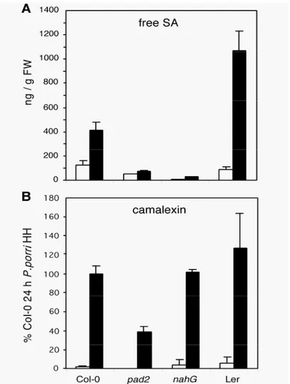
PR-1 expression was completely blocked in pad2. This nahG-like phenotype suggested that the pad2 mutation might have a negative effect on SA accumulation. To test this hypothesis, the effect of P. porri inoculation on SA levels was measured in Col-0, Ler, nahG and pad2. The results of the SA measurement 24h post inoculation are shown in Figure 5A. Within 24h following inoculation the level of free SA increased about 3 times in the resistant Col-0 and more than 10 times in the susceptible Ler. The SA levels of nahG plants were very low in control plants and did hardly increase following inoculation with P. porri. A similar SA-minus phenotype was found for pad2. Even uninfected pad2 plants had a 3 times lower SA content than Col-0 plants. This value did only slightly increase following inoculation and remained lower than the SA content in untreated Col-0. The pattern of SA levels 36h and 48h post inoculation remained qualitatively unchanged from the one shown in Figure 5 (data not shown). Pad2 clearly shows a nahG-like SA-minus phenotype. The values for conjugated SA for Col-0, nahG and pad2 24h post inoculation were in the range of control plants (500-800 ng/g FW) indicating that the lack of accumulation of free SA was not caused by an increased SA-conjugation rate. The level of conjugated SA was raised to 1800ng/g FW in the susceptible Ler (data not shown).
Because pad2 was originally described as a camalexin mutant (Glazebrook and Ausubel, 1994), its ability to produce camalexin was tested 24h post inoculation (Figure 5B). Inoculation of Col-0 with P.
porri isolate HH lead to a 60-fold increase in the level of camalexin compared to uninoculated control plants. Very similar results were found for nahG plants while the levels of camalexin in the susceptible accession Ler were slightly higher. The increase in camalexin production appears to be independent of SA accumulation and the occurence of HR. In contrast to Col-0 the level of camalexin in uninoculated pad2 plants was found to be below the limit of detection. Camalexin accumulation was reduced in inoculated pad2 plants to about 40% of the values found in Col-0.
Figure 4. PR-1 and PDF1.2 marker gene expression in different Arabidopsis genotypes in response to inoculation with P. porri.
PR-1 and PDF1.2 gene specific probes were used for RNA gel blot analysis of the
indicated genotypes (Col-0, nahG, npr1 , etr1, jar1, pad2, pad3). Ethidium bromide staining
of the gel was used as an estimation of equal sample loading (rRNA). Plants were either
uninoculated (control), inoculated with P. porri isolate HH or P. parasitica isolate EMWA.
RNA was extracted 24h post inoculation. Resistance phenotypes of the respective
interactions are indicated for P. porri in the top line and for P. parasitica in the bottom line,
Figure 5. Accumulation of free SA and camalexin in Col-0, pad2, nahG and Ler after
inoculation with P. porri isolate HH.
Five weeks old plants were inoculated with P. porri isolate HH and leaves were harvested 24h later. The values represent the average of two independent samples ± SE.
(A) Levels of free salicylic acid (SA).
(B) Camalexin levels. Because of the lack of a pure standard, values are expressed in relation
to the value of Col-0 24h post inoculation.
DISCUSSION
The Arabidopsis-Phytophthora Pathosystem
An experimental system for the analysis of the interaction of Arabidopsis with the phytopathogenic oomycete Phytophthora porri was established. Fifteen accessions of Arabidopsis were screened for their reaction to 7 isolates of P. porri known to be pathogenic on family members of the Brassicaceae. Accession-isolate combinations were identified that result in either complete resistance or in complete susceptibility. Accessions susceptible to a given isolate of P. porri are completely colonized by P. porri within a few days (Figure 2). In the initial phase, the pathogen developped in the intercellular space and no host reaction was observed. In a later phase, the host cells were macerated, oospores formed inside the colonized tissues and hyphae grew out of the stomata to give rise to zoosporangia. Thus, P. porri can complete its whole life cycle in a susceptible host and Arabidopsis can therefore be considered a true host of this pathogen. The compatible interaction showed all the characteristics of a facultative biotrophic interaction very similar to P. infestans on potato and other agronomically important diseases
caused by Phytophthora (Erwin and Ribeiro, 1996). In incompatible host-pathogen combinations different degrees of resistance were observed (Figure 1). The plant reacted either with a HR comprising one to a few cells or the pathogen was able to grow to some extent into the tissue triggering a HR visible macroscopically as a necrotic fleck. The formation of callose-containing papillae was frequently observed at the site of penetration. Interestingly, callose production and cell wall apositions were also found in the cells adjacent to cells undergoing HR. These extensive appositions are presumably produced by the neighbouring cells and were restricted to walls with direct contact to the dying cells. It is not known how this directional callose deposition process is regulated.
The observation of a HR in resistant hosts suggested that the interaction of some combinations of Arabidopsis accessions and P. porri isolates might follow a gene-for-gene type of interaction. The segregation analysis of the phenotypes of the F2 progeny of crosses between the resistant accession Col-0
and the susceptible accession Ler as well as backcrosses of F1 progeny with the susceptible accession
indicated that resistance is inherited as a single dominant mendelian trait. The successful infection of plants by Phytophthora strongly depends on environmental conditions. This is also the case in the Arabidopsis system. In order to generate reproducible results it is necessary to strictly control the environmental conditions (see Methods). The strong dependence on environmental conditions made it difficult to screen larger numbers of inoculated plants. The tentative scoring of intermediate interactions lead to an increased uncertainty. Presumably because of these practical problems in upscaling, our first attempt to map the putative resistance gene in Col-0 was not successful.
The major advantage of the novel Phytophthora-pathosystem is its use of Arabidopsis as a host. The availability of complete sequence information, the ease of mutational analysis, the extensive mutant collection and the possibility to use microarrays for gene expression analysis is expected to lead to an acceleration in data generation. Phytophthora is an agronomically much more important pathogen than the obligate biotrophic Peronospora parasitica which is frequently used as a model oomycete pathogen of Arabidopsis (Koch and Slusarenko, 1990; Holub et al., 1994). Phytophthora has the advantage that it can be cultured in vitro. Both sexual and asexual spores can be produced in vitro by P. porri (data not shown). Phytophthora is therefore much more accessible to molecular analysis. Phytophthora species including P. porri (Si-Ammour et al., unpublished results) are transformable (Judelson, 1991). The genome size of P. porri is relatively small (Si-Ammour et al., unpublished results) compared to P. infestans (Tooley and Therrien, 1987).
The major disadvantage of the novel system is based on an inherent property of the oomycetes. Oomycetes are diploid during most phases of their life cycle (Brasier and Sansome, 1975; Boccas, 1976). The only haploid stages occur in the gametangia formed immediately prior to fertilization. This fact considerably complicates the genetic analysis of Phytophthora since the phenotype of recessive mutations can only be discovered after selfing in the F2 generation. This step would be very difficult if not
impossible to achieve with the heterothallic P. infestans. Most Phytophthora species are heterothallic and require strains of different mating types for genetic crosses (Erwin et al., 1983). In contrast, P. porri, like P. sojae, is a homothallic species forming oospores by selfing (Erwin and Ribeiro, 1996). This is a clear