HAL Id: tel-02993716
https://tel.archives-ouvertes.fr/tel-02993716
Submitted on 7 Nov 2020
HAL is a multi-disciplinary open access
archive for the deposit and dissemination of sci-entific research documents, whether they are pub-lished or not. The documents may come from teaching and research institutions in France or abroad, or from public or private research centers.
L’archive ouverte pluridisciplinaire HAL, est destinée au dépôt et à la diffusion de documents scientifiques de niveau recherche, publiés ou non, émanant des établissements d’enseignement et de recherche français ou étrangers, des laboratoires publics ou privés.
Natural Killer Cells and Pre-B Acute Lymphoblastic
Leukemia : Evidence for an Unconventional Cytotoxicity
Pathway
Simon Nicoletti
To cite this version:
Simon Nicoletti. Natural Killer Cells and Pre-B Acute Lymphoblastic Leukemia : Evidence for an Unconventional Cytotoxicity Pathway. Cellular Biology. Université Paris Saclay (COmUE), 2017. English. �NNT : 2017SACLS383�. �tel-02993716�
Natural Killer cells and
pre-B acute lymphoblastic
leukemia: evidence for
an unconventional
cytotoxicity pathway
Thèse de doctorat de l'Université Paris-Saclaypréparée à l’Université Paris Sud
École doctorale n°582 Cancérologie : Biologie – Médecine – Santé (CBMS)
Spécialité de doctorat : aspects moléculaires et cellulaires de la biologie
Thèse présentée et soutenue à Paris, le 6 novembre 2017, par
M. Simon Nicoletti
Composition du Jury : M. Éric Solary
Professeur des Universités – Praticien Hospitalier
Université Paris Sud (Institut Gustave Roussy, UMR 1170) Président
Mme Catherine Blish
M.D., Ph.D., Professeure
Stanford University (Stanford School of Medicine) Rapporteur
M. Thierry Walzer
Directeur de recherche, ENS, Université de Lyon
Centre International de Recherche en Infectiologie Rapporteur
M. Alain Fischer
Professeur des Universités – Praticien Hospitalier
Institut Imagine UMR 1163, Collège de France Examinateur
M. Felipe Suarez
Professeur des Universités – Praticien Hospitalier
Institut Imagine UMR 1163 Examinateur
M. Olivier Hermine
Professeur des Universités – Praticien Hospitalier
Institut Imagine UMR 1163 Directeur de thèse
M. Élie Haddad
Professeur titulaire
CHU Sainte-Justine, Université de Montréal Co-Directeur de thèse
NNT : 2 0 1 7 S A C L S 3 8 3
À mamie Jeanne)e, une grand-mère provençale pleine d’amour et d’élégance
ACKNOWLEDGEMENTS
The study that is presented here is a synthesis of a story that started in August 1st, 2010, when I first arrived in Canada. Since then, many different people contributed to make it more than a research work.
I want to thank Élie Haddad for such incredible relaNonships. In my work, he made me learn experimental controls, rigor, experimental controls, creaNvity, experimental controls, analogies, experimental controls, and so much more. More than that, I believe I have been very lucky to meet a pediatrician, physician, with a very simple and direct contact. Someone that, as a paNent or a family, I know I would like to meet. Un immense merci Élie, ce travail avec vous a été simplement génial et le meilleur moment de ma vie.
Canada has been an amazingly welcoming country. The very beginning was a complicated start, arriving alone in an unknown land. But I was lucky enough to meet people that I definitely keep in my mind and in my heart.
I want to thank: Silvia Selleri, the Italian source of inspiraNon to whom I owe many knowledges in science and immunology; Mame Massar Dieng and Vimal Krishnan, incredible friends, a family; Françoise Le Deist, for her help, her rigor, her mentorship, many thanks to her; Isabelle Louis, Panojot Bifsha, François Fontaine, Kathie Béland, Renée Dicaire, Ludovic Durrieu, Joëlle Grégoire-Gauthier, Arnaud Duval, Yuanyi Li, Mark Abramovitz, Zoulfia Allakhverdieva, Didier Doranger, Aurélien ColamarNno, Chloé Colas, Hugo Roméro, in my lab. Et quel laboratoire!
I also need to thank my roommates: Ma)hew Morantz, actually as a part of my family; Marigona Uka, a sweet friend. I wish them both the best. And George Fisher as well.
Looking at these names, you understand Canada and its strength. I’m proud to have been part of it.
Back to France, I have to thank Olivier Hermine for his help and mentorship in this project. Thanks to him, we were able to pursue the work and I was allowed to do my PhD. His humanity and his devoNon are precious for paNents. Merci Olivier pour ces fulgurances et ces échanges plus qu’enrichissants, parfois tardifs pour ne pas dire nocturnes! Je garde en mémoire un départ dans la nuit parisienne en vélo, comme un moment privilégié.
I want to thank in my lab: Mathilde Lamarque and her delicious cakes; Hervé Souchet, a sailor climbing partner; Morgane Cheminant, very efficient and a clever mind; Moussab Tagi,
a surprising friend. The lab 217 was an amazing atmosphere. Thank you to Francine Côté, a bridge between Montreal and Paris, Joël Babdor, a climbing friend sharing interesNng ideas. Thanks to all the members of the team Hermine: Flavia Guillem, Geneviève Courtois, Guilleme)e Fouquet, Leila Maouche-ChréNen, Lucile Couronné, Yves LepelleNer… and all the others.
I also need to thank many people from Imagine insNtute, Olivier Pellé and Aude Magerus for their help with the Fortessa, asso YR2I, and colleagues and friends from many labs (Benoît, Antoine, David, Clarisse, Christelle…).
In Lyon, I thank Laurent Jacqueroud, a true friend with whom we have been through the challenging steps of the MSc before we started our PhD. Besides, Pierre Cochat, Charles Dumontet, Caroline Moyret-Lalle, Yves Bertrand and Alexandre Belot were very important for me, especially to build a consistent medical and scienNfic career path. I also want to thank our collaborators worldwide. Their help is crucial. It makes our science feasible. Thanks to the ARC FoundaNon for their financial support for the 4th year of PhD; The Liliane Be)encourt School of Inserm, and the Be)encourt-Schueller FoundaNon for their scholarship during the medical school. Thanks to Catherine Blish, Alain Fischer, Eric Solary, Felipe Suarez and Thierry Walzer for their role in the evaluaNon of this manuscript. It’s a great honor and a privilege for me. My parents and my brother are the cornerstones of my personality. I owe them everything. Merci à la famille Nicoler, nous quatre, un solide repère, une direcNon.
I started these acknowledgements wriNng it was more than a research work. I’m looking forward to the next stopovers.
TABLE OF CONTENTS LIST OF ABBREVIATIONS AND ACRONYMS . . . . p 6 LIST OF FIGURES . . . . p 9 LIST OF SUPPLEMENTAL FIGURES . . . . p 11 LIST OF TABLES . . . . p 11 LIST OF SUPPLEMENTAL TABLES . . . . p 11 1. INTRODUCTION AND PROBLEM STATEMENT . . . . p 13 1.1. IMMUNE FUNCTION OF NATURAL KILLER CELLS . . . . p 13
1.2. THE INTERACTION BETWEEN CYTOTOXIC LYMPHOCYTES AND TARGET
CELLS . . . . p 15 1.3. THE MAIN RECOGNIZED EFFECTOR PATHWAYS . . . . p 16 1.3.1. CONTACT-INDEPENDENT PATHWAYS . . . . p 18 1.3.2. CONTACT-DEPENDENT PATHWAYS . . . . p 19 1.3.2.1. GRANULE EXOCYTOSIS . . . . p 19 1.3.2.2. DEATH RECEPTORS . . . . p 23 1.4. EFFECTOR CELLS HETEROGENEITY AND DIVERSITY . . . . p 25 1.5. PRE-B ACUTE LYMPHOBLASTIC LEUKEMIA AS A TARGET CELL’S MODEL . p 27
1.6. PRIMARY IMMUNE DEFICIENCIES TO DECIPHER EFFEC TOR
LYMPHOCYTES’ MOLECULAR PATHWAYS . . . . p 29 1.7. AIMS . . . . p 29 2. METHODS . . . . p 32 2.1. PATIENTS RECRUITMENT . . . . p 32 2.2. PERIPHERAL BLOOD MONONUCLEAR CELLS ISOLATION . . . . p 34 2.3. CELL CULTURE . . . . p 34 2.3.1. CELL LINES . . . . p 34
2.3.2. IN VIVO EXPANSION OF PRIMARY PRE-B ALL BLASTS AND CELL
CULTURE . . . . p 35 2.4. NK ACTIVATION AND EXPANSION SYSTEMS (NKAES) . . . . p 36 2.5. CYTOTOXICITY ASSAY . . . . p 36 2.5.1. PKH26 STAINING OF THE TARGET CELLS . . . . p 38 2.5.2. CD3 POSITIVE CELLS DEPLETION . . . . p 38 2.5.3. FLOW CYTOMETRY-BASED CYTOTOXICITY (FCC) ASSAY . . . . p 38 2.6. DEVELOPMENT OF A VERSATILE LENTIVIRAL PLATFORM . . . . p 39 2.6.1. TRANSDUCTION OF PRE-B ALL CELL LINES . . . . p 41 2.6.2. TRANSDUCTION OF PRIMARY ACTIVATED NK CELLS . . . . p 42 2.6.3. CRISPR/CAS9-BASED GENOME ENGINEERING . . . . p 42 2.6.3.1. UNIPLEX GENOME ENGINEERING . . . . p 42 2.6.3.2. MULTIPLEX GENOME ENGINEERING . . . . p 43 2.6.3.3. SGRNA CLONING . . . . p 44 2.6.3.4. IMMUNOBLOT SCREENING OF CANDIDATE CLONES . . p 46 2.7. CELL CONJUGATION ASSAY . . . . p 46 2.8. DEGRANULATION ASSAY . . . . p 46 2.9. BLOCKING EXPERIMENTS . . . . p 47 2.10. FLOW CYTOMETRY STAININGS . . . . p 48 2.10.1. ANNEXIN V STAINING . . . . p 48 2.10.2. MITOCHONDRIAL DEPOLARIZATION ASSAY . . . . p 49 2.11. PRIME FLOW RNA ASSAY . . . . p 49 2.12. MEASUREMENT OF INTRACELLULAR CASPASES ACTIVATION . . . . p 50 2.13. CHARGE-BASED CAPILLARY NANO-IMMUNOASSAY (NANOPRO ASSAY) . p 50
2.14. SEAHORSE ANALYSIS . . . . . p 51 2.15. RNA-SEQUENCING . . . . . p 51 2.15.1. EXPERIMENTAL DESIGN . . . . p 51 2.15.2. RNA EXTRACTION, LIBRARY PREPARATION AND SEQUENCING . p 52 2.15.3. DATA PROCESSING . . . . p 53 2.16. STATISTICAL ANALYSIS . . . . p 56 3. RESULTS AND DISCUSSION . . . . p 58
3.1. PRE-B ALL CELLS ARE SENSITIVE TO NK CELL KILLING IN A LONG-TERM
CYTOTOXICITY ASSAY . . . . p 58 3.2. CHARACTERIZATION OF THE EFFECTOR PATHWAY . . . . p 59 3.2.1. A CONTACT-DEPENDENT MECHANISM . . . . p 60 3.2.2. A DEGRANULATION-INDEPENDENT MECHANISM . . . . p 64 3.2.3. APOPTOSIS-LIKE CELL DEATH OF THE TARGET CELLS . . . . p 67 3.2.4. A DEATH RECEPTOR PATHWAY-INDEPENDENT MECHANISM . . . p 71
3.2.5. MINIMAL MOLECULAR REQUIREMENTS AMONG EFFECTOR
CELLS . . . . p 73
3.2.6. MINIMAL MOLECULAR REQUIREMENTS AMONG TARGET
CELLS . . . . p 76 3.2.7. ROLE OF REACTIVE OXYGEN SPECIES (ROS) . . . . p 79 3.2.8. EVIDENCE FOR A NADPH COMPLEX IN NK CELLS . . . . p 81 3.3. METABOLIC SUPPORT OF NK CELLS ACTIVITY . . . . p 91 4. CONCLUDING REMARKS AND PERSPECTIVES . . . . p 97 4.1. CONCLUDING REMARKS . . . . p 97 4.2. PERSPECTIVES . . . p 103 5. SUPPLEMENTARY MATERIAL AND APPENDICES . . . . p 112 5.1. SUPPLEMENTARY MATERIAL . . . . p 112 5.2. APPENDICES . . . p 117 6. REFERENCES . . . . p 126
LIST OF ABBREVIATIONS AND ACRONYMS 7-AAD : 7-aminoac)nomycine ALL: Acute lymphoblas)c leukemia AML: Acute myeloid leukemia APAF-1: Apopto)c protease ac)va)ng factor 1 APC : Allophycocyanine BCA: Bicinchoninic acid B-EBV LCL: Epstein-Barr virus-transformed human B-lymphoblastoid cell lines BID: BH3 interac)ng domain death agonist BMMCs: Bone marrow mononuclear cells BSA : Bovine serum albumin CCCP: Carbonyl cyanide m-chlorophenylhydrazone CD: Cluster of differen)a)on CFSE : 5-6 Carboxy fluorescein diacetate succimidyl ester CGD: Chronic granulomatous disease CIK: Cytokine-induced killer cells CPDA: Citrate phosphate dextrose adenine solu)on CRISPR: Clustered regularly interspaced short palindromic repeats CTL : Cytotoxic T lymphocytes DAP10: DNAX-ac)va)ng protein 10 DCs: Dendri)c cells DHR: Dihydrorhodamine
DKO: Double knockout
DMSO: Dimethylsulfoxide D-PBS: Dulbecco’s phosphate buffer saline DRs: Death receptors ECAR: Extracellular acidifica)on rate E:T ra>o: Effector to target ra)o FACS : Fluorescence-ac)vated cell sor)ng FADD: FAS-associated death domain protein FAS: Cluster of differen)a)on FBS: Fetal bovine serum FCC: Flow cytometry-based cytotoxicity assay FHL: Familial hemophagocy)c lymphohis)ocytosis FITC: Fluorescein isothiocyanate FLICA: Fluorescent labelled inhibitors of caspases GFP : Green fluorescent protein GM-CSF: Granulocyte macrophage colony-s)mula)ng factor GvHD: GraR-versus-host disease GvT: GraR-versus-tumor HCMV: Human cytomegalovirus HIES: Hyper-IgE syndrome HLA : Human leukocyte an)gen HLH: Hemophagocy)c lymphohis)ocytosis syndrome HSCT: Hematopoie)c stem cell transplanta)on HTS: High throughput sampler
ICAM-1: Intercellular adhesion molecule 1 IFN-γ: Interferon-gamma ILCs: Innate lymphoid cells IL-X : Interleukin-X (X depicts a number) IRES : Internal ribosome entry site IU: Interna)onal unit KIR(L): Killer immunoglobulin-like receptors (ligand) KO: Knockout LAMP-1: Lysosomal-associated membrane protein 1 LFA-1: Lymphocyte func)on-associated an)gen 1 MFI : Mean fluorescence intensity MHC: Major histocompa)bility complex MOMP: Mitochondrial outer membrane permeabiliza)on MPO: Myeloperoxidase MTOC: Microtubule-organizing centre mTOR: mammalian Target of rapamycin NAC: N-acetylcystein NADPH oxidase: nico)namide adenine dinucleo)de phosphate-oxidase NCRs: Natural cytotoxicity receptors NEMO: NFκB essen)al modulator NFκB: Nuclear factor-kappa B NK: Natural Killer cells NKAES : Natural Killer ac)va)on and expansion system NSG: Nod/SCID/IL2Rγ-/- mice OCR : Oxygen consump)on rate OXPHOS : Oxida)ve phosphoryla)on PBMCs: Peripheral blood mononuclear cells PCA: Principal component analysis PCR: Polymerase chain reac)on PD-(L)1: Programmed cell death-(ligand) 1 PDX: Pa)ent-derived xenograR PE : Phycoerythrin PIDs: Primary immune deficiencies RAG: Recombina)on-ac)va)ng gene RNAseq: RNA sequencing ROS: Reac)ve oxygen species SCID: Severe combined immunodeficiency sgRNA: single guide RNA SLAM: Signaling lymphocyte ac)va)on molecule TCR: T cell receptor TGF-β: Transforming growth factor-beta (mb)TNF-α: (membrane-bound) Tumor necrosis factor-alpha TNFR superfamily: Tumor necrosis factor-receptors superfamily TRADD: Tumor necrosis factor receptor type 1-associated death domain protein TRAIL: Tumor-necrosis-factor related apoptosis inducing ligand UCOEs: Ubiquitous chroma)n opening elements UPR: Unfolded protein response
VSV-G: Vesicular stoma))s virus G-protein WPRE: Woodchuck hepa))s virus poseranscrip)onal regulatory element XHIGM: X-linked immunodeficiency with hyper-IgM XLP: X-linked lymphoprolifera)ve disease X-MAID: X-linked moesin-associated immunodeficiency XMEN: X-linked immunodeficiency with magnesium defect, Epstein-Barr virus infec)on, and neoplasia syndrome
LIST OF FIGURES
Figure 1: Innate lymphoid cells and their adap2ve lymphocytes counterpart: a
mirrored model . . . p 14 Figure 2: A model of NK cells ac2va2on: signal integra2on and outcome depending on target cell type . . . p 16 Figure 3: Different effector pathways can be engaged by cytotoxic lymphocytes . . p 17 Figure 4: Cytotoxic killing of a cognate target cell through the granule exocytosis . p 19 Figure 5: Induced target cell death through the granule exocytosis pathway . . . p 22 Figure 6: Extrinsic and intrinsic apopto2c signaling pathways . . . p 24 Figure 7: An updated view of human NK cell subsets (proposed by Frank Cichocki et al.) . . . . p 26 Figure 8: AML and B-ALL cancer stem cell divergent models . . . p 28 Figure 9: NK cells ac2va2on and expansion system (NKAES) from PBMCs . . . p 36
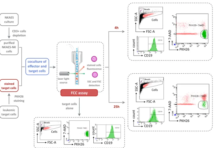
Figure 10: Flow cytometry-based cytotoxicity (FCC) assay (representa2ve dot plots
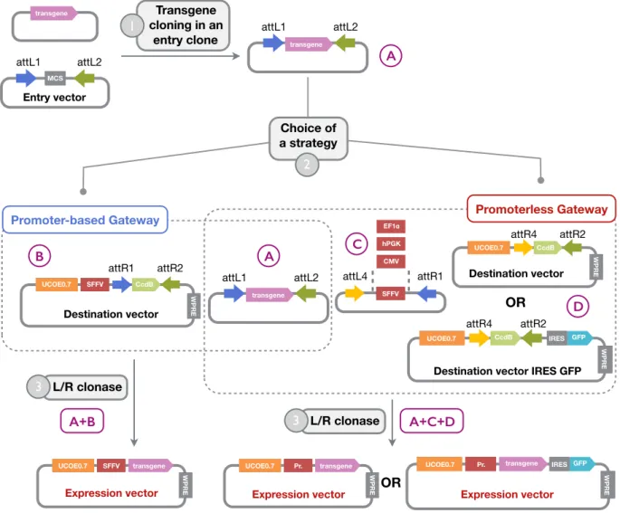
with REH cell line as target and an E:T ra2o = 4:1) . . . p 37 Figure 11: Homemade Gateway based len2viral plaZorm . . . . p 40 Figure 12: Uniplex genome engineering strategy . . . . p 42 Figure 13: Mul2plex genome engineering strategy . . . . p 43 Figure 14: NCRs blockade using Fc chimeras . . . . p 47 Figure 15: Parameters for the annexin-V staining . . . . p 48 Figure 16: Principle of the Prime Flow RNA assay . . . . p 49
Figure 17: Experimental design for sample processing in the RNA-sequencing
experiment . . . p 52 Figure 18: Group design in the RNA-sequencing experiment analysis . . . . p 53 Figure 19: Pre-B ALL are not intrinsically resistant to NK cell mediated cytotoxicity . p 58 Figure 20: A simplified three-step model of the NK killing process . . . . p 60 Figure 21: Killing ini2a2on and effector phase are contact-dependent . . . . p 61 Figure 22: NK cell killing is cumula2ve and requires permanent cell adhesion . . . . p 63
Figure 23: NK cell killing is independent of the perforin and degranula2on pathway p 65 Figure 24: Apopto2c-like flow cytometry profile of pre-B ALL cells . . . . p 68 Figure 25: Apopto2c pre-B ALL cells ac2vate both intrinsic and extrinsic pathways . p 69 Figure 26: Hierarchical ordering of the ac2va2on of the apopto2c extrinsic pathway p 70 Figure 27: Pre-B ALL cell killing is independent of the death receptor pathway . . . . p 71 Figure 28: NK cell mediated cytotoxicity towards pre-B ALL targets: PIDs’ screening . p 74 Figure 29: Ac2ve caspases and mitochondrial depolariza2on in pre-B ALL cell death are not mandatory for NK cell induced killing . . . p 77 Figure 30: Preliminary experiment sugges2ng a poten2al implica2on of ROS . . . . . p 79
Figure 31: Different sensi2vity to H2O2 among leukemic cell lines . . . p 79
Figure 32: Catalase and SOD2 over-expressing targets do not show increased
resistant to NK killing . . . p 80
Figure 33: CYBB deficient NK cells display significantly decreased cytotoxicity
against pre-B ALL cells . . . p 82
Figure 34: NK cells express gp91phox and p22phox but do not display a phagocyte-type
NADPH oxidase complex . . . p 83
Figure 35: CYBB mRNA is ac2vely regulated by NK cells . . . . p 86
Figure 36: NK cells from X-CGD pa2ents and normal donors have different
transcriptomic expression profile . . . p 87
Figure 37: Gene ontology func2onal network of biological process and cellular
components terms . . . p 88
Figure 38:
Oxygen Consump2on Rate (OCR) among NK cells from AR47-CGD and X-CGD pa2ents (compared to controls) . . . p 91
Figure 39: Extracellular Acidifica2on Rate (ECAR) among NK cells from AR47-CGD
and X-CGD pa2ents (compared to controls) . . . p 92
Figure 40: NK cell metabolic profile discriminates s2mula2on with K562 or pre-B
ALL cell lines . . . p 94
Figure 41: Metabolic profile of target cell lines . . . . p 95
Figure 42: A proposed schema2c representa2on of the steps involved in the NK
LIST OF SUPPLEMENTAL FIGURES
LIST OF TABLES
LIST OF SUPPLEMENTAL TABLES
Figure 43: Comparison of effect sizes for response to REH (and RS4;11, and their
difference) for NKAES-NK cells from controls (n = 4 per condi2on) and a X-CGD pa2ent . . . p 106 Figure 44: Proposed models for NK cell effector responses at the single cell level: a metabolic fingerprin2ng? . . . p 107 Figure 45: Experimental design for a genome-scale CRISPR/Cas9-based screening . . p 109 Figure 46: Alterna2ve models for resistance to NK cell mediated cytotoxicity . . . . . p 110 Figure S1: Killing increase against leukemic target cells is not restricted to mbIL-15 ac2vated NK cells . . . p 112 Figure S2: Pre-B ALL cell lines are sensi2ve to TRAIL mediated apoptosis . . . p 113
Figure S3: CIK effector cells cytotoxicity toward pre-B ALL targets increases
following a prolonged incuba2on . . . p 116 Table 1: Characteris2cs of gene2c disorders associated with occurrence of HLH . . p 21 Table 2: Pa2ents and cytotoxicity assays characteris2cs . . . p 32 Table 3: List of oligonucleo2des related to sgRNA cloning . . . p 44 Table 4: Differen2al expression contrasts retrieved from Model 1 . . . p 55 Table 5: Differen2al expression contrasts retrieved from Model 2 . . . p 55 Table 6: Tested gene2c defects with published NK cell deficiency . . . p 75 Table S1: Number of hits using the Emma nested models 1 and 2 . . . p 114 Table S2: Enrichment of GO Biological process terms . . . p 115 Table S3: Enrichment of GO Cellular components terms . . . p 115
CHAPTER 1.
INTRODUCTION AND
PROBLEM STATEMENT
1. INTRODUCTION AND PROBLEM STATEMENT
1.1. IMMUNE FUNCTION OF NATURAL KILLER CELLS
In 1975, two ar.cles reported the discovery of large granular lymphocytes capable of killing a target cell spontaneously, without requiring neither a priming step nor the expression of major histocompa.bility complex (MHC) molecules on target cells (Herberman et al., 1975). Since then, the so-called Natural Killer (NK) cells have been the subject of intensive inves.ga.on, from their ontogeny to their effector func.ons. It is now well-established that these cells contribute to defending the organism against infec.ons and cancer, and also play a role in immune homeostasis through selec.ve elimina.on of cells that were involved in the immune response (Lieberman, 2016).
The propor.on of systemic NK cells among peripheral blood mononuclear cells (PBMCs) is generally considered to be between 5-15% (Lotze and Thomson, 2009). NK cells are also found in some organs in which they can represent a more significant part of all lymphocytes, such as secondary lymphoid .ssues, mucosal .ssues, liver (where they can encompass up to 30-50% of all lymphocytes), lungs, skin, uterus, pancreas, joints and central nervous system
(Björkström et al., 2016; Shi et al., 2011).
Because they exhibit features reminiscent of innate immunity such as the ability to be rapidly ac.vated and respond to mutated or infected cells, and the absence of priming step requirement or of receptor rearrangement from their germline configura.on, NK cells have been considered innate lymphocytes (Caligiuri, 2008; Vivier et al., 2016). However, recent works have challenged this view (Cerwenka and Lanier, 2016; Holmes and Bryceson, 2016; Paust and Andrian, 2011; Sun et al., 2009): adapta.ve or memory NK cells have been iden.fied in humans in response to cytomegalovirus infec.on (Schlums et al., 2015) following pioneering work in mice, but also murine hapten-specific liver NK cells (Paust and Andrian, 2011), as well as an.gen-specific NK cells in non human primates (Reeves et al., 2015; Walzer and Marçais, 2016). Also, cytokine-induced memory NK cells, that can be generated in vitro, show increased prolifera.on, cell recovery, and effector func.on in vivo (Ni et al., 2012; Romee et al., 2016). Furthermore, the expression of the recombina.on-ac.va.ng gene proteins RAG1 and RAG2 (collec.vely, RAG) and of RAG endonuclease ac.vity during NK cell ontogeny, which play a cri.cal role in T and B cells development as shown in the related severe combined immunodeficiency (SCID) (Fischer et al., 2015), appears to impact NK cell phenotype, fitness and func.on both in mice (Karo et al., 2014) and humans (Dobbs et al., 2017). Thus, NK cells blur the well-established func.onal dis.nc.ons between
the two arms of the immune system, namely the innate and adap.ve immuni.es (Vivier et al., 2011).
Recently, several groups have redefined the conven.onal immune dichotomy to further dis.nguish adap.ve lymphocytes from innate-like ones, with evidence for overlapping func.ons (Bedoui et al., 2016). Among the laeer, innate lymphoid cells (ILCs) were conceptually theorized as a large family of cells that mirror the phenotype and ac.vity of differen.ated T cells (Eberl et al., 2015; 2014). In this model, NK cells are the counterpart of the cytotoxic CD8+ T cells due to their shared ability to kill infected, stressed, or transformed cells (Figure 1).
NK cells are the only cytotoxic ILCs and the major circula.ng subset although all ILC groups can be detected in peripheral blood from children and adults (Vély et al., 2016). As with
T-Figure 1. Innate lymphoid cells and their adapFve lymphocytes counterpart: a mirrored model.
Signals from injured or infected .ssues ac.vate ILCs whose func.on will mirror those of CD8+ and CD4+ T cells. The main difference between both cell types is the kine.cs of the immune response.
Adapted from Eberl et al., 2015; 2014.
NK cells (Killer ILCs)
Innate Lymphoid Cells
ILC1 ILC2 ILC3
CD8 cytotoxic T cells Th1 CD4+ T cells Th2 CD4+ T cells Th17 CD4+ T cells AdapFve Lymphocytes
Group 1 ILC Group 2 ILC Group 3 ILC
IL-12 IL-15 IL-18 IL-25 IL-33 TSLP IL-1β IL-23 Tissue signals Effector molecules cytotoxicity perforin granzymes TRAIL IFN-γ TNF-α IFN-γ TNF-α IL-4 IL-5 IL-9 IL-13 Areg IL-17A IL-22 GM-CSF LT-α1β2 IFN-γ Func.ons Immunity to viruses Tumor surveillance Chronic inflamma.on Immunity to bacteria Inflamma.on Immunity to helminth Allergic diseases Metabolism Immunity to bacteria Intes.nal homeostasis Chronic inflamma.on Lymphoid development
helper cells, plas.city has been demonstrated among ILCs (Ar.s and Spits, 2015); for instance, local TGF-β can drive conversion of NK cells into ILC1s with defec.ve cytotoxic func.on, thus contribu.ng to tumor immunoevasion (Gao et al., 2017).
Overall, many parallels can be drawn between NK cells and CD8+ T cells due to their shared molecular ac.va.on pathways and effector responses, and dis.nct ways of recognizing target cells (Narni-Mancinelli et al., 2011; Sun and Lanier, 2011).
1.2. THE INTERACTION BETWEEN CYTOTOXIC LYMPHOCYTES AND TARGET CELLS
In order to avoid inappropriate immune reac.on promo.ng auto-immunity, i.e. a break of tolerance toward the self, it is of major importance for cytotoxic lymphocytes to efficiently discriminate between infected or transformed cells and normal cells. While CD8+ T cells
recogni.on is restricted to an.gens presented by target cells through the major histocompa.bility complex (MHC) class I protein, NK cells immune recogni.on is driven by the "missing self" model, preferen.ally killing cells with low or no MHC class I expression (Watzl et al., 2014). Such recogni.on allows NK cells to complement T cell-mediated MHC class I-dependent immunosurveillance through so-called natural cytotoxicity (Kärre et al., 1986).
This unique feature implies that cells which express MHC-I are protected from NK cell-mediated damage by engaging their inhibitory receptors, while cells lacking or with considerably reduced levels of MHC-I are not because they display a "missing-self" phenotype (Figure 2). As a corollary, NK cells also target healthy allogenic cells because they lack self-MHC-I molecules (Kärre, 2008).
Nevertheless, the laeer model has been expanded by the recogni.on that both ac.va.ng and inhibitory receptors play a role in NK cell func.on: the expression of "induced self" ligands by target cells can overcome the inhibi.on induced by MHC class I-specific inhibitory
receptors (Mar.net and Smyth, 2015). Thus, NK cell ac.va.on is controlled by a wide range
of germline-encoded receptors, either inhibitory or ac.va.ng, depending on the nature of the signal they transmit. As a result of the engaged immune synapse, effector cells integrate both types of signals in their "decision to kill" (Figure 2).
Appropriate progression through cri.cal cell biological steps is essen.al for the triggering of the effector phase and subsequent target cell death. These checkpoints include three consecu.ve stages iden.fied respec.vely as recogni.on, effector and termina.on steps (Mace et al., 2014). Cell adhesion to the target cell is the first and crucial step both to ensure the subsequent stages of the immune synapse and to avoid uncontrolled cytotoxic response.
LFA-1 is a key molecule for NK cell adhesion. Indeed, other NK ac.va.ng receptors induce so-called inside-out signals to dynamically regulate LFA-1 conforma.on and spa.al distribu.on, highligh.ng its key role (Bryceson et al., 2009).
Overall, the interac.on between effector cytoly.c cells and target cells is of major importance as it will determine whether the targets have to be eliminated. For NK cells, both ac.va.ng and inhibi.ng signals are integrated in an equilibrium that controls the triggering of subsequent effector responses.
1.3. THE MAIN RECOGNIZED EFFECTOR PATHWAYS
We previously emphasized the similari.es between NK cells and CD8+ T cells which also share complementary immune func.ons (Narni-Mancinelli et al., 2011; Sun and Lanier, 2011). The major dis.nc.on between the two is the induced pathways for ac.va.on aoer target recogni.on. As shown by transcriptomic studies in mice (Bezman et al., 2012) and to some extent in humans (Hidalgo et al., 2008), many transcripts are shared between both cell
MHC-I (autologous) MHC-I (allogeneic) Ac.va.ng ligand Inhibi.ng receptor Ac.va.ng receptor
+
+
-
>
+
-
<
+
++
Signal integra.on NK cell Target cellNormal Missing-self Induced self
Effector response (outcome)
No killing Killing Killing Killing
Healthy cell Stressed cell Allogeneic cell Stressed cell
Figure 2. A model of NK cells acFvaFon: signal integraFon and outcome depending on target cell type.
Both ac.va.ng and inhibi.ng signals are integrated following the adhesion of NK cells to target cells. Self MHC-I molecule provides strong inhibi.on through the engagement of inhibitory receptors. In an allogeneic sepng or in case of infec.on or tumoral transforma.on, ac.va.ng signals become predominant and trigger NK cell effector response (mainly granule exocytosis). Of note, cytokine produc.on is regulated in the same way.
types while NK-specific ones were found to be principally related to NK receptors (Hidalgo et al., 2008) and were some.mes expressed by other cell types of the immune system, apart from T cells (Bezman et al., 2012).
At the func.onal level, similari.es have been documented as well, especially for granule exocytosis with common molecular requirements, although cytokine produc.on was notably different in its kine.cs and frequency (Chiang et al., 2013). Deep immune profiling by mass cytometry also showed that parallels in cytotoxic molecule expression can be drawn between T and NK cells, with granulysin, perforin and various granzymes (A, B, K and M) detected in both cell types (Bengsch et al., 2017).
Moreover, these works established dynamic changes linked to the ac.va.on state of the effector cell. Res.ng NK cells are poor effectors at steady state but can acquire their full effector poten.al once the have been primed (Narni-Mancinelli et al., 2011), which implies a key role for dendri.c cells in secondary lymphoid organs through membrane-bound IL-15 trans-presenta.on (Hun.ngton et al., 2009; Lotze and Thomson, 2009). Among CD8+ cells,
cytotoxic molecule expression paeerns are also linked to T cell differen.a.on (Bengsch et al., 2017).
Focusing on effector func.ons, NK cells do not require prolifera.on for acquisi.on of effector func.ons but rather exhibit a "pre-armed" state with cons.tu.ve expression of perforin, granzyme A, granzyme B and IFN-γ allowing quick response, similarly to memory CD8+ T cells
(Narni-Mancinelli et al., 2011). Furthermore, the epigene.c control of NK cell func.ons, that has been linked to some viral infec.ons like the human cytomegalovirus (HCMV) (Cichocki et al., 2013; Schlums et al., 2015), introduces a supplemental level of complexity in the ac.ve regula.on of their responses which parallels CD8+ cells.
Apart from their control, effector pathways poten.ally engaged by cytotoxic lymphocytes can be categorized depending on their dependence to cell contact. On one hand, the secre.on of cytokines (such as TNF-α and IFN-γ) is likely to induce target cell changes
A contact-independent pathways (cytokine secretion) granule exocytosis contact-dependent pathways (FASL/FAS and TRAIL interactions) target cell NK cell C B
Figure 3. Different effector pathways can be engaged by cytotoxic lymphocytes.
(A) Granule exocytosis mainly involves perforin, granzymes and granulysin secre.on.
(B) Death receptors engagement are part of contact-dependent pathways.
(C) Cytokine secre.on (TNF-α and IFN-γ) can sensi.ze target cells to cytotoxic lymphocytes.
through the local milieu, while, on the other hand, granule exocytosis as well as death receptors pathways rely on a direct cellular contact between effector and target cells (Lieberman, 2016; Marsnez-Lostao et al., 2015) (Figure 3).
1.3.1. CONTACT-INDEPENDENT PATHWAYS
A vast array of cytokines can be produced by NK cells, including IFN-γ, GM-CSF, TNF-α and IL-10, as well as chemokines such as CCL3, CCL4 and CCL5, and also bactericidal pep.des like α-defensins (Chalifour et al., 2004; Clark et al., 2016; Perona-Wright et al., 2009; Rajasekaran et al., 2013; Reefman et al., 2010). Although IFN-γ produc.on has been intensively linked to the control of viruses, however it is interes.ng to note that NK cells not only release both pro and an.-inflammatory factors, but also chemoaeractant mediators and molecules with an.-infec.ous proper.es, thus providing a link between NK cell effector and regulatory func.ons within the immune system.
Cytokine produc.on is not equally shared among NK cell subsets although all NK cells can produce cytokines. Thus, CD56bright cells have been classically defined as an immunoregulatory subpopula.on because of their greater (and faster) ability to produce a wide range of cytokines in response to monokine s.mula.on (Cooper, 2001). However, further work on the regula.on of cytokine produc.on upon target cell recogni.on revealed that the CD56dim subset is a prominent producer in this sepng (Fauriat et al., 2010).
Another key ques.on is whether cytotoxicity and cytokine produc.on are differen.ally controlled by NK cells. Indeed, ac.va.on of NK cells by tumor or infected cells classically leads to both phenomenons, and interac.on with a suscep.ble target cell line can induce a pro-inflammatory profile of chemokine and cytokine secre.on (Fauriat et al., 2010; Jenkins et al., 2015). Colocaliza.on and trafficking studies of IFN-γ and TNF-α in human NK cells have suggested that compartments and vesicles do not overlap with perforin or other late endosome granule markers (Reefman et al., 2010). However, the defini.ve answer was provided by a work demonstra.ng that a dedicated signaling pathway consis.ng of the tyrosine kinase Fyn, the adaptor ADAP and the CBM signalosome (which consists of the adaptors Carma1, Bcl-10 and MALT1) was exclusively responsible for the produc.on of inflammatory cytokines but not for cytotoxicity, uncoupling both effector responses (Rajasekaran et al., 2013; Vivier et al., 2013).
Importantly, cytokines which are produced by NK cells can impact targets’ sensi.vity and the outcome of NK mediated cytotoxicity. For instance, IFN-γ and TNF-α were shown to act synergis.cally to promote NK cell cytotoxicity through NFκB-dependent up-regula.on of
ICAM-1 expression in target cells (Wang et al., 2012). Contradictory reports have also been
published regarding the role of IFN-γ either sensi.zing or altering NK cell mediated cytotoxicity among targets through the up-regula.on of various NK ligands such as PD-L1, MHC-I or FAS (Aquino-López et al., 2017). Taken together, this suggests that cytokine effect may vary depending on target cell type.
1.3.2. CONTACT-DEPENDENT PATHWAYS
1.3.2.1. GRANULE EXOCYTOSIS
The main effector mechanisms of cytotoxic lymphocytes are strongly associated with their ability to engage an immune synapse through a membrane cellular contact with target cells. Cytotoxic granule exocytosis is a major contribu.ng pathway to contact-dependent effector func.on. Pore-forming proteins (perforin and granulysin) and proteases (granzymes) are the two types of molecular effectors contained in specialized secretory lysosomes, the so-called cytotoxic granules (Lieberman, 2016; Voskoboinik et al., 2015). A serglycin matrix holds them in the granule in an acidic environment where they are maintained inac.ve to avoid self-destruc.on of the killer cells. Death-inducing enzymes are synthesized as proenzymes which are inac.ve precursors, and will be processed in an ac.ve form only once within the cytotoxic granule (Lieberman, 2016). Cytosolic serpins (serine protease inhibitors which inac.vate the granzymes) protect the effector cell from any poten.al leak from the granules. The granule-mediated death is a well-orchestrated process leading, from the ini.al cell adhesion and signal transduc.on from an ac.va.ng immune synapse, to the cytotoxic granule mobiliza.on and exocytosis (Figure 4).
Target binding and recognition, effector cell activation MTOC target cell NK cell nucleus microtubule cytotoxic granule MTOC mobilization
to the synapse Granule exocytosis
Effector cell release and target cell death
There are four main steps to this interac.on (de Saint Basile et al., 2010; Krzewski and Coligan, 2012; Voskoboinik et al., 2015): (1) the ini.al cell contact between the killer cell and its target, (2) the mobiliza.on of a microtubule-organizing centre (MTOC, to which cytotoxic granules are anchored) toward the cell contact area, (3) the cytotoxic granule delivery and release of the molecular death-inducing effectors into the synap.c cleo and (4) the target cell death per se. At the molecular level, the sequen.al stages are the following. First, cytotoxic granules move along the microtubules toward the MTOC and the immune synapse (polariza.on step). Then, they switch to the filamentous ac.n network and navigate toward the cell membrane (docking step), where they undergo a series of changes and become primed (priming step), before eventually fusing with the cell membrane (fusion step) to release their effector molecules. The synapse is .ghtly sealed and different mechanisms have been shown to contribute to effector cell protec.on toward its own death-inducing molecules. Among them, it has been proposed that the lysosomal-associated membrane protein LAMP-1 (CD107a), which is used as a surrogate marker to assess NK cell degranula.on (Alter et al., 2004) and plays an important role for perforin trafficking to ly.c granules (Krzewski et al., 2013), protects cytotoxic lymphocyte from damage consecu.ve to degranula.on (Cohnen et al., 2013).
Gene.c defects impairing granule exocytosis highlight its importance in host defense toward infec.ons but also for immune homeostasis (de Saint Basile et al., 2015): hemophagocy.c syndromes (hemophagocy.c lymphohis.ocytosis syndrome or HLH, also named macrophage ac.va.on syndrome) are caused by impaired NK and cytotoxic CD8+ T cell func.on (
Ramos-Casals et al., 2014). HLH is a rare disease which can occur at any age and generally has an infec.ve agent as a trigger. Familial HLH (or FHL) are a subgroup of inherited hemophagocy.c syndromes: their study allowed the iden.fica.on of key molecular effector of granule exocytosis, and demonstra.on of the link between cytotoxicity and immune homeostasis (de Saint Basile et al., 2010; 2015; Sieni et al., 2014). Several gene.c disorders have now been associated with occurrence of HLH (Table 1).
PRF1 and UNC13D muta.ons account for ∼70% of FHL cases (Sieni et al., 2014). They illustrate gene.c defects in cytotoxic granule content (perforin deficiency) or granule priming (Munc13-4 deficiency) and show that altera.on of the granule exocytosis pathway at different steps can lead to severe pathology (Feldmann et al., 2003; Stepp et al., 1999). Of note, the degree of degranula.on defect can change, from total abroga.on to a rela.ve decrease, par.cularly with hypomorphic muta.ons affec.ng one or mul.ple genes involved in the pathway. Interes.ngly, it has been suggested that the importance of the cytotoxic
ac.vity defect correlates with clinical HLH severity (de Saint Basile et al., 2015). This postulate could explain the variable age-onset of the disease, with a higher threshold for triggering the HLH in older people which would be reached through cumula.ve environmental factors (de Saint Basile et al., 2015; Ramos-Casals et al., 2014).
Released death-inducing molecules include perforin, granzymes and granulysin. Perforin is a protein that oligomerizes to form membrane-spanning pores at the target cell membrane. Granzymes are serine proteases and abundant cons.tuents of the cytotoxic granules, released together with perforin (among others). There are five iden.fied granzymes in humans, namely granzymes A, B, H, K and M (Lieberman, 2016). However, coexpression paeerns of cytotoxic molecules reveal that each single CD8+ T cell or NK cell does not express all the types of granzymes, nor perforin or granulysin (at least for uns.mulated cells) (Bengsch et al., 2017). Granzymes act synergis.cally with perforin to induce target cell death (Voskoboinik et al., 2015). To date, no gene.c disorders affec.ng granzymes have been described in humans (Lieberman, 2016). Finally, granulysin is another membrane-perturbing effector whose expression has been reported in many mammals but not in rodents (Lieberman, 2016).
How do these molecular actors interact together? Mechanis.cally, at the target cell level, once effector cells have degranulated they release these death-inducing molecules in the synapse. Perforin will then form pores in the target cell membrane: whether granzymes enter directly into the cytosol or through an endocytosis process was the subject of debate (Voskoboinik et al., 2015).
Table 1. CharacterisFcs of geneFc disorders associated with occurrence of HLH.
Subtype GeneFc defect Affected protein
FHL type 1 unknown unknown
FHL type 2 PRF1 Perforin
FHL type 3 UNC13D Munc13-4
FHL type 4 STX11 Syntaxin 11
FHL type 5 STXBP2 Munc18-2
Griscelli syndrome type 2 RAB27A RAB27A
Chediak-Higashi syndrome LYST LYST
Hermansky-Pudlak syndrome type 2 ADTB3A AP-3
XLP-1 SH2D1A SAP
XLP-2 XIAP XIAP
Granzyme BID tBID Inactive BAX/BAK BCL2 family Active BAX/BAK MOMP low Δψm high Δψm cytochrome c Pro-caspase 9 Active caspase 9 Apoptosome formation Active caspases 3 and 7 Pro-caspases 3 and 7 Perforin Apoptosis Intracellular parasite Intracellular bacteria Infection Pathogen killing Granulysin Endocytosis Endosome Figure 5. Induced target cell death through the granule exocytosis pathway.
Perforin, granzyme and granulysin are released by cytotoxic lymphocytes in the synapse. Direct entry of granzyme through cell membrane perforin pores and the "endosomolysis" hypothesis are depicted. Once in the cytosol, granulysin acts through a similar mechanism, inducing pores in intracellular bacteria and parasites which allows granzyme to induce pathogen elimina.on.
Indeed, an "endosomolysis" hypothesis has been suggested proposing a first step where transient pores in the cell membrane trigger the endocytosis of granzyme, perforin and granulysin, and then pore forma.on in endosomes followed by a second step triggering cytosolic release and subsequent cell death (Thiery et al., 2011). However, the role of granulysin is now appreciated for infec.on with intracellular bacteria and parasites with a two-step process to deliver granzyme into the pathogen (Do.wala et al., 2016; Walch et al., 2014) (Figure 5).
Perforin and granulysin respec.ve roles are closely related to their biochemical ac.vity: while the former is only ac.ve in cholesterol-containing membranes, the laeer preferen.ally disrupts membranes that does not contain cholesterol (which is the case for bacteria, fungi and parasites) (Lieberman, 2016). Granulysin induces a pathogen cell death which is largely oxida.ve and based on two complementary processes: superoxide anion produc.on and inac.va.on of the microbial an.oxidant defenses (Do.wala et al., 2016; Lieberman, 2016; Walch et al., 2014). Once delivered in the cytosol of the target cell, granzyme cleaves BID to trigger the intrinsic apopto.c cascade, or ac.vates directly the terminal caspases 3 and 7 (Figure 5 and §1.3.2.2). Overall, these molecular mechanisms are highly relevant in the sense
that they confer mul.ple possible outcomes following the same ini.al process (granule exocytosis) which are appropriate for various pathogens (virus, bacteria, fungi and parasites) as well as transformed or stressed target cells.
1.3.2.2. DEATH RECEPTORS
Another cytotoxic weapon of the killer cells' arsenal is the expression of death ligands at their cell surface. These death ligands can engage death receptors and induce the so-called death receptor mediated cell death in target cells. Death receptors (DRs) form a subgroup within the TNFR superfamily, characterized by the shared presence among all DRs of an approximately 80 amino acid long cytoplasmic death domain (Ashkenazi and Salvesen, 2014). To date, six human DRs have been iden.fied: TNF-R1, CD95 (Fas/APO-1), TRAIL-R1 (DR4), TRAIL-R2 (APO-2/TRICK/DR5/KILLER), DR3 (TRAMP/APO-3) and DR6 (Walczak, 2013). Their known respec.ve ligands are TNF, CD95L (FasL), TRAIL, TL1A, and APP respec.vely.
FasL, TRAIL and membrane bound TNF expression have been reported for NK cells in conjunc.on with their ac.va.on profile (Bengsch et al., 2017; Caligiuri, 2008; Caron et al., 1999; Kashii et al., 1999). FasL exposure at the effector cell surface is linked to the mobiliza.on of lysosome-related vesicles dis.nct from the degranula.on-associated ones (He and Ostergaard, 2007; Leeau et al., 2015). TRAIL expression by NK cells plays a role in the
control of cancer cells (Kashii et al., 1999; Takeda et al., 2002) but also in immune homeostasis through the interac.on between NK cells and autologous immature dendri.c cells (DCs) (Peppa et al., 2013) as well as autologous ac.vated CD8+ T cells (Melki et al.,
2010).
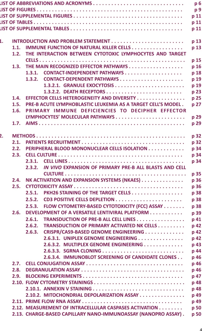
Figure 6. Extrinsic and intrinsic apoptoFc signaling pathways.
Once bound to the appropriate ligand, death receptors signal through adaptor molecules such as FADD (FAS-associated death domain protein) and ac.vate ini.ator caspase-8 and caspase-10. These either directly ac.vate the effector caspase-3 and caspase-7 or will cleave BID, providing a link toward the mitochondria-dependent cell death signaliza.on. In the laeer, truncated BID ac.vate BAX and BAK which will mediate mitochondrial outer membrane permeabiliza.on (MOMP) and subsequent release of cytochrome c. Through its interac.on with APAF1 (apopto.c protease ac.va.ng factor 1), it leads to the apoptosome assembly and caspase-9 ac.va.on which will ac.vate then the effector caspase-3 and caspase-7 triggering apoptosis. FADD Pro-caspases 8 and 10 Active caspases 8 and 10 Pro-caspases 3 and 7 Active caspases 3 and 7 BID tBID Inactive BAX/BAK BCL2 family Active BAX/BAK MOMP low Δψm high Δψm cytochrome c Pro-caspase 9 Active caspase 9 Apoptosome formation Active caspases 3 and 7 Pro-caspases 3 and 7 Apoptosis
DR mediated signaling can induce target cell death through the extrinsic pathway of apoptosis (Ashkenazi and Salvesen, 2014) (Figure 6). Of note, DRs can also mediate non-apoptosis-related func.ons which include regula.on of cell prolifera.on and differen.a.on, chemokine produc.on and inflammatory responses (Guicciardi and Gores, 2009). Regula.on between these signaling pathways is expected to be linked to the adaptor molecules: thus, the FAS-associated death domain protein (FADD) is more likely to trigger cell death, while signaling with the TNF type 1–associated death domain (TRADD) is more likely to ac.vate cell prolifera.on and inflamma.on (Lieberman, 2016).
It is interes.ng to note that granule exocytosis and death receptor mediated pathways both par.ally overlap and diverge in the molecular mechanisms which are implied in target cell death (Figures 5 and 6).
1.4. EFFECTOR CELLS HETEROGENEITY AND DIVERSITY
How cytotoxic effector cells are dis.nct from each other is an interes.ng ques.on to address. As previously discussed, CD8+ T cells and NK cells have similari.es in their development, homeostasis and immune role (Sun and Lanier, 2011) and can be considered as complementary effectors in their mechanism for target cells recogni.on.
As opposed to adap.ve immunity, innate immune response has been considered as a set of non-specific early defenses, regardless of the threat. Nevertheless, this view has now been challenged. Although the T cell receptor (TCR) confers T cell specificity toward the recognized an.gen, not all the NK cells will react to a threat, underlying their heterogeneity and diversity. The laeer can be defined at the phenotypic as well as the behavioral levels, both being intrinsically correlated.
Regarding the phenotypic differences, some have been long known: CD56dim CD16+ and CD56bright CD16- NK cell subsets are classically dis.nguished as cytotoxic and immunoregulatory cells respec.vely (Caligiuri, 2008), based on their func.onal ability to engage cytotoxicity or cytokine produc.on more efficiently. It has been proposed that these two subsets correspond to different stages of NK cell differen.a.on, with CD56bright CD16- cells being less mature precursors of CD56dim CD16+ cells, although some groups have found evidence for a different ontogeny between them (Wu et al., 2014). Another dis.nc.on can be made whether circula.ng or .ssue resident NK cells are considered, which deeply impact the rela.ve propor.on of these two subsets (Björkström et al., 2016). In that regard, liver NK cells are a good example of organ-adapted effector cell func.on with an important propor.on of immunoregulatory NK cells within this specific site of immunosurveillance.
Although NK cells lack a recombined an.gen-specific receptor, they express a complex array
of germline-encoded receptors whose combinatorial expression defines a massive degree of
NK cell diversity. Using a mul.parametric study by mass cytometry, it has been es.mated that one could dis.nguish between 6000 and 30,000 phenotypically NK subpopula.ons within an individual and more than 100,000 NK cell subpopula.ons within a cohort of 22 people (Horowitz et al., 2013). One can consider that this combinatorial diversity and the mul.plicity of ac.va.ng and inhibitory receptors confers a wide range of triggering thresholds for effector responses.
We have previously discussed cytokine produc.on and cytotoxicity, and it has been shown that they can be uncoupled (Rajasekaran et al., 2013; Vivier et al., 2013). From a "behavioral" point of view, the rela.ve contribu.on in the killing of target cells varies between individual NK cells. Indeed, in a degranula.on-dependent model of cytotoxicity, others demonstrated that a subset of "serial killer" NK cells were responsible for an important propor.on of the total number of kills (Choi and Mitchison, 2013; Vanherberghen et al., 2013). Overall, a classifica.on into five categories was proposed based on the contact number and their outcomes: (1) NK cells that don't interact with target cells; (2) NK cells that interact with targets but don't kill them; (3) NK cells that kill all target cells encountered; (4) exhausted NK cells (i.e. that stop killing aoer a certain number of interac.ons); and (5) NK cells that kill stochas.cally (a strong minority of the effector cells).
NK cell subsets also exhibit divergent adapta.on with regard to their local environment. For instance, it has been showed that the CD56bright CD16- cells are more resistant to reac.ve
oxygen species-induced stress (Thoren et al., 2007), which was shown responsible for tumoral immune escape (Aurelius et al., 2012). Figure 7. An updated view of human NK cell subsets (proposed by Frank Cichocki et al.). Adapted from Cichocki et al., 2015. TranscripFon factor expression Surface receptor expression Granule content CirculaFng CD56bright NK cell EOMEShigh PLZFlow CD62L+ CD49a–/ CD103– Canonical CD56dim NK cell AdapFve CD56dim NK cell Tissue-resident CD56bright NK cell EOMEShigh PLZF+ EOMEShigh PLZF -EOMESlow PLZF? CD62L+/- CD49a–/ CD103– CD62L– CD49a–/ CD103– CD62L– CD49a+/ CD103+
Furthermore, HCMV infec.on drives the expansion of a so-called adap.ve subset with dis.nct func.onal capaci.es which include on one hand reduced effector responses toward autologous ac.vated T cells, and on the other hand enhanced func.on against virus-infected cells (Lee et al., 2015; Schlums et al., 2015).
Among others, Frank Cichocki et al., taking in considera.on these recent discoveries, proposed an updated model of NK cell subsets that dis.nguish four main subgroups with a dichotomic contribu.on to either immunoregula.on or immunosurveillance (Figure 7). These studies provide further evidence of the complexity of NK cell responses at the cellular level. 1.5. PRE-B ACUTE LYMPHOBLASTIC LEUKEMIA AS A TARGET CELL’S MODEL Among neoplasia, pre-B cell acute lymphoblas.c leukemia (pre-B ALL) is the most common form of childhood cancer. Long-term survival has improved to reach 85-90% in children, although intensified systemic and central nervous system-directed chemotherapy causes acute and long-term treatment-related complica.ons (Hunger and Mullighan, 2015; Inaba et al., 2013; Oskarsson et al., 2016). However, relapse, which is observed in about 20% of pediatric and in more than 60% of adult pa.ents (Perova et al., 2014), is s.ll a major concern with a poor prognosis that has not significantly improved since the 80s (Bhojwani and Pui, 2013). Thus, relapsed ALL ranks as the fourth most common childhood malignancy and only 30% of the pa.ents survive (Mullighan et al., 2008).
Pre-B cell ALL development is a complex mul.step process and the disease e.ology is likely
different between young infant and older children or adults, although no single causal
mechanism is expected (Greaves, 2006). Following a classical view of cancer development, a
prenatal origin has been proposed for ALL arising in childhood (Marshall et al., 2014) while the disease in adults could result from acquired muta.ons (Hanahan and Weinberg, 2011). This view is supported by the observa.on of dis.nct molecular gene.cs between both groups of pa.ents. For example, BCR-ABL1 (or Philadelphia chromosome) is detected in over 25% of adult versus only 3% to 5% of pediatric ALL cases (Mullighan, 2012). A causal link between childhood pre-B ALL and infec.on has been reported (Greaves, 2006; Greaves and Muschen, 2015; Marsn-Lorenzo et al., 2015; Swaminathan et al., 2015). At the pre-B cell stage of B lymphopoiesis, cells exhibit an increase suscep.bility to gene.c lesions that can be exacerbated by abnormal repe..ve or chronic infec.ons, through the associated cytokine signaling and inflamma.on. However, there is no precise view of the individual species of viruses, bacteria, or other pathogens involved. An infec.on-triggered selec.on of
pre-leukemic clones has been suggested by the occurrence, among 2% of the pediatric pa.ents, of a transient clinical phase of aplasia in the few months preceding ALL. This phase, which is expected to favor the emergence of preleukemic clones (Greaves, 2006), was almost always associated with an infec.on (albeit either cause or effect).
Whether pre-B ALL follows the classical cancer stem cell model has been inves.gated by several groups. According to this model, tumors are heterogeneous but hierarchically organized en..es. In the par.cular case of hematological malignancies, it is temp.ng to apply a normal hematopoiesis-derived model in which (immature) cancer stem cells (leukemia stem cells), by analogy with hematopoie.c stem cells, retain stemness-related proper.es, i.e. the ability to self-renewing and differen.a.on into more differen.ated cancer cells. This considera.on is important since immature cells are also the most quiescent ones and thus less sensi.ve to conven.onal chemotherapy that targets prolifera.ve cells.
While this model seems coherent with acute myeloid leukemias (AML) (Shlush et al., 2017), it does not apply to pre-B ALL (McClellan and Maje., 2013). Strong evidence was brought by using a xenotransplant model in NOD/scid immunodeficient mice (le Viseur et al., 2008). Sorted blast popula.ons defined with the CD34, CD19 and CD20 surface markers shared the ability to recons.tute and reestablish the complete leukemic phenotype in vivo. These results and others establish a clear difference between AML and B-ALL (Belderbos et al., 2017; Rehe et al., 2013): AML arises within the normal HSC compartment and retains a hierarchy similar to normal hematopoiesis while pre-B ALL is highly polyclonal and stochas.c in its stemness (Figure 8). As a corollary, relapsed ALL clonal diversity is comparable to the one observed at the moment of the diagnosis while clonal survival from diagnosis to relapse is not associated with a muta.on burden (Ma et al., 2015; Mullighan et al., 2008). Figure 8. AML and B-ALL cancer stem cell divergent models. CD34+ CD38-CD34+ CD38+ CD34-CD34+ CD19-CD34+ CD19+ CD19+ CD20+ immature mature AML B-ALL
In this work, we have been interested in studying the immune control of pre-B ALL cells (that we used as targets) by cytotoxic lymphocytes and more specifically NK cells.
1.6. PRIMARY IMMUNE DEFICIENCIES TO DECIPHER EFFECTOR LYMPHOCYTES’
MOLECULAR PATHWAYS
One par.cular aspect of our approach was the use of effector cells from pa.ents suffering
from various primary immune deficiencies (PIDs), as natural human knockout models.
By defini.on, a cytotoxicity between effector NK cells and targets involves two cellular types. To study the precise molecular mechanisms governing NK mediated killing, many previous
works used chemical approaches like the inhibi.on of the granule exocytosis pathway by
EGTA/MgCl2 (EGTA is a chela.ng agent of divalent ca.ons including Ca2+ to block exocytosis
while the presence of Mg2+ in excess allows to maintain adherence between effector and target cells), or by concanamycin A that degrades perforin by increasing the pH of ly.c granules. However, a major concern with this strategy is the lack of specificity of these methods that affect both the effector and the target cells. In addi.on, the molecular selec.vity of these drugs is ques.onable as many different processes can be impacted. For
example, with EGTA/MgCl2, Ca2+ chela.on also inhibits FasL expression on the cell surface of
CD8+ cytotoxic T cells (He and Ostergaard, 2007), thus rendering any interpreta.on equivocal.
To circumvent these limits, we aimed to separate effector and target cell study. For this purpose, we used NK cells obtained from primary immune deficient pa.ents to study effector cells, and knockouts generated by CRISPR/Cas9 genome edi.ng technology to study the target leukemic cell lines. We used PIDs whose molecular defects could impact: (1) NK cell ac.va.on, (2) the integra.on of the ac.va.ng and inhibitory signals and (3) the effector response per se (Long et al., 2013).
1.7. AIMS
Allogeneic HSCT was ini.ally developed for two reasons (Copelan, 2006). First, it allows complete replacement of an abnormal hematopoie.c system by a normal one (which is of par.cular interest for severe primary immune deficiencies as well as hematological malignancies) (Gooley et al., 2010). Second, it allows dose-intensive/myeloabla.ve therapy for pa.ents suffering from cancer, for whom these high dose regimens increase tumoral cell elimina.on but also induce a permanent bone marrow failure that can be rescued by HSCT.
The grao is as an immunocompetent .ssue considering the immune cells it contains which are able to mediate ac.ve immune responses, either deleterious or beneficial, the so-called grao-versus-host disease (GvHD) and grao-versus-tumor (GvT) effect respec.vely. Both T cells and NK cells have been shown to contribute to GvT.
In 2002, Loredana Ruggeri et al. published an important study establishing a role for NK cell
alloreac.vity in mismatched hematopoie.c stem cell transplanta.on (HSCT) (Ruggeri, 2002).
Their work laid the founda.ons for this phenomenon which was aeributed to a mismatch between killer immunoglobulin-like receptors (KIR) expressed by donor NK cells within the grao, and KIR ligands (KIRL) expressed at the surface of recipient cells, including tumor cells. Interes.ngly, they provided evidence in AML but not in pre-B ALL that they found to be resistant to NK-mediated killing (Ruggeri, 2002; Ruggeri et al., 1999). Since then, different models have been proposed to predict the rela.ve risk of relapse taking into account donor and recipient’s phenotype, including the "ligand-ligand" and the "receptor-ligand" models
suggested by the Perugia and the Memphis groups respec.vely (Handgre.nger et al., 2016).
If the former considers KIRL expressed both by the donor and the recipient to establish a poten.al mismatch, the laeer considers the KIRs from the donor and the absence of the corresponding KIRLs in the recipient. Of note, alterna.ve models exist such as the “gene-gene” model which includes KIR genes from both the donor and the recipient in its predic.on (Handgre.nger et al., 2016). Altogether, each model performs beeer in specific condi.ons sugges.ng that there might be no unique rule for NK alloreac.vity but instead several mechanisms which could account differently depending on the disease type and pa.ents (childhood versus adults).
Deciphering the mechanisms governing it is important from both a fundamental point of view (i.e. to understand molecular pathways which contribute to NK cell ac.va.on and/or inhibi.on against this type of target cells as well as control their effector func.ons), and considering poten.al future developments as an immunotherapeu.c tool.
We worked on understanding NK cell alloreacFvity against pre-B ALL and why do these blasts exhibit an increase resistance to effector cell killing?
CHAPTER 2.
METHODS
2. METHODS
2.1. PATIENTS RECRUITMENT
Several centers around the world were involved in recrui2ng pa2ents suffering from various primary immunodeficiencies (PID). Because of the scarcity of PIDs that were interes2ng for our study, we obtained samples from pa2ents followed in many ins2tu2ons over the world. Hence, we could get samples from Hôpital Necker-Enfants Malades (Paris, France), CHU Jus2ne (Montreal, QC, Canada), Hospices Civils de Lyon (Lyon, France), Hôpital Saint-Louis (Paris, France) and Cincinna2 Children’s Hospital Medical Center (Cincinna2, OH, USA). Healthy control donors were sampled at the same 2me as the pa2ents bearing confirmed muta2ons, and samples were carried together. This study was accepted by the CHU Sainte-Jus2ne IRB (protocole number 3195) and all the involved subjects (pa2ents and controls) signed informed consent forms reviewed and approved by the appropriate ins2tu2onal medical research ethics commiZee (Table 2). Table 2. Pa7ents and cytotoxicity assays characteris7cs. Pa7ent ID Recrui7ng center Disorder Gene7c defect Muta7on Sample’s type Cell lines tested in cytotoxicity assay P01 CHUSJ X-linked lymphoprolifera2ve disease, Pur2lo syndrome (XLP1) SH2D1A PBMCs from fresh blood K562, REH P02 CHUSJ Type 2 familial hemophagocy2c lymphohis2ocytosis (FHL2) PRF1 [c.116C>A ] +[c.445G>A], [p.Pro39His] +[p.Gly149Ser] PBMCs from fresh blood K562, NALM6, REH, 697 P03 CHUSJ NEMO deficiency syndrome IKBKG PBMCs from fresh blood K562, REH P04 CHUSJ X-linked chronic granulomatous disease (X-CGD)
CYBB c.252G>A, p.Ala84Ala Frozen
PBMCs K562, NALM6, REH P05 CHUSJ Autosomic recessive chronic granulomatous disease (AR47-CGD) NCF1 [c.75_76delGT] +[c.75_76delGT], [p.Tyr26HisfsX26] +[p.Tyr26HisfsX26] PBMCs from fresh blood K562, NALM6, REH P06 CHUSJ X-linked chronic granulomatous disease (X-CGD) CYBB c.252G>A, p.Ala84Ala PBMCs from fresh blood K562, NALM6, REH P07 NCK X-linked chronic granulomatous disease (X-CGD) CYBB c.217C>T, p.Arg73X PBMCs from fresh blood K562, NALM6, REH P08 NCK X-linked chronic granulomatous disease (X-CGD) CYBB c.1610_1611delGT, p.Cys537TrpfsX3 PBMCs from fresh blood K562, NALM6, REH