Platelet-rich plasma and tendon healing:
rat model
KAUX JF 1, Drion P 2, Pascon F 3, Libertiaux V 3, Le Goff C 4, Gothot A 4, Cescotto S 3, Defraigne JO 5, Rickert M 6, Crielaard JM 1 1. Physical Medicine Service, Department of Motility Sciences, University Hospital of Liège, University of Liège, Belgium – [email protected] 2. Animal Facility of University Hospital of Liège, ULg-GIGA-R, University of Liège, Belgium.
3. Department Argenco, University of Liège, Belgium.
4. Department of Clinical Biology, University Hospital of Liège, University of Liège, Belgium. 5. CREDEC, Laboratory of Experimental Surgery, University of Liège, Belgium. 6. Department of Orthopaedic Surgery, University of Heidelberg, Germany.
Introduction
The aim of our study was to determine if a Platelet-Rich Plasma (PRP) injection could improve the healing process of ruptured Achilles tendons of rats.
Methods
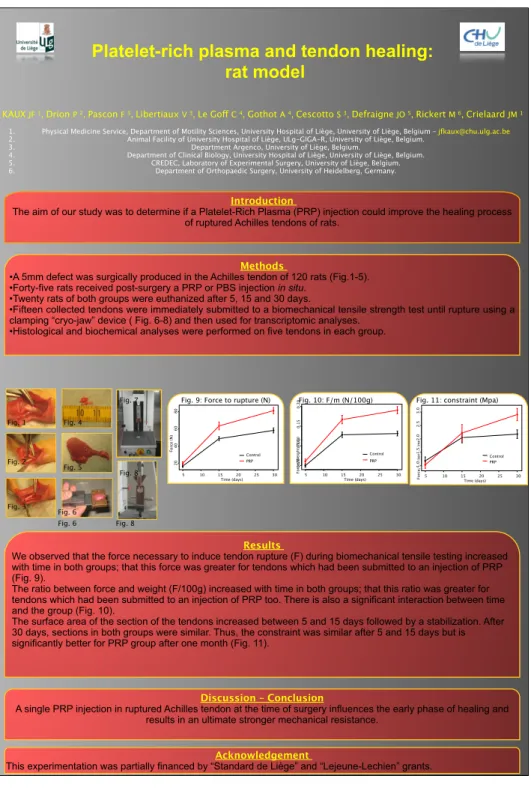
•A 5mm defect was surgically produced in the Achilles tendon of 120 rats (Fig.1-5). •Forty-five rats received post-surgery a PRP or PBS injection in situ.
•Twenty rats of both groups were euthanized after 5, 15 and 30 days.
•Fifteen collected tendons were immediately submitted to a biomechanical tensile strength test until rupture using a clamping “cryo-jaw” device ( Fig. 6-8) and then used for transcriptomic analyses.
•Histological and biochemical analyses were performed on five tendons in each group.
Results
We observed that the force necessary to induce tendon rupture (F) during biomechanical tensile testing increased with time in both groups; that this force was greater for tendons which had been submitted to an injection of PRP (Fig. 9).
The ratio between force and weight (F/100g) increased with time in both groups; that this ratio was greater for tendons which had been submitted to an injection of PRP too. There is also a significant interaction between time and the group (Fig. 10).
The surface area of the section of the tendons increased between 5 and 15 days followed by a stabilization. After 30 days, sections in both groups were similar. Thus, the constraint was similar after 5 and 15 days but is significantly better for PRP group after one month (Fig. 11).
Discussion – Conclusion
A single PRP injection in ruptured Achilles tendon at the time of surgery influences the early phase of healing and results in an ultimate stronger mechanical resistance.
Acknowledgement
This experimentation was partially financed by “Standard de Liège” and “Lejeune-Lechien” grants. Time (days) Force (N) 5 10 15 20 25 30 20 40 60 80 Control PRP Time (days) Fo rc e/ W ei g h t (N /g ) 5 10 15 20 25 30 0.05 0.10 0.15 0.20 Control PRP Time (days) Fo rc e/ Se cti o n ( N /m m ²) 5 10 15 20 25 30 1.0 1.5 2.0 2.5 3.0 Control PRP Fig. 1 Fig. 2 Fig. 3 Fig. 4 Fig. 5 Fig. 6 Fig. 7 Fig. 8
Fig. 9: Force to rupture (N) Fig. 10: F/m (N/100g) Fig. 11: constraint (Mpa)
Fig. 6