Université de Montréal
Biopacemaker acceleration without increased
synchronization by chronic exposure to phorbol myristate
acetate
par
Yashar Alami Alamdari
Département de physiologie moléculaire et intégrative Faculté de médecine
Thèse présentée à la Faculté des études supérieures et postdoctorales en vue de l’obtention du grade de Maîtrises en science (M.Sc.)
en physiologie moléculaire et intégrative
Décembre 2015
i
Résumé
L'activité électrique du coeur est initiée par la génération spontanée de potentiels d'action venant des cellules pacemaker du noeud sinusal (SN). Toute dysfonction au niveau de cette région entraîne une instabilité électrique du coeur. La majorité des patients souffrant d'un noeud sinusal déficient nécessitent l'implantation chirurgicale d'un pacemaker électronique; cependant, les limitations de cette approche incitent à la recherche d'une alternative thérapeutique. La base moléculaire des courants ioniques jouant un rôle crucial dans l'activité du noeud sinusal sont de plus en plus connues. Une composante importante de l'activité des cellules pacemakers semble être le canal HCN, responsable du courant pacemaker If. Le facteur T-box 3 (Tbx3), un facteur de transcription conservé durant le processus de l'évolution, est nécessaire au développement du système de conduction cardiaque. De précédentes études ont démontré que dans différentes lignées cellulaires le Phorbol 12-myristate 13-acetate (PMA) active l'expression du gène codant Tbx3 via des réactions en cascade partant de la protéine kinase C (PKC).
L'objectif principal de cette étude est de tester si le PMA peut augmenter la fréquence et la synchronisation de l'activité spontanée du pacemaker biologique en culture. Plus précisément, nous avons étudié les effets de l'exposition chronique au PMA sur l'expression du facteur de transcription Tbx3, sur HCN4 et l'activité spontanée chez des monocouches de culture de myocytes ventriculaires de rats néonataux (MVRN). Nos résultats démontrent que le PMA augmente significativement le facteur transcription de Tbx3 et l'expression ARNm de HCN4, favorisant ainsi l'augmentation du rythme et de la stabilité de l'activité autonome. De plus, une diminution significative de la vitesse de conduction a été relevée et est attribuée à la diminution du couplage intercellulaire. La diminution de la vitesse de conduction pourrait expliquer l'effet négatif du PMA sur la synchronisation de l'activité autonome du pacemaker biologique. Ces résultats ont été confirmés par un modèle mathématique multicellulaire suggérant que des fréquences et résistances intercellulaires plus élevée pourraient induire une activité plus stable et moins synchrone. Cette étude amène de nouvelles connaissances très importantes destinées à la production d'un pacemaker biologique efficient et robuste.
ii
Mots-clés : pacemaker biologique, activité autonome, synchronisation, stabilité
iii
Summary
The normal heartbeat is initiated by the spontaneous generation of action potentials in pacemaker cells of the sinoatrial node (SAN) region. Dysfunction of this region leads to electrical instability of the heart. The majority of the patients with sinus node dysfunction require surgical implantation of electronic pacemaker devices; however, limitations of this therapeutic approach lead to a need to search for alternatives. To date, the molecular basis of the ionic currents which play pivotal role in SAN action potential has been discovered. It is thought that an important component of the pacemaker cells are HCN channels, responsible for the funny current (If) in the SAN. Meanwhile, T-box factor 3 known as an evolutionary conserved transcription factors is necessary for development of the conduction system. In previous studies, it has been shown that Phorbol 12-myristate 13-acetate (PMA) activates Tbx3 gene expression in a PKC-dependent manner in several cell lines.
The main objective of this study is to test if PMA can increase the frequency and synchronization of spontaneous activity of cultured biopacemakers. More precisely, we studied the effects of chronic exposure to PMA on the expression of the Tbx3 transcription factor and HCN4 in neonatal rat ventricular myocytes monolayers and how spontaneous activity was altered. Our results show that PMA significantly increases the Tbx3 transcription factor and HCN4 mRNA expression favoring an increased in the rate and spatial-temporal stability of the spontaneous activity. In addition, a significant decrease in conduction velocity was found that is attributed to decrease electrical intercellular coupling of the cells. The decrease in the conduction velocity could explain the negative effect PMA has on synchronization of spontaneous activity of the biopacemaker. These findings are confirmed by a multicellular mathematical model implying that faster frequency and higher intercellular resistance of the pacemaker cells may lead to a more stable and less synchronous activity. This study provides important new knowledge to produce efficient and robust biological pacemakers.
iv
Table of contents
Résumé ... i Summary ... iii Table of contents ... iv List of Figures ... viList of abbreviations ... vii
Acknowledgments... x 1 Introduction ... 1 1.1 Heart physiology ... 3 1.1.1 Cardiomyocyte ... 3 1.1.2 Pacemaker cells ... 3 1.1.3 Fibroblasts ... 4 1.1.4 Spontaneous activity ... 5
1.2 Sino-atrial node (SAN) ... 5
1.2.1 Primary pacemaker cells ... 5
1.2.2 Secondary pacemakers (AVN and Purkinje fibers) ... 7
1.2.3 Rhythm ... 7
1.2.4 Clocks ... 9
1.2.5 Pathology of the rhythm... 20
1.3 Pacemakers ... 21
1.3.1 Electronic pacemakers ... 21
1.3.2 Biopacemakers ... 22
1.4 Phorbol 12-myristate 13-acetate (PMA) ... 28
1.4.1 PMA link with Tbx3 ... 28
1.4.2 Structural and physiological features ... 28
v 1.5 HCN blockers... 30 1.6 Project hypothesis ... 30 2 ARTICLE ... 31 Abstract ... 33 Introduction ... 33 Methods... 36 Results ... 41
Discussion and conclusion ... 45
References ... 48
Figure captions ... 55
Table and figures... 58
3 DISCUSSION AND CONCLUSION ... 71
3.1 PMA increases the rate of spontaneous activity through Tbx3 re-expression ... 71
3.2 PMA modulates the spatial-temporal activity in multicellular patches ... 72
3.3 The increase in frequency of spontaneous activity does not yield greater synchronization ... 74
3.4 CONCLUSION ... 74
vi
List of Figures
Figure 1: Schematic of sarcomeric structure ... 4 Figure 2: Electrical system of the heart ... 6 Figure 3: Schematic model of the embrionic heart presenting the various gene expression zones ... 15 Figure 4: SAN action potential traces underling the Ca2+ transactions ... 16 Figure 5: Schematic model of the calcium clock of the heart ... 17 Figure 6: Different electrical features and various distributions of ion channels and gap junctions between central and peripheral cells of the SAN ... 19 Figure 7: Different gene therapy-based strategies ... 23
vii
List of abbreviations
AC: adenylyl cyclase Ach: acetylcholine ACP: artificial pacing AKAP: A-kinase anchor AP-1: activator protein-1 APD: AP duration
AVN: atrioventricular node bpm: beats per minute CaM: Calmodulin
cAMP: cyclic adenosine monophosphate CCT: cardiac cell therapy
CFs: cardiac fibroblasts
CLL: chronic lymphocytic leukemia CMs: cardiomyocytes
cPKC: classical PKC
CRT: cardiac resynchronization therapy cTnT: Cardiac Troponin-T
DAG: 1, 2-diacylglycerol
DD: diastolic depolarization Discoidin domain receptor 2 (DDR2) DHP: dihydropyridines
EPCs: endothelial progenitor cells GPCR: G-protein-coupled receptor Gs: G proteins
HCN: Hyperpolarization-activated cyclic nucleotide-gated channels hESCs: human embryonic stem cells
HIV-1: human immunodeficiency virus-1 hMSCs: human mesenchymal stem cells HSCs: hematopoietic stem cells
viii ICD: implantable cardioverter-defibrillators LCP: local Ca2+ period
LCRs: local Ca2+ releases LTCC: L-type Ca2+ Channel MDP: maximum diastolic potential NCX: Na+/Ca2+ exchanger
NE: norepinephrine
NFκB: nuclear factor-kappa-B nPKC: novel PKC
NRVMs: neonatal rat ventricular myocytes ODC: ornithine decarboxylase
PIP2: phosphatidylinositol 4,5-bisphosphate PKA: protein kinase A
PMA: phorbol 12-myristate 13-acetate PPM: permanent pacemakers
PS: phosphatidylserine RyRs: ryanodine receptors
SACPs: specialized sinoatrial conduction pathways SAN: sinoatrial node
SERCA: ATPase pumps of the sarcoplasmic reticulum SR: Sarcoplasmic Reticulum
SSS: sick sinus syndrome
TPA: 12-O-Tetradecanoyl-phorbol-13-acetate TTX: tetrodotoxin
ix
x
Acknowledgments
It would not have been possible to write this thesis without the help and support of the kind people around me to only some of whom it is possible to give particular mention here.
First and foremost, I would like to thank my supervisor Dr. Philippe Comtois for his guidance and persistent help without which this project would not have been possible.
I wish to express appreciation to my colleagues for discussion and communications, among them James Elber Duverger and Alireza Aghighi who spent extra time helping me in writing the thesis, Alexandre Blanchette, Feng Xiong and Jonathan Béland. I devote my special gratitude to James for his attention, patient and unfailing supports during my master program.
It is my privilege to thank my wife, Laleh Abbassi, for supporting me all the way. I would certainly be remiss to not mention and sincerely thanks my parents for their love, encouragement and profound contribution in providing financial assistance for me to help with my studies. Their sacrifices have brought me to where I am today.
I am grateful to the authors for allowing us to use figures from their publications. Above all, I thank God for his many blessings.
1
1 Introduction
Normal function of the heart is extremely dependent on the flawless activity of the sinoatrial node (SAN). Abnormal propagation from the sinoatrial node leads to serious arrhythmias called sick sinus syndrome (SND). Nowadays, the prevalence of SND is increasing. In spite of prominent developments of electrical pacemakers, there are a lot of shortcomings. The need to create alternative therapies and the biological pacemaker is among them.
Different concepts will be presented in the introduction (Chapter 1). Starting with section 1.1, an overview of the heart physiology is introduced with a focus on the different cell types including the pacemaker cell. Primary pacemaker cells are located in the sinoatrial node (SAN, section 1.2) which is the structure responsible for initiating the normal heart beat.
In brief, pacemaker cells differs from cardiomyocytes (CMs) having reduced maximum diastolic potential (MDP), slower action potential (AP) upstroke velocity, high intercellular resistance which leads to slow conduction velocity and spontaneous diastolic depolarization (DD) (Clocks, section 1.2.4). An important components of the pacemaker cells are HCN channels (Clocks, section 1.2.4) which conduct the funny current (If). Mutations or knockout of these proteins in human or mice linked to SND (Pathology of the rhythm, section 1.2.5). Expression of HCNs is linked to T-box factor 3 (Tbx3) which is one of the evolutionary conserved transcription factors necessary for development of many tissues. Moreover, connexin 40 and 43 expressions were down regulated in ectopic Tbx3 activated embryonic atrial myocytes (Clocks, section 1.2.4).
Probably, it happens because of the current-to-load mismatch between HCN over-expressing cells and the surrounding tissue.
The most important therapeutic approach to date for patients with bradycardia and unstable heart rate remains the electronic pacemaker (section 1.3.1). An alternative being developed, termed the biopacemaker, is based on generating pacemaking activity of cells through different approaches (presented in section 1.3.2).
2
Gene therapy is a key approach to modulate expression of proteins. Chemical conditioning can also be an interesting alternative to modulate transcription factors. Phorbol 12-myristate 13-acetate (PMA, section 1.4) is an extracellular PKC activator which can activate the protein-1 (AP-1) family of transcription factors. In the previous studies, it has been shown that PMA activates Tbx3 gene expression in a PKC-dependent manner via the AP-1 transcription factors in several cell lines but its effect on spontaneous activity in cardiomyocytes remains unknown.
A widely used cardiac model is rat ventricular myocytes (NRVMs) which have been shown to be useful a model system for analyzing states of cellular hypertrophy and contractile protein gene expression. More interesting is that there is a transient regenerative phase in neonatal murine heart related to cardiomyocyte DNA-synthesis activity which declines during the first 1–2 weeks of life in rodents making this model a close relative to cardiomyocyte induced from pluripotent cells. (Biopacemakers, section 1.3.2).
We hypothesized that chronic conditioning of neonatal cardiomyocytes with PMA could upregulate the Tbx3 and HCN channels expression and facilitates the development of the cardiomyocytes to the pacemaker cells (Project hypothesis, section 1.6).
To test this hypothesis, we isolated ventricular cardiomyocytes from 1- to 3-day-old Sprague-Dawley rats. Cells were cultured in the pre-coated glass bottom dishes. Cultured dishes were exposed with PMA after 24h and the experiments were done 24h and 48 h after the start of conditioning with PMA (Article, section 2). The results of chronic exposure of the NRVMs with PMA showed an increase in expression of Tbx3 and HCN4 channels which was concordant with increasing the spatial-temporal stability of the spontaneous activity. In addition, the conduction velocity of the impulse propagation decreased providing an evidence of electrical uncoupling of the cells that led to decreased synchronization of the spontaneous activity. To confirm our experimental results, we implemented a mathematical model which mimics the PMA effect in increasing of cell spontaneous rate of activity and decreasing the electrical coupling in the experimental monolayers. The results of the computational model confirmed spatial-temporal stability and lower synchronous activity of our monolayers (Article, section 2).
3
In the last part of the thesis, the implication of our results on the biopacemaker approach is further discussed (Discussion and conclusion, section 3).
1.1 Heart physiology
1.1.1 Cardiomyocyte
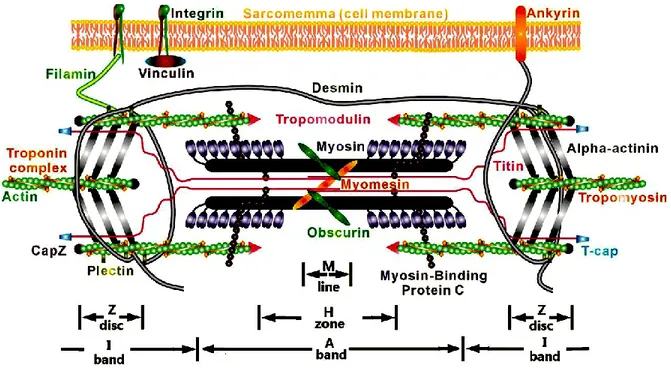
The human heart is built of different cell types including: cardiomyocytes, fibroblasts/myofibroblasts, vascular and immune cells, vascular smooth muscle cells and vascular endothelial cells (1). A cardiac myocyte is enclosed by a sarcolemma as cell membrane that contains a nucleus (2). Myocytes are composed of numerous mitochondria, and they supply the needed ATP for the contraction of muscles. In addition, cardiac muscles contain the machinery to perform the intact contraction including contractile proteins actin (thin filaments) and myosin (thick filaments) accompanied by troponin and tropomyosin as regulatory proteins (Fig. 1). Cardiac muscle has a striated shape, however the pattern is a little bit different with skeletal muscle (2). Cardiac myocytes composed of approximately 75% of normal heart tissue volume, but they occupy only 30–40% of the total cell population in the heart. The rest of the heart tissue comprises non-myocytes, including fibroblasts, as a predominant population and other cell types like vascular or endothelial smooth muscle cells (3). Cardiac Troponin-T (cTnT) could be used to identify the myocardial cells, as it is specifically expressed in the myocardium (4, 5).By differentiation of the cardiac chambers, the automaticity of the mature myocytes entirely disappears and they display a fast conduction velocity (6, 7).
1.1.2 Pacemaker cells
In addition to the contractile myocytes, there are other types of the excitable cells in the heart with specific electrophysiological characteristics privileged them to generate and propagate electrical pulses. The pacemaker cells and their related conduction system are composed of two main nodes called sinoatrial (SAN) and atrioventricular (AVN) nodes in addition to the specified conduction system. The features of the pacemaker cells and their electrical pathways will be discussed comprehensively in the following chapters.
4
Figure 1: Schematic of sarcomeric structure. Actin and myosin filaments as contractile
components and sarcomeric titin as a structural component are shown in the diagrammatic model. (Reproduced with permission fromLiu H. et al. 2013 (8))
1.1.3 Fibroblasts
Fibroblasts are defined as flat and spindle shaped cells with numerous protrusions arising from the cell membrane. One of the outstanding morphological features of the fibroblasts is the lack of a basement membrane, and it distinguishes these kinds of cells from the other cell types of the heart. Generally, fibroblasts play a critical role in chemical, mechanical, and electrical signaling in the heart, and any pathological change of these signaling pathways can lead to cardiac dysfunction (9). Normal development and aging increase the fibroblast contents (10, 11). In fact, 5–6% of the volume of the normal adult myocardium is composed of connective tissue which is in parallel with increasing the fibroblast contents during the life time, however the fraction of the connective tissue in sinoatrial node (SAN) is more than 50% in the adult human heart (12, 13). Although cardiac fibroblasts are basically unexcitable cells, they are able to affect the electrophysiological communication of myocytes as effective components (14). In spite of non-excitable properties,
5
these type of cells play an important role in electromechanical functions of the heart in addition to compose of the anatomical and biochemical integrity of the heart tissue. Discoidin domain receptor 2 (DDR2) is specific marker of the cardiac fibroblasts which is not expressed on other types of the cardiac cells (15).
1.1.4 Spontaneous activity
Cardiomyocytes development during embryonic period affects their electrical properties notably (16). Automaticity is a dominant aspect of cardiomyocytes in very early prenatal period. However it is going to disappear by transforming the cells into the ventricular myocytes. In the late embryonic period, sinoatrial node takes the responsibility of controlling the automaticity coincident with the complete differentiation of the cardiomyocytes to the working ventricular cells (17). Recent studies based on patch clamp techniques demonstrated increase in amplitude of several cardiac currents including the fast Na+ channel (18) current and the L-type Ca2+ channel (19) current during the rat embryonic developing phase which is related to increase in their channel expression level. In addition, there are some reports corresponding with increasing the outward currents from the middle to late embryonic period. Maybe these increments are to be responsible for the interruption of automaticity of working ventricular myocytes. Some studies (20, 21) have reported an increment of the inwardly rectifying background K+ current (IK1) in fetal working ventricular myocytes. Thereby, it could be proposed a hypothesis suggesting that the hyperpolarization effect of IK1 augmentation leads to the cessation of the spontaneous activity in fetal ventricular myocytes, nonetheless it would be beneficial to reduce or abolish some pacemaker inward currents.
1.2 Sino-atrial node (SAN)
1.2.1 Primary pacemaker cells
In the mammalian heart, SAN is laid at the junction of the superior vena cava and right atrium. The size of the SAN in the adult human heart is 12–20 mm long and 2–6 mm wide which is detectable by its ellipsoidal shape and intramural position. The head of the node is located around 1 mm under the epicardium isolated by a layer of lipid and connective tissue (22). The head part of the SAN extends inferiorly for 10–20 mm stretching beneath the sulcus
6
terminalis till the crista terminalis and has multiple extensions into the bordering atrial myocardium, which composes the specialized sinoatrial conduction pathways (SACPs) (23) (Fig. 2). The sinoatrial node is the main commander of the human heart rhythmicity by producing and propagating the electrical impulses that plays a critical role in regularity of the heart beats (22).
Figure 2: Electrical system of the heart. In normal heart electrical pulses originate from
SAN and propagate through the AVN to the His bundles and Purkinje fibers. (Reproduced with permission from Harvey et al. 2002 (24))
It is well known that the structure of the human and canine SAN is a complex multi-compartment (25). The SAN of mammalians heart, is composed of clusters of developed cardiomyocytes, which are engulfed within the matrix of connective tissue including the mixture of fibroblasts and some kinds of connective proteins like collagen and elastin. Such a fibrotic structure insulates the pacemaker cells from the hyperpolarizing effect of the bordering atrial cells in addition to mechanical protection (23).
7
1.2.2 Secondary pacemakers (AVN and Purkinje fibers)
In the intact heart, electrical impulse originates from the SAN and propagates to the surrounding atrial myocardium and reaches the atrioventricular node (AVN) after then electrical wave passes through the Purkinje bundles to depolarize the ventricles and initiate the contraction of the heart thus perform the pumping function (26).
Anatomical position of the AVN is at the junction of the atrial and ventricular septum which is dubbed as the triangle of Koch (27) and it is bounded by tendon of Todaro, the coronary sinus ostium and the tricuspid valve. There are two different pathways which lead into the AVN the first one is transitional zone, fast pathway, and the second one is inferior nodal extension defined as slow pathways (28).
As a matter of fact, there are two electrical pathways (29). The fast route of propagation passes through the atrial septum and transitional zone, indicating the normal route. On the other hand, there is another way to conduct the propagation wave from the SAN through the AVN using the terminal crest and inferior nodal extension (30). There is also a penetrating bundle, at the position distal to the AVN, engulfed in the central fibrous body and extrudes on the ventricular spectrum crest where the His bundle begins. The AVN plays critical roles in the heart. First of all, it serves as a conduction delay barrier which is essential to perform the atrial contraction before the ventricular one. Secondly, long refractory period of the AVN helps to block the high frequency activity in atrial arrhythmias to minimize the ventricular tachycardia. Furthermore, AVN has also a potential ability to emerge as a first initiation site when the normal activity of the SAN disappears to prevent the heart arrest (31).
1.2.3 Rhythm
In the intact physiological condition, the SAN which located in the right atrium initiates the rhythmic pacing discharge. Indeed, the SAN is a part of the intrinsic conduction system included in the heart. Cardiac activation starts with the SAN or pacemaker and propagates to the surrounding cells resulting in depolarization and contraction of the atrial tissue and extends in order of rate to the internodal pathway, the AVN (where the impulse is delayed), AV bundle, the left and right branches of the bundle of His and lastly the Purkinje
8
fibers, consequently leads to ventricular depolarization and contraction. Autonomous rhythmicity is one of the pivotal characteristic of the intrinsic conduction system, thereby in the absence of extrinsic neural or hormonal stimulation, the SAN enables to impulse with the pacing rate about 100 beats per minute (bpm). Nonetheless, the heart rate and cardiac output must be changed in response to the needs for oxygen and nutrients supply under varying conditions. In order to make an immediate response to be concordant with body’s request, it is vital to provide an interacting system to control the heart rate and contractility. The SAN has two regulating systems: the sympathetic and parasympathetic systems. The sympathetic nervous system releases norepinephrine (NE) while the parasympathetic nervous system releases acetylcholine (ACh). Adrenergic β1 receptors are expressed in the SAN, AVN, besides in atrial and ventricular cardiomyocytes. The activation of β1 receptors, mediating NE, increases intracellular calcium concentrations and calcium release by the sarcoplasmic reticulum (SR) which leads to increase in contractility as a consequence and increase AVN conduction velocity. Thus, sympathetic nervous system stimulation is responsible for: ―1) Positive chronotropic effect (increase in heart rate); 2) Positive inotropic effect (increase of contractility); and 3) Positive dromotropic effect (enhancement of conduction)‖ (32).
The parasympathetic nervous system has limited modulatory effect on the heart in contrast to sympathetic activity. Acetylcholine, as a main neurotransmitter of the parasympathetic system, produces effects that are in opposite to the sympathetic activation which includes: negative chronotropic and dromotropic effect. However, there are a lot of controversies about the probable negative inotropic effects of parasympathetic stimulation, recent in vivo studies in the atrium may suggest otherwise (32).
The catecholaminergic control of the heart rate and contractility is mediated by the protein-cAMP-PKA signaling pathway (32). In fact, activation of β1-adrenoceptor as a G-protein-coupled receptor (GPCR) is the source of the sympathetic stimulation-induced effects in the heart. NE binding to β1 receptors activates stimulatory G proteins (Gs) and consequently activates adenylyl cyclase (AC) which mediates dephosphorylation of ATP into cyclic adenosine monophosphate (cAMP). After then, cAMP accomplishes numerous functions and it plays an important role in regulating ion channels, transcription factors, or enzymes. Regarding to the cardiovascular system, protein kinase A (PKA) is the most important enzyme
9
activated by cAMP. In consequence, PKA phosphorylates proteins, such as contractile machinery including troponin C, I, and sarcoplasmic proteins and L-type Ca2+ channels (LTCC). In addition, cAMP binds directly to hyperpolarization-activated cyclic nucleotide-gated (HCN) channels, thereby increasing the heart rate (33).
G-protein-coupled receptor, muscarinic (M2) receptors in the heart, plays also a pivotal role in the parasympathetic system in the heart resulting from activation by ACh as a parasympathetic neurotransmitter. ACh binding to M2 receptors causes a conformational change within the Gi subunit of the receptor facilitating inhibitory activity of the G protein, consequently the disassociated αi subunit can bind to and inhibits AC. M2 receptors decrease cAMP formation due to negatively coupling to AC. As a result, M2 receptors inhibit PKA activity and have an opposite effect on ion channels, Ca2+ handling proteins, and contractile machinery, in contrast to sympathetic stimulation (32).
1.2.4 Clocks
Briefly, spontaneous excitation of ―pacemaker‖ cells in the sinoatrial node initiates the normal cardiac electrical activity. The activation wave then travels to the adjacent atrial myocytes through intercellular gap junctions, and finally causes atrial excitation. After then, the excitation wave passes via the atrioventricular node and the Purkinje fibers to the ventricles and leads to ventricular myocyte depolarization. The self-activating characteristic of the SAN is attributed to the precise contribution between sarcolemma and sarcoplasmic membranes ion channels regulation termed as membrane and calcium clocks, respectively. Inward or outward direction of ion currents is related to the electrochemical gradient of the corresponding ions. Normally, there is a linear relationship between current amplitude (I), membrane potential (V) and the conductivity (G) of the responsible ion channels. This relation is defined in equation form as ―I=VG (R as resistance is the reverse of conductivity: I=V/R [Ohm’s law])‖ (26), indicating that membrane potential changes affect the current amplitude. Nonetheless, there are types of membrane channels that show non-ohmically behavior called voltage-dependent channels. One such current that is important for the resting potential is the rectifying currents (IK1). The rectifying channels carry dynamic currents which vary nonlinearly at different membrane potentials. It is the interplay between inward and outward
10
currents that leads to the spontaneous formation of an action potential (26). It is believed that the cooperation of both the membrane and calcium clocks in SAN is responsible for the pacemaker cell activity with characteristics including:
1- ―Reduced maximum diastolic potential (MDP)‖ (34).
2- ―A slow action potential (AP) upstroke velocity‖ (34) which is regulated by the L-type Ca2+ current, ICaL, in central sino-atrial nodal cells (35).
3-High intercellular resistance (34). Indeed, the lack of fast-propagating connexins including Cx40 (Gja5) and Cx43 (Gja1), besides distribution of slow-propagating connexins such as Cx30.2 (Gjd3) and Cx45 (Gjc1) (36, 37) leads to the electrical uncoupling resulting in slow conduction velocity in SAN.
4- ―Spontaneous diastolic depolarization (DD)‖ (34). The animal studies, in particular on rabbit heart demonstrated that there is a precise control system to depolarize the membrane potential over the activation threshold.
Cellular processes are thus central to the spontaneous activity and interplay between membrane and intracellular calcium regulation would play a role. The differences between the membrane and calcium clocks are described below.
1.2.4.1 Membrane clock
According to the contribution of the variety of ionic currents in spontaneous pacemaking, and due to their time-dependent behavior and localization in the cell membrane, it has been dubbed ―the membrane clock‖(35). The hyperpolarization-activated cation current, or ―funny‖ current (If) is the dominant ionic current in the membrane clock (34).
The membrane clock theory of pacemaking states that precise cooperation of time- and voltage-dependent membrane ion channels enhances diastolic depolarization from MDP to threshold potential which opens L-type voltage-dependent Ca2+ channels to create the upstroke phase of the action potential. Consequently, activation of outward repolarizing potassium channels including transient outward (Ito), fast delayed rectifier (IKr), and slow delayed rectifier (IKs) K+ currents initiate repolarization phase by reducing the membrane potential to the maximum diastolic potential after then the next cycle starts again (35).
11
1.2.4.1.1 HCN channels
Genes isoforms HCN1 through HCN4 are responsible to encode the voltage-gated ion channels (38, 39) although HCN2 and HCN4 are found in neonatal hearts. Different subunits possess various characteristics which proper them to do their biophysical duties such as phosphorylation by tyrosine kinases, voltage dependency and regulation by low molecular weight factors like phosphatidylinositol 4,5-bisphosphate (PIP2) and cAMP (40). Regulation of If by cAMP is one the most important features of the HCN channels with increment of the membrane resting potential thus increasing the current activation (41). Hence, rate of activation of the HCN4 channels is augmented with cAMP (42). Moreover, higher level of phosphorylation can increase the sensitivity of the cells to the β-adrenergic stimulation in which maximal conductance of the pacemaker cells increases in a voltage independent manner (41). Funny current is defined as a mixed inward Na+ and K+ current which is activated slowly at membrane potential range of -50 to -65 mV (43, 44). All of the channels in cardiac cells are activated by membrane depolarization except If known as funny current (35).
1.2.4.1.2 CaT channels
Transient type Ca2+ (CaT) channels, including Cav3.1 through Cav3.3, have been encoded by three different genes, CACNA1G through CACNA1I (45). The first hypothesis regarding the role of ICaT in automaticity was described by Bean who proposed that fast Ca2+ channels have more important effect on generating spontaneous activity, due to their activation kinetics at negative potentials, while the Na+ channels show inactivated behavior in less negative potentials (46). In addition, the previous studies on rabbit heart proved that ICaT block with 40 μM Ni2+ has a negative chronotropic effect on pacemaker cells as a result of decreasing the slope of the late phase of DD. These types of channels show activity in more negative activation potentials in comparison with L-type Ca2+ channels (47).
1.2.4.1.3 CaL channels
L-type Ca2+ (CaL) channels are other types of the calcium conductors defined as L-type Ca2+ channels. They include of the variety of subunits such as α1-, β- and α2-δ (48). Their selectivity to the Ca2+ ions is dependent on α1-subunit of the channel pore. Such capability is due to the high affinity of the pore for Ca2+ ions (49). Different isoforms of the α1-subunit are
12
encoded by different genes resulting in various types of the channels such as Cav1.2 (CACNA1C) and Cav1.3 (CACNA1D) which are presented in the human SAN. It is shown that Cav1.2 mRNA is expressed in atrial and nodal cells, whereas Cav1.3 is distributed much more in the SAN comparing in atrial myocytes (50, 51).
Cav1.2 channels basically have some specific characteristics including requirement of higher membrane voltage for activation, long-lasting activity performance. In addition, lower concentrations of L-type calcium channel blockers including phenylalkylamines, dihydropyridines (DHP) and benzothiazepines would be able to block the channels (52-54). L-type Ca2+ channels are targeted by hormones in spite of their voltage dependent characteristic (55). According to the Reuters studies (56), there is a correlation between increasing in ICaL and the positive effect of NE on cardiac muscle contraction. Recent investigations demonstrated that such inotropic effect occurs due to the up-regulation of intracellular cAMP levels as a consequence of β-adrenergic receptor stimulation resulting in activation of cAMP-dependent protein kinase (PKA). In agreement with these studies, the experiments on guinea pig cardiomyocytes substantiated increment of action potential duration (57) and ICaL (58) due to the catalytic subunit of PKA.
1.2.4.1.4 NaK pump
Sarcolemmal NaK pump plays a critical role in regulation of the automaticity by carrying three Na+ ions outward and transporting two K+ ions inward the cells. The activity of the ATPase pump decreases the rate of the spontaneous activity by producing a net outward and hyperpolarizing current (INaK or Ip) (59). NaK pump is highly sensitive to the intracellular Na+ concentration; thereby there is a close relationship between the activity of the pump and If-derived Na+ ions in SAN (60). Therefore, INaK has a pivotal role in maintaining the maximum diastolic potential (61).
1.2.4.1.5 K+ channels
1.2.4.1.5.1 Transient outward potassium channels
There are two different subtypes of transient outward current in the heart one of them passes K+ ions (Ito1) and other one conducts Cl- ions (Ito2) (62). Distribution of three subunits
13
of the potassium transient channels has been recognized in human (50) and murine (51) SAN including Kv4.2, Kv1.4 and, in particular, Kv4.3. Fast activation and inactivation after depolarization kinetics is recognized as a specific feature of K+ transient outward channels, therefore they play a critical role in early phase of repolarization of ventricular cells in most of mammals, except for guinea pig (63) and pig (64). There is a big difference in action potential kinetics and duration between epicardium and endocardium tissue which is attributed to the various density in distribution of K+ transient outward channels (65).
1.2.4.1.5.2 Delayed rectifying channels
These types of channels divided to two different components, rapid (IKr) and slow (IKs) components. They trend to pass the current inwardly rather than outward direction, duded as inward rectification. In rapid rectifying channels, very rapid inactivation of the channel occurs upon activation of the channel after membrane depolarization. Hence the effect of such a little amplitude of IKr on plateau phase of action potential would be very low. However, they play a significant role in late repolarization phase due to their recovery from inactivation which results in a huge amplitude of outward current. This outward current will be vanished gradually after slow deactivation of the channels (66).
Another component of the rectifying channels which caused a lot of debates during the last decades was termed as slow component, IKs. The gene of the channel, called minK (KCNE1) was expressed on Xenopus oocytes in 1988 (67). The KCNQ1 subunit is the main component of delayed rectifying channels carrying the slow rectifying current in the heart. Thereby, they put their effect on the repolarization phase of cardiac action potential (68, 69). Due to the voltage gated characteristic of the channels, they activate gradually by augmenting membrane potential and result in increasing K+ current, then their slow inactivation kinetics leads to decrease the current progressively (70). Bounding an additional protein called A-kinase anchor (AKAP), to the cytosolic surface of the KvLQT subunits would be able the channels to be regulated by PKA (71). Thereby, slow rectifying current would be increased following sympathetic stimulation (72) as a consequence of stimulating their AKAP domain with PKA which is upregulated by β-adrenergic receptor activation (71). As a result of slow activation behavior, amplitude of IKs augments during the plateau phase of the action potential,
14
however it falls gradually when the membrane potential reach to 0 mV because of inactivation of the channel (73).
1.2.4.1.5.3 Inwardly rectifying K+ channels
There are only two subfamilies of the inwardly rectifying K+ channels expressed in heart cells (74). Kir2 channels including Kir2.1 and Kir2.2 which are responsible to IK1 (75, 76) and Kir3 channels consist of Kir3.1 and Kir3.4 conducting IKACh. IK1 is remarkable in ventricular cardiomyocytes and IKACh, a receptor-activated Kir current, has been shown in atrial and SAN cells. In fact vagal nerve and AV node govern the heart rate by mediating the receptor activated channels (74).
1.2.4.1.6 Sustained inward current
Sustained inward current (Ist) has been found in rabbit SAN in 1995 (77). There is little information about the molecular structure of the channels. It is known as an inward current conducted by some types of channels which possess pharmacological features of voltage-gated Ca2+ channels such as reactivity to nicardipine and resistance to tetrodotoxin (TTX), whereas is permeable to Na+ ions. These channels are open in membrane potentials around –60 mV during depolarization phase of action potential (77). Activation of Ist around diastolic potential range proposes the role of sustained inward current in membrane depolarization (78).
1.2.4.1.7 Tbx3
Tbx3 is defined as one of the critical transcription factors of T-box family which plays a transcriptional repressor role during embryonic period (79, 80). It is established that homozygous mutations of Tbx3 in murine embryos would be lethal (81). Meanwhile, mammary gland abnormalities and limb deformations are reported due to the Tbx3-heterozygous mutations. Moreover, haplo-insufficiency of Tbx3 causes the ulnar-mammary syndrome in human (82, 83).
It needs to be mentioned tumorigenic effect of Tbx3 as well as its developmental role. There are obvious evidences regarding its expression in some types of malignancies such as melanoma, breast and bladder cancers, melanoma (84, 85). The origin of pacemaker activity is SAN and AVN in intact adult heart. One of the specific characteristics of these regions is slow
15
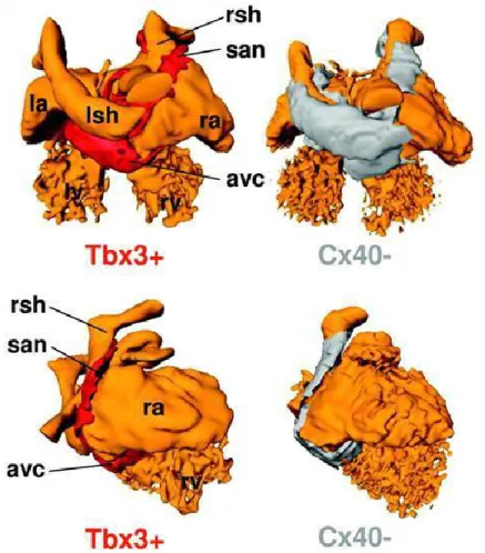
conduction velocity. The mechanism by which the SAN and AVN differentiate during embryonic period and perform their confined spontaneous activity has been the source of significant interest. Recently, progressive developments have been reported regarding the detection of new cellular mediators including transcription factors correlating with the differentiation regime of sinoatrial cells. Tbx3 is one of those pivotal structures which facilitate the SAN formation (6, 86-88). According to the Mommersteeg et al. investigations (88) the SAN stems from the inside region of the fetal heart. Tbx3 and HCN4 expression is one of the specific characteristic of the SAN region. This region is characterized by expression of the T-box transcription factor Tbx3 and the HCN4 ion channel gene. In addition Cx43 which is lead to faster propagation in ventricular cells has not been expressed in embryonic SAN (Fig. 3).
Figure 3: Schematic model of the embrionic heart presenting the various gene expression zones. Three-dimensional computational models of E14.5 wild-type embryos which present the heart lumen (red color) from two different prospective including dorsal view (top panels)
16
and right side view (bottom panels). Myocardium structure defined as Cx40-negative myocardium (gray areas) and Tbx3-positive myocardium (red areas) which is enclosed in the Cx40-negative myocardium. avc, atrioventricular canal; la/ra, left/right atrium; lsh/rsh, left/right sinus horn; san, sinoatrial node. (Reproduced with permission from Mommersteeg M.T. et al. 2007 (88))
1.2.4.2 Calcium clock
According to the calcium clock theory, SAN cells possess an intracellular mechanism to modulate the Ca2+ release from sarcoplasmic reticulum (SR) which is termed local Ca2+ releases (LCRs). In fact, there is a close interplay between LCR and membrane electrical polarization. The period between the previous peak of membrane Ca2+ release and the initiation of sarcoplasmic Ca2+ release is named the local Ca2+ period (LCP) (Fig. 4) (35).
Figure 4: SAN action potential traces underling the Ca2+ transactions. Normal action
potential traces of SAN spontaneous activity labeled with different phases of the AP is shown in top red panel. Different components of the ―Ca2+ clock‖ are presented at the bottom. It should be noticed concurrency of phase 4 of AP indicating diastolic depolarization with LCR outflowed from the sarcoplasmic reticulum. Calcium emanating from SR increase suddenly as a result of Ca2+-induced Ca2+ release called whole cell Ca2+ transient. (Adapted from Monfredi O. et al. 2013 (35))
SR ryanodine receptors (RyRs) play an important role in creating local calcium sparks in sinoatrial cells (89, 90). Neural stimulation or Ca2+ overload has no effect on LCR. Generally, local Ca2+ release starts during the diastolic depolarization concomitant with dissipation of global action potential-induced Ca2+ transient. It is believed to lead to activation
17
threshold of SR ryanodine receptors. Consequently, the signals called Ca2+-induced-Ca2+ -release goes up gradually which in turn leads to overall Ca2+ release of the SR, activating the Na+-Ca2+ exchanger, depolarizing the cell membrane and leading to activation of the action potential. Then, a new cycle begins with activation of ATPase pumps of the sarcoplasmic reticulum (SERCA) with replenishing the Ca2+ stores (Fig. 5) (35). Briefly, the reason of nomenclature of the SR as ―Ca2+ clock‖ is the periodic regime of LCR (91).
Figure 5: Schematic model of the calcium clock of the heart. RyRs and SERCA pump are
the main components of the Ca2+ clock which are responsible to the release and restoring of the Ca2+ ions, respectively. (Reproduced with permission from Monfredi O. et al. 2013 (35))
1.2.4.2.1 SERCA
Morphologically, the SERCA protein as a transmembrane protein is composed of 3 different components. Its molecular weight is 110 kD. The calcium binding kinetics of the protein is related to the transmembrane part including 2 sites to bind Ca2+ ions (92).Another domain of the protein is cytoplasmic head. The head domain is divided to 3 separate units including actuator, phosphorylation and nucleotide domains. Each of them plays an important role in function of the pump. The actuator domain acts as a Ca2+ binding site. While, junction of nucleotide and the phosphorylation domains facilitates ATP hydrolysis (93). SERCA is the most important pump in mammals facilitates refilling of the Ca2+ capacity of the SR (94). Indeed, it mediates around 92% of mouse and 70% of human cardiac Ca2+ removal, hence plays a remarkable role in heart contraction activity (94, 95).
18
1.2.4.2.2 RyR
Ryanodine receptors (RyRs) are composed of four subtypes (96). They mediate the join of t-tubules and junctional SR in both cardiac and skeletal myocytes (97, 98). Mammalian tissues are composed of three subfamilies of ryanodine receptors. Skeletal muscles express ubiquitously RyR1 and heart cells are composed of RyR2 and RyR3 isoforms, however RyR3 quantity in the heart is negligible and its function is not critical in cardiac cells function (99). RyRs are essential in cardiac automaticity, so that prenatal knockout of RyR2 could be fatal in mice embryos (100). Cardiac-specific RyR knockout mice in which RyR expression decreased around 50% show cardiomyopathy symptoms in addition to severe arrhythmias and bradycardia (101). RyR2 channels regulation is mediated by several parameters including Calmodulin (CaM), Ca2+ ions, phosphorylation, thiol oxidation and nitrosylation (102).
1.2.4.2.3 NCX
Na+/Ca2+ exchanger (NCX) is one of critical membrane components of the cardiac cells that regulates intracellular Na+ and Ca2+ concentrations. Indeed, NCX acts as a transporter passing 3 Na+ ions for outflowing 1 Ca2+ ion through the cell membrane (103). It is demonstrated even NCX contribution with the L-type Ca2+ current in regulating the subsarcolemmal Ca2+ concentration in up-regulated levels (104). Previous studies show the existence of a fuzzy space between the junctional SR and cytoplasmic layer. It could be a proper explanation for occurrence of calcium sparks in limited zones called ―diadic clefts‖ (105). Ca2+-induced Ca2+-release prominently is confined to the diadic clefts. It is concordant with the ubiquitous distribution of NCX in the T-tubular membranes (106).
1.2.4.3 Multicellular spontaneous activity
One of the important issues correspondent with proper function of the SAN is coordination of the dominant pacemaker cells with the surrounding tissue which is termed the source-sink relationship. Nonetheless, how the depolarizing ―source‖ current generated by the SAN propagates and activates the surrounding atrial tissue (current ―sink‖) remains to be resolved. It has been corroborated that the SAN is not functionally and anatomically continuous with the surrounding myocardium, but rather areas of functional or anatomical conduction block exist, providing discrete pathways at which SAN electrical pulses pass
19
through to activate the atrial myocardium (107). Previous studies in rabbit, canine, and human SAN have demonstrated the presence of such functional block and discrete exit pathways (108-110). Such source-sink mismatch facilitates electrical insulation of the SAN from the surrounding atrial myocardium. Histological studies over the years have failed to confirm proof of insulating fibrotic sheath surrounding the SAN in the human heart (111, 112) which could increase the probability of the existence of a functional barrier rather than anatomical mismatch. However a recent study showed that the SAN border is composed of fibrosis, fat, and/or discontinuous fibers between SAN and atria (113). It remains that various ion channel and gap junction expression in SAN would be extremely critical in performance of pacemaker cells (114). Therefore, three-dimensional architecture of SAN provides a complex structure comprised of central and peripheral or ―paranodal‖ components composed of variety of ion channels and gap junctions. It is shown that there is a significant difference between electrical characteristics and conduction properties of central and peripheral cells (Fig. 6) (31).
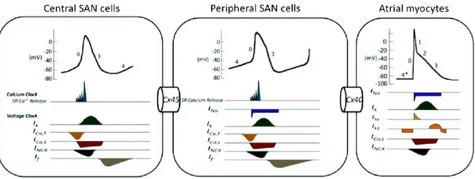
Figure 6: Different electrical features and various distributions of ion channels and gap junctions between central and peripheral cells of the SAN. Lack of the fast activating Na+
channels in the central cells of the sinoatrial node disposes them to have a slow upstroke velocity than peripheral cells, in addition distribution of slow propagating connexin proteins like Cx45 decrease the conduction velocity of the wave propagation in the SAN. (Reproduced with permission from Park D.S. et al. 2011 (115))
Regarding the experimental and computational studies, heterogeneity of the pacemaker cells gives an eminent capability to the SAN to perform the normal pacemaking activity and
20
impulse conduction. To date, two different hypotheses have been proposed to explain the electrical machinery of the SAN. Boyett and colleagues proposed the ―gradient model‖ indicating that AP properties show a gradual transition from the central to peripheral SAN (116), whereas some of the other researchers proposed ―mosaic model‖ suggesting that there are only a few different types of nodal cells which are interspersed with each other and with atrial cells (117). Apart from the cell heterogeneity model, it is well known that central SAN APs show slower rate of upstroke velocity, longer AP duration (APD), and lower potential level of negative maximum diastolic in comparison with peripheral SAN and atrial APs (118). These AP changes are contributed to the differential expression of several ion channels between these areas, as cited above (114).
1.2.4.3.1 Gap junctions
Conduction of impulse among heart muscles and consequently their coordinated contractions is highly dependent on the heart cells electrical association (119).
Gap junctions are responsible for electrical communication among heart cells. These types of junctions are made up of a network of membrane proteins complexes named as connexons (120). The counterpart complexes from adjacent cells form head-to-head connections with each other, and therefore create gaps with 2-3 nm of diameter between every two neighbor cells. There are six protein subunits in each connexon that surround a central aqueous pore which is 1.5-2 nm in diameter. This pore is wide enough to allow ions, second messengers, and any molecule smaller than ~1 kDa to pass through and reach the cytosol of the neighboring cell (119). It has been shown that Cx43 (the principal connexin of the working myocardium) is not expressed at the center of the SAN while there is expression of Cx45 and Cx30.2, however at the periphery of the SAN, Cx43 as well Cx45 is found (121) . It has been shown that in co-cultured cardiac fibroblasts (CFs) and cardiomyocytes (CMs) obtained from neonatal rat ventricles, both Cx43 and Cx45 are expressed (122).
1.2.5 Pathology of the rhythm
One of the congenital or acquired diseases of the SAN is sinus node dysfunction which is also called sick sinus syndrome (SSS). The clinical symptoms of SAN dysfunction include
21
bradycardia, pause, and arrest of sinus as well as tachy-brady syndrome. In addition, patients with permanent pacemaker implantation usually suffer from SAN dysfunction (123). It has been shown that sinoatrial node remodeling leading to heart failure and fibrillation of atrium is a potential factor involved in the formation of SAN dysfunction in some patients (124-126). Although SAN dysfunction is usually developed in older adults, it can occur at any age, as well. In fact, old age in addition to a couple of cardiovascular diseases is considered as risk factors for the formation of SA node dysfunction. Indeed, it has been shown that there is a positive correlation between the prevalence of cardiovascular disorders due to aging and the prevalence of clinical symptoms of sick sinus syndrome (127).
1.3 Pacemakers
Before the middle of 20th century, the mortality rate of patients with complete heart block was dramatically high (128). Although typical therapeutic approaches have shown encouraging results, numerous limitations remain to be solved (129).
1.3.1 Electronic pacemakers
It is well understood that the heart pacemaker cells are trigger the normal heartbeat, maintain the blood circulation, and adjust the rhythm of heart muscles contractions (130). Any disease that disables or damages pacemaker cells could result in circulatory collapse which must be compensated by the implantation of artificial electronic pacemaker (131). To date, a huge number of patients in the world are candidate for the treatment with artificial cardiac pacemaker devices annually, thereby artificial pacing (ACP) is considered as a safe and proper treatment strategy. Even though, patients treated by permanent pacemakers (PPM) and implantable cardioverter-defibrillators (ICD), associated or not with cardiac resynchronization therapy (CRT), show encouraging prognosis, cardiomyopathy has been reported in some cases due to the artificial anti-physiological ventricular activation induced by the ACP (132). In spite of prominent achievements in electronic pacemaker approaches, there are important drawbacks which need to be solved such as insufficient autonomic reactions, necessity for changing the battery due to its limited life, infections that might arise from the presence of pocket/lead, likely fractures of lead, electromagnetic interference, pacing-induced remodeling,
22
and troubles in dealing with pediatric patients (133). These complications highlight the interference of alternative methods compatible with the normal and physiological condition.
1.3.2 Biopacemakers
Recent studies on biological pacemakers have provided promising results to prevail the shortcomings of the electronic pacemakers. The SAN as a natural biopacemaker is an appropriate template to fabricate the biological pacemakers, since all types of the essential channels and transporters to produce the electrical impulse in mammals’ heart are included in the SA node myocyte membrane. Although direct applicability of gene/cell therapy to arrhythmia prevention and treatment is questionable yet, there are encouraging developments in gene and cell transfer techniques promising the feasibility to generate pacemaker cells and related conduction system to trigger and propagate the electrical impulses through the heart (129). The main approach in designing the biological pacemakers is efficiency in generating the heart beats and an optimal safety for the patients, even if the native SAN is not a perfect pacemaker necessarily. The final aim is to produce functional pacemaker cells by generating a net inward current during diastole. One of the methods is gene overexpression for specific proteins, however there are some other alternatives like overexpression of transcription factors to reprogram cardiac myocytes towards induced SAN cells (134) (Fig. 7).
As mentioned above, the ratio of the net inward and outward currents determine the rate of diastolic depolarization. Indeed, any increase in outward currents due to the inward rectifier current IK1 decreases the rate of the spontaneous activity. Therefore, in atrial cells, a constant inwardly pacemaker current will induce the spontaneous activity in a faster rate comparing to the ventricular cells, considering the lower IK1 environment of the atrium versus ventricle. This is applicable in both single cells and multicellular preparations. In multicellular patterns, rate of pacemaker activity in pacemaker cells is affected by the electrical (hyperpolarizing) load generated by surrounding non-pacemaker syncytium (134). Ideal biological pacemaker possesses some characteristic including: 1) stability in generating spontaneous rhythm for whole life of the patients; 2) independency to the external management such as battery or electrode replacement; 3) effective competition in direct comparison with electronic pacemakers; 4) tolerance against inflammation/infection; 5)
non-23
neoplastic; 6) capability to make rapid changes in heart rate in response to the physiological changes of the body condition; 7) generate electrical pulses propagating through an optimal pathway of activation to maximize efficiency of contraction and cardiac output; 8) possessing no arrhythmic potential; 9) performing a permanent effect rather than temporary palliation (129).
Figure 7: Different gene therapy-based strategies. There are two different gene-therapy
approaches including pacemaker function-related genes overexpression (left) and transcription factors overexpression (right). (Reproduced with permission from Boink G.J. et al. 2015 (134))
1.3.2.1 Gene therapy
Gene therapy is applied to remove a genetic problem or to promote the therapeutic process in target cells. To this point, related genes included in biological vectors (e.g. plasmid DNA or sRNA) are transferred to cells, tissues or organs (135). Gene therapy is applicable through different methods such as inducing the overexpression of a certain molecule,
24
manipulating the transporting paths of the host cells by means of decoy molecules, deactivating processes by the use of dominant negative molecules or small RNAs. It is also performed through the fixation of genes which have been affected by mutations/deletions or even by introduction of the genetically modified donor cells (136). One of the considerable differences between gene therapy and pharmaceutical treatment is that the gene therapy is able to exclusively change the disease mechanisms while pharmaceutical methods directly aim the symptoms and need long-term treatment. However, it should be taken into consideration that applying gene therapy with the goal of simulation of growth signaling pathways may increase the risk of overgrowth and tumorigenesis (137). One of the other aspects of gene therapy usage is in the field of biopacemaker generation. Some benefits of using gene transfer to generate biomedical pacemakers are high consistency of the generated pacemakers with sympathetic and parasympathetic stimuli, capacity of forming multiple initiation sites within the heart tissue, and permanent effect during the whole life-time. Related preliminary studies were mostly done in the area of inducting a single gene created according to the individual SAN ion channels (134).
The genes expressing HCN channels were transferred to pigs and dogs by means of adenovirus and the result was the generation of a biopacemaker which was sensitive to β-adrenergicmodulation. However, significant variations of heart beating rate was diagnosed in biopacemaker rhythms which is considered as a disadvantage of this approach (138, 139). Adenoviral vectors possess strong capability in being transferred to cardiomyocytes, however these vectors can induce only transient gene expression, hence there is doubt about their efficacy as a potential therapeutic tool (140). Apart from adenoviral vectors, lentiviral vectors obtained from the human immunodeficiency virus-1 (HIV-1) are also potent to be introduced to cardiomyocytes (141, 142). However, a significant advantage of lentiviral comparing to adenoviral vectors is that lentiviral vectors are able to be merged with target cell genome. This exclusivity of lentiviral vectors allows them to insert long-term gene expression which makes them to be considered as a suitable candidate in treatment of chronic cases such as SAN dysfunction (143).
25
Generation of biological pacemaker function is possible through three approaches including increase of neurohormonal activity on heart rate (143, 144), decrease of the current of repolarization (145), and elevation of diastole inward current (146).
1.3.2.2 Cell therapy
Because of the lack of clear-cut successes with viral vectors for gene therapy in particular feeling of dismay associated to the transmission of viral based illnesses, other strategies of creating biological pacemakers have been revised (147).
To this point, either of human embryonic stem cells (hESCs) (which need to be differentiated into a pacemaker cell line through cell culture) (148), or adult human mesenchymal stem cells (hMSCs) could be used (149, 150). The main limitation of using hMSCs in the cell cultures is the need to prescribe the immunosuppressive medications in the animals and this is also a general problem with hESCs. Other issues which should be considered are the risk of evolving neoplastic cells or generation of the other cardiac cell types like ventricular myocytes rather than intact pacemaker cells (147). In contrary to hESCs, multipotent hMSCs do not have the essential subunits of ion channel and therefore are not able to produce cardiac action potential, however, the presence of gap junctions which are built up of Cx43 and Cx40 allow hMSCs to develop pathways for the flow of electric current among neighbor cells (151).
The adult heart has been known as a postmitotic organ for years (152, 153). In fact, it was believed that heart endothelial, smooth muscle, and fibroblast cells could undergo mitosis whereas myocardium cells are fully differentiated and thus do not have the capacity of proliferation any more (152-154). However, it has been revealed that the adult heart include a population of stem and progenitor cells which are capable of proliferating to new cardiomyocytes (155-159).
Cardiac cell therapy (CCT) is a newly introduced technique. ESCs can differentiate into cardiomyocytes. However, using human ESCs is complicated due to a couple of issues. First of all, since human ESCs must be derived from conception products, ethical permission is required. Secondly, immune system should be repressed as ESCs have allogenic effect. Thirdly, following the graft of ESC-derived cardiomyocytes into the host myocardium, there is
26
a high risk of grafted ESCs death due to ischemia. Fourthly, it has been shown that human ESCs are able to develop to teratomas. Taken together all the above difficulties, the necessity of applying undifferentiated cells, instead of ESCs, for CCT is evident. Considering that adult stem cells can be used as autologous counterparts and show plasticity nature, these cells could be interesting candidates. However, it is not yet confirmed and there are controversies concerning this idea(160-162).
The most challenging issue in the therapeutic approach is to identify the ideal cells. Some of the key points which have to be taken into account are: 1) accessibility and capacitance of the source of cells 2) the period of time needed between yielding and differentiation of cells; and 3) consistency and efficiency of cells in the host organ (163).
In order to reconstitute the damaged myocardium following infarcts in experimental animals, several types of cells have been used including embryonic stem cells, fetal myocytes, skeletal myoblasts, endothelial progenitor cells (EPCs), and mesenchymal or hematopoietic stem cells derived from bone marrow (164).
Although embryonic stem cells are identified as the most favorable and appropriate source for cardiac cell therapy approaches, their disposition to generate teratomas is one of the important limitations for their use. Meanwhile, there are a lot of ethical controversies to use of human source embryonic cells in the field of clinical medicine. Furthermore, the necessity of using immunosuppressive medications in the patients treated by this technic is one the major drawbacks (165). To date, it is supposed that hematopoietic stem cells (HSCs) are the best option in terms of versatility which has been provided new insight into the subject of cardiac cell therapy. Hence, adult HSCs may possess the intrinsic potency to develop the new cell lines as well as embryonic stem cells.
In spite of all endeavors, achieving the ideal approach to cellular therapy for myocardial injury is still a matter of debate (163).
1.3.2.3 Re-expression of Tbx3
In embryonic, post-natal, and adult heart Tbx3 is expressed in any member of the conduction system except from the Purkinje fibres (166). What Tbx3 does in the embryonic heart is that it inhibits the differentiation of sinus node and atrioventricular bundle into
27
myocardium and therefore allows these cells to become pacemaker (87, 167). One of the other roles of Tbx3 isthat, in its high dosage, it promotes the functional development and post-natal homoeostasis of the heart conduction system (168). However in vivo culture, it has been shown that abnormal function of Tbx3 in embryonic atrial myocytes causes Gja5 and Gja1 inhibition, HCN4 stimulation, and the development of pacemaker in abnormal places within atrial myocardium (87). Tbx3 is able to repress the gene expression of working myocardium, stimulate DD, change the basic mechanism of spontaneous function, and reduce the conduction velocity. In addition, Tbx3 inhibits the conduction of INa and IK1 among neighboring cells. Altogether, it can be concluded that Tbx3 has the capacity to change working myocardial phenotype to a pacemaker phenotype and thus scientists can benefit from this property of Tbx3 in developing biological pacemaker. The heart rate in HCN channel gene transferred cases is lower than the optimal rate. This suboptimal function may be due to the mismatch between the HCN over-expressed cells and the normal surrounding cells. The best solution to bypass this mismatch could be the expression of Tbx3 and HCN or other pacemaker genes, at the same time (34).
1.3.2.4 Monolayer of neonatal cardiomyocytes as a biopacemaker model
Mammalian cardiomyocytes mostly terminate their differentiation after birth. Fully differentiated cardiomyocytes are not able to proliferate any more although they can keep their hypertrophic growth during the whole life-time. Cardiomyocytes undergo synchronized changes during differentiation. These changes include a decrease in the expression level of cell division and embryonic markers in CMs (169), an increase in the expression of CM differentiation genes; and development of sarcomeres which are involved in the formation of the myoskeletal system in CMs. Other compartments involved in establishment of mechanical and electrical communication between adjacent CMs are intercalated discs, which consist of gap junctions, adherents junctions and desmosomes (170, 171). After birth, heart tissue becomes stiffer through an increase in the level and cross-linking of the extracellular matrix (ECM) proteins (172-174). During the first 1-2 weeks of murine postnatal life, heart undergoes a temporary regenerative phase, before the reduction in the DNA-synthesis of cardiomyocytes (175, 176) and this regenerative period disappears during the first week after birth (177). In addition, appearance of binuclear cardiomyocyte happens in mice and rats