i
Université de Montréal
Pro-inflammatory and angiogenic activities of VEGF and angiopoietins in murine sponge/Matrigel model
par
Tharsika Sinnathamby
Département de pharmacologie Faculté de Médecine
Mémoire présenté à la Faculté des Études Supérieures en vue de l’obtention du grade de Maître ès sciences (M.Sc.) en pharmacologie
Juin 2014
ii
Université de Montréal Faculté des Études Supérieures
Ce mémoire intitulé :
Pro-inflammatory and angiogenic activities of VEGF and angiopoietins in murine sponge/Matrigel model
a été évalué par un jury composé des personnes suivantes :
Jonathan LEDOUX , Ph.D. --- Président-rapporteur Martin G. SIROIS, Ph.D. --- Directeur de Recherche Jean-François GAUCHAT, Ph.D. --- Membre du jury
iii
RÉSUMÉ
La dérégulation de la formation et l'intégrité des vaisseaux sanguins peut conduire à un état pathologique tel qu’observé dans de nombreuses maladies ischémiques telles que: la croissance de tumeur solide, l’arthrite rhumatoïde, le psoriasis, les rétinopathies et l'athérosclérose. Par conséquent, la possibilité de moduler l'angiogenèse régionale chez les patients souffrant d'ischémie est cliniquement pertinente. Un élément clé dans l'induction de l'angiogenèse pathologique est une inflammation qui précède et accompagne la formation des nouveaux vaisseaux. Ce phénomène est démontré par l'augmentation de la perméabilité vasculaire et le recrutement de monocytes/ macrophages et cellules polynucléaires (neutrophiles). En collaboration avec d'autres groupes, nous avons montré que différents facteurs de croissance tels que le facteur de croissance endothélial vasculaire et les angiopoïétines peuvent non seulement promouvoir l'angiogenèse mais aussi induire diverses étapes connexes au processus de la réaction inflammatoire, y compris la synthèse et la libération des médiateurs inflammatoires et la migration des neutrophiles. Les objectifs de notre étude étaient d'adresser si le vascular endothelial growth factor (VEGF) et les angiopoïétines (Ang1 et Ang2) sont capables de promouvoir la formation des nouveaux vaisseaux sanguins au fil du temps et d'identifier la présence de différentes cellules inflammatoires dans ce processus. Des éponges d'alcool polyvinylique stérilisées et imbibées de Matrigel appauvri en facteur de croissance (contenant PBS, VEGF, Ang1 ou Ang2 (200 ng/200 µl)) ont été insérées sous la peau de souris C57/Bl6 anesthésiées. Les éponges ont ensuite été retirées aux jours 4, 7, 14 ou 21 après la procédure pour des analyses histologiques, immunohistologiques et cytométriques. La formation des nouveaux vaisseaux a été validée par la coloration au Trichrome de Masson et des analyses histologiques et immunohistologiques contre les cellules endothéliales (anti-CD31). De plus, la maturation des vaisseaux a été démontrée par la coloration séquentielle contre les cellules endothéliales (anti-CD31) et musculaires lisses (anti-alpha-actine). Nous avons effectué la même procédure pour caractériser le recrutement de neutrophiles (anti-MPO), et de macrophages (anti-F4/80). Afin de mieux délimiter la présence de différents sous-ensembles de leucocytes recrutés dans les éponges, nous avons utilisé une technique de cytométrie en flux sur des préparations de cellules isolées à partir de ces éponges. Nous avons observé que le VEGF et les angiopoïétines
iv
favorisent le recrutement de cellules endothéliales et la formation de nouveaux vaisseaux plus rapidement qu’en présence de PBS. Une fois formé au jour 7, ces nouveaux vaisseaux restent stables en nombre, et ne subissent pas une réorganisation importante de leur surface. Ces vaisseaux maturent grâce au recrutement et au recouvrement par les cellules musculaires lisses des néovaisseaux. En outre, le micro-environnement angiogénique est composé de cellules inflammatoires, principalement de neutrophiles, macrophages et quelques cellules de type B et T. Donc, le VEGF, l’Ang1 et l’Ang2 induisent séparément la formation et la stabilisation de nouveaux vaisseaux sanguins, ainsi que le recrutement de cellules inflammatoires avec des puissances différentes et une action temps-dépendante dans un modèle d’éponge/Matrigel.
Mots-clés: VEGF, angiopoïétines, angiogenèse, maturation, inflammation, neutrophiles,
v
SUMMARY
A deregulation in blood vessel formation and integrity can lead to a pathological state as seen in many ischemic diseases such as tumor growth, rheumatoid arthritis, psoriasis, retinopathies and atherosclerosis. Therefore, the possibility to modulate regional angiogenesis in patients suffering from ischemia is clinically relevant. One key feature in the induction of pathological angiogenesis is that inflammation precedes and accompanies the formation of neovessels as evidenced by increased vascular permeability and the recruitment of monocytes/macrophages and neutrophils. Along with other groups, we have previously shown that selected growth factors, namely vascular endothelial growth factor (VEGF) and angiopoietins (Ang1 and Ang2) can not only promote angiogenesis but can also induce inflammatory responses, including the synthesis/release of inflammatory mediators and neutrophil migration. The objectives of our study were to address how VEGF and angiopoietins are capable of promoting the formation of neovessels over time and to identify the presence of different inflammatory cells in this event. Sterilized polyvinyl alcohol (PVA) sponges soaked in growth factor-depleted Matrigel containing PBS, VEGF, Ang1 or Ang2 (200 ng/200 µl) were subcutaneously inserted into anesthetized C57/Bl6 mice. The sponges were then removed at day 4, 7, 14 or 21 post-procedure for histological, immunohistological (IHC) and flow cytometric analyses. The formation of neovessels was validated by Masson’s Trichrome staining and by IHC against endothelial cells (anti-CD31) and its maturation was elucidated by sequential IHC staining against endothelial cells and smooth muscle cells (anti-alpha-actin). Likewise, we performed IHC to characterize the recruitment of neutrophils (anti-MPO), and macrophages (anti-F4/80). To better delineate the presence of different leukocyte subsets recruited in the sponges, we utilized multicolor flow cytometry procedure on single cell preparation from the sponges. We observed that both VEGF and angiopoietins favors the recruitment of endothelial cells and the formation of new vessels more rapidly as compared to PBS. Once formed by day 7, these neovessels remain stable in number, do not undergo reorganization in their cross sectional area and mature through the recruitment and ensheathing of smooth muscle cells. In addition, the angiogenic micro-environment is comprised of inflammatory cells, mainly neutrophils, macrophages, and sparsly T and B cells. Hence, VEGF, Ang1 and Ang2 individually promote the formation and stabilisation of
vi
neovessels and the venue of inflammatory cells with different potency in a temporal dependant manner in a sponge/Matrigel model.
Key words: VEGF, angiopoietins, angiogenesis, maturation, inflammation, neutrophils,
vii TABLE OF CONTENTS Résumé ………...iii Summary ………...v
Table of Contents ………...vii
List of Figures and Tables……….……...ix
List of Abbreviations ………...x
Acknowledgements ………...………...xii
1.0 INTRODUCTION ………..………...1
1.1 Angiogenesis ……….………...…….2
1.1.1 Origin and mechanism of blood vessel formation.………..…...2
1.1.2 Physiological angiogenesis ………5
1.1.3 Pathological angiogenesis ………..…6
1.2 Regulation of angiogenesis ………...6
1.2.1 Vascular endothelial growth factor (VEGF)…….……….………….7
1.2.1.1 VEGF in angiogenesis.………..………...11
1.2.2 Angiopoietins……….………...……..…..14
1.2.2.1 Structure, localization and function of Ang1 and Ang2……...14
1.2.2.2 Structure, localization and function of Ang3 and Ang4…………....…18
1.2.2.3 Structure, localization and regulation of Tie receptors…...19
1.2.2.4 Ang1 and Ang2 in development angiogenesis: genetic evidence …….22
1.2.2.5 Ang1 and Ang2 on the endothelium………...24
1.2.2.6 The role of Ang1 and Ang2 in angiogenesis ……….…...…27
1.3 Inflammation……….………...…31
1.3.1 Regulation of inflammation………..…...33
1.3.1.1 Polymorphonuclear leukocytes (PMNs)………..…...33
1.3.1.2 Mononuclear phagocytes……….……...38
1.4 Angiogenesis and inflammation………..44
1.4.1 The paradox of angiogenic growth factors in inflammation ………45
viii
1.4.1.2 Angiopoietins..………..……….46
1.4.2 The paradox of inflammatory cells in angiogenesis ………...………….49
1.4.2.1 Neutrophils ………....49
1.4.2.2 Monocytes and macrophages ………....51
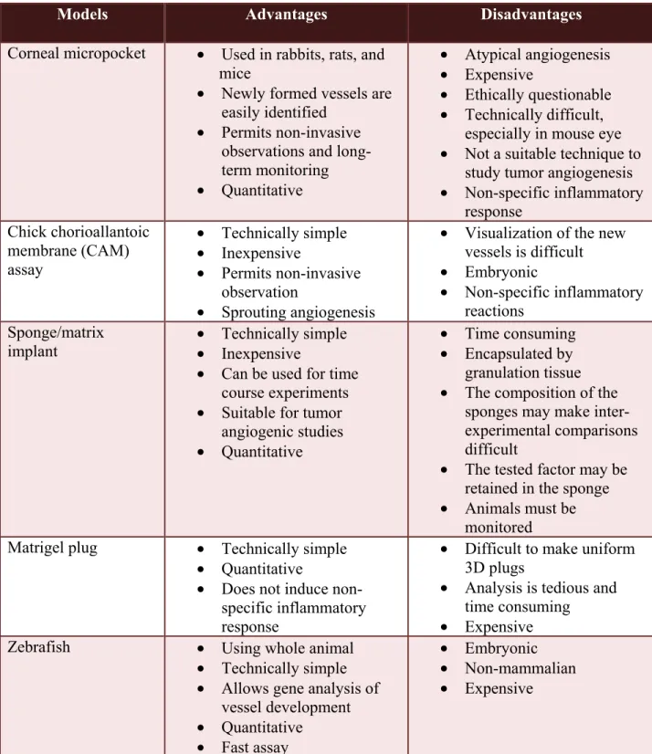
1.5 Angiogenic model………53
1.6 Project reasoning and purpose ………...………...……..57
2.0 ARTICLE…..…………..………...………...….59
ABSTRACT ………...….61
INTRODUCTION ………...………...62
MATERIALS & METHODS ………...………66
RESULTS ……….71
DISCUSSION ………...77
ACKNOWLEDGEMENTS ………..………84
REFERENCES ……….85
FIGURE LEGENDS ………...91
CONFLICT OF INTEREST DISCLOSURE ………...94
FIGURES ………...95
3.0 DISCUSSION ………..………...……..…...………101
3.1 Suitability of the sponge/Matrigel model for angiogenic study ………..………..101
3.2 Inflammatory cells potentiate the pro-angiogenic activities of VEGF and angiopoietins 104 3.3 Future perspective ………..………..….109
4.0 CONCLUSION ………...……….………..….112
5.0 BIBLIOGRAPHY ………..………..……...…………113
6.0 APPENDIX 1: Authorization, permission and the published manuscript in the Journal of Cellular Biochemistry………..……...133
ix
LIST OF FIGURES AND TABLES
Figure 1: Anatomy of blood vessels
Figure 2: Schematic overview of vasculogenesis and angiogenesis
Figure 3: Schematic representation of predominant VEGF-A isoforms in humans
Figure 4: Schematic representation of the intracellular pathways activated through VEGFR-2
Figure 5: Schematic representation of angiopoietins and their isoforms
Figure 6: Schematic representation of the intracellular pathways activated through Tie2
Figure 7: Steps in leukocyte infiltration
x
LIST OF ABBREVIATIONS
Ang: angiopoietin
APCs: antigen presenting cells
COX-2: cyclooxygenase-2
CXCL: (C-X-C motif) ligand
DC: dendritic cell
EC: endothelial cell
EGF: epidermal growth factor
FGF: fibroblast growth factor
HUVEC: human umbilical vein endothelial cell
ICAM and VCAM: intracellular and vascular cellular adhesion molecules,
respectively
IL: interleukin
LPS: lipopolysaccharide
MHCII: major histocompatibility class II
MMP: metalloproteinase
MPO: myeloperoxidase
NO: nitric oxide
NRP: neuropilin
PAF: platelet-activating factor
PDGF: platelet-derived growth factor
PECAM-1: platelet endothelial-cell adhesion molecule-1
PI3K: phosphoinositide 3-kinase
PlGF: placenta growth factor
RTK: receptor tyrosine kinase
SMC: smooth muscle cell
Tie: tyrosine kinase with immunoglobulin and epidermal
growth factor homology domains
xi
VEGF: vascular endothelial growth factor
VEGFR: vascular endothelial growth factor receptor
xii
ACKNOWLEDGEMENTS
First and foremost, I would like to thank my project supervisor, Dr. Martin G. Sirois, for giving me the wonderful opportunity to explore the intricate world of research. His patience, guidance, active participation and effort were essential to the birth of this document and to my formation as a student in scientific research. Thank you very much for your encouragements and motivations.
I am also grateful to Dr. Cheolho Cheong for his guidance as a collaborator of this project and for showing me new techniques in obtaining the objective of this thesis. All of his advices and suggestions have been greatly appreciated.
I owe my sincere thanks to the Department of pharmacology and to the Faculty of graduate studies of Université de Montréal for providing me with high quality education. I am especially indebted to thank the committee members, Dr. Jonathan Ledoux, Dr. Martin G. Sirois and Dr. Jean-François Gauchat, who are evaluating my thesis. Similarly, I would like to thank the Montreal Heart Institute for providing me a wonderful platform for a bright future. Moreover, I gratefully acknowledge the funding sources, Montreal Heart Institute and the department of pharmacology of Université de Montréal, which have made my M.Sc. work possible.
I would like to extent my appreciation to my fellow lab mates: Pauly, Elizabeth, Damien, Marie-Elaine and Diana for providing me a cordial working environment during these years. Thank you Pauly and Damien for the humor and the friendly sarcasms that always makes me laugh. Inevitably, the small everyday chit chats with Elizabeth and the coffee breaks with Diana will always be remembered. Finally, I devote my special gratitude to Marie-Elaine for her ideas in optimizing my staining and for her help during my experiments.
I would like to equally thank my parents for their love, care, guidance, encouragement and invaluable support throughout my academic trajectory. Their sacrifices have brought me to
xiii
where I am today. I would also like to thank my brother and sister for being my best friends all my life and for listening to both my enthusiasms and my disappointments. I love you guys.
xiv
1
1.0 INTRODUCTION
The endothelium of the vascular system is a disseminated organ forming a physical barrier between the vessel lumen and the extracellular space. It is comprised of quiescent endothelial cells (ECs) interconnected by junctional proteins and ensheathed by perivascular cells (e.g. pericytes and vascular smooth muscle cells (SMCs); Figure 1). ECs play a pivotal role in maintaining tissue homeostasis, which includes the controlling of vasomotor tone and cellular and molecular trafficking across quiescent cells, and in the maintenance of blood fluidity and vascular permeability [1] . In addition, ECs have been shown to participate as key players in tissue vascularization following injury in a process termed angiogenesis. They are also implicated in physiological inflammatory response where their surface adhesion glycoprotein (glycocalyx) expression allows transendothelial migration of blood cells and plasma proteins to the site of infection or injury [2]. Thus, the endothelium is not merely a static physical barrier but indeed an active cell system.
Apart from its various functions, ECs show remarkable heterogeneity in structure, time and space allowing the endothelium to mold itself based on the needs of the underlying tissue. In fact, the phenotype of ECs can differ between different organs, between different segments of a vascular loop and yet between adjacent cells in the same organ. Thus, the constant response of the endothelium to an array of agonists and environmental challenges can lead to, under certain circumstances, a perturbed state and contribute to the development of numerous vascular diseases involving angiogenesis (reviewed in [1]).
2
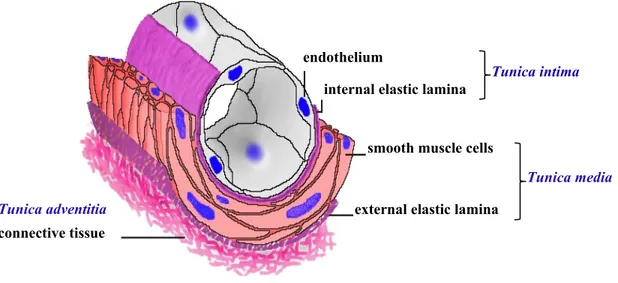
Figure 1: Anatomy of blood vessels. Blood vessels of the vascular network are comprised of
three concentric layers (tunics). The inner layer called tunica intima is composed of endothelial cells and a thin layer of supporting connective tissue. The middle muscular and/or elastic layer, termed tunica media, is comprised of perivascular cells (pericytes and smooth muscle cells). Finally, the outer layer is comprised of fibrous connective tissue and is called tunica adventitia. (Adapted from http://www.siumed.edu/~dking2/crr/cvguide.htm Copyright © 2005, Board of Trustees, Southern Illinois University)
1.1 Angiogenesis
1.1.1 Origin and mechanism of blood vessel formation
The cardiovascular system, comprised of the heart and the circulatory system (the network of blood vessels), is the first functional system to develop in vertebrate embryo [3]. The luminal surface of the circulatory system is comprised of ECs derived from the mesoderm germ layer during development. Precisely, hemangioblasts in the yolk sac differentiate from mesodermal
endothelium
internal elastic lamina
smooth muscle cells
external elastic lamina connective tissue
Tunica intima
Tunica media Tunica adventitia
3
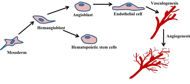
progenitor cells giving rise to hematopoietic stem cells and angioblasts. Angioblasts in turn proliferates, migrates and differentiates into ECs assembling the primitive de novo vascular plexus of veins and arteries – a process termed vasculogenesis. Subsequent sprouting ensures expansion of the vascular network of larger vessels ramifying into smaller ones, known as angiogenesis (Figure 2). The latter is a tightly regulated process that occurs throughout life, from utero to old age, ensuring adequate oxygen and nutrient supply to all body cells.
Classic angiogenesis, or sprouting angiogenesis is a multistep process as detailed by Dr. Folkman in 1971 [4]. Overall, the steps in angiogenesis include enzymatic degradation of capillary basement membrane, EC proliferation, directed migration of ECs, EC tube formation and perivascular cell stabilization [5]. In fact, ECs are equipped with oxygen sensor for the maintenance of vascular homeostasis. Hence, local hypoxia due to poor tissue perfusion is detected by these sensors and initiates the angiogenic process. Firstly, the perivascular cells covering the pre-existing vessel detach from the basement membrane by metalloproteinase (MMP)-mediated proteolytic degradation. This loosens the EC junctions and dilates the nascent vessel. Simultaneously, the release of growth factors and inflammatory cytokines by ECs and inflammatory cells increases the permeability of EC layer causing the plasma proteins to extravasate and lay down a provisional extracellular matrix scaffold. Next, the proteases that are present at the angiogenic site remodel the extracellular matrix into an angio-competent milieu. One EC, known as the tip cell, is selected to lead the tip towards the angiogenic signal. The neighbors of the tip cell become stalk cells, which divide to elongate the stalk and establish the lumen. Interestingly, the tip cells are equipped with filopodia to sense environmental cues while the stalk cells release molecules to convey spatial information
4
Angioblast Endothelial cell Hemangioblast
Angiogenesis
Mesoderm
Hematopoietic stem cells
Vasculogenesis
about their position to their neighbors. Finally, the newly formed vessels become functional, mature and stable through the ensheathing of perivascular cells, entering a quiescent state. Once functional, occlusions in arteries can cause a pressure difference in the arterioles. This calls for the growth of collateral arteries or “natural bypass” from pre-existing arterio-arteriolar anastomoses to overcome the shear forces in a process termed arteriogenesis [6]. Although, angiogenesis remains a necessity under physiological circumstances, this complex process involving multiple factors and exerting specific activities at different phases, becomes a culprit under numerous pathological conditions.
Figure 2: Schematic overview of vasculogenesis and angiogenesis. Extraembryonic
mesoderm, located in the blood islands of the yolk sac, differentiates into hemangioblast giving rise to angioblast and hematopoietic stem cell lineages. Following the commitment to EC lineage, angioblasts gather and rearrange to form capillary-like tubes forming the primary circulatory network, termed vasculogenesis. Angiogenesis is the formation of neovessels from preexisting vessels which occurs both during embryogenesis and during an organism’s life.
5
1.1.2 Physiological angiogenesis
In vertebrate development, angiogenesis plays a pivotal role in nourishing the growing organs with oxygen and in providing instructive trophic signals to promote organ morphogenesis [7]. Though angiogenesis continues to contribute to organ and somatic growth after birth, most blood vessels remain in a quiescent state as of adulthood. In fact, EC turnover in healthy adults is remarkably low. It is only reactivated under certain conditions namely during wound healing, intense physical activity and menstrual cycle.
In wound healing, a natural restorative response to tissue injury, angiogenesis takes place during the proliferative phase where it provides the necessary nutrients to sustain the newly formed tissue [8]. Likewise, during prolonged physical exercises, the increased oxygen demand in skeletal muscles is compensated in the long-term through the formation of new blood vessels [9]. Finally, physiological angiogenesis is fundamental to the female reproductive system (ovaries, uterus) and it is the only organ in adult humans which undergoes a regular cycle of growth and regression of blood vessels. Indeed, during the menstrual cycle, angiogenesis generally takes place in the growing corpus luteum and the endometrium. When fertilization does not occur, the endometrial lining is shed along with the newly formed blood vessels. However, upon fertilization, the placenta takes over the angiogenic process sustaining the developing embryo throughout pregnancy [10].
6
1.1.3 Pathological angiogenesis
Angiogenesis is a tightly regulated process that requires the maintenance of a balance between pro-angiogenic (stimulatory) and anti-angiogenic (inhibitory) factors. A perturbation in this equilibrium can result in either excessive angiogenesis or insufficient angiogenesis leading to a pathological state [11]. Numerous disorders have been associated with excessive angiogenesis including solid tumor, psoriasis, rheumatoid arthritis, retinopathy and atherosclerosis. High fat diet has also been demonstrated to promote angiogenesis in adipose tissue which in turn stimulates adipogenesis, the generation of adipocytes; hence, creating a vicious cycle [12, 13]. Alternately, other disorders have been identified for abnormal vessel regression and maturation such as purpura, scleroderma and nephropathy. Moreover, local loss of blood supply in patients results in tissue ischemia leading to death or disability.
Presently, several medications have been identified to inhibit angiogenesis [14-16] but efforts to therapeutically generate new blood vessels have not been as successful. Therefore, understanding the molecular mechanism of angiogenesis is crucial for the development of therapeutic strategies to combat these inflammatory, malignant and ischemic disorders.
1.2 Regulation of angiogenesis
Over the years, a plethora of endogenous mediators, including growth factors (vascular endothelial growth factor (VEGF), transforming growth factor (TGF)-α and -β, fibroblast growth factor (FGF), epidermal growth factor (EGF) and angiopoietins), matrix
7
metalloproteinases (e.g. MMP-2, MMP-7, MMP-9 and MMP-12), cytokines (e.g. tumor necrosis factor; TNF-α), chemokines (interleukins; IL-8), and integrins (e.g. α1β1, α2β1, α4β1,
α5β1, αvβ5 and αvβ3) have been discovered to regulate angiogenesis. Among these angiogenic
factors, VEGF and angiopoietins are well-established to exhibit both distinct and overlapping expression patterns that collaborate to regulate the different stages of physiological angiogenesis. The following sections will summarize the properties and functions of these growth factors and their places in the angiogenic process.
1.2.1 Vascular endothelial growth factor (VEGF)
In 1989, VEGF (initially identified as vascular permeability factor (VPF) [17]) was isolated and identified [18] as a potent, diffusible and EC-specific mitogen. Its discovery arose the hypothesis that it may possess a significant role in the regulation of physiological and pathological growth of blood vessels [18-20]. Indeed, it is presently well-established that VEGF is the most potent, versatile and ubiquitous vascular growth factor known to date.
The VEGF family is comprised of seven members: VEGF-A, VEGF-B, VEGF-C, VEGF-D, VEGF-E, VEGF-F, and placenta growth factor (PlGF) all containing a highly preserved VEGF homology domain. VEGF-A, being the first one to be identified is a homodimeric glycoprotein of approximately 45 kDa [18]. In healthy adults, the VEGF-A mRNA is highly expressed in lung, kidney, heart and adrenal gland and marginally expressed in liver, spleen and gastric mucosa [21]. It is also expressed by cultured SMCs [22], macrophages [23] and ECs [24]. At the cellular level, VEGF-A functions as a major paracrine
8
regulator of the angiogenic response, which modulates EC proliferation, migration, sprouting and survival [25].
The human gene coding for VEGF-A is localized on chromosome 6p21.3 [26] and consists of eight exons separated by seven introns [27, 28]. Alternative exon splicing of this gene results in the generation of four major isoforms: VEGF-A121, VEGF-A165, VEGF-A189
and VEGF-A206 [27, 28] (Figure 3). Other less frequent splice variants such as VEGF-A145
[29], VEGF-A183 [30], VEGF-A162 [31]and VEGF-A165b [32] have also been reported. Exons 1
to 5 are conserved in all isoforms. The splice variation among the isoforms depends on the presence or absence of exon 6 and 7 which encode distinct heparin binding domains. The acidic polypeptide, VEGF-A121, lacking both exons 6 and 7 does not bind to heparin and is
highly diffusible [33, 34]. The highly basic variants, VEGF189 and VEGF206 contain both exon
6 and exon 7 and bind heparin with high affinity [33]; hence they are completely sequestered in the extracellular matrix. VEGF165 being the predominant isoform [33] lacks exon 6 but
contains exon 7 in its coding sequence. It is only moderately diffusible while a significant amount of itremains bound to the cell surface of the extracellular matrix [35]. The difference in the diffusibility of these splice variants create a gradient of VEGF expression that is responsible for guiding and shaping the vascular network during angiogenesis.
Although structurally similar to VEGF-A, the other members of the VEGF family with their splice variants is differentially expressed in many cell types and display different biological activities. For example, PlGF in adults is a master switch in pathological angiogenesis. It is predominantly expressed in the placenta, heart and lungs [36]. Indeed, mice
9
lacking PlGF gene are phenotypically inert but show impaired angiogenesis, plasma extravasation and collateral growth during ischemia, inflammation, wound healing and cancer [37]. In contrast, PlGF overexpressed transgenic mice produce increased vascularization, inflammation and vascular permeability in the skin [38, 39]. VEGF-B is abundantly expressed in adult myocardium, skeletal muscle and pancreas [40]. Its expression is also observed in developing heart, brown fat, muscle and spinal cord during mouse embryogenesis. VEGF-B is postulated to be involved in controlling VEGF-A bioavailability; its exact function however, is unknown [21]. The expression of VEGF-C is exclusive in regions of developing lymphatic vessels and in lymph nodes [42, 43]; hence, its function is associated with the lymphatic system during development. In adult tissues however, VEGF-C is expressed in heart, placenta, ovary, small intestine and thyroid gland [21]. VEGF-D is largely identified in the lung and skin during embryogenesis [44] and in the lung, heart, skeletal muscle, colon and small intestine in adults. In humans, expression of VEGF-D is used as a prognostic marker for lymphatic metastasis [45]. Lastly, the VEGF-E isoform is encoded by Orf virus and is involved in inducing pathological angiogenesis in virus-infected lesions [46]. The later splice variant does not have a mammalian homologue.
The VEGF isoforms stimulate various cellular responses by binding to VEGF receptors. VEGF receptors belong to class V receptor tyrosine kinases (RTK) and is comprised of VEGFR-1 (also known as Flt-1; 180kDa), VEGFR-2 (also known as KDR or Flk-1; 200-230kDa) and VEGFR-3 (also known as Flt-4; 195kDa). Each receptor is constructed of seven immunoglobulin-like domains in the extracellular domain, a single transmembrane domain and a consensus tyrosine kinase sequence containing intracellular domain [47]. The signaling
10
by VEGF receptors is initiated upon the binding of a covalently linked VEGF dimer to the extracellular receptor domain. This interaction promotes receptor homo- and heterodimerization (VEGFR-1 with VEGFR-2 and VEGFR-2 with VEGFR-3) followed by kinase activation and autophosphorylation of specific tyrosine residues located in the intracellular juxtamembrane domain, the kinase insert domain, and the carboxylic tail of the receptor [48]. Subsequently, a variety of signaling molecules interact at specific sites of the VEGF receptor dimers to activate distinct downstream cellular pathways [49].
Among the three receptors, VEGFR-2 is considered to be the major mediator of many physiological effects of VEGF-A on ECs. It is expressed on vascular ECs, lymphatic cells, megakaryocytes and haematopoietic stem cells [50] and binds VEGF-A, the processed form of VEGF-C and VEGF-D, and VEGF-E. VEGFR-2 signaling is modulated through co-receptors termed heparin sulfated proteoglycans, which also interact with several isoforms of VEGF [51]. In fact, neuropilin-1 (NRP-1), one such modulator, is the primary co-receptor of VEGF-A/VEGFR-2 ligand/receptor complex [52]. Although, this putative receptor was previously identified to bind collapsin/semaphorin family mediating neuronal guidance, it is now evident that NRP-1 can also amplify the effectiveness of VEGF-A/VEGFR-2 signal transduction [53]. NRP-1 is highly expressed in vascular ECs and its presence in these cells promotes VEGFR-2 phosphorylation and activation. Indeed, our laboratory has demonstrated that NRP-1 enhances VEGF-A/VEGFR-2-mediated EC migration and proliferation, and platelet activating factor (PAF) synthesis, a pro-inflammatory molecule [54].
11
Figure 3: Schematic representation of predominant isoforms of VEGF-A in humans. The
different exon (indicated with numbers) compositions as a result of alternative splicing gives rise to the various isoforms of VEGF-A. Exons 1 to 5 are conserved in all VEGF-A splice variants. Exon 3 is implicated in the homodimerization of VEGF-A and in its interaction with VEGFR-1 while exon 4 is required for the binding of VEGF-A onto VEGFR-2. Exons 6 and 7 are both involved in binding heparin. Exon 7 additionally links VEGF-A onto their co-receptor neuropilin-1 (NRP-1). Finally, exon 8b is present only on inhibitory isoforms of VEGF-A. (Adapted from Fearnley, G.W. et al. 2013 [41])
1.2.1.1 VEGF in angiogenesis
VEGF-A (here after referred to VEGF) has been revealed to induce angiogenesis in a variety of in vivo and in vitro models. Knockout mice containing one allele of VEGF exhibit a number of development anomalies such as defective vascularization in several organs and reduced
12
nucleated red blood cell number within the blood islands in the yolk sac [55]. Consequently, these mice die within the first few days of development. Yet more, partial inhibition of VEGF using inducible gene targeting in early postnatal life leads to stunted body growth and impaired organ development leading to death in mice [56]. Similarly in adults, VEGF elicits pronounced angiogenesis in various in vivo models including the rabbit cornea [57], the primate iris [58] and the Matrigel plug in mice [59]. Concomitantly, in vitro studies revealed the ability of VEGF to promote the growth of vascular ECs derived from arteries, veins and lymphatics [18, 19, 60-64].
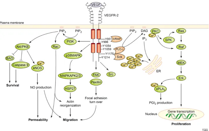
It is now evident that VEGF regulates and orchestrates the angiogenic cascade (EC proliferation, survival, migration and permeability) through activated VEGFR-2 post-receptor signaling pathways. The binding of VEGF-A to VEGFR-2 results in the autophosphorylation of the following tyrosine residues in the intracellular domain of the receptor: Tyr951 and Tyr996 (located in the kinase domain), Tyr1054 and Tyr1059 (located in the kinase domain), and Tyr1175 and Tyr1214 (located in the C-terminal tail) (Figure 4). Phosphorylation of Tyr951 and Tyr1175 creates a binding site for VEGFR-associated protein (VRAP), and Sck [65] and PLCγ1 [66], respectively, which subsequently activate Raf via protein kinase C (PKC)
in a Ras-independent manner [67]. This in turn induces the activation of the extracellular regulated kinase (ERK) pathway (p42/44 mitogen activated protein kinase (MAPK)) leading to gene transcription required for EC proliferation. VEGFR-2 also activates phosphoinositide 3-kinase (PI3K), which results in an increase of lipid phosphatidylinositol (3,4,5)P3 (PIP3),
conducting the activation of protein kinase B (Akt/PKB) and small GTP-binding protein, Rac. The Akt/PKB pathway, 1) promotes EC survival by inhibiting pro-apoptotic factors such as
B-13
cell lymphoma-2 associated death promoter homologue (BAD) and caspase-9 [68], and 2) increases vascular permeability and cellular migration through the activation of endothelial nitric oxide synthase (eNOS) [69, 70]. Rac [71], on the other hand, along with other adapter proteins including p38MAPK [72] and focal kinase (FAK) with its substrate paxillin [73] have been demonstrated to promote cellular migration. In addition, the activation of VEGFR-2 on ECs promotes the production of PAF by ECs, which apart from its role during inflammation (involved in promoting the rolling and adhesion of leukocytes) also potentiate the migration of cultured ECs and increase vascular permeability [54]. Although the exact mechanism is not known, Src family members interact with activated VEGFR-2 and induce vascular permeability in mice [74].
In addition to its vast activities on the endothelium via VEGFR-2, VEGF is also responsible for guiding and shaping of the vascular tree during the angiogenic process [75-77]. It does so through the differential spatial distribution of VEGF isoforms in the extracellular space which creates a gradient of VEGF expression. Such gradient is crucial for selecting the tip cells and the stalk cells during the sprouting of nascent blood vessels. This patterning is controlled at the level of transcription, isoform splicing and cell surface retention. For instance, the splice variants that bind heparin remain on the cell surface or in the extracellular matrix, while the splice variants lacking retention motif diffuse away from the surface. Furthermore, the transcriptional level of VEGF determines the site of sprouting. For instance, tip cell formation and sprouting only occurs at regions of highest VEGF concentration via the induction of gene expression of the NOTCH signaling pathway ligand Delta-like ligand 4 (Dll4) [78, 79]. Dll4 expression is restricted to developing arteries and at the tip of vascular
14
sprouts [80, 81] and it is almost never expressed on the neighboring stalk cells. Together, the differential expression of Dll4 mediated by VEGF gradient favors the appropriate formation of filopodia, sprouting and branching of nascent vessels creating functional blood vessels at the end of an angiogenic response.
1.2.2 Angiopoietins
Angiopoietins, identified in mid 1990s, are a novel class of angiogenic growth factors that exert a crucial regulatory role in the maintenance of vascular integrity and quiescence. In fact, the different cellular functions of angiopoietins (responsible for the assembling and disassembling of the EC lining of blood vessels) in concert with VEGF coordinate precise morphogenic events in angiogenesis.
1.2.2.1 Structure, localization and function of Ang1 and Ang2
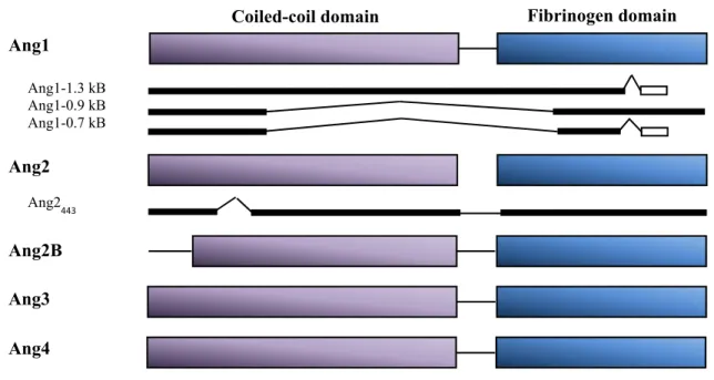
The angiopoietin protein family is comprised of four members, Ang1, Ang2, Ang3 and Ang4 (Figure 5), all of which having a size of about 500 amino acids [83-85]. Structurally, the angiopoietins are composed of two domains: a fibrinogen-like C-terminal domain and an alpha-helical rich coiled-coil N-terminal domain [86]. The receptor binding sequence located in the fibrinogen-like domain of these growth factors enables their binding to the receptor
tyrosine kinase with immunoglobulin and epidermal growth factor homology domains 2
(Tie2) while the coiled-coil motif promotes angiopoietin oligomerisation, a prerequisite for Tie2 receptor activation [86].
15
The human gene coding for Ang1 and Ang2, the predominant isoforms, is localized on chromosome 8q22 and 8q23, respectively [87, 88]. Alternative exon splicing of the Ang1 gene results in the generation of four isoforms: Ang1-1.5 kb (the prototypic form), Ang1-1.3 kb, Ang1-0.9 kb and Ang1-0.7 kb [89]. The Ang1 splice variants, Ang1-1.3 kb, Ang1-0.9 kb and Ang1-0.7 kb, bind Tie2 with similar potency but do not activate Tie2 signaling. Instead, they serve as a dominant negative regulator of the full length Ang1 (Ang1-1.5 kb) activity, which both binds and activates Tie2 [89]. Similarly, alternative exon splicing of the Ang2 gene gives rise to a splice variant of 443 amino acids (Ang2443) [90]. It lacks part of its coiled-coil motif
and as a consequence renders it incapable of inducing Tie2 autophosphorylation upon its binding. Ang2443 nonetheless, pre-occupies the angiopoietin binding site on Tie2 and prevents
the binding of the full length Ang1 and Ang2 to their receptor; hence, sequestering Tie2 activation [90]. Recently, an additional isoform of Ang2 (Ang22B) with a partial truncated
amino terminal coiled-coil domain has also been identified in chicken as a result of 5’ intron alternative splicing [91].
In adults, Ang1 is constitutively expressed in pericytes, SMCs and fibroblasts [83]. Its expression in these cells enables them to regulate the adjacent endothelium through a paracrine interaction. In addition, Ang1 expression has also been noted in neuronal cells [92] and in some types of tumor cells [92, 93]. During embryonic development however, Ang1 expression is temporal dependent. It is first expressed in the myocardium and later in mesenchymal cells surrounding the developing vessels [84, 94, 95]. Recently, our laboratory discovered the expression of Ang1 in the cytoplasm of neutrophils [96]. In contrast, presynthesized Ang2 is almost exclusively stored within the storage granules of ECs termed
16
Figure 4: Schematic representation of the intracellular pathways activated through VEGFR-2. Upon the activation of VEGFR-2 receptors on ECs, adopter proteins including
VEGFR-associated protein (VRAP), Sck and PLC-γ bind specific tyrosine residues on the receptors via their SH2 domain. Their binding in turn phosphorylates and activates the aforementioned adaptor proteins. Activation of PLC-γ induces the second messengers, DAG and IP3. While DAG activates PKC, IP3 induces the release of Ca2+ and in turn act on the
endoplasmic reticulum. In addition, other proteins are also activated by VEGFR-2 such as Src, PI3K, FAK and p38MAPK. Together, these signaling pathways promote EC migration, proliferation, survival and vascular permeability. (Adapted from Cross, M.J. et al. 2003 [82])
17
Weibel-Palade bodies (WPB) that allows the rapid release of Ang2 in response to various stimuli [97]. The release of Ang2 by activated ECs operates as a built-in autocrine switch, which promotes the transition of the quiescent endothelium to an activated state. For instance, under physiological conditions, Ang2 is only strongly upregulated in regions undergoing vascular remodeling such as during the menstrual cycle and wound healing [98]. Their expression is also elevated in tumor cells [99-101] and in ECs of tumor blood vessels [93, 102, 103]. Furthermore, marginal level of Ang2 expression has been detected in Kaposi’s sarcoma cells [104] and in Muller cells of the retina [105].
Although the well-characterized ligands, Ang1 and Ang2, bind their receptor Tie2 at the same site with similar affinities, Ang2 was originally identified as a competitive antagonist of Ang1/Tie2 ligand/receptor signaling axis in ECs [97, 106-111]. It is now apparent that the biology of Ang2 is less straight-forward; it can behave both, as an agonist or as an antagonist of Tie2 signaling depending on the concentration and the spatial-temporal context it is in. Indeed, Ang2 can activate Tie2 receptor on ECs promoting its migration, survival and capillary tube formation at high concentration [112] or after prolonged incubation [113]. Interestingly, recent studies in our laboratory have identified other agonistic activities of Ang2/Tie2 complex both on ECs (e.g. promotes PAF synthesis, P-selectin translocation, neutrophil adhesion) and on neutrophils (e.g. promotes PAF synthesis, CD11b/CD18 integrin activation and its migration) following a short incubation and at same concentration as Ang1 [115-118]. We have observed that neutrophils treated with either Ang1 or Ang2 alone for 7.5 minutes promotes PAF synthesis at a concentration of 10-9 M [115]. Nonetheless, on normal vascular endothelium, Ang2 acts as a Tie2 antagonist to destabilize the endothelium and
18
empowers the initiation of the angiogenic response [95, 119-121]. Ang1 on the other hand, is an agonistic ligand of the Tie2 receptor where its binding to Tie2 contributes to blood vessel maturation and stability. In addition to the same agonistic effects exerted by Ang2 on ECs and neutrophils, Ang1 also promotes the survival of ECs and neutrophils, inhibits VEGF-mediated adhesion molecule (intercellular adhesion molecule-1 (ICAM-1), vascular cell adhesion molecule-1 (VCAM-1) and E-selectin) increase on ECs and promotes the synthesis of IL-8 and IL-1β [122, 123]. In fact, the group of Yu, X. et al identified a short loop within the angiopoietin fibrinogen domain, which may potentially confer the differential agonistic and antagonistic properties of these biologically important molecules [124].
1.2.2.2 Structure, localization and function of Ang3 and Ang4
In 1999, Valenzuela et al. reported the existence of Ang3 and Ang4, inter-species homologues found in mice and humans, respectively [85]. These orthologues, sharing 65% amino acid homology, are found at the same gene locus in mice and humans [85]. Similar to Ang1 and Ang2, the protein structure of Ang3 and Ang4 contains an N-terminal coiled-coil domain and a C-terminal fibrinogen domain. Both Ang3 and Ang4 are predominately synthesized as dimers connected by disulfide bonds, which resemble the native structure of Ang2 [83, 85]. Although, much less is known about them till this date, the expression of Ang3 has been identified to be distributed in multiple tissues in mice while the expression of Ang4 has been identified to be restricted to the lungs in humans [85]. However, the expression of Ang4 can be significantly increased in response to hypoxia and to various growth factors [125,
19
126]. Likewise, Ang3 expression in rats has also been demonstrated to be enhanced in lungs, brain and heart in response to hypoxia [127].
Ang3 and Ang4 exert similar activities upon their binding to Tie2 receptor [128]. Initially, Ang3 was characterized as an antagonist of Tie2 based on the observation that it, when expressed in a chimeric form, inhibited the activity of Ang1 in humans [85]. However, an in vivo study using murine corneal micropocket assay revealed that these orthologs (Ang3 and Ang4) both have the capacity to induce angiogenesis [128]; hence, classifying them as Tie2 receptor agonists [85]. In fact, it is now accepted that the action of Ang3 is context-dependent which can act both as an agonist and as an antagonist while Ang4 is an agonistic ligand of Tie2.
1.2.2.3 Structure, localization and regulation of Tie receptors
In the early 1990s, a novel class of RTK, termed Tie receptors, was identified consisting of two members, Tie1 (135 kDa) and Tie2 (150 kDa) [129-131]. These receptors of ≈1100 amino acids consist of an extracellular, transmembrane and an intracellular/cytoplasmic domain and are expressed on both vascular and lymphatic EC surface. The extracellular amino-terminal domain of Tie receptors is comprised of three EGF-like cysteine rich repeats, two immunoglobulin-like domains flanking the EGF-like repeats and three fibronectin-type III repeats. The intracellular portion of Tie receptors contains two highly conserved tyrosine kinase domains with 76% sequence homology that allows the activation of various downstream effectors following its autophoshorylation [129-133]. Although structurally
20
similar, the extracellular portions of these receptors demonstrate numerous differences (sequence homology of 33%) [133].
During the early stages of development, Tie1 is expressed in differentiating angioblasts of the head mesenchyme, in the splanchnopleura (a layer of embryonic cells lining the walls of the visceral organs formed by the association of mesoderm and endoderm), dorsal aorta and in migrating ECs of the developing heart [134]. In adults, Tie1 expression is predominantly observed on EC surface along with Tie2 [135]. While the angiopoietins have been identified as the ligands of Tie2 receptor, Tie1 remains as an orphan receptor. Nonetheless, Tie1 has been demonstrated to modulate Tie2 mediated signaling through intracellular heterotypic interaction [135]. Tie1 on its own does not activate downstream intracellular signaling. In agreement with this notion, Tie1 undergoes proteolytic cleavage in presence of VEGF and inflammatory cytokines (e.g. TNF-α) releasing its soluble extracellular domain. This portion of the receptor then binds Tie2 forming the Tie1:Tie2 complex and thereby modulates the downstream signaling pathways [136, 137]. Yet, Kontos et al. recently identified the existence of a chimeric form of Tie1, c-fms-Tie1, which upon ligand stimulation results in Tie1 autophosphorylation and activation of downstream P13K/Akt pathway similar to Tie2; thus, suggesting that Tie1 receptors may potentially exert biological activities under certain circumstances [138].
Tie2 receptors are expressed in embryonic and adult endothelium [139], haemopoietic endothelial progenitors [129], lens epithelial cells [130], leukemia cells [131, 132, 140], eosinophils [141] and in circulating human neutrophils [117, 118]. In addition, Tie2 positive
21
Figure 5: Schematic representation of angiopoietins and their isoforms. The angiopoietin
superfamily consisting of Ang1, Ang2, Ang3 and Ang4 are structurally related with a coiled-coil domain and a fibrinogen domain. Four splice variants of Ang1 have been identified; Ang1-1.5 kB (the full length), Ang1-1.3 kB, Ang1-0.9 kB and Ang1-0.7 kB. Ang2 on the other hand are found in their full form or as Ang2443 lacking part of its coiled-coil domain.
Ang2B is a splice variant observed in chicken. Ang3 and Ang4 are inter-species homologues found in mice and humans, respectively. (Adapted from Jones, N. et al. 2001 [114])
subpopulation of monocytes, termed Tie2-expressing monocytes (TEM), has been identified to exist in concert with tumor-associated macrophages during tumor formation [142]. The expression of Tie2 is induced by hypoxia and pro-inflammatory mediators (e.g. TNF-α and IL-1β) [143] and is noted to be highly upregulated in tumoral angiogenesis [139, 144, 145]. Activation of Tie2 receptor follows the same basic steps as all other RTKs (e.g. VEGFRs). The binding of Ang1 per se to Tie2 with a binding constant of ≈3 nM leads to receptor
Fibrinogen domain Ang1 Ang2 Ang3 Ang4 Ang1-1.3 kB Ang1-0.9 kB Ang1-0.7 kB Ang2B Ang2 443 Coiled-coil domain
22
dimerization followed by rapid activation of the cytoplasmic kinase domain through autophosphorylation of specific tyrosine residues, which subsequently activates intracellular signaling pathways [139, 146].
1.2.2.4 Ang1 and Ang2 in developmental angiogenesis: genetic evidence
Over the years, numerous studies utilizing transgenic mouse models have provided concrete evidence that the angiopoietin/Tie signalling pathway is essential for vessel remodelling and stabilisation during angiogenesis. Indeed, conventional knockout of Ang1 (Ang1-/-) in mice resulted in the formation of dilated vasculature lacking complexity (reduced numbers of small vessel formation and diminished branching) [94]. The nascent blood vessels were prone to ruptures as they were constructed with fewer ECs and failed to anchor onto the extracellular matrix [94]. As a result, Ang1-/- mice died very early during embryogenesis. Conversely, transgenic mice overexpressing Ang1 in the skin produced large vessels (presumably capillaries) with marked improvement in vascular integrity; the newly formed vessels were ensheathed by pericytes [147, 148]. Moreover, as mice deficient of Tie2 receptor (Tie2-/-)
produced similar phenotype as Ang1-/- mice, it is now well-established that Ang1 is an agonistic ligand of Tie2 [149-152]. Furthermore, double-transgenic mice overexpressing both Ang1 and VEGF in the skin of mice did not only produce highly structured vessels in greater numbers and diameter but the vessels were covered by pericytes and SMCs, and lacked leakiness and permeability which were absent in mice expressing VEGF alone [153]. Together, these results concluded that VEGF and Ang1 regulates angiogenesis at distinct
23
levels where VEGF induces vessel sprouting and growth while Ang1 through Tie2 mediates remodelling and maturation of newly formed blood vessels.
Ang2, being the antagonistic member of the angiopoietin family at the receptor level (under certain conditions) during angiogenesis also behaves to counteract Ang1 activity in transgenic mice models. For instance, mice overexpressing Ang2 were embryonic lethal and possessed similar phenotypes as mice deficient in Ang1 or Tie2 expression [84]. In agreement with these observations, Ang2 was demonstrated to disrupt the interaction between EC monolayer and SMCs in culture [154]. Ang2-null mice in contrast, were born normal but developed chylous ascites (an accumulation of milky chyle in the peritoneal cavity as a result of lymphatic disruption [155]) within the first few days postnatal. In fact, depending on the genetic background of the mice, Ang2-deficient mice died within the first 14 days of their life or developed normally to adulthood but with persistent vascular defects (such as impaired responses to inflammatory challenges). Hence, it was concluded that the release of Ang2 by ECs induces vascular regression.
The effects of the angiopoietins are not solely limited to the vascular system. It is now evident that the angiopoietins also have redundant roles in lymphatic vascular development through a process termed lymphangiogenesis [95]. The lymphatic vascular system is the body’s second vascular system that is involved in the maintenance of normal tissue fluid homeostasis, immune surveillance, and absorption of fatty acids and lipid soluble vitamins in the gut. Although, very little is known about the role of Ang1 during lymphangiogenesis, mice overexpressing Ang1 has been shown to induce the formation of large lymphatic vessels in
24
increased numbers [156] either directly via Tie2 [157] or indirectly via VEGF-C/VEGFR3 [156]; Ang1 induces lymphatic vessel enlargement, sprouting and proliferation. Interestingly, Ang2 has been demonstrated to play a crucial role in keeping the lymphatic vessels under a quiescent state. For instance, Ang2 null mice have been shown to develop severe lymphatic defects [95], but minor blood vessel defects. The vessels formed in these mice failed to remodel or form a structured network and did not support the ensheathing of SMCs. Thus, Ang2 acts as an agonist on lymphatic vessels but also as an antagonist on blood vessels [95, 158].
1.2.2.5 Ang1 and Ang2 on the endothelium
While both Ang1 and Ang2 bind to the same site in the extracellular domain of Tie2, only Ang1 was initially characterized as the principal ligand of Tie2 receptor. Yet, it is now evident that Ang1 and Ang2 can multimerize prior to their binding onto Tie2, which promotes Tie2 receptor dimerization and subsequently activates the autophosphorylation of adjacent tyrosine residues on the carboxyl terminal of the receptor [159, 160]. The activation of Tie2 by Ang1 promotes vessel survival, migration and reorganization, inhibits vascular leakage and suppresses inflammatory gene expression.
Autophosphorylation of the tyrosine kinase domain on Tie2 receptor leads to the phosphorylation of p85 subunit of PI3K, which activates Akt and in turn phosphorylates and inhibits the forkhead transcription factor, FKHR, in ECs. FKHR in the endothelium is responsible for inducing EC apoptosis through the phosphorylation and inactivation of
pro-25
apoptotic factors including BAD and pro-caspase-9 [114, 161-164]. It also promotes the expression of genes involved in vascular destabilization and remodeling such as Ang2 [114, 161-163, 165]. Thus, the inhibitory effect of Ang1 on FKHR prevents EC apoptosis and vascular destabilization. In addition, Akt activation leads to an increased expression of survivin, a classical inhibitor of apoptosis, and promotes cell survival [166, 167]. Along with its anti-apoptotic and stabilization effects, the PI3K/Akt signaling pathway promotes blood vessel maturation. It maintains vascular quiescence by enhancing the interaction between perivascular cells and the endothelium which thereby suppresses inflammatory phenotypes. Concurrently, the NF-κB pathway is inhibited upon the activation of Tie2 by Ang1 through A20-binding inhibitor of NF-κB activation-2 (ABIN-2) which simultaneously exerts protective effect on the endothelium by preventing EC apoptosis [168, 169]. Ang2 on the other hand, antagonize the anti-apoptotic effect of Ang1. It induces EC apoptosis leading to the regression of the vasculature through the recruitment of macrophages.
Furthermore, Ang1 signaling stimulates Tie2-dependent EC migration via the adaptor protein Dok-R [170, 171]. The recruitment of Dok-R to the activated Tie2 receptor (p-Tyr1107) requires both the phosphotyrosine binding (PTB) and pleckstrin homology domains located on the adaptor protein. Phosphorylated Dok-R then creates interaction site for Nck and the serine kinase, p21-activating kinase (Pak), leading to the activation of EC migration and the reorganization of the cytoskeleton [146]. In addition, the activation of Tie2 on ECs induces the phosphorylation of focal adhesion kinase (FAK) which leads to the phosphorylation and activation of paxilline and p42/44 MAPK (ERK) [172]. Their activation in turn contributes to EC migration. In fact, blocking Tie2 activation inhibits ERK mediated Ang1 activation of EC
26
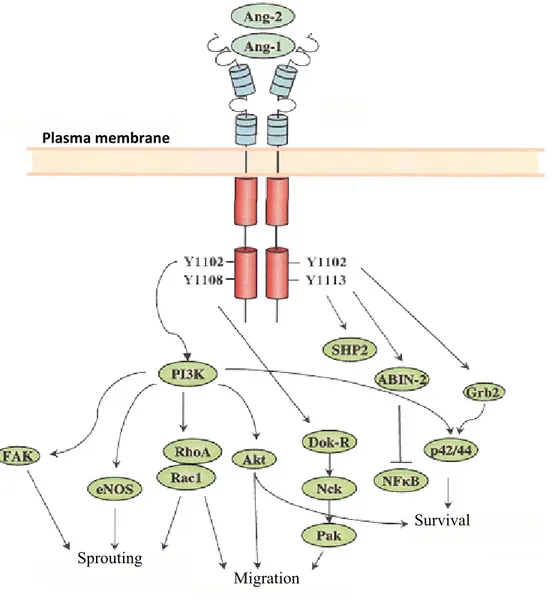
migration [173]. Other mediators including the adaptor protein ShcA, the GTPases RhoA and Rac1, growth factor receptor bound-2 (Grb2), Grb-7, p85 subunit of PI3K, eNOS and SH2 domain containing phosphatase (SHP2) are also recruited to Tie2 in ECs which have been identified to exert an important role not only in promoting EC migration but also in promoting EC proliferation and differentiation in the presence of Ang1 [146, 170, 174, 175]. SHP2 in particular additionally functions as a negative regulator of Tie2 phosphorylation [170, 176].
Ang1 also plays an important role in the reorganization of ECs into tubule-like structures. In fact, Ang1-stimulated reorganization of cultured ECs into tubules and their invasion into 3D matrices is Tie2-dependent [177]. Reorganization of ECs was not observed in ECs lacking Tie2 but was rescued by Tie2 activating antibody in the rat aortic ring assay [178]. Furthermore, the use of inhibitors and dominant-negative constructs has indicated a number of signaling intermediates involved in Ang1 induced reorganization of endothelium and motility including PI3K, SchA, focal adhesion kinase, and endothelial NO synthase [172, 179-181]. Consistent with such remodeling effects, Ang1 stimulates the production of proteases, including plasmin and matrix metalloproteases, which decrease the EC-substratum interaction allowing the ECs to reshape the vessel lumen [182]. Ang2 has also been demonstrated to promote vascular remodeling by inducing an inflammatory response [183]. Such response activates the endothelium and thereby increases vascular permeability and renders the endothelium susceptible to inflammatory cytokine. In fact, long-term incubation of EC by Ang2 activates PI3K/Akt pathway, which promotes EC survival, sprouting and migration [184].
27
Ang1 regulates EC-cell and cell-matrix interactions through which it is well-known to exert anti-inflammatory biological activities. The intercellular expression of VE-cadherins on ECs forms a complex, which can be broken by VEGF, decreasing vascular permeability. This effect can in fact be antagonized by Ang1. It is mediated through the translocation of Tie2 to EC surface in response to Ang1, which subsequently interact with Tie1 and activate the signaling pathways reducing vascular permeability. Similarly, Ang1, under inflammatory conditions is capable of reducing vascular permeability. It promotes a tight interaction between adhesion molecules at EC junctions (PECAM-1 and VE-cadherins).
It is well established that Ang1 exerts its function during the later phase of angiogenesis in promoting vessel maturation [147, 185]. However, few studies demonstrate that Ang1 can also promote EC chemotaxis [186] and induce EC sprouting under appropriate conditions [177, 186, 187]. The mitogenic effect of Ang1 on the other hand remains controversial. While Kanda, S et al. demonstrated that Ang1 can induce EC proliferation [188], several other groups have shown otherwise, that Ang1 has few or no mitogenic activity on EC [177, 186, 189].
1.2.2.6 The role of Ang1 and Ang2 in angiogenesis
The angiopoietin/Tie2 signaling axis is a key interaction during sprouting angiogenesis, which regulates the transition of ECs from a quiescent state to an activated state. In fact, the constitutive expression of Ang1 in adult perivascular cells covering the vasculature functions as a default pathway to maintain the quiescence resting state of the endothelium. It does so by
28
clustering with homotypic Tie2 complexes present at inter-EC-cell junctions and initiating signaling pathways promoting cell-cell adhesion, anti-permeability and cell survival. Interestingly, Ang1 does not only protect and seal the endothelium but also limits the endothelium from being activated by exogenous cytokines. For instance, Ang1 has been shown under certain experimental conditions to inhibit VEGF-induced blood vessel formation and adhesion molecule expression in order to avoid vascular homeostasis perturbation [119, 190].
29
Figure 6: Schematic representation of the intracellular pathways activated through Tie2.
The binding of Ang1 and Ang2 onto Tie2 induces the dimerization and autophosphorylation of the receptor. Phosphorylated tyrosine activates the PI3K pathway which subsequently activates Akt and RhoA/Rac1 implicated in cellular migration. FAK and eNOS also activated by PI3K leads to vessel sprouting. In addition, the pro-survival activity of angiopoietins is mediated by the p42/44 MAPK which is activated by both PI3K and Grb2/Ras/Raf pathways. Finally, the anti-inflammatory activity of Ang1 is mediated through the inhibiton of NFκB. (Adapted from Brindle, N.P. et al. 2006 [197])
Sprouting
Migration
Survival Plasma membrane
30
This state is however antagonized by Ang2 in vessels undergoing angiogenesis. While almost undetectable in quiescent vasculature, Ang2 expression is dramatically upregulated at the transcriptional level and stored in the Weibel-Palade bodies by the pro-angiogenic stimuli including growth factors (e.g. FGF-2 and VEGF), cytokines (e.g. TNF) and environmental cues (hypoxia, high glucose levels and superoxides) [92, 109, 191-193]. They are then rapidly released in response to phorbal esters, thrombin and histamine leading to the destabilization of the quiescent endothelium (loosening of the attachment between ECs and perivascular cells) [84]. The unstable endothelium facilitates the infiltration of proteases, cytokines and angiogenic myeloid cells, which consequently primes the vasculature to respond to numerous exogenous stimuli for a robust angiogenic response in presence of VEGF [194, 195]. Interestingly, in absence of VEGF, high Ang2 levels lead to vessel regression and not to destabilization as it occurs during the menstrual cycle. Hence, Ang2 functions as a built-in switch controlling the transition of the resting quiescent endothelium towards the activated responsive endothelium. Indeed, it is the Ang1:Ang2 ratio which determines the functional status of the vasculature [196]. In resting state, a vasculature remains quiescent as the Ang1:Ang2 ratio is in favor of Ang1. Following EC activation, this ratio is locally shifted in favor of Ang2. The ECs of unstable vessels may die in the absence of VEGF or migrate and proliferate, initiating the angiogenic cascade in presence of VEGF. Pericytes use the developing sprouts as migration guidance cues and cover the newly formed vessel [198, 199]. Subsequently, the endothelium switches back to the quiescent state in the absence of additional stimulus.
31
1.3 Inflammation
Inflammation or an inflammatory response is a natural defensive reaction in response to tissue damage caused by micro-organisms, noxious stimuli, nutritive deficiency and pro-inflammatory mediators (secreted by apoptotic, necrotic and damaged cells). The maintenance of tissue integrity is vital to human health as we are continuously exposed to such molecular threats. Under physiological conditions, the inflammatory response is tightly regulated by soluble proteins (cytokines, chemokines and growth factors) that create an interactive network between inflammatory cells and vascular ECs within the affected site. The clinical manifestation of such complex system of chemical cues and cell interactions include rubor (redness), calore (heat), dolore (pain), tumor (swelling) and functio laesa (loss of function). When inflammation is not properly regulated, the balance between pro- and anti-inflammatory molecules is shifted away from the initiation of the healing process and towards the occurrence of persisting inflammation. In fact, inflammation is classified as either acute or chronic.
An inflammatory response to any cellular insult begins acutely, that is, it occurs rapidly and is of short duration (within few days). Regardless of the nature of the initial trigger, the series of events involved in acute inflammation can be categorized into different phases. Firstly, a variety of chemical mediators cause local microvessel dilation at the site of injury to increase blood flow (causing erythema and heat release). These mediators equally increase local vessel permeability in order to allow the recruitment of proteins and leukocytes (e.g. neutrophils and monocytes/macrophages) to the site of infection (causing swelling of the
32
tissue or edema). In concert, mast cells, platelets, ECs, dendritic cells (DCs), macrophages and other tissue resident cells also release chemoattractants to recruit neutrophils to the affected area. The latter takes place via local biosynthetic changes of adhesion molecules in ECs allowing the transmigration of neutrophils from the circulation to the site of action. Monocytes follow later, and once in the tissue they undergo anatomical and functional changes that transform them into macrophages. These leukocytes (neutrophils and macrophages) then destroy invading microbes and clear cellular debris through the process of phagocytosis. Finally, the local acute inflammatory response, in the absence of further inflammatory stimuli, is ceased with tissue repair.
If the stimulus persists, inflammation can last days, months or even years and is said to be chronic. In fact, chronic inflammation is abnormal and does not benefit the body. It is primarily mediated by monocytes/macrophages, lymphocytes and plasmocytes through the enhanced production of proteolytic enzymes, reactive oxygen species, MMPs, serine proteases and growth factors and is characterized by increased fibroblast proliferation, collagen deposition, fibrosis and angiogenesis. Yet, the inflammatory and morphological profiles of chronic inflammation differ significantly depending on the nature of the initial trigger. Nonetheless, persistent aggressive stimuli in chronic inflammation leads to a pathological state as observed in rheumatoid arthritis, atherosclerosis, ischemia and solid tumors [200, 201].
33
1.3.1 Regulation of inflammation
As briefly mentioned above, neutrophils and monocytes/macrophages play a crucial role in coordinating the inflammatory response (reviewed in [202]). They arise in a process termed myelopoiesis and are of haematopoietic pluripotent stem cell origin. These stem cells in the bone marrow give rise to myeloid (neutrophils, basophils, eosinophils, monocytes/ macrophages and DCs) and lymphoid lineages (T cells, B cells, DCs, and natural killer (NK) cells). Following tissue injury or infection, an “emergency myelopoiesis” takes place in humans, which generates large pools of neutrophils and monocytes in the bone marrow beyond the normal requirement of a healthy person.
1.3.1.1 Polymorphonuclear leukocytes (PMNs)
Morphologically, neutrophils are classified as PMNs and granulocytes together with basophils and eosinophils due to their multi-lobulated nucleus and the presence of specific granules in the cytoplasm. These leukocytes can be histologically distinguished based on the staining capacity of their granules to different dyes. While basophil granulocytes are stained by basic dyes, eosinophil and neutrophil granulocytes are stained by acidic and neutral dyes, respectively. Basophils are the least numerous of the granulocytes and account for about 0.01-0.3% of circulating leukocytes. They are involved in mediating hypersensitivity reactions (e.g. allergic reactions) of the immune system. Specifically, when activated, basophils release histamine (a pro-inflammatory mediator) and other enzymes that lead to a vast array of allergic symptoms. Eosinophils on the other hand account for 2-4% of circulating leukocytes
34
and are the major participant in the development of allergic inflammation. They also function as cytotoxic effector cells against parasitic infections [203]. Lastly, neutrophils, the well-studied leukocyte,function as the body’s primary line of defense against invading pathogens (e.g. bacteria) in order to safeguard the body from infections.
Neutrophils represent about 50-70% of total leukocytes and are the most abundant inflammatory cell in the blood circulation. In humans, the neutrophil count represents approximately 100-700 million cells/100 mL of blood with a daily production of 0.8-1.6 x 10⁹ neutrophils/kg of body mass. Such ample production of neutrophils is required in order to maintain a constant neutrophil concentration in the blood as their half-life is relatively short (10-24 hours) [204]. However, under an inflammatory state, the number of neutrophils increases significantly with an increased life span as they are one of the first responders to tissue injury and infection. This is made possible as neutrophils under steady state belong to one of two pools in the circulation; the circulating pool, which consists of neutrophils that are freely circulating or the marginated pool, which consists of neutrophils that are bound to the endothelium of small vessels [205]. Upon an inflammatory response, the marginated pool of neutrophils, serving as a reserve, is quickly mobilized to the circulating pool increasing the concentration of neutrophils in the blood.
In healthy adults, circulating neutrophils exist in a resting state in order to protect the host tissue from damage by accidental release of neutrophil toxic intracellular contents. The resting neutrophils become activated via a two-stage process. First, they become primed by agents such as bacterial products and cytokines or chemokines (e.g. granulocyte-macrophage
35
colony-stimulating factor (GM-CSF), TNF-α, IL-8 and IFN-γ) [206] and then they get mobilized to the site of infection or inflammation where they encounter activating signals to trigger bacterial killing.
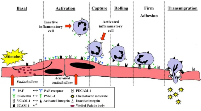
The migration of neutrophils from the circulation across the endothelium (also referred to as neutrophil extravasation) to the site of inflammation can be divided into three stages: rolling, firm adhesion and transmigration [207]. Neutrophil extravasation occurs mainly in the post-capillary venules as the low haemodynamic shear force in these regions along with the expression of selectins on neutrophils facilitate the tethering of the neutrophils onto the activated endothelium. This slows down the flow of neutrophils and enables it to move in a position close to the endothelium and away from the central blood stream. The selectin adhesion family is comprised of L-selectin, P-selectin and E-selectin. L-selectin is constitutively expressed on neutrophils and they bind the immunoglobulin superfamily proteins glyCAM and MadCAM on ECs. This mediates weak constitutive neutrophil-endothelium interaction under normal conditions. However, the expression of L-selectin is significantly increased during inflammation following P-selectin translocation to the endothelium. P-selectin is normally stored in the Weibel Palade body of ECs and is rapidly translocated to the cell surface following the release of an inflammatory stimulus such as histamine, thrombin or phorbol esters [208-210]. The primary ligand of P-selectin is P-selectin glycoprotein ligand-1 (PSGL-1) which is constitutively expressed on all leukocytes. The binding of P-selectin to PSGL-1 activates the p42/44 MAPK and induces the release of IL-8 by leukocytes, which recruits more leukocytes [211, 212]. Finally, E-selectin has only been shown to be expressed on activated ECs upon the inflammatory stimulus lipopolysaccharide