HAL Id: dumas-02950811
https://dumas.ccsd.cnrs.fr/dumas-02950811
Submitted on 28 Sep 2020HAL is a multi-disciplinary open access
archive for the deposit and dissemination of sci-entific research documents, whether they are pub-lished or not. The documents may come from teaching and research institutions in France or abroad, or from public or private research centers.
L’archive ouverte pluridisciplinaire HAL, est destinée au dépôt et à la diffusion de documents scientifiques de niveau recherche, publiés ou non, émanant des établissements d’enseignement et de recherche français ou étrangers, des laboratoires publics ou privés.
Tolérance de la chimio-embolisation pour le traitement
du carcinome hépatocellulaire chez les patients âgés
Olivier Hernandez
To cite this version:
Olivier Hernandez. Tolérance de la chimio-embolisation pour le traitement du carcinome hépatocellu-laire chez les patients âgés. Médecine humaine et pathologie. 2020. �dumas-02950811�
AVERTISSEMENT
Ce document est le fruit d'un long travail approuvé par le
jury de soutenance.
La propriété intellectuelle du document reste entièrement
celle du ou des auteurs. Les utilisateurs doivent respecter le
droit d’auteur selon la législation en vigueur, et sont soumis
aux règles habituelles du bon usage, comme pour les
publications sur papier : respect des travaux originaux,
citation, interdiction du pillage intellectuel, etc.
Il est mis à disposition de toute personne intéressée par
l’intermédiaire de
l’archive ouverte DUMAS
(Dépôt
Universitaire de Mémoires Après Soutenance).
Si vous désirez contacter son ou ses auteurs, nous vous
invitons à consulter la page de DUMAS présentant le
document. Si l’auteur l’a autorisé, son adresse mail
apparaîtra lorsque vous cliquerez sur le bouton « Détails »
(à droite du nom).
Dans le cas contraire, vous pouvez consulter en ligne les
annuaires de l’ordre des médecins, des pharmaciens et des
sages-femmes.
Contact à la Bibliothèque universitaire de Médecine
Pharmacie de Grenoble :
UNIVERSITÉ GRENOBLE ALPES UFR DE MÉDECINE DE GRENOBLE
Année : 2020
SAFETY OF TRANSARTERIAL CHEMO-EMBOLIZATION FOR HEPATOCELLULAR CARCINOMA IN ELDERLY PATIENTS
THÈSE
PRÉSENTÉE POUR L’OBTENTION DU TITRE DE DOCTEUR EN MÉDECINE DIPLÔME D’ÉTAT
Olivier HERNANDEZ
THÈSE SOUTENUE PUBLIQUEMENT À LA FACULTÉ DE MÉDECINE DE GRENOBLE
Le mardi 22 septembre 2020
DEVANT LE JURY COMPOSÉ DE Président du jury :
M. le Professeur DECAENS Thomas Membres :
M. le Professeur GAVAZZI Gaëtan M. le Docteur SEIGNEURIN Arnaud M. le Docteur GHELFI Julien
Mme le Docteur COSTENTIN Charlotte (directrice de thèse)
L’UFR de Médecine de Grenoble n’entend donner aucune approbation ni improbation aux opinions émises dans les thèses ; ces opinions sont considérées comme propres à leurs auteurs.
Remerciements
À Monsieur le Professeur Thomas DECAENS, merci pour ton engagement dans l’hépato-gastro-entérologie, merci pour ton implication dans la formation des internes. C’est pour moi un honneur de te voir présider mon jury de thèse.
À Monsieur le Professeur Gaëtan GAVAZZI. Je vous remercie de participer à ce jury, d’apporter votre expertise de gériatre et je vous sais gré pour l’enseignement de qualité que vous dispensez à la faculté de médecine.
À Monsieur le Docteur Arnaud SEIGNEURIN, merci d’avoir accepté de juger ce travail et d’y apporter votre expertise.
À Monsieur le Docteur Julien GHELFI, merci pour l’aide précieuse que tu m’as spontanément apportée dans ce travail. Je suis heureux de te compter parmi ce jury.
À Madame le Docteur Charlotte COSTENTIN. Alors que tu n’étais pas encore en poste à Grenoble, je te suis très reconnaissant d’avoir accepté d’encadrer ce travail. Merci pour ta gentillesse et ta grande disponibilité.
À Madame Najeh DAABEK, merci pour votre disponibilité, pour votre aide précieuse et avisée dans les analyses statistiques.
À toute l’équipe d’hépato-gastro-entérologie du CHU de Grenoble, merci pour votre accompagnement durant ces années d’apprentissage. Travailler à vos côtés fut à la fois un plaisir et une expérience gratifiante.
À toute l’équipe d’hépato-gastro-entérologie du CH de Chambéry, merci de m’avoir accompagné dans mes premiers pas d’interne. Je suis particulièrement heureux de vous rejoindre pour exercer à vos côtés.
À toute l’équipe d’hépato-gastro-entérologie du CH d’Annecy, merci pour votre accueil, votre convivialité et la richesse de votre enseignement.
Aux équipes d’oncologie médicale et de radiothérapie du CH d’Annecy, merci de m’avoir fait découvrir vos belles spécialités. Votre travail d’équipe est pour moi un exemple.
À mes co-internes, merci pour votre complicité tant dans les moments difficiles que dans les moments les plus heureux. Je souhaite à chacun d’entre vous le meilleur dans votre vie personnelle et dans votre vie professionnelle.
À Amélie, merci pour ton écoute, ton soutien et ta bienveillance. Ta présence est pour moi très précieuse.
À toutes les belles rencontres et les amitiés nées pendant mon internat. Merci pour ce premier semestre à Saint-Hélène qui restera parmi mes meilleurs souvenirs.
A mes amis de la faculté, Sophie, Aude, Vinciane, Claire, Corentin, Benoît et les autres. Merci pour tous les bons moments passés et à venir. C’est à chaque fois un bonheur de vous retrouver.
À Maxime et Julien, merci pour votre amitié fidèle malgré les années, les distances et nos parcours si différents. Je sais qu’elle perdurera.
À mes parents et à ma sœur, merci pour votre soutien constant et sans faille dans toutes les étapes de ma vie. Merci de m’avoir particulièrement accompagné dans mes études, de la première année au point final de ma thèse.
Sommaire
Page Remerciements ………... 6 Résumé ………... 9 Abstract ……… 11 Abbreviations ……….. 13Article : Safety of transarterial chemo-embolization for hepatocellular carcinoma in elderly patients Introduction ……… 14
Materials and Methods ……… 17
Results ……… 21 Discussion ………. 27 Conclusion ………. 30 Tables ……….. 31 Appendix ……….. 40 References ……….. 43 Conclusion ………... 47 Serment d’Hippocrate ………. 48
Résumé
Introduction : La chimio-embolisation est le traitement du carcinome hépatocellulaire
(CHC) le plus pratiqué dans le monde. Son efficacité est démontrée par plusieurs études. En revanche, peu de données sont disponibles concernant sa tolérance, en particulier chez les sujets âgés, qui représentent une part croissante des patients avec CHC. Le but de notre étude est de décrire le profil de tolérance de la chimio-embolisation pour CHC chez les patients âgés de 70 ans ou plus comparé aux patients plus jeunes.
Matériels et méthodes : Dans cette étude rétrospective, les patients ayant bénéficié
d’une première chimio-embolisation pour carcinome hépatocellulaire au CHU de Grenoble de janvier 2012 à mars 2017 ont été inclus. Ils sont répartis en deux groupes selon l’âge : les plus et les moins de 70 ans. Les taux de complications précoces (pendant l’hospitalisation initiale), tardives (entre la sortie d’hospitalisation et la réévaluation oncologique) et globales sont évalués dans chaque groupe puis comparés. Les facteurs prédictifs d’évènements indésirables sévères sont étudiés par des analyses univariées et multivariées.
Résultats : 271 patients ont été inclus dans notre étude : 88 dans le groupe des 70
ans ou plus et 183 dans le groupe des moins de 70 ans. Le taux d’évènements indésirables sévères chez les patients âgés est de 20,5% contre 21,3 % chez les plus jeunes. Il n’y a pas de différence significative entre les deux groupes (p = 0.87). Les facteurs prédictifs d’évènements indésirables sévères sont : un score de Child-Pugh ≥
B7 (p < 0.0001), un score OMS ≥ 1 (p = 0.0019) et un score de MELD ≥ 9 (p = 0.0415). Un CHC multifocal et l’utilisation l’idarubicine comme chimiothérapie sont associés à une moindre survenue d’évènements indésirables sévères (respectivement p = 0.0396 et p = 0.0351). L’âge de 70 ans ou plus n’est pas associé à la survenue d’évènements indésirables sévères (p = 0,87).
Conclusion : Nos résultats montrent qu’il n’y a pas de différence de tolérance de la
chimio-embolisation pour le traitement du carcinome hépatocellulaire entre les patients âgés de 70 ans ou plus et les patients plus jeunes.
Mots clés : Carcinome hépatocellulaire ; Chimio-embolisation ; Tolérance ; Patients
Abstract
Introduction : Transarterial chemo-embolization is the most widely used treatment for
hepatocellular carcinoma (HCC) in the world. Its efficacy has been demonstrated by several studies. However, safety data are limited, especially in the elderly. The aim of our study is to describe the safety profile of chemo-embolization for hepatocellular carcinoma in patients aged 70 years or more compared to younger patients.
Materials and Methods : In this retrospective study, patients who received a first
transarterial chemo-embolization for hepatocellular carcinoma at Grenoble University Hospital from January 2012 to March 2017 were included. They are divided into two groups according to age greater or below 70 years. The rates of early (during initial hospitalization), late (between discharge from hospital and oncological reassessment) and overall complications are assessed in each group and compared. Predictive factors of severe adverse events are studied using univariate and multivariate analyzes.
Results : 271 patients were included in our study : 88 aged 70 or more and 183 in the
younger group. The rate of severe adverse events in elderly patients is 20.5% compared to 21.3% in younger patients. There is no significant difference between the two groups (p = 0.87). The predictive factors of severe adverse events are a Child-Pugh score ≥ B7 (p < 0.0001), a PS ≥ 1 (p = 0.0019) and a MELD score ≥ 9 (p = 0.0415). Multifocal HCC and the use of idarubicin as chemotherapy are associated with reduced occurrence of severe adverse events (p = 0.0396 and p = 0.0351,
respectively). Age 70 years or older is not associated with the occurrence of severe adverse events (p = 0.87).
Conclusion : Our results show that safety profile of transarterial chemo-embolization
for hepatocellular carcinoma in elderly is comparable to that of younger patients.
Key words : Hepatocellular carcinoma ; Transarterial chemo-embolization ; Safety ; Elderly patients.
Abbreviations
AASLD : American Association for the Study of Liver Diseases AUR : Acute Urinary Retention
AKI : Acute Kidney Injury
BCLC : Barcelona Clinic Liver Cancer BMI : Body Mass Index
CKD : Chronic Kidney Disease
CTCAE : Common Terminology Criteria for Adverse Events
DEB – TACE : Drug-Eluting Bead Transarterial Chemo-Embolization
DSM – TACE : Degradable Starch Microspheres Transarterial Chemo-Embolization HCC : Hepatocellular Carcinoma
HE : Hepatic Encephalopathy HTN : Hypertension
MCM / RCP : Multidisciplinary Concertation Meeting / Réunion de Concertation Pluridisciplinaire
MELD : Model for End-Stage Liver Disease NASH : Non-Alcoholic Steatohepatitis OR : Odds Ratio
PR : Prothrombin Ratio PS : Performance Status SAS : Sleep Apnea Syndrome
TACE : Transarterial Chemo-Embolization WHO : World Health Organization
Safety of transarterial chemo-embolization for
hepatocellular carcinoma in elderly patients
Introduction
Definition and epidemiology
Hepatocellular carcinoma (HCC) is the most common type of primary liver cancer. It most often develops in cirrhosis, more rarely in chronic non-cirrhotic liver disease and exceptionally in a healthy liver [1].
Worldwide in 2018, primary liver cancers were the 7th cancer in terms of incidence with 841,000 new cases per year [2]. In France in 2018, there were 10,850 new annual cases (9th cancer). This number has been steadily increasing since 1990 [3].
The prognosis is poor with overall survival at 5 years estimated at 10-15% all stages combined. In 2018, with 782,000 annual deaths, primary liver cancer ranked third in terms of mortality [2]. In France in 2018, 8,700 annual deaths were reported (4th cancer). Mortality tends to decrease since 1990 [3]. The incidence of HCC increases with age. The median age at diagnosis is 69 years [3].
Treatments for hepatocellular carcinoma
Treatment modalities for HCC depend on three equally important parameters : tumor burden, condition of the underlying liver and the patient’s general condition.
Although imperfect, there are several prognostic classifications used to formulate clinical practice guidelines [4]. The most widely used is the Barcelona Clinic Liver Cancer (BCLC) classification [5] (Appendix 1-2).
A minority of patients receives curative treatment such as liver transplantation, surgical resection or percutaneous destruction techniques. However, the majority of patients are not eligible for these therapies due to an advanced stage at time of diagnosis and/or the existence of comorbidities contraindicating these treatment options. The alternatives are transarterial chemo-embolization (TACE) for intermediate stages, systemic therapies for advanced stages or supportive care, especially in cases of liver failure and/or poor general condition.
TACE is a procedure in which embolic and chemotherapy agents are injected into the nourishing artery of the tumor causing ischemic necrosis and a cytotoxic effect [6]. This treatment is the most frequently used worldwide. It is the first-line palliative treatment of intermediate stage HCC (BCLC B) which corresponds to multifocal HCC without extrahepatic spread or significant anomaly of the portal flow, in Child-Pugh A or B7 patients, asymptomatic and in good general condition (PS 0) [7]. It is also used as a bridging or a downstaging therapy before liver transplantation [1].
Several randomized trials and meta-analyzes have demonstrated that TACE increases overall survival of patients with unresectable intermediate stage HCC [8-11]. However, associated morbidity and mortality cannot be overlooked [8-9]. The mortality rate is estimated around 0.5%, often related to the deterioration of liver function. Post-embolization syndrome including fever, abdominal pain and nausea/vomiting is frequent after the procedure. It concerns up to 30 to 40% of the patients. Liver failure is common but usually transient. More severe complications are possible but rare (< 5% of cases) and include complications at the puncture site, lesions of the hepatic artery, ischemic manifestations linked to accidental embolization of extrahepatic arteries, cholangitis and biliary stenosis, hepatic abscesses and renal failure [12].
Elderly patients and cancer
Aging is a universal, progressive and multifactorial process corresponding to a set of physiological and psychological events that modify the structure and functions of the body with advanced age. The liver undergoes various changes with aging, such as decreased blood flow, volume, cytochrome P 450 activity which can affect drug metabolism and increase the risk of induced toxicities. Immune responses against pathogens or neoplastic cells are also weaker in the elderly, which could modify the pathogenesis of HCC according to some authors [13].
There is no consensual definition of the elderly. The World Health Organization (WHO) sets the threshold at 65. However, therapeutic trials in oncology use other thresholds ranging from 65 to 80 years, the majority agreeing on the age of 70 years [14].
The benefits and toxicities of anti-cancer treatments are less studied in the elderly since this population is often less represented in clinical trials. Furthermore, optimal cancer treatment is sometimes precluded in the elderly for fear of potential toxicities [15-16].
Owing to increasing life expectancy and incidence of cancer with age, the geriatric population in oncology is increasing. Tools to guide the management of cancer in the elderly population have been developed. The most widely used in France is the G8 score (ONCODAGE study) (Appendix 3). It identifies patients who require onco-geriatric assessment before initiating treatment. In the ONCODAGE study, the age used at inclusion is 70 years [17].
Elderly patients and TACE
At the beginning of TACE era, advanced age was considered by some authors to be a contraindication [18-19].
Few data are available on the efficacy and safety of TACE in the elderly. Most studies focused on survival data. Well documented safety data are scarce and limited to descriptive analyzes of the most frequent complications [20-24]. Furthermore, the clinical practice guidelines do not specify if outcomes are different depending on age. Due to the scarcity of safety data in the elderly and the growing number of elderly patients with HCC, clinicians frequently encounter difficulties in their treatment decisions. It is therefore a need for well documented safety data to guide clinician’s decisions when dealing with HCC in the elderly.
The aim of our study is to describe the safety profile of chemo-embolization for hepatocellular carcinoma in patients aged 70 years or more compared to younger patients.
Materials and Methods
Patients
This is a retrospective study including patients who received a first TACE for HCC at the Grenoble University Hospital between January 01, 2012 and March 02, 2017. The inclusion criteria are :
- age over 18 ;
- HCC diagnosed either by liver biopsy or according to non-invasive criteria validated by the AASLD (American Association for the Study of Liver Diseases) updated in 2011 [25] ;
The exclusion criteria are :
- liver transplant patients before TACE ; - history of TACE for HCC.
Treatment
Patients are managed in accordance with the establishment's internal procedure (HGE.PRO.003 : Chimio-embolisation, 12/08/2010).
The TACE is performed by an interventional radiologist after validation of the indication in the Multidisciplinary Concertation Meeting (RCP : Réunion de concertation
pluridisciplinaire). Patient consent is obtained after clear, fair and appropriate
information. Hospitalization is scheduled the day before the procedure in order to confirm absence of contraindications.
In supine position and after local anesthesia, the femoral artery is punctured and then a catheterization probe is placed using an introducer.
At first, performing an angiography allows to monitor portal permeability and to know the local arterial mapping. The main arteries are identified (aorta, celiac trunk and its branches, superior mesenteric artery), accessory tumor vasculature (example : diaphragmatic, mammary, etc.) as well as anatomical variants.
An emulsion of Lipiodol and chemotherapy is prepared by the operator. The two drugs used at Grenoble University Hospital are doxorubicin dosed at 50 mg or idarubicin dosed at 10 mg with possible supplement according to the radiologist's opinion. Depending on the findings per procedure, a global or more selective hepatic catheterization is performed followed by injection of the emulsion and then embolic agents (such as CURASPON®, GELISPON®, GELITASPON®).
Drug-eluting bead transarterial chemo-embolization (DEB-TACE) is an alternate modality. It was developed in order to standardize the technique by the use of embolization beads (type HEPASPHERE® or EMBOZENE®) which have the capacity to take charge of the chemotherapy drug and to release it slowly after the beads are in the target tissue [26].
After the procedure, hydration and systematic prophylactic antibiotic treatment with Ceftriaxone 1g are provided. Unless significant complications arise, discharge from hospital is possible on D2 after TACE (D0 = TACE), thus after a usual total hospital stay of 4 days.
Data collection and assessment
For each patient, from the computerized medical record, data relating to comorbidities, liver disease, HCC and TACE were collected retrospectively.
The safety data collected are qualified as early complications if they occur during the initial hospitalization or late if they occur between discharge from hospital and oncological reassessment, usually 3 months after TACE.
The complications collected are the post-embolization syndrome (defined by the occurrence of abdominal pain and/or fever and/or nausea or vomiting), the appearance of asthenia or its aggravation if present prior to the procedure, the occurrence of a post-puncture hematoma, ischemic cholecystitis, ischemic pancreatitis, ischemic gastroduodenal ulcer, other ischemic complications (ischemic cholangitis, mesenteric ischemia, splenic ischemia, limb ischemia, spinal cord ischemia or stroke), thromboembolic events, bacterial infections, cardiovascular complications (including cardiac decompensations, arrhythmias or severe hypertension), diaphragmatic
paralysis, acute urinary retention (AUR), acute kidney injury (AKI), diabetes imbalance, severe ionic disorders, ascites and hepatic encephalopathy (HE).
Laboratory parameters including total bilirubin and prothrombin ratio (PR) are collected at their most severe level in the early phase (during hospitalization) and during follow-up until reassessment. Albumin is assessed only after initial hospitalization. Liver function is assessed by the Child-Pugh score at its highest value during post-hospitalization follow-up.
We also report the PS score after discharge, the rate of death during hospitalization and patient follow-up, the length of initial hospitalization, the rate of prolonged hospitalizations beyond 4 days as well as the re-hospitalizations.
From these parameters, we define the following composite variables :
- global complications corresponding to any complication occurring during hospitalization or follow-up until oncological reassessment ;
- the major deterioration in general condition corresponding to patients whose PS score reaches 3 or 4 during follow-up until the oncological reassessment ; - global hepatic decompensations defined as any ascites and/or HE (early
and/or late) and/or Child C patients at a distance from the TACE ;
- Severe adverse events are defined by the occurrence of death and/or hepatic decompensation and/or a major deterioration in general condition.
Statistical analysis
Descriptive analyzes are used for the characterization of the two groups : the quantitative variables are presented by medians/interquartile ranges and the qualitative variables by frequency (%).
At first, we compare the occurrence of adverse events between the two groups. A c2 test or a Fisher test is used for the comparison of qualitative variables and a non-parametric Wilcoxon test for quantitative variables.
The second step is to determine the predictive factors of severe adverse events. Univariate analysis is performed using a logistic model to identify factors associated with the occurrence of severe adverse events. Variables with p-value < 0.2 in univariate analysis are subjected to a stepwise selection method and then included in the final multivariate model. Collinearity between factors is tested and if necessary corrected by combining the two strongly correlated variables into one, or by keeping the variables with the least amount of missing data. Data with p-value < 0.05 are considered statistically significant. Finally, sensitivity analyzes are carried out in order to not overlook an association between an older age (75 or 80 years) and the occurrence of severe adverse events.
These analyzes are performed with SAS 9.4 software (2013).
Results
During the study period, 271 patients received a first TACE for HCC at the Grenoble University Hospital : 88 patients aged 70 or more and 183 in the younger group.
Baseline characteristics
Comorbidities and liver disease
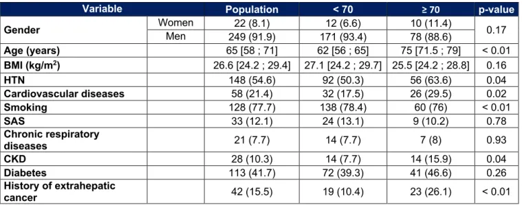
The characteristics of the patients are shown in Table 1. The median age is 75 years in the group of elderly patients and 62 years in the group of younger patients (p < 0,01). The sex-ratio is not different between the two groups with a large male predominance (SR = 9 : 1 ; p = 0.17).
Elderly patients have significantly more extrahepatic comorbidities. We find more arterial hypertension (HTN) (63.6% vs 50.3%, p = 0.04), more cardiovascular diseases (29.5% vs 17.5%, p = 0.02), more chronic kidney disease (CKD) (15.9% vs 7.7%, p = 0.04) and more history of extrahepatic cancer (26.1% vs 10.4%, p < 0.01). On the other hand, smoking (former or current) is less frequent in the elderly (76% vs 78.4%, p < 0.01). The general condition of elderly patients is more impaired (PS 1 : 29.5% vs 19.1% ; PS 2 : 5.7% vs 0.5%, p < 0.01).
There is no significant difference for other parameters, including body mass index (BMI), chronic respiratory diseases, sleep apnea syndrome (SAS) or diabetes.
In our population, the elderly have less cirrhosis (71.6% vs 91.8%, p < 0.01) and less history of hepatic decompensation (21.6% vs 36.6%, p = 0.01) than younger patients. Non-alcoholic steatohepatitis (NASH) and rare causes of chronic liver disease are more frequent in the elderly (respectively 46.6% vs 32.2%, p = 0.02 and 9.1% vs 3.3%, p = 0.04). Alcohol (54.5% vs 73.8%, p < 0.01) and viral causes (15.9% vs 36.1%, p < 0.01) are however less often observed in the elderly compared to younger people. In cirrhotics, the distribution of the Child-Pugh score is not different between the two groups.
About the biological variables there is a lower bilirubin level (12 µmol/l vs 16 µmol/l, p < 0.01), a higher PR (81% vs 78%, p < 0.01) and a higher albuminemia level (38 g/l vs 36 g/l, p = 0.03).
Characteristics of HCC
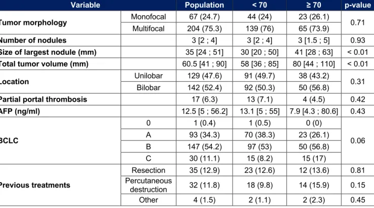
The characteristics of the patients relating to their HCC are shown in Table 2. The tumor volume is significantly larger in the elderly with a main nodule whose median size is 41 mm vs 30 mm (p < 0.01) and a median total tumor volume of 80 mm vs 58 mm (p < 0.01). The tumor stage tends to be more advanced (BCLC B : 56.8% vs 53% ; BCLC C : 17% vs 8.2%, p = 0.06).
There is no significant difference for the other parameters, in particular for multifocality, for the number of nodules, for the presence of a partial portal thrombosis, for the level of alpha-fetoprotein or for treatments received previously.
Characteristics of TACE
The patient characteristics relating to TACE are shown in Table 3. Elderly patients receive a DEB - TACE more often than younger patients (13.8% vs 4.4%, p < 0.01). There is no significant difference in the chemotherapy used, the area treated or the accessory tumor vasculature embolized.
Complications
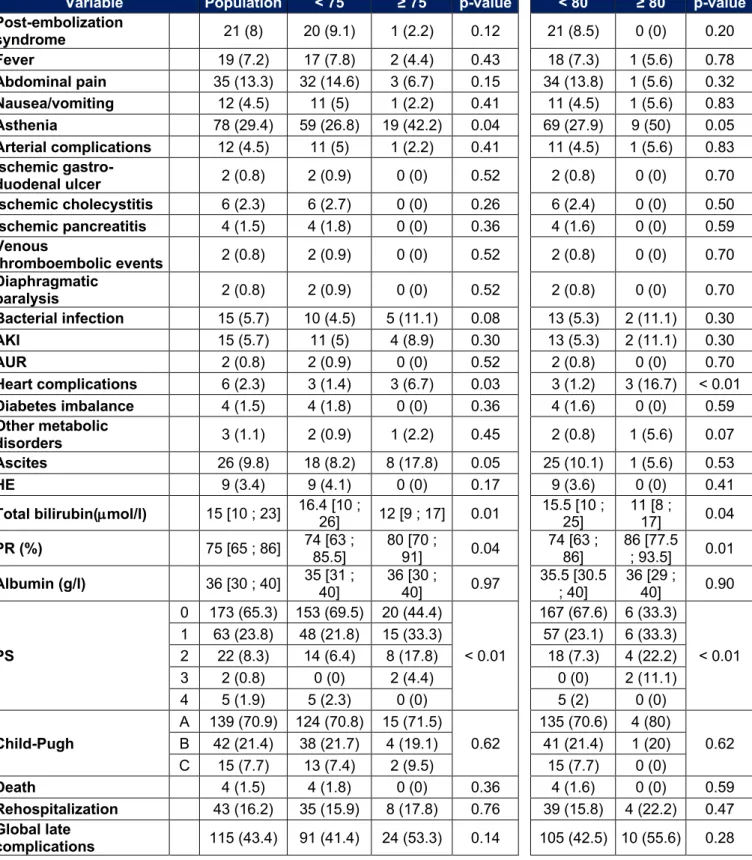
Early adverse events
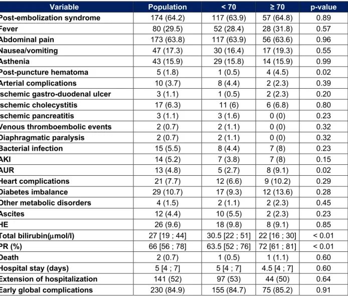
The adverse events occurring during the initial hospitalization are shown in Table 4. In our series, the elderly presented more post-puncture hematoma (4.5% vs 0.5%, p = 0.02) and more acute urinary retention (AUR) (9.1% vs 2.7%, p = 0.02). Control
parameters of liver function measured during hospitalization are better with a median bilirubin of 22 μmol/l vs 30.5 μmol/l and a median PR of 72% vs 63.5% (p < 0.01). No significant difference noted for the other complications, in particular, there is only one death in each group and there is no more prolongation of hospitalization in the elderly. The median length of hospital stay is 4.5 days in those over 70 years old compared to 5 days in younger patients (p = 0.60).
Late adverse events
Adverse events occurring between discharge from hospital and oncologic reassessment are indicated in Table 5. No patient aged 70 years or older shows signs of encephalopathy compared to 5% of younger (p = 0.04). The onset or worsening of pre-existing asthenia is more often reported in the elderly following TACE (40% vs 24.4%, p < 0.01). The PS assessed at a distance from hospitalization differs between the two groups (PS 0 : 51.8% vs 71.7% ; PS 1 : 31.8% vs 20% ; PS 2 : 14.1% vs 5.6% ; PS 3 : 2.4% vs 0% ; PS 4 : 0% vs 2.8%, p < 0.01). The bilirubin level is significantly lower in the elderly (12 μmol/l vs 17 μmol/l, p < 0.01).
There is no significant difference for the other parameters. No deaths were reported in the elderly compared to 4 (2.2%) in younger patients (p = 0.16). There is also no significant difference in the re-hospitalization rate before oncological reassessment : 11.8% in elderly patients and 18.3% in younger patients (p = 0.18).
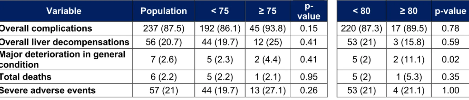
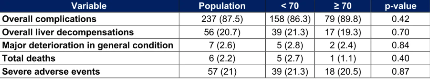
Major adverse events and overall complications
The major adverse events and the overall complications are shown in Table 6 : 20.5% of patients over 70 years of age have a severe adverse event against 21.3% in younger.
There is no statistically significant difference between the two groups (p = 0.87). There was also no difference in the rates of total death, overall complications, liver decompensations or major deterioration in general condition.
Predictive factors of severe adverse events
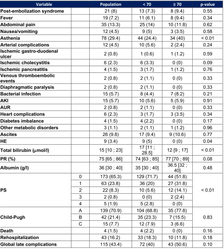
The results of univariate and multivariate analyzes for the study of predictive factors of severe adverse events are indicated in Tables 7 and 8. In univariate analysis, the factors associated with the occurrence of severe adverse events are : a history of chronic alcoholism (p = 0.041), a history of hepatic decompensation (p < 0.0001), a PS greater than or equal to 1 (p = 0.0003), a MELD score greater than or equal to 9 (p = 0.0008) and a BCLC stage greater than or equal to B (p = 0.0364). A Child-Pugh score below B7 is associated with a lower risk of serious adverse events (p < 0.0001). In contrast, there is no statistically significant association between an age of 70 years or older and the occurrence of severe adverse events (p = 0.87).
In multivariate analysis, a Child score greater than or equal to B7 (p < 0.0001), a PS greater than or equal to 1 (p = 0.0019), a MELD score greater than or equal to 9 (p = 0.0415) are associated with an increased risk of severe adverse events. A multifocal tumor (p = 0.0396) and the administration of idarubicin as chemotherapy (p = 0.0351) are associated with a lower occurrence of severe adverse events.
Analysis for patients over 75 and 80 years of age
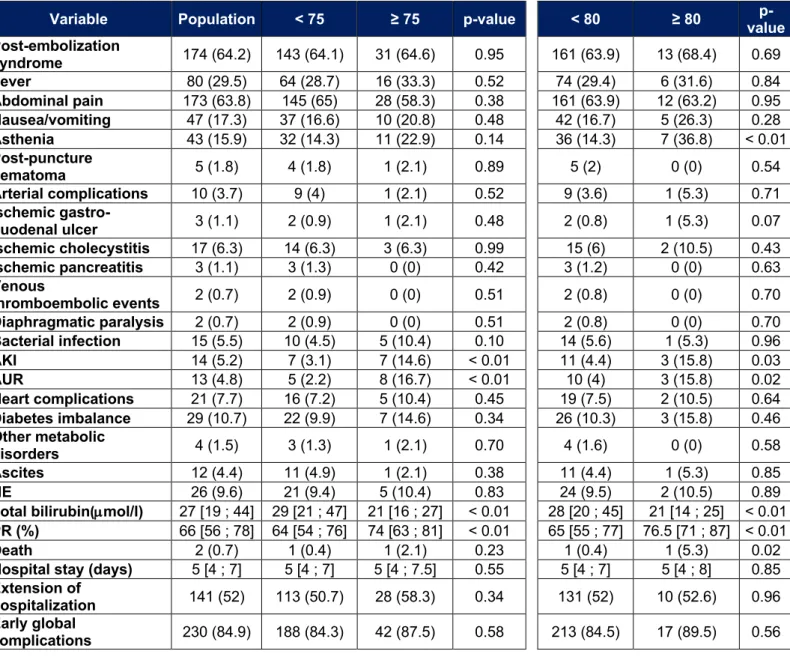
In our population there are 48 patients over 75 years old and 19 over 80 years old. Adverse events that occurred in these subgroups are listed in Tables 9, 10 and 11. In people over 75 years of age, we observe more acute kidney injury (14.6% vs 3.1%, p < 0.01) and more acute urinary retention (16.7% vs 2.2%, p < 0.01) during the initial
hospitalization. Their post-TACE liver function is also better. At a distance from initial hospitalization, there is more asthenia (42.2% vs 26.8%, p < 0.01) and more cardiac complications (6.7% vs 1.4%, p = 0.04). The rate of ascites after initial hospitalization tends to be higher in patients over 75 years of age (17.8% vs 8.2%, p = 0.05). The PS is more often high (p < 0.01). For the other parameters, especially the death rate, the length of hospitalization or the rate of severe adverse events, no significant difference was noted.
In those over 80, the results are similar. We find more acute kidney injury (15.8% vs 4.4%, p = 0.03), more acute urinary retention (15.8% vs 4%, p = 0.02) as well as more asthenia (36.8% vs 14.3%, p < 0.01) during the initial hospitalization. Their post-TACE liver function is also better. One death is observed in each group (5.3% vs 0.4%, p = 0.02). After hospitalization, more cardiac complications are observed (16.7% vs 1.2%, p < 0.01). A major deterioration in general condition is more frequently observed (11.1% vs 2%, p = 0.02). There is no significant difference for other variables, including length of hospital stay, overall death rate, or rate of severe adverse events.
In univariate analysis, neither the age of 75 nor 80 years are associated with an increased risk of occurrence of severe adverse events (OR : 1.51 [0.738 ; 3.095] ; p = 0.2591 and OR : 1.01 [0.319 ; 3.143] ; p = 0.9983) (Table 7). Including the age threshold of 75 years in the multivariate model, no significant association is demonstrated either (OR : 2.55 [0.898 ; 7.239] ; p = 0.0786) (Table 12). Due to a lack of patients, it is not possible to include the 80-year threshold in the multivariate model.
Discussion
The aim of our study was to describe the safety profile of TACE for HCC in patients over 70 years of age. We have shown safety profile of TACE in the elderly was comparable to that of younger patients. Age, even advanced, is not a predictive factor of severe adverse events. This criterion should therefore not be considered as an obstacle in the therapeutic decision.
Comparison with data from the literature
Few data are available on the safety of TACE in the elderly. We have shown that the elderly do not have more severe complications such as death or liver decompensations. These results are confirmed by the studies of Cohen et al. (2013, 2014) and Nishikawa et al. (2015) [22 - 24]. Identical findings are reported in a 2009 retrospective study by Yau et al. [20]. In this series, the differences with our results concern only minor adverse events. The post-procedure ischemic ulcer rate was higher in patients over 70 years of age (2.5% vs 0.5%, p = 0.01). Data relating to episodes of acute urinary retention, post-puncture hematoma or the onset of asthenia were not analyzed in their work.
To our knowledge, no publication has investigated the predictive factors of severe adverse events secondary to TACE. However, the analysis of factors associated with survival and tumor response is better understood. Age is not one of those factors. The absence of association between occurrence of adverse events or tumor response and age supports our idea that age should not be considered as a contraindication for TACE.
Our analysis in the oldest patient subgroups (75 and 80 years or older) does not show any difference in the occurrence of severe adverse events. In 2019, Cheng et al. studied the efficacy and safety of TACE for HCC in patients over 80 years of age [27]. Note that there was no younger age control group. Of the 86 patients included, apart from post-TACE liver test abnormalities, only one patient (1.2%) had grade III or IV toxicity according to the CTCAE classification (tumor rupture) [28]. Our results are supported by these findings.
Strength and limitation of our study
For this study, we performed a systematic and structured data collection of potential complications after TACE. The information provided are not limited to only hepatic adverse events, but also concerns cardiac, respiratory, urinary, infectious complications or venous thromboembolic events which are not described in other publications. It was also relevant to distinguish immediate tolerance from late tolerance after TACE, this approach not being systematically followed in the other available studies. In order to synthesize the large amount of data obtained and provide relevant information for clinical practice, we choose to focus on the most severe adverse events by creating composite variables.
To strengthen our conclusions, we studied the predictive factors of severe adverse events after TACE. The data available in the literature are limited to the results of descriptive analyzes. In order not to overlook the effect of age in older patients (75 and 80 years or older), we have chosen to carry out sensitivity analyzes in order to provide results for the different age groups. To our knowledge, only one publication without a control group and using a different methodology had studied the tolerance of people over 80 years of age [27].
Our study has several limitations. Due to its retrospective aspect, a bias in the assessment of some parameters is possible. Indeed, the gradation of severity is not systematically registered in the medical file. Some events may be missing. Furthermore, our definition of severe adverse reactions can be challenged since it has not been established according to the CTCAE classification. Although probably rare, some severe extrahepatic complications, not leading to death and not responsible for a major deterioration in general condition (PS 3 or 4), may have been overlooked in our definition.
In our population, the number of older patients, especially those over 80, is small. Due to a lack of statistical power, the relevance of our results may be affected for these patients.
Finally, although TACE techniques are relatively standardized, specific practical habits to each center are possible. As this is a single-center study, it is possible that the data on safety may be different in other establishments.
Research perspectives
The geriatric population has specificities which require the development of tools adapted to their evaluation and their management. In oncology, the G8 score is a validated example [17]. Although it is not a lengthy procedure, it is unfortunately still underused, especially in HCC. In our work the G8 score was not available for any of the elderly patients. Only one of them had undergone a thorough geriatric assessment. The training of clinicians in the management of the geriatric population is critical for improving practices. Other assessment parameters could be proposed as an
alternative. Walking speed over 4 meters is a test recommended by some authors for its performance close to the G8 score and for its great simplicity [29].
In order to improve the safety of TACE and to be able to offer it to the most fragile patients, technical improvements have been proposed. Although its results are debated, the drug-eluting bead TACE (DEB-TACE) could according to some authors, allow to reduce toxicity [30-32]. However, the tolerance of this technique doesn’t seem to differ between the elderly and the younger patients [33].
A recent technical improvement consisting in the use of microparticles containing iodine atoms, visible in fluoroscopy, would allow chemotherapy to be delivered with precision and control during treatment in order to increase its efficacy and limit its adverse events [34]. This technique is currently being evaluated.
Moreover, patients with intermediate HCC and hepatic impairment (except for Child C patients) or portal thrombosis may receive in some centers a TACE with degradable starch microspheres (DSM-TACE) which carry the chemotherapy. These are rapidly digested in the hepatic bloodstream, which may reduce the ischemic effect and improve safety [35-36].
Conclusion
Our study shows that there is no significant difference in the occurrence of severe adverse events after chemo-embolization for hepatocellular carcinoma between elderly and younger patients. Advanced age is not a predictive factor of severe adverse events and should not be considered as an obstacle in the treatment decision. Further assessment of TACE safety in patients over 80 years of age whose number are constantly increasing is however needed.
Table 1 : Patient characteristics
Cardiovascular diseases = ischemic heart disease and/or peripheral artery disease and/or stroke ; Smoking = former or current ; SAS = with CPAP or not ; Chronic respiratory disease = asthma and/or
chronic obstructive pulmonary disease ; History of liver decompensation = Ascites and/or HE and/or hemorrhage and/or hepato-renal syndrome.
Variable Population < 70 ≥ 70 p-value
Gender Women Men 22 (8.1) 12 (6.6) 10 (11.4) 0.17
249 (91.9) 171 (93.4) 78 (88.6) Age (years) 65 [58 ; 71] 62 [56 ; 65] 75 [71.5 ; 79] < 0.01 BMI (kg/m2) 26.6 [24.2 ; 29.4] 27.1 [24.2 ; 29.7] 25.5 [24.2 ; 28.8] 0.16 HTN 148 (54.6) 92 (50.3) 56 (63.6) 0.04 Cardiovascular diseases 58 (21.4) 32 (17.5) 26 (29.5) 0.02 Smoking 128 (77.7) 138 (78.4) 60 (76) < 0.01 SAS 33 (12.1) 24 (13.1) 9 (10.2) 0.78 Chronic respiratory diseases 21 (7.7) 14 (7.7) 7 (8) 0.93 CKD 28 (10.3) 14 (7.7) 14 (15.9) 0.04 Diabetes 113 (41.7) 72 (39.3) 41 (46.6) 0.26 History of extrahepatic cancer 42 (15.5) 19 (10.4) 23 (26.1) < 0.01 Cirrhosis 231 (85.2) 168 (91.8) 63 (71.6) < 0.01 Cause of hepatopathy Chronic alcoholism 183 (67.5) 135 (73.8) 48 (54.5) < 0.01 NASH 100 (36.9) 59 (32.2) 41 (46.6) 0.02 Chronic viral hepatitis 80 (29.5) 66 (36.1) 14 (15.9) < 0.01 Other 14 (5.2) 6 (3.3) 8 (9.1) 0.04 History of liver decompensation 86 (31.7) 67 (36.6) 19 (21.6) 0.01 PS 0 204 (75.3) 147 (80.3) 57 (64.8) < 0.01 1 61 (22.5) 35 (19.1) 26 (29.5) 2 6 (2.2) 1 (0.5) 5 (5.7) MELD 9 [8 ; 11] 10 [8 ; 12] 9 [8 ; 10] 0.13 Child-Pugh A 176 (76.9) 121 (72.4) 55 (88.7) 0.10 B 49 (21.4) 42 (25.2) 7 (11.3) C 4 (1.7) 4 (2.4) 0 (0) Ascites 20 (7.4) 16 (8.7) 4 (4.5) 0.22 HE 4 (1.5) 4 (2.2) 0 (0) 0.16 Albumin (g/l) 37 [32 ; 41] 36 [31 ; 41] 38 [34 ; 41] 0.03 PR (%) 79 [67 ; 89.5] 78 [64 ; 87] 81 [72 ; 93] < 0.01
Table 2 : Characteristics of HCC
Variable Population < 70 ≥ 70 p-value Tumor morphology Monofocal 67 (24.7) 44 (24) 23 (26.1) 0.71
Multifocal 204 (75.3) 139 (76) 65 (73.9)
Number of nodules 3 [2 ; 4] 3 [2 ; 4] 3 [1.5 ; 5] 0.93
Size of largest nodule (mm) 35 [24 ; 51] 30 [20 ; 50] 41 [28 ; 63] < 0.01
Total tumor volume (mm) 60.5 [41 ; 90] 58 [36 ; 85] 80 [44 ; 110] < 0.01
Location Unilobar 129 (47.6) 91 (49.7) 38 (43.2) 0.31
Bilobar 142 (52.4) 92 (50.3) 50 (56.8)
Partial portal thrombosis 17 (6.3) 13 (7.1) 4 (4.5) 0.42
AFP (ng/ml) 12.5 [5 ; 56.2] 13.1 [5 ; 55] 7.9 [4.3 ; 80.6] 0.43 BCLC 0 1 (0.4) 1 (0.5) 0 (0) 0.06 A 93 (34.3) 70 (38.3) 23 (26.1) B 147 (54.2) 97 (53) 50 (56.8) C 30 (11.1) 15 (8.2) 15 (17) Previous treatments Resection 35 (12.9) 23 (12.6) 12 (13.6) 0.81 Percutaneous destruction 32 (11.8) 18 (9.8) 14 (15.9) 0.15 Other 4 (1.5) 2 (1.1) 2 (2.3) 0.45
Table 3 : Characteristics of TACE
Variable Population < 70 ≥ 70 p-value Area treated Global TACE 156 (57.8) 106 (57.9) 50 (57.5) 0.94
Selective TACE 114 (42.2) 77 (42.1) 37 (42.5)
Type of TACE c-TACE 249 (92.6) 174 (95.6) 75 (86.2) < 0.01
DEB-TACE 20 (7.4) 8 (4.4) 12 (13.8)
Embolization of accessory tumor vasculature Diaphragmatic arteries 27 (10.2) 14 (7.8) 13 (15.1) 0.07 Mammary artery 2 (0.8) 2 (1.1) 0 (0) 0.33 Other arteries 10 (3.8) 7 (3.9) 3 (3.5) 0.87 Chemotherapy Doxorubicin 230 (87.5) 153 (85.5) 77 (91.7) 0.16 Idarubicin 33 (12.5) 26 (14.5) 7 (8.3)
Table 4 : Early adverse events
Variable Population < 70 ≥ 70 p-value Post-embolization syndrome 174 (64.2) 117 (63.9) 57 (64.8) 0.89 Fever 80 (29.5) 52 (28.4) 28 (31.8) 0.57 Abdominal pain 173 (63.8) 117 (63.9) 56 (63.6) 0.96 Nausea/vomiting 47 (17.3) 30 (16.4) 17 (19.3) 0.55 Asthenia 43 (15.9) 29 (15.8) 14 (15.9) 0.99 Post-puncture hematoma 5 (1.8) 1 (0.5) 4 (4.5) 0.02 Arterial complications 10 (3.7) 8 (4.4) 2 (2.3) 0.39
Ischemic gastro-duodenal ulcer 3 (1.1) 1 (0.5) 2 (2.3) 0.20
Ischemic cholecystitis 17 (6.3) 11 (6) 6 (6.8) 0.80
Ischemic pancreatitis 3 (1.1) 3 (1.6) 0 (0) 0.23
Venous thromboembolic events 2 (0.7) 2 (1.1) 0 (0) 0.32
Diaphragmatic paralysis 2 (0.7) 2 (1.1) 0 (0) 0.32 Bacterial infection 15 (5.5) 8 (4.4) 7 (8) 0.23 AKI 14 (5.2) 7 (3.8) 7 (8) 0.15 AUR 13 (4.8) 5 (2.7) 8 (9.1) 0.02 Heart complications 21 (7.7) 12 (6.6) 9 (10.2) 0.29 Diabetes imbalance 29 (10.7) 17 (9.3) 12 (13.6) 0.28
Other metabolic disorders 4 (1.5) 2 (1.1) 2 (2.3) 0.45
Ascites 12 (4.4) 10 (5.5) 2 (2.3) 0.23
HE 26 (9.6) 18 (9.8) 8 (9.1) 0.85
Total bilirubin(µmol/l) 27 [19 ; 44] 30.5 [22 ; 51] 22 [16 ; 30] < 0.01
PR (%) 66 [56 ; 78] 63.5 [52 ; 76] 72 [61 ; 81] < 0.01
Death 2 (0.7) 1 (0.5) 1 (1.1) 0.60
Hospital stay (days) 5 [4 ; 7] 5 [4 ; 7] 4.5 [4 ; 7] 0.60
Extension of hospitalization 141 (52) 97 (53) 44 (50) 0.64
Early global complications 230 (84.9) 155 (84.7) 75 (85.2) 0.91
Arterial complications = Mesenteric ischemia, splenic ischemia, ischemic cholangitis, spinal cord
ischemia, stroke, arterial dissection ; Heart complication : cardiac decompensation and/or arrhythmia and/or severe hypertension ; Other metabolic disorders = severe ionic disorders ; Extension of
hospitalization = hospitalization > 4 days ; Early global complications = any complication occurring
Table 5 : Late adverse events
Variable Population < 70 ≥ 70 p-value Post-embolization syndrome 21 (8) 13 (7.3) 8 (9.4) 0.55 Fever 19 (7.2) 11 (6.1) 8 (9.4) 0.34 Abdominal pain 35 (13.3) 25 (14) 10 (11.8) 0.62 Nausea/vomiting 12 (4.5) 9 (5) 3 (3.5) 0.58 Asthenia 78 (29.4) 44 (24.4) 34 (40) < 0.01 Arterial complications 12 (4.5) 10 (5.6) 2 (2.4) 0.24 Ischemic gastro-duodenal ulcer 2 (0.8) 1 (0.6) 1 (1.2) 0.59 Ischemic cholecystitis 6 (2.3) 6 (3.3) 0 (0) 0.09 Ischemic pancreatitis 4 (1.5) 3 (1.7) 1 (1.2) 0.76 Venous thromboembolic events 2 (0.8) 2 (1.1) 0 (0) 0.33 Diaphragmatic paralysis 2 (0.8) 2 (1.1) 0 (0) 0.33 Bacterial infection 15 (5.7) 8 (4.4) 7 (8.2) 0.21 AKI 15 (5.7) 10 (5.6) 5 (5.9) 0.91 AUR 2 (0.8) 2 (1.1) 0 (0) 0.33 Heart complications 6 (2.3) 3 (1.7) 3 (3.5) 0.34 Diabetes imbalance 4 (1.5) 4 (2.2) 0 (0) 0.17
Other metabolic disorders 3 (1.1) 2 (1.1) 1 (1.2) 0.96
Ascites 26 (9.8) 17 (9.4) 9 (10.6) 0.77
HE 9 (3.4) 9 (5) 0 (0) 0.04
Total bilirubin (µmol/l) 15 [10 ; 23] 17 [11 ; 28.5] 12 [9 ; 17] < 0.01
PR (%) 75 [65 ; 86] 74 [63 ; 85] 77 [70 ; 89] 0.08 Albumin (g/l) 36 [30 ; 40] 35 [30 ; 40] 36.5 [32 ; 40] 0.48 PS 0 173 (65.3) 129 (71.7) 44 (51.8) < 0.01 1 63 (23.8) 36 (20) 27 (31.8) 2 22 (8.3) 10 (5.6) 12 (14.1) 3 2 (0.8) 0 (0) 2 (2.4) 4 5 (1.9) 5 (2.8) 0 (0) Child-Pugh A 139 (70.9) 104 (68.8) 35 (77.8) 0.83 B 42 (21.4) 35 (23.3) 7 (15.5) C 15 (7.7) 12 (7.9) 3 (6.6) Death 4 (1.5) 4 (2.2) 0 (0) 0.16 Rehospitalization 43 (16.2) 33 (18.3) 10 (11.8) 0.18
Global late complications 115 (43.4) 72 (40) 43 (50.6) 0.10
Global late complications = Any complication occurring during follow-up until the oncological
Table 6 : Major adverse events and overall complications
Variable Population < 70 ≥ 70 p-value Overall complications 237 (87.5) 158 (86.3) 79 (89.8) 0.42
Overall liver decompensations 56 (20.7) 39 (21.3) 17 (19.3) 0.70
Major deterioration in general condition 7 (2.6) 5 (2.8) 2 (2.4) 0.84
Total deaths 6 (2.2) 5 (2.7) 1 (1.1) 0.40
Severe adverse events 57 (21) 39 (21.3) 18 (20.5) 0.87
Overall complications = any early and/or late complication ;
Overall liver decompensations = Any HE (early and/or late) and/or ascites (early and/or late) and/or
Child C score during the follow-up ;
Major deterioration in general condition: PS 3 or 4 during follow-up until oncology reassessment ; Total deaths = early and/or late deaths ;
Severe adverse events = any liver decompensation and/or death and/or major deterioration in general
Tableau 7 : Predictive factors of severe adverse events (univariate analysis) Variable OR 95% CI p-value Age Age ≥ 70 0.949 [0.507 ; 1.778] 0.8712 Age ≥ 75 1.511 [0.738 ; 3.095] 0.2591 Age ≥ 80 1.001 [0.319 ; 3.143] 0.9983 Gender Women 1.114 [0.393 ; 3.162] 0.8389 BMI BMI ≥ 26.5 0.763 [0.424 ; 1.372] 0.3666 HTN 0.756 [0.421 ; 1.358] 0.3496 Cardiovascular diseases 0.736 [0.346 ; 1.565] 0.4254 Smoking 1.480 [0.673 ; 3.254] 0.3299 SAS 0.481 [0.162 ; 1.431] 0.1884
Chronic respiratory diseases 0.875 [0.282 ; 2.709] 0.8163
CKD 1.287 [0.518 ; 3.197] 0.5873
Diabetes 0.774 [0.424 ; 1.413] 0.4035
History of extrahepatic cancer 0.716 [0.3 ; 1.709] 0.4517
Cirrhosis 1.606 [0.639 ; 4.037] 0.3141
Chronic alcoholism 2.065 [1.03 ; 4.141] 0.0410
NASH 0.743 [0.399 ; 1.385] 0.3498
Chronic viral hepatitis 1.387 [0.746 ; 2.58] 0.3009
Other causes 2.190 [0.704 ; 6.813] 0.1757
History of liver decompensation 3.793 [2.066 ; 6.961] < 0.0001
PS ≥ 1 3.199 [1.717 ; 5.962] 0.0003
MELD ≥ 9 3.804 [1.745 ; 8.294] 0.0008
Child-Pugh A 0.152 [0.076 ; 0.303] < 0.0001
Tumor morphology Monofocal 1.729 [0.914 ; 3.271] 0.0923
Number of nodules ≥ 3 1.396 [0.768 ; 2.535] 0.2739
Size of largest nodule (mm) ≥ 35 0.981 [0.545 ; 1.768] 0.9504
Location Unilobar 1.080 [0.602 ; 1.938] 0.7958
Partial portal thrombosis 1.619 [0.546 ; 4.799] 0.3852
AFP (ng/mL) ≥ 12.5 1.399 [0.76 ; 2.576] 0.2804
BCLC ≥ B 2.061 [1.047 ; 4.059] 0.0364
Previous treatments 0.875 [0.43 ; 1.781] 0.7121
Area treated Global 0.736 [0.408 ; 1.329] 0.3088
Type of TACE c-TACE 1.532 [0.433 ; 5.426] 0.5082
Embolization of accessory tumor vasculature Diaphragmatic arteries 0.638 [0.211 ; 1.927] 0.4253 Mammary artery 1.000 [1 ; 1] 0.9998 Other artery 1.673 [0.418 ; 6.693] 0.4669 Chemotherapy Doxorubicin 2.921 [0.857 ; 9.959] 0.0867
Tableau 8 : Predictive factors of severe adverse events (multivariate analysis)
Variable OR 95% CI p-value
Child-Pugh ≥ B 5.034 [2.278 ; 11.126] < 0.0001
PS ≥ 1 3.556 [1.6 ; 7.903] 0.0019
MELD ≥ 9 2.450 [1.035 ; 5.798] 0.0415
Tumor morphology Multifocal 0.423 [0.186 ; 0.96] 0.0396
Chemotherapy Idarubicin 0.177 [0.036 ; 0.886] 0.0351
Tableau 9 : Early adverse events for patients over 75 or 80 years old
Variable Population < 75 ≥ 75 p-value < 80 ≥ 80 value p-Post-embolization syndrome 174 (64.2) 143 (64.1) 31 (64.6) 0.95 161 (63.9) 13 (68.4) 0.69 Fever 80 (29.5) 64 (28.7) 16 (33.3) 0.52 74 (29.4) 6 (31.6) 0.84 Abdominal pain 173 (63.8) 145 (65) 28 (58.3) 0.38 161 (63.9) 12 (63.2) 0.95 Nausea/vomiting 47 (17.3) 37 (16.6) 10 (20.8) 0.48 42 (16.7) 5 (26.3) 0.28 Asthenia 43 (15.9) 32 (14.3) 11 (22.9) 0.14 36 (14.3) 7 (36.8) < 0.01 Post-puncture hematoma 5 (1.8) 4 (1.8) 1 (2.1) 0.89 5 (2) 0 (0) 0.54 Arterial complications 10 (3.7) 9 (4) 1 (2.1) 0.52 9 (3.6) 1 (5.3) 0.71 Ischemic gastro-duodenal ulcer 3 (1.1) 2 (0.9) 1 (2.1) 0.48 2 (0.8) 1 (5.3) 0.07 Ischemic cholecystitis 17 (6.3) 14 (6.3) 3 (6.3) 0.99 15 (6) 2 (10.5) 0.43 Ischemic pancreatitis 3 (1.1) 3 (1.3) 0 (0) 0.42 3 (1.2) 0 (0) 0.63 Venous thromboembolic events 2 (0.7) 2 (0.9) 0 (0) 0.51 2 (0.8) 0 (0) 0.70 Diaphragmatic paralysis 2 (0.7) 2 (0.9) 0 (0) 0.51 2 (0.8) 0 (0) 0.70 Bacterial infection 15 (5.5) 10 (4.5) 5 (10.4) 0.10 14 (5.6) 1 (5.3) 0.96 AKI 14 (5.2) 7 (3.1) 7 (14.6) < 0.01 11 (4.4) 3 (15.8) 0.03 AUR 13 (4.8) 5 (2.2) 8 (16.7) < 0.01 10 (4) 3 (15.8) 0.02 Heart complications 21 (7.7) 16 (7.2) 5 (10.4) 0.45 19 (7.5) 2 (10.5) 0.64 Diabetes imbalance 29 (10.7) 22 (9.9) 7 (14.6) 0.34 26 (10.3) 3 (15.8) 0.46 Other metabolic disorders 4 (1.5) 3 (1.3) 1 (2.1) 0.70 4 (1.6) 0 (0) 0.58 Ascites 12 (4.4) 11 (4.9) 1 (2.1) 0.38 11 (4.4) 1 (5.3) 0.85 HE 26 (9.6) 21 (9.4) 5 (10.4) 0.83 24 (9.5) 2 (10.5) 0.89
Total bilirubin(µmol/l) 27 [19 ; 44] 29 [21 ; 47] 21 [16 ; 27] < 0.01 28 [20 ; 45] 21 [14 ; 25] < 0.01
PR (%) 66 [56 ; 78] 64 [54 ; 76] 74 [63 ; 81] < 0.01 65 [55 ; 77] 76.5 [71 ; 87] < 0.01
Death 2 (0.7) 1 (0.4) 1 (2.1) 0.23 1 (0.4) 1 (5.3) 0.02
Hospital stay (days) 5 [4 ; 7] 5 [4 ; 7] 5 [4 ; 7.5] 0.55 5 [4 ; 7] 5 [4 ; 8] 0.85
Extension of
hospitalization 141 (52) 113 (50.7) 28 (58.3) 0.34 131 (52) 10 (52.6) 0.96 Early global
Tableau 10 : Late adverse events for patients over 75 or 80 years old
Variable Population < 75 ≥ 75 p-value < 80 ≥ 80 p-value Post-embolization syndrome 21 (8) 20 (9.1) 1 (2.2) 0.12 21 (8.5) 0 (0) 0.20 Fever 19 (7.2) 17 (7.8) 2 (4.4) 0.43 18 (7.3) 1 (5.6) 0.78 Abdominal pain 35 (13.3) 32 (14.6) 3 (6.7) 0.15 34 (13.8) 1 (5.6) 0.32 Nausea/vomiting 12 (4.5) 11 (5) 1 (2.2) 0.41 11 (4.5) 1 (5.6) 0.83 Asthenia 78 (29.4) 59 (26.8) 19 (42.2) 0.04 69 (27.9) 9 (50) 0.05 Arterial complications 12 (4.5) 11 (5) 1 (2.2) 0.41 11 (4.5) 1 (5.6) 0.83 Ischemic gastro-duodenal ulcer 2 (0.8) 2 (0.9) 0 (0) 0.52 2 (0.8) 0 (0) 0.70 Ischemic cholecystitis 6 (2.3) 6 (2.7) 0 (0) 0.26 6 (2.4) 0 (0) 0.50 Ischemic pancreatitis 4 (1.5) 4 (1.8) 0 (0) 0.36 4 (1.6) 0 (0) 0.59 Venous thromboembolic events 2 (0.8) 2 (0.9) 0 (0) 0.52 2 (0.8) 0 (0) 0.70 Diaphragmatic paralysis 2 (0.8) 2 (0.9) 0 (0) 0.52 2 (0.8) 0 (0) 0.70 Bacterial infection 15 (5.7) 10 (4.5) 5 (11.1) 0.08 13 (5.3) 2 (11.1) 0.30 AKI 15 (5.7) 11 (5) 4 (8.9) 0.30 13 (5.3) 2 (11.1) 0.30 AUR 2 (0.8) 2 (0.9) 0 (0) 0.52 2 (0.8) 0 (0) 0.70 Heart complications 6 (2.3) 3 (1.4) 3 (6.7) 0.03 3 (1.2) 3 (16.7) < 0.01 Diabetes imbalance 4 (1.5) 4 (1.8) 0 (0) 0.36 4 (1.6) 0 (0) 0.59 Other metabolic disorders 3 (1.1) 2 (0.9) 1 (2.2) 0.45 2 (0.8) 1 (5.6) 0.07 Ascites 26 (9.8) 18 (8.2) 8 (17.8) 0.05 25 (10.1) 1 (5.6) 0.53 HE 9 (3.4) 9 (4.1) 0 (0) 0.17 9 (3.6) 0 (0) 0.41
Total bilirubin(µmol/l) 15 [10 ; 23] 16.4 [10 ; 26] 12 [9 ; 17] 0.01 15.5 [10 ; 25] 11 [8 ; 17] 0.04
PR (%) 75 [65 ; 86] 74 [63 ; 85.5] 80 [70 ; 91] 0.04 74 [63 ; 86] 86 [77.5 ; 93.5] 0.01 Albumin (g/l) 36 [30 ; 40] 35 [31 ; 40] 36 [30 ; 40] 0.97 35.5 [30.5 ; 40] 36 [29 ; 40] 0.90 PS 0 173 (65.3) 153 (69.5) 20 (44.4) < 0.01 167 (67.6) 6 (33.3) < 0.01 1 63 (23.8) 48 (21.8) 15 (33.3) 57 (23.1) 6 (33.3) 2 22 (8.3) 14 (6.4) 8 (17.8) 18 (7.3) 4 (22.2) 3 2 (0.8) 0 (0) 2 (4.4) 0 (0) 2 (11.1) 4 5 (1.9) 5 (2.3) 0 (0) 5 (2) 0 (0) Child-Pugh A 139 (70.9) 124 (70.8) 15 (71.5) 0.62 135 (70.6) 4 (80) 0.62 B 42 (21.4) 38 (21.7) 4 (19.1) 41 (21.4) 1 (20) C 15 (7.7) 13 (7.4) 2 (9.5) 15 (7.7) 0 (0) Death 4 (1.5) 4 (1.8) 0 (0) 0.36 4 (1.6) 0 (0) 0.59 Rehospitalization 43 (16.2) 35 (15.9) 8 (17.8) 0.76 39 (15.8) 4 (22.2) 0.47 Global late complications 115 (43.4) 91 (41.4) 24 (53.3) 0.14 105 (42.5) 10 (55.6) 0.28
Tableau 11 : Major adverse events and overall complications for patients over 75 or 80 years old
Variable Population < 75 ≥ 75 value p- < 80 ≥ 80 p-value Overall complications 237 (87.5) 192 (86.1) 45 (93.8) 0.15 220 (87.3) 17 (89.5) 0.78
Overall liver decompensations 56 (20.7) 44 (19.7) 12 (25) 0.41 53 (21) 3 (15.8) 0.59
Major deterioration in general
condition 7 (2.6) 5 (2.3) 2 (4.4) 0.41 5 (2) 2 (11.1) 0.02 Total deaths 6 (2.2) 5 (2.2) 1 (2.1) 0.95 5 (2) 1 (5.3) 0.35
Severe adverse events 57 (21) 44 (19.7) 13 (27.1) 0.26 53 (21) 4 (21.1) 1.00
Tableau 12 : Predictive factors of severe adverse events (multivariate analysis including the age threshold of 75)
Variable OR 95% CI p-value Age ≥ 75 2.550 [0.898 ; 7.239] 0.0786 Child-Pugh ≥ B 5.788 [2.543 ; 13.175] < 0.0001 PS ≥ 1 3.118 [1.381 ; 7.039 0.0062 MELD ≥ 9 2.651 [1.101 ; 6.386] 0.0297
Tumor morphology Multifocal 0.441 [0.192 ; 1.014] 0.0538
Appendix 1
BCLC Staging classification
Stage Performance status Tumor stage Liver function
Very early HCC 0 0 Single < 2cm No portal hypertension and normal bilirubin
Early HCC
A1 0 Single < 5cm No portal hypertension and normal bilirubin
A2 0 Single < 5cm Portal hypertension and normal bilirubin
A3 0 Single < 5cm Portal hypertension and abnormal bilirubin
A4 0 3 tumors < 3cm Child-Pugh A-B
Intermediate HCC B 0 Large multinodular Child-Pugh A-B
Advanced HCC C 1-2 Vascular invasion or extra-hepatic spread Child-Pugh A-B
End-stage HCC D 3-4 Any Child-Pugh C
41
Appendix 2
EASL Clinical guidelines of HCC
complicates prognostic assessments.87,255In addition, the
pres-ence of cancer-related symptoms has consistently shown an impact on survival. Finally, any system aimed at being clinically meaningful should link prognostic prediction to treatment indication.
Staging systems for HCC should be designed with data from two sources. Firstly, prognostic variables obtained from studies describing the natural history of cancer and cirrhosis. Secondly, treatment-dependent variables obtained from evidence-based studies providing the rationale for assigning a given therapy to patients in a given subclass.
The main clinical prognostic factors in patients with HCC, based on studies reporting the natural history of the disease, are related to tumour status (defined by number and size of nodules, presence of vascular invasion, extrahepatic spread), liver function (defined by Child-Pugh’s class, bilirubin, albumin, clinically relevant portal hypertension, ascites) and general tumour-related health status (defined by the Eastern Coopera-tive Oncology Group [ECOG] classification and presence of symptoms).256–260Aetiology has not been identified as an
inde-pendent prognostic factor.260
Several staging systems have been proposed to provide a
TNM edition in accordance with the American Joint Committee on Cancer,233which was obtained from the analysis of a series
of patients undergoing resection, has several limitations.261,262
Firstly, pathological information is required to assess microvas-cular invasion, which is only available in patients treated by sur-gery (!20%). In addition, it does not capture information regarding liver functional status or health status. Finally, its prognostic value in non-early tumours is limited.262 Among
more comprehensive staging systems, six have been broadly tested, three European (the French classification,263the Cancer
of the Liver Italian Program [CLIP] classification,257and the
Bar-celona-Clínic Liver Cancer [BCLC] staging system87,264) and
three Asian (the Chinese University Prognostic Index [CUPI] score,265the Hong-Kong Liver Cancer [HKLC] staging system266
and the Japan Integrated Staging [JIS], which was refined includ-ing biomarkers (alpha-fetoprotein [AFP], des-c -carboxypro-thrombin [DCP] AFP-L3) (bm-JIS)267). CUPI and the CLIP scores
largely sub-classify patients at advanced stages, with a small number of effectively treated patients. Overall, most of these systems or scores have been externally validated, but only three include the three types of prognostic variables (BCLC, CUPI, and HKLC) and only two assign treatment allocation to specific
prog-HCC in cirrhotic liver Very early stage (0)
Single <2 cm Preserved liver function1,
PS 0
Early stage (A)
Single or 2-3 nodules <3 cm Preserved liver function1, PS 0
Intermediate stage (B)
Multinodular, unresectable Preserved liver function1,
PS 0
Advanced stage (C)
Portal invasion/ extrahepatic spread Preserved liver function1,
PS 12-2
Terminal stage (D)
Not transplantable HCC End-stage liver function
PS 3-4 Solitary Optimal surgical candidate3 Yes No Transplant candidate 2-3 nodules ≤3 cm Prognostic stage Treatment4 Survival Yes No
Ablation Resection Transplant Ablation Chemoembolization Systemic therapy5 BSC
>5 years >2.5 years ≥10 months 3 months Fig. 3. Modified BCLC staging system and treatment strategy.1‘‘Preserved liver function” refers to Child-Pugh A without any ascites, considered conditions
to obtain optimal outcomes. This prerequisite applies to all treatment options apart from transplantation, that is instead addressed primarily to patients with decompensated or end-stage liver function.2PS 1 refers to tumour induced (as per physician opinion) modification of performance capacity.3Optimal surgical
candidacy is based on a multiparametric evaluation including compensated Child-Pugh class A liver function with MELD score <10, to be matched with grade of portal hypertension, acceptable amount of remaining parenchyma and possibility to adopt a laparoscopic/minimally invasive approach. The combination of the previous factors should lead to an expected perioperative mortality <3% and morbidity <20% including a postsurgical severe liver failure incidence <5%.4The
stage migration strategy is a therapeutic choice by which a treatment theoretically recommended for a different stage is selected as best 1st line treatment option. Usually it is applied with a left to right direction in the scheme (i.e. offering the effective treatment option recommended for the subsequent more advanced tumour stage rather than that forecasted for that specific stage). This occurs when patients are not suitable for their first line therapy. However, in highly selected patients, with parameters close to the thresholds defining the previous stage, a right to left migration strategy (i.e. a therapy recommended for earlier stages) could be anyhow the best opportunity, pending multidisciplinary decision.5As of 2017 sorafenib has been shown to be effective in first line,
while regorafenib is effective in second line in case of radiological progression under sorafenib. Lenvatinib has been shown to be non-inferior to sorafenib in first line, but no effective second line option after lenvatinib has been explored. Cabozantinib has been demonstrated to be superior to placebo in 2nd or 3rd line with an improvement of OS from eight months (placebo) to 10.2 months (ASCO GI 2018). Nivolumab has been approved in second line by FDA but not EMA based on uncontrolled phase II data. ASCO, American Society of Clinical Oncology; BCLC, Barcelona Clinic Liver Cancer; EMA, European Medicines Agency; FDA, Food and Drug Administration; MELD, model for end-stage liver disease; PS, performace status; OS, overall survival. Modified with permission from87.
Appendix 3
G8 Score
Questionnaire G8
Test de dépistage du recours au gériatre chez un patient âgé atteint de cancer
Questions (temps médian de remplissage = 4,4 minutes) Réponses Cotations
Le patient présente-t-il e e e d a i ?
A-t-il mangé moins ces 3 derniers mois par manque d a i , bl me dige if , diffic l de ma ica i ou de déglutition? Anorexie sévère Anorexie modérée Pa d a e ie ☐ 0 ☐ 1 ☐ 2 Perte de poids dans les 3 derniers mois >3 Kg
Ne sait pas Entre 1 et 3 Kg Pas de perte de poids
☐ 0 ☐ 1 ☐ 2 ☐ 3
Motricité Lit Fauteuil
A me l i ie
Sort du domicile
☐ 0 ☐ 1 ☐ 2
Troubles neuro-psychiatriques Démence ou dépression sévère
Démence ou dépression modérée Pas de trouble psychiatrique
☐ 0 ☐ 1 ☐ 2 Indice de Masse Corporelle
= Poids/(Taille)2 < 19 19 21 21 23 > 23 ☐ 0 ☐ 1 ☐ 2 ☐ 3
Plus de 3 médicaments Oui
Non ☐ 0 ☐ 1
Le patient se sent-il en meilleure ou en moins bonne santé que la plupart des personnes de son âge?
Moins bonne Ne sais pas Aussi bonne Meilleure ☐ 0 ☐ 0,5 ☐ 1 ☐ 2 Age > 85 ans 80 85 ans < 80 ans ☐ 0 ☐ 1 ☐ 2 Score total /17
Interprétation > 14 = Prise en charge standard
< 14 = Evaluation gériatrique spécialisée
D a Soubeyran P. Validation of G8 screening tool in geriatric oncology: The ONCODAGE
References
1 : Blanc JF, Barbare JC, Baumann AS, Boige V, Boudjema K, Bouattour M, Crehange G, Dauvois B, Decaens T, Dewaele F, Farges O, Guiu B, Hollebecque A, Merle P, Roth G, Ruiz I, Selves J. « Carcinome hépatocellulaire ». Thésaurus National de
Cancérologie Digestive, Mars 2019, [En ligne] [http://www.tncd.org]
2 : Bray F, Ferlay J, Soejormatarum I, Siegel RL, Torre LA, Jemal A. Global cancer statistics 2018 : GLOBOCAN estimates of incidence and mortality worlwide for 36 cancers in 185 countries. A Cancer Journal for Clinicians. 2018 ; 68 (6) : 394-324 3 : Defossez G, Le Guyader-Peyrou S, Uhry Z, Grosclaude P, Colonna M, Dantony E, Delafosse P, Molinié F, Woronoff AS, Bouvier AM, Bossard N, Remontet L, Monnereau A. Estimations nationales de l’incidence et de la mortalité par cancer en France métropolitaine entre 1990 et 2018 – Volume 1 : Tumeurs solides : Etude à partir des registres des cancers du réseau Francim. 2019, [En ligne] [http://www.santepubliquefrance.fr]
4 : EASL guidelines : European Association For The Study Of The Liver. EASL clinical practice guidelines: management of hepatocellular carcinoma. Journal of Hepatology 2018 ; 69 : 182-236
5 : Llovet JM, Brú C, Bruix J. Prognosis of hepatocellular carcinoma: the BCLC staging classification. Semin Liver Dis. 1999 ; 19 : 329-339
6 : Takayasu K, Arii S, Ikai I, Omata M, Okita K, Ichida T, Matsuyama Y, Nakanuma Y, Kojiro M and Makuuchi M: Prospective cohort study of transarterial chemoembolization for unresectable hepatocellular carcinoma in 8510 patients. Gastroenterology 2006 ; 131: 461-469
7 : Bruix, J. and M. Sherman. Management of hepatocellular carcinoma. Hepatology 42 (5) : 1208-1236
8 : Lo CM, Ngan H, Tso WK, and al. Randomized controlled trial of transarterial Lipiodol
chemoembolization for unresectablehepatocellular carcinoma. Hepatology. 2002 ; 35 : 1164-1171
9 : Llovet JM, Real MI, Montana X, and al. Arterial embolisation or chemoembolisation versus symptomatic treatment in patients with unresectable hepatocellular carcinoma : a randomised controlled trial. Lancet. 2002 ; 359 : 1734-1739
10 : Cammà C, Schepis F, Orlando A, Albanese M, Shahied L, Trevisani F, Andreone
P, Craxì A and Cottone M: Transarterial chemoembolization for unresectable hepatocellular carcinoma: meta-analysis of randomized controlled trials. Radiology 2002 ; 224 (1) : 47-54
![Tableau 7 : Predictive factors of severe adverse events (univariate analysis) Variable OR 95% CI p-value Age Age ≥ 70 0.949 [0.507 ; 1.778] 0.8712 Age ≥ 75 1.511 [0.738 ; 3.095] 0.2591 Age ≥ 80 1.001 [0.319 ; 3.143] 0.9983 Gender Women 1.11](https://thumb-eu.123doks.com/thumbv2/123doknet/5431177.127278/38.892.110.833.189.1029/tableau-predictive-factors-adverse-univariate-analysis-variable-gender.webp)