Photochemistry and Photobiology Vol. 53, No. 3. pp. 323-321, 1991 Printed in Great Britain. All rights reserved
0031-8655/91 $03.00+0.00 Copyright
0
1991 Pergamon Press picSULFONATED PHTHALIMIDOMETHYL ALUMINUM
PHTHALOCYANINE: THE EFFECT OF HYDROPHOBIC
SUBSTITUENTS ON THE in
vitro
PHOTOTOXICITY OF
PHTHALOCYANINES
BENOIT
PAQUETTE',
Ross W. BOYLE',
HASRAT
ALI', ALEXANDER
H. MACLENNAN~,
T. GEORGE
TRUSCO+
and JOHAN
E.
VANLIER'*
I MRC Group in the Radiation Sciences, Faculty of Medicine, University of Sherbrooke,
Technology, High St., Paisley, Strathclyde PA1 2BE, UK
(Received 9 May 1990; accepted 12 September 1990)
Abstract-The photocytotoxicity of sulfonated phthalimidomethyl aluminum phthalocyanine, a more hydrophobic photosensitizer as compared to phthalocyanine substituted with sulfonate
groups
only, was investigated. Inclusion of 1-2 phthalimidomethyl groups into disulfonated aluminum phthalocyanine, resulted in increased partition coefficients between n-octanol and water, and a six-fold increase in both cellular uptake and photocytotoxicity towards Chinese hamster lung fibroblast cells (line V-79).Reducing the number of phthalimidomethyl groups, or increasing the degree of sulfonation, lead to a decrease in the partition coefficient, cellular uptake, and phototoxicity. The quantum yield of singlet oxygen was comparable for all dyes tested in this series, indicating that no significant change in this photophysical parameter resulted from phthalimidomethylation. These results suggest that the addition of 1-2 phthalimidomethyl groups to disulfonated aluminum phthalocyanine improves cellular uptake, but, as the relative efficiency of cell killing was not effected, the intracellular distribution on photo- sensitive molecules may not be modified.
Sherbrooke, QuCbec, Canada
J1H
5N4
and Department of Chemistry, Paisley College ofINTRODUCTION
Photodynamic therapy (PDT)t has been proposed
as an alternative therapy of neoplastic disease
(Dougherty
et af.,1978)
and due to the advan-
tageous relationship between the absorption
maximum and light penetration in tissue, the phthal-
ocyanines are studied as potential photosensitizers
for use in this treatment
(for
recent reviews see
Rosenthal and Ben-Hur, 1989;
van Lier and Spikes,
1989;
van Lier, 1990). These well-defined com-
pounds, when sulfonated to render them water sol-
uble, and chelated with an appropriate metal in
order to maximize generation of the reactive species
singlet oxygen (Girotti, 1983: Langlois
etal., 1986)
efficiently destroyed transformed cells (Brasseur
etal., 1985,1987;
Ben-Hur and Rosenthal, 1985;
Chan
etaf., 1986).
Using V-79 Chinese hamster cells,
amphiphilic phthalocyanines with two sulfonate
groups on adjacent benzenoid rings interacted with
the lipid portion of the plasma membrane and were
shown to enter into the cytoplasm after
24h incu-
bation in vitro (Paquette
etaf.,
1988).
This amphi-
philic property explained their high photo-
cytotoxicity under in vitro conditions.
In vivo, using Photofrin
I1
or phthalocyanines,
*
To whom correspondence should be addressed.t
Abbreviations: AIOH-PcS, sulfonated aluminum phthal- ocyanine; AIOH-PcSP, sulfonated phthalimidomethyl aluminum phthalocyanine; LDL, low density lipopro- tein; PBS, phosphate-buffered saline; PDT, photo- dynamic therapy;Ba,
quantum vield of singlet oxygen.necrosis mainly results from tumor microcirculation
stasis (Fingar and Henderson, 1987; Selman et al.,
1985;
Reed
etal., 1989). Such an indirect effect on
the tumor could allow tumor regrowth unless nor-
mal blood vessels surrounding the tumor were
alsodestroyed (Star et al., 1986). The effect also
appeared to be non-specific as both normal and
tumoral vessels were damaged to a similar degree
(Reed
etal., 1989). Among the sulfonated alumi-
num phthalocyanines (AIOH-PcS) the lower sulfon-
ated amphiphilic dyes have a superior potential for
direct tumor cell inactivation while being relatively
sparing to the vasculature (Henderson and Bellnier,
With the aim of augmenting direct tumor response
on neoplastic cells, we synthesized a more hydro-
phobic sulfonated aluminum phthalocyanine by
introducing phthalimidomethyl groups concurrently
at the sulfonation stage (Fig.
1).
In this study we
report the partition coefficient, cellular uptake,
photocytotoxicity, and quantum yield of single oxy-
gen for sulfonated phthalimidomethyl aluminum
phthalocyanine (AIOH-PcSP), and compare these
results with those obtained for unmodified AIOH-
PCS
.
1989).
MATERIALS AND METHODS
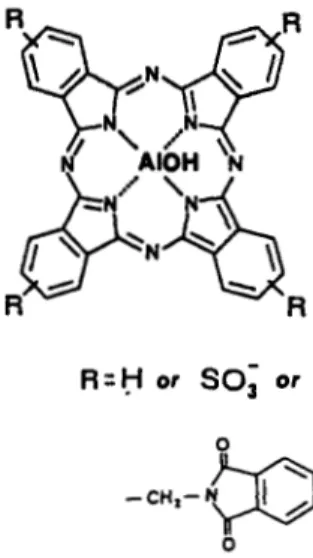
Synthesb. Sulfonated aluminum phthalocyanine was prepared as previously described (Ali er al., 1988). Sulfon- ated phthalimidomethyl aluminum phthalocyanine was prepared as follows: 1.34 g (2.32 mmol) of aluminum phthalocyanine (Ciba Geigy) was added to 5 mL of con- centrated sulfuric acid. the mixture was stirred for 20 min
0
Figure 1. Chemical structure of sulfonated phthalimido- methyl aluminum phthalocyanines (AIOH-PcSP).
R
= H , SO; or CH,-phthalimide, depending on the degree ofsubstitution and sulfonation.
then 0.41 g (2.79 mmol) phthalimide, 0.12 g (4 mmol) par- aformaldehyde, and 15 mL of oleum containing 30% free SO, were added. The mixture was stirred at 80°C for 5 h. The reaction mixture was poured on crushed ice (200 mL) and the resulting precipitate recovered by filtration, washed with 1
M
H2S04 (200 mL), redissolved in 1M
KOH and filtered again before being neutralized. After removal of water by evaporation in vacuo, salt was removed from the dried product by dissolution in dry methanol and filtration to yield AIOH-PcSP (1.67 g). Molar absorption coefficients of the AIOH-PcSP (c,,,, = 1.5 x I F at A,,, = 676 nm) were slightly lower than those found for the AIOH-PcS (eMEOH = 1.9 x 10⁵ at A,,,
= 674 nm).
Fractionarion of sulfonated phthalimidomethyl aluminum
phthalocyanine (AIOH-PcSP). “Crude” AIOH-PcSP (1.1
g) was fractionated by preparative medium pressure reverse phase chromatography on a 30 cm long by 2 cm ID glass column packed with (2-18 reverse phase, particle size 25-40 pm (Macherey-Nagel, Diiren, Germany) using a linear gradient from 0 to 95% MeOH in 10
mM
sodium phosphate buffer, pH 5 (Ali et al., 1988). Samples were analyzed by reverse phase HPLC, eluting phthalocyanines being detected spectroscopically at 670 nm, and pooled to yield six main fractions (A-F). The relative level of substitution was determined by oxidative degradation of 1 mg of compound using cerium ammonium nitrate (0.01 mmol; 4-5 mL) in hot, acidic aqueous solution, followed by reverse phase HPLC analysis (Ali et al., 1988). The eluting phthalimide, and substituted phthalimide units, resulting from the oxidation, were detected spectroscopi- cally at 215 nm. The identity of phthalimide degradation products was determined by coupled HPLUmass spec- trometry. Unsulfonated material (phthalimideR,
= 29 min; phthalimidomethylphthalimideR,
= 35 min) was characterized by direct comparison of chromatographic properties with those of authentic samples, whereas the sulfonated moieties were found indirectly by HPLC com- parison of AICI-PcP and AIOH-PcSP degradation frag- ments (sulfophthalimideR,=
16 min; sulfophthal- imidomethylphthalimideR,
= 20 min). Under the reaction conditions some sulfonation of the benzenoid ring of the phthalimidomethyl group also resulted. The level of this sulfophthalimidomethyl moiety in each fraction was found to be constant with a relative value of 0.2 mol per mol Pc which was taken into account as sulfonate substitution in the molar composition of the various frac-tions. In this manner the following compositions were assigned to fractions A-F: A
(R,
= 18 min), AIOH- PcS,.,P0.,; B(R,
= 20 min), A I O H - P C S ~ , ~ P ~ . ~ ; C(R,
=25
min),
A I O H - P C S ~ . ~ P ~ , ~ ; D(R,
= 28-33 min), AIOH- P&.2P0.4; E(R,
= 37 min), AIOH-Pc&,,P,,,; F(R,
= 40 min), AIOH-PCS~,,P,,~. For the in vitro studies only fractions D and F were evaluated.Partitioning studies. Dyes (25-300 nM) were partitioned
between n-octanol and 100 mM Tris buffer pH 7.4. After shaking for 2 min, the phases were separated by centrifug- ation. The dye concentration in each phase was measured by fluorescence, after &fold dilution in 100% methanol, using a SLM-Aminco SPF-5OOC spectrofluorometer (AIOH-PcS: A,, = 664 nm, ,A, = 684 nm; AIOH-PcSP:
A,, = 666 nm,
,
A
,
= 686 nm).Cell uptake. 3 x 10⁶ Chinese hamster lung fibroblasts
cells (line V-79) were plated in 60 mm Petri dishes. After an incubation period of 3 h under 5% CO,, to allow cell attachment, 10 KM dye in medium (1 mL) containing 1% serum was added and the cells were incubated for 1 h. Absorbed dyes were extracted and quantified as previously described (Paquette el al., 1990). Briefly, after removal of the medium, cells were washed three times with PBS and detached with 600 pL trypsin-EDTA 0.25% (4-5 min incubation) and dishes were washed with 600 pL PBS. Cells were collected by centrifugation in 1.5 mL plastic tubes (5 min, 600 g) and resuspended in 200 pL of buffer
(Tris
0.2 M, MgCI, 10 mM, CaCI, 1 mM, pH 7.8). After three freezethaw cycles in liquid nitrogen, cell debris was incubated in the dark, overnight, with 0.5 A, unit of proteinase K (Sigma Chemical Co, St. Louis, MO) and diluted in 100 pL of Tris buffer. DNA was digested for 2 h at 37°C with 1 A, unit of micrococcal nuclease (Worthington, Freehold, NJ). To eliminate hydrophobic and ionic interactions with cellular fragments, 50 pL DMF was added, whereafter the mixture was incubated for 30 min followed by the addition of 50 pL of 1.0 N NaOH and a 10 min incubation period at 37°C. After 40-fold dilution in 100% methanol, dye concentrations were meas- ured by fluorescence as described above.Photocytotoxicity assuy. Cell survival of Chinese hamster
lung fibroblasts (line V-79) was determined using a colony forming assay as described by Brasseur et al. (1985). After the attachment of 200 cells in Petri dishes, the cells were rinsed with PBS, and incubated for 1 h with 1 mL of medium containing 1% serum and the dye. After removal of the dye and washing with PBS, the cells were exposed at room temperature for 4 min to red light. The irradiation device consisted of a 500
W
tungsten/halogen lamp equipped with a 10 crn water filter and a red filter (26-4390, Ealing). From the emission spectrum monitored with a monochromator (Instruments S.A., Inc., Jobin Yvon Division, Metuchen,
NJ)
the fluence rate over the absorp- tion peak of the photoactive monomeric dye (Amax f 20 nm) was calculated and set at 100 W/m2 for a final dose of 24 kJ/m2. Experiments were repeated three times using three dishes per concentration point.Quantum yield of singlet oxygen. Singlet oxygen quan-
tum yields were determined for the fractionated product and AIOH-PcS. (Porphyrin Products) in deuterated meth- anol using the infrared luminescence technique (Rodgers and Snowden, 1982). Excitation of the soluton was by a Q switched, frequency doubled ruby laser (347114. Hematoporphyrin hydrochloride (Porphyrin Products) was used as standard, the absolute singlet oxygen quantum yield for this compound having been determined pre- viously by the time resolved thermal lensing technique (Redmond and Braslavsky, 1988).
RESULTS
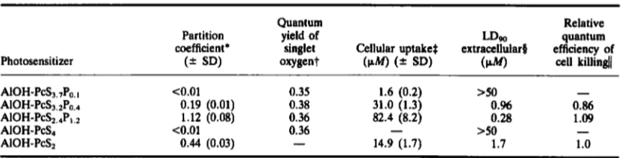
Partition coefficient data (Table 1)
showthat
decreasing the number of sulfonate groups and
Sulfonated phthalimidomethyl aluminum phthalocyanine 325 Table 1. Properties of differently substituted AIOH-PcSP and AIOH-PcS
Photosensitizer
Quantum Relative
Partition yield of LDw quantum
coefficient' singlet Cellular uptake$ extracellular8 efficiency of
(2 SD) oxygent
(PW
(= SD)(W)
cell killindl>50
-
AIOH-PcS3.7Po. I co.01 0.35 1.6 (0.2)
AIOH-P&,2Po,.+ 0.19 (0.01) 0.38 31.0 (1.3) O.% 0.86
A I O H - P C S ~ , ~ P ~ , ~ 1.12 (0.08) 0.36 82.4 (8.2) 0.28 1.09
AIOH-PCS~ co.01 0.36
-
>so
-
AIOH-PCS~ 0.44 (0.03)
-
14.9 (1.7) 1.7 1.0'Dyes (25-300 nM) were partitioned between n-octanol and 100 mM Tris buffer pH 7.4.
tQuantum yields of singlet oxygen were obtained relative to hematoporphyrin dihydrochloride
(k
= 0.64) using the IR $Cellular uptake was measured after incubating Chinese hamster cells (V-79) with 10F M
of dyes in 1% serum media during $The extracellular LDw against V-79 cells were calculated from the survival curves presented in Fig. 2.[ m e relative quantum efficiency of cell killing was calculated from the LDw corrected for the amount of dye in the cells,
luminescence technique. 1 h.
taking the value for AIOH-PcS2 as unity.
increasing the number of phthalimidomethyl sub-
stituents increase the hydrophobicity of the dyes.
Selecting the most hydrophobic of the water soluble
AlOH-PcSP fractions, i.e. fraction
F which con-
tained an average of
2.4
sulfonate and
1.2
phthalimi-
domethyl groups per Pc molecule (AIOH-
P C S ~ . ~ P ~ . ~ ) ,
we find that the effect of incorporation
of the phthalimidomethyl group results in an
increase of the partition coefficient by
2.6
times.
Comparing data for cellular uptake with partition
coefficients (Table l), a relationship between hydro-
phobicity and cell uptake is observed. Higher hydro-
phobicity of dyes correlates well with an improved
cellular uptake of 60 times between AIOH-
PcS3.,P0. and AIOH-PcSz.4P1.z.
The isolated effect
of phthalimidomethyl addition on disulfonated dyes
can be seen from the
5.6times higher cellular uptake
of A I O H - P C S ~ . ~ P ~ . ~
as compared to AlOH-PcSz.
Fraction D, i.e. AIOH-PCS~.~P~.~,
was the only one
which did not follow the hydrophobicity-cellular
uptake relationship. The three sulfonate groups con-
tributed to partitioning the dye more in the aqueous
phase while the addition of an average of
0.4phthal-
imidomethyl allowed a cellular uptake
2.1
times
higher than the amphiphilic AlOH-PcS2. This
increase in cellular uptake corresponds with an
increase in photocytotoxicity of the dyes
invitro
(Fig.
2).
A ~ O H - P C S ~ . ~ P ~ . ~
and A I O H - P C S ~ . ~ P ~ . ~
exhibited LDw values (extracellular)
of
0.96
and
0.28
p Mrespectively, while AIOH-PcS3,,P0, failed
to achieve
90%
cell mortality even at the highest
concentrations used (50
p M ) .The influence of the
phthalimidomethyl group on photocytotoxicity was
clearly indicated by comparing A I O H - P C S ~ . ~ P ~ . ~
with AlOH-PcSz where inclusion of the phthalimi-
domethyl moiety resulted in an increased photo-
dynamic potency of more than &fold (AlOH-PcSz,
LDw
=1.7
p M ;
Paquette et
al.,
1988).
The dif-
fering phototoxic effects do not relate to the
efficiency of singlet oxygen production for the
PAP 5313-C
0 0.2 OA
ae
od 1.0 13 Coneontratlon pMFigure 2. Survival of
V-79
cells incubated for 1 h at37°C
with AIOH-PcS3.zP,.4 (0) and A I O H - P C S ~ . ~ P , . ~(A)
fol- lowed by exposure t o red light. Experiments were repeated three times using three dishes per concentration points.monomerized fractions of the various AIOH-PcSP
in homogeneous solution, since all the dye prep-
arations gave similar quantum yields (Table
1).
The
effect of inclusion of phthalimidomethyl groups
in
AIOH-PcS did correlate very well with the relative
quantum efficiency of cell killing of the various dye
preparations (Table
1).
After expressing photo-
toxicity as a function of cellular uptake, AIOH-PcSz
and A I O H - P C S ~ . ~ P ~ . ~
exhibited the same relative
efficiency, while AIOH-PCS~.~P~.,
was only
14%
less
phototoxic.
DISCUSSION
These results show clearly that the higher cellular
uptake of differently substituted AIOH-PcSP
invitro
is associated with the more hydrophobic
derivatives of sulfonated phthalocyanines. Compar-
ing dye fractions AIOH-PC$,,P,,~ and AIOH-PcS2,
the fraction substituted with phthalimidomethyl
groups partitioned more in the n-octanol phase,
and exhibited both increased cellular uptake and
photocytotoxicity by about a factor of six without
showing significant differences in the quantum yield
of singlet oxygen.
Thus,
the strategy of limiting
the degree of sulfonation to obtain an amphiphilic
photosensitizer, whilst increasing the number of
phthalimidomethyl substituents to increase hydro-
phobicity, and hence membrane penetration, results
in an increased photodynamic effect.
It has been reported that amphiphilic dyes inter-
act with the lipid portion of plasma membranes
(Chatelier et
al., 1985), thereby facilitating transport
of these molecules into the cell (Paquette
eral.,
1988).
This same amphiphilic property allows more
efficient binding to low density lipoprotein (LDL),
which has been implicated in the transport of por-
phyrin based sensitizers to tumorous tissue (Kessel
er
al., 1987). Tetraphenylporphine monosulfonate,
and the more hydrophobic of the disulfonated iso-
mers bound to a greater extent with LDL, and
were preferentially localized in neoplastic cells. In
contrast, the tetra-, tri- and less hydrophobic disul-
fonated isomer were associated more with the albu-
min fraction of the serum proteins and distributed
mainly in the stroma of the tumor. Although little
additional data has been published to corroborate
these results, with regard to phthalocyanines, it
might be expected on the basis
of sensitizer struc-
ture, that both tetraphenylporphine and Pc’s, with
the same degree of sulfonation, would exhibit simi-
lar affinities for the different serum lipoproteins and
thence be distributed in a like manner. Thus the
probability of a direct tumor response should be
greater if the hydrophobicity of the molecule is
increased whilst retaining water solubility.
Addition of hydrophobic phthalimidomethyl
groups to AlOH-PcS results in a dramatic increase
in cell uptake. A one hour incubation period of
V-
79
cells with 10
F M
AI-OH-PCS~.~P~.~
did result
in an 8-fold higher intracellular as compared to
extracellular, dye concentration (Table 1). The
phthalimidomethyl group did not however, appear
to modify the intracellular distribution of the dye
as
the relative quantum efficiency of cell killing of
AIOH-PCS~.,P~.~
and A10H-PcS2 were similar.
Kessel (1986) and Moan et
al. (1983) have
reported that plasma membrane is an important
target following
in vitroPDT. This assumption on
the nature of the intracellular target is supported
by a correlation between plasma membrane damage
and
loss of cell viability after a short incubation
period with HpD. Our results with both AIOH-
PcS~..,P~.~
and AIOH-PCS~.~P,,.~
were consistent with
this assumption. A more hydrophobic amphiphilic
dye, such as AIOH-PCS~.~P~.~,
should interact with
the lipid portion of plasma membrane (Chatelier
etal.,
1985).
It therefore seems logical to obtain a
higher membrane interaction, and thus optimal cell
uptake, by increasing the hydrophobicity of an
amphiphilic dye. In the case of AIOH-PCS~.~P~.~,
the relative yield of cell killing was only 14% lower
than that observed for AIOH-Pcs. This result
seemed surprising, since it has been previously
established that phthalocyanines substituted with
three sulfonate groups interact poorly
in virrowith
the plasma membrane of V-79
cells, resulting, even
after
24h incubation, in little cell uptake and photo-
cytotoxicity (Brasseur
etal., 1987; Paquette
eral.,
1988).
The relatively low partition coefficient of
A I O H - P C S ~ . ~ P ~ . ~ ,
as compared to the amphiphilic
AIOH-PcS2, also suggests a weak interaction with
the membrane, however, both the cellular uptake
and phototoxic effect were improved. Thus, these
results indicate that the addition of phthalimidome-
thy1 groups on AIOH-PCS~.~P,,.~
compensates for the
adverse hydrophilic effect of the sulfonate sub-
stituents, and induces phototoxicity by permitting
interaction with the plasma membrane, sub-
sequently mediating photoinduced damage to the
tumor cell.
In summary, these
in virrodata with V-79 cells
confirm the correlation between hydrophobicity,
cellular uptake and cell killing of the photo-
sensitizer, and demonstrate the relevance of intro-
ducing phthalimidomethyl groups into amphiphilic
sulfonated phthalocyanines to improve cellular
uptake and photodynamic action. The involvement
of this correlation, with regard to both serum distri-
bution of dye and tumor response
in vivo,is cur-
rently being investigated in our laboratory.
Acknowledgements-This work was supported by the Medical ResearchCouncil
of Canada. R. W. B. thanks the C. K. Marr Educational Trust for a scholarship. The authors thank Dr. C. Campbell and Professor I. MacPher- son for considerable advice on Pc synthesis and Miss Huguette Savoie for expert technical assistance.REFERENCES
Ali, H., R. Langlois, J. R. Wagner, N. Brasseur, B. Paquette and J. E. van Lier (1988) Biological activities of phthalocyanines
-
X. Syntheses and analyses of sulfonated phthalocyanines. Phorochem. Phorobiol. 44,Ben-Hur, E. and I. Rosenthal(1985) Photosensitized inac- tivation of Chinese hamster cells by phthalocyanines. Phorochem. Photobiol. 42,
192-193.
Brasseur, N., H. Ah, D. Autenrieth, R. Langlois and J.
E. van Lier
(1985)
Biological activities of phthalocyanines-111. Photoinactivation ofV-79
Chi- nese hamster cells by tetrasulfophthalocyanines. Phoro- chem. Photobiol. 42,515-521.
Brasseur, N., H. Ali, R. Langlois and J. E. van Lier
(1987)
Biological activities of phthalocyanines-VII. Photoinactivation of V-79 Chinese hamster cells by selectively sulfonated phthalocyanines. Phorochem. Photobiol. 45,587-594.
Chan, W. S., R. Svensen, D. Phillips and I. R. Hart (1986)
Sulfonated phthalimidomethyl aluminum phthalocyanine 327 Cell uptake, distribution and response to aluminum
chloro sulphonated phthalocyanine, a potential antitu- mour photosensitizer. Br. J. Cancer 53, 255-263. Chatelier, R. C., W. H. Sawyer, A. G. Swincer and A. D.
Ward (1985) Merhods in Porphyrin Photosensitization
(Edited by D. Kessel), p. 169. Plenum Press, New York. Dougherty, T. J., G. Lawrence, J. E. Kaufman, D. G. Boyle, K. R. Weishaupt and A. Mittelman (1978) Pho- toradiation therapy for the treatment of malignant tumors. Cancer Res. 36, 262b2635.
Fingar, V. H. and B. W. Henderson (1987) Drug and light dose dependence of photodynamic therapy: a study of tumor and normal tissue response. Phorochem. Phoro- Girotti, A. W. (1983) Mechanism of photosensitization.
Photochem. Photobiol. 38, 745-751.
Henderson, B. W. and D. A. Bellnier (1989) Tissue local- ization of photosensitizers and the mechanism of photo- dynamic tissue destruction. In Photosensitizing Com-
pounds: Their Chemistry, Biology and Clinical Use
(Edited by G. Bock and
S.
Harnett), Ciba Foundation Symposium 146, pp. 112-125. Wiley, Chichester. Kessel, D. (1986) Sites of photosensitization by derivativesof hematoporphyrin. Phorochem. Phorobiol. 44, 489-493.
Kessel, D., P. Thompson, K. Saatio and K. D. Nantwi (1987) Tumor localization and photosensitization by sul- fonated derivatives of tetraphenylporphine. Photochem.
Photobiol. 45, 787-790.
Langlois, R., H. Ah, N. Brasseur, R. Wagner and J. E. van Lier (1986) Biological activities of phthalocyanines-IV. Type I1 sensitized photooxidation of L-tryptophan and cholesterol by sulfonated metal- lophthalocyanine. Photochem. Phorobiol. 44, 117-125. Lier, J. E. van (1990) Phythalocyanines as sensitizers for
PDT of cancer. In Photodynamic Therapy of Neoplasric Disease (Edited by D. Kessel), Vol. I, pp. 279-291. CRC Press, Boca Raton, FL.
Lier, J. E. van and J. D. Spikes (1989) The chemistry, photophysics and photosensitizing properties of phthalo- cyanines. In Photosensitizing Compounds: Their Chem-
istry, Biology and Clinical Use (Edited by G. Bock
and S. Harnett), Ciba Foundation Symposium, 146, pp. 17-26. Wiley, Chichester.
bid. 46, 837-841.
Moan, J., T. Christensen and P. B. Jacobsen (1984) Photo- dynamic effects on cells in virro labelled with hemato-
porphyrin derivative. Photobiochem. Photobiophys. 7, 349-358.
Moan, J., J. McGhie and P. B. Jacobson (1983) Photodyn- amic effects on cells in vitro exposed to hematoporphy- rin derivative and light. Phorochem. Phorobiol. 37, 599-604.
Paquette, B., H. Ali, R. Langlois and J.
E.
van Lier (1988) Biological activities of phthalocyanines-VIII. Cellular distribution in V-79 Chinese hamster cells and phototoxicity of selectively sulfonated aluminum phthal- ocyanines. Phorochem. Phorobiol. 47, 215-220. Paquette, B., H. Ali, R. Langlois and J. E. van Lier(1990) Biological activities of phthalocyanines-XI. Phototoxicity of sulfonated aluminum naphthalocyan- ines towards
V-79
Chinese hamster cells. Phorochem.Photobiof. 51, 313-318.
Redmond, R.
W.
and S. E. Braslavaky (1988) Phorosensir-izarion. Molecular, Cellular and Medical Aspects (Edited
by G. Moreno, R. H. Pottier and T. G. Truscott), Nato AS1 Series, Vol. H15, pp. 93-97. Springer, Berlin. Reed, M.
W.
R., T. J. Wieman, D. A. Schuschke, M. T.Tseng and F.
N.
Miller (1989) A comparison of the effects of photodynamic therapy on normal and tumor blood vessels in the rat microcirculation. Radiation Res. Rodgers, M. A. J. and P. T. Snowden (1982) Lifetime of 02('A,) in liquid water as determined by time-resolved infrared luminescence measurements. J. Am. Chem. Rosenthal, I. and E. Ben-Hur (1989) Phthalocyanines in photobiology. In Phthalocyanines, Properties and Appli-cations (Edited by C. C. Leznoff and A. B. P. Lever),
pp. 393425. VCH, New York.
Selman,
S.
H., M. Kreimer-Bimbaum. J. E. Klaunig, P. J. Goldblatt, R.W.
Keck and S. L. Britton (1985) Blood flow in transplantable tumors treated with hematopor- phyrin derivative and light. Cancer Res. 45, 1924-1927. Star, W. M., H. P. A. Marunissen, A. E. van den BergBlok, J. A. C. Versteeg, K. A. P. Franken and H. S. ,
Reinhold (1986) Destruction of rat mammary tumor and normal tissue microcirculation by hematoporphyrin derivative photoradiation observed in vivo in sandwich observation chambers. Cancer Res. 46, 2532-2540. 119, 542-552.