HAL Id: hal-02446214
https://hal.sorbonne-universite.fr/hal-02446214
Submitted on 20 Jan 2020HAL is a multi-disciplinary open access archive for the deposit and dissemination of sci-entific research documents, whether they are pub-lished or not. The documents may come from teaching and research institutions in France or
L’archive ouverte pluridisciplinaire HAL, est destinée au dépôt et à la diffusion de documents scientifiques de niveau recherche, publiés ou non, émanant des établissements d’enseignement et de recherche français ou étrangers, des laboratoires
perinatally or during childhood
Solène Fastenackels, Delphine Sauce, Corinne Vigouroux, Véronique
Avettand-Fenoel, Jean-Philippe Bastard, Soraya Fellahi, Laura Nailler, Elisa
Arezes, Christine Rouzioux, Josiane Warszawski, et al.
To cite this version:
Solène Fastenackels, Delphine Sauce, Corinne Vigouroux, Véronique Avettand-Fenoel, Jean-Philippe Bastard, et al.. HIV-mediated immune aging in young adults infected perinatally or during childhood. AIDS, Lippincott, Williams & Wilkins, 2019, 33 (11), pp.1705-1710. �10.1097/QAD.0000000000002275�. �hal-02446214�
Concise Communication
HIV mediated immune aging
in young adults infected perinatally
or during childhood
Solène FASTENACKELS1, Delphine SAUCE1, Corinne VIGOUROUX2,3, Véronique
AVETTAND-FÈNOËL4, Jean-Philippe BASTARD2,5, Soraya FELLAHI2,5, Laura NAILLER6,
Elisa AREZES6, Christine ROUZIOUX4, Josiane WARSZAWSKI6, Jean Paul VIARD7, Victor
APPAY1,8, for the ANRS Co19 COVERTE Study Group
1 Sorbonne Université, INSERM, Centre d’Immunologie et des Maladies Infectieuses
(CIMI-Paris), F-75013, Paris, France
2 Sorbonne Université, Inserm UMR_S 938, Centre de Recherche Saint-Antoine, Paris, France
3 Assistance Publique-Hôpitaux de Paris, Hôpital Saint-Antoine, Service d’Endocrinologie,
Diabétologie et Endocrinologie de la Reproduction, Laboratoire Commun de Biologie et Génétique Moléculaires, Centre National de Référence des Pathologies Rares de l’Insulino-Sécrétion et de l’Insulino-Sensibilité (PRISIS), Paris, France
4 APHP, Laboratoire de Virologie, Hôpital Necker, EA 7327, Université Paris Descartes, Paris,
France
5 Assistance Publique-Hôpitaux de Paris, Hôpital Tenon, UF Bio-marqueurs Inflammatoires et
Métaboliques, Service de Biochimie et Hormonologie, Paris, France
6 CESP 1018 INSERM, Team HIV/Pediatry, Le Kremlin-Bicêtre, France, Univ Paris-Saclay,
France, AP-HP Hôpital Bicêtre, Le Kremlin-Bicêtre, France
7 AHP, Centre de Diagnostic et de Thérapeutique, Hôpital de l’Hôtel-Dieu, EA 7327, Université
Paris Descartes, Paris, France
8 International Research Center of Medical Sciences (IRCMS), Kumamoto University, Kumamoto
860-0811, Japan
Address correspondence to:
Victor Appay, CIMI-Paris, INSERM U1135, 91 Bd de l’Hôpital, 75013 Paris, France. Phone: +33-1-40-77-81-83. Email: victor.appay@upmc.fr
Running head: Immune aging in young HIV-1 infected adults
Funding: This work was supported by the French ANRS (Co19 COVERTE Cohort), the FRM (project DEQ20120323690), and Sidaction.
Abbreviations
APC, allophycocyanin; FITC, fluorescein isothiocyanate; PE, phycoerythrin; IFN, Interferon; TNF, Tumor necrosis factor; CMV, cytomegalovirus
Counts
Text: 1742 words abstract: 211 words
Abstract
Background: HIV-infected patients progressing towards disease present a premature immune aging
profile, characterized by the exhaustion of lymphopoiesis. The development of these anomalies may be prevented in young HIV-infected patients owing to their robust immune resources and lymphocyte regeneration capacities.
Methods: An immunomonitoring substudy was designed for young adults aged between 18 and 25
years, living with HIV since childhood included in the national ANRS Co19 COVERTE Cohort. We compared markers associated with immune aging, including the frequency of circulating hematopoietic progenitors and the phenotype of lymphocyte populations, with those of patients infected with HIV in adulthood.
Results: HIV-infected young adults displayed decreasing numbers of CD34+ hematopoietic progenitors and mature lymphocytes, indicative of general lymphopenia and reminiscent of the alterations found in patients infected in adulthood or uninfected elderly people. This highlights the strong impact of HIV on the immune system despite patient young age at infection. Immune aging related alterations were particularly obvious in young patients who presented high viral loads.
Conclusions: HIV-infected young adults can present increased markers of immune activation and
senescence, related to uncontrolled viral replication. This highlights the issue of non-compliance to antiretroviral therapy in patients at a young age, resulting in loss of viral control, premature immunosenescence, and potentially irreversible damage of their lymphopoietic system.
Keywords
INTRODUCTION
Massive depletion of CD4+ T-cells is the hallmark of HIV disease progression. However, this
decline is not the only immune alteration occurring during the course of HIV-1 infection. HIV disease progression is also associated with a marked decrease in primary immune resources
(encompassing CD34+ hematopoietic progenitors[1, 2] and naïve T-cells[3, 4]), the failure to maintain
adequate counts of all types of lymphocytes (including B cells[5, 6] and NK cells[7, 8]) and the
accumulation of highly differentiated T-cells (i.e. CD57+) often considered as cells approaching
cellular senescence[9, 10]. These alterations highlight an exhaustion of lymphopoiesis, i.e. the
capacity to produce or renew the lymphocyte compartment, and reflect a premature process of
immune aging[11]. Systemic immune activation has emerged as a major driver of this altered
lymphopoiesis, which can therefore be limited owing to antiretroviral therapy[1]. Nonetheless, HIV
disease progression despite viral suppression, naturally (i.e. in HIV controllers) or due to antiretroviral treatment (i.e. also referred as to immunological failure), was associated with
profound and persistent exhaustion of lymphopoiesis[1]. This highlights further the importance of
preserving lymphopoietic capacity in HIV infected patients, which is a primary determinant for the
maintenance of adequate CD4+ T-cell levels during HIV infection.
In this context, young adults infected with HIV at birth or during childhood (YAHIC) represent a particularly interesting group to study. The development of immune aging-like anomalies related to HIV infection may possibly be limited in these patients because of the large immune resources and
robust lymphocyte regeneration capacity associated with young age[12]. We therefore decided to
assess the impact of HIV infection on the immune system of YAHIC, in comparison with patients infected later during adulthood, and age-matched uninfected donors. For this purpose, we analyzed
HIV replication, HIV-DNA levels and blood frequencies of CD34+ hematopoietic progenitor cells,
Immunological studies in these young HIV-infected patients provide important insights into the consequences of the infection at early stages of life onwards, which is important for the care of this vulnerable population.
MATERIALS AND METHODS Study subjects and samples
Blood samples of young adults living with HIV since childhood (n=134) were obtained from the ANRS COVERTE-CO19, a national prospective cohort of adults diagnosed with HIV infection before 13 years of age, enrolled in 88 sites since 2010, annually followed, with multiple clinical and
physiopathological objectives[13, 14]. Patients were infected perinatally or during childhood, and
were diagnosed with HIV on average three years after birth (with a 4 year SD) and were aged 18 to 25 years at study inclusion. Frozen PBMC samples (n=84) were collected for in depth immunological evaluation at inclusion. For comparison, blood samples of older adults aged 25 to 55 years (n=61) or aged 65 to 81 years (n=28) infected with HIV-1 during adulthood were obtained from patients attending HIV clinics in France and treated with antiretroviral therapy for more than 3 years. Blood samples were also obtained from age-matched uninfected individuals, including young (n=24), middle aged (n=34), and elderly (n=13) adults. Elderly individuals with malignancies, acute diseases, or advanced stages of severe chronic diseases, such as chronic inflammatory disease, atherosclerotic disease, congestive heart failure, poorly controlled diabetes mellitus, renal or hepatic disease, or chronic obstructive pulmonary disease, as well as individuals under immunosuppressive therapy were excluded from the study. All participants gave their written informed consent. The study received the approval of the local institutional ethics committees (at Cochin and Pitié Salpêtrière Hospitals, Paris). Peripheral blood mononuclear cells (PBMCs) were isolated over a Lymphoprep gradient and cryopreserved until use. High-sensitivity C-reactive protein (hsCRP) was
measured from frozen serum samples by immunonephelometry on an IMMAGE analyzer (Beckman-Coulter, Brea, California, USA).
Quantification of plasma HIV-RNA and cell-associated HIV-DNA
HIV-RNA was quantified in blood plasma with the Cobas Ampliprep Cobas Taqman assay v2 (Roche, France). Total DNA was extracted from thawed PBMCs by using the QIAamp DNA microkit (Qiagen, Courtaboeuf, France) and HIV-DNA was quantified with an ultrasensitive real-time PCR method (Generic HIV-DNA assay, Biocentric, Bandol France) with a detection limit of
five copies per PCR[15]. To standardize results, the total DNA in extracts was quantified using
fluorescence readings at 260 nm (Nanodrop, Labtech,Ringmer, UK). Each entire DNA extract was
tested in two replicates. Results were reported as the number of HIV-DNA copies per 106 PBMCs.
DNA extracts were stored at -20°C.
Flow cytometry
Directly conjugated antibodies were obtained from the following vendors: BD Biosciences (San Jose, CA): CD4 (APC-cyanin7), CCR7 (PE-Cy7), CD38 (APC), CD21 (PE-Cy7), CD34 (FITC and PE), CD56 (FITC), CD45RA (V450), Ki67 (FITC), and lineage cocktail (CD3, CD14, CD16, CD19, CD20, CD56 / FITC); Beckman Coulter: CD45 (ECD), CD57 (FITC); Caltag (Burlingame, CA): CD8 (Alexa405); Dako (Glostrup, Denmark): CD3 (Cascade Yellow); BioLegend (San Diego, CA): CD27 (AlexaFluor700). Stainings were performed on thawed PBMCs by addition of the live/dead Aqua marker (Thermofisher, Illkirch, France) and the respective antibodies for 15 min at room temperature. After incubation, stained PBMCs were washed in PBS and then fixed with 2%
paraformaldehyde. CD4/CD8 ratios were calculated based on the absolute counts of CD4+ and
LSR Fortessa flow cytometer (Becton Dickinson) and data analyzed using FlowJo v10.6 (Tree Star, Inc).
Statistical analyses
Statistical analysis was performed using GraphPad prism software. Groups were compared using the non-parametric Kruskal Wallis or Mann-Whitney tests. Spearman’s rank test was used to determine correlations. P values above 0.05 were considered not significant.
RESULTS
Altered lymphopoiesis and premature immune aging in YAHIC
Young adults HIV infected during childhood (YAHIC) were aged between 18 and 25 years and all received antiretroviral therapy. For the analyses, patients were divided according to disease
progression stage (i.e. CD4+ T-cell count >500, <500-200> and <200 cells/µl) and compared with
equivalent groups of older HIV-infected patients, including middle aged (between 25 and 55 years - mean of 44 y) and old (above 65 years - mean of 70 y) individuals, as well as age-matched HIV-uninfected adult controls. All HIV-infected patient age groups presented with equivalent time since diagnosis (mean of 17 y) and time since treatment initiation (mean of 11 y).
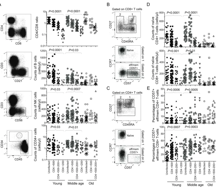
Similar to patients infected with HIV during adulthood, YAHIC displayed decreasing CD4/CD8
ratio, numbers of progenitor cells (CD34+CD45+Lin-), as well as B (CD21+) and NK (CD56+CD3-)
lymphocytes with lowering CD4+ T-cell counts (Figure 1A). This suggests a disruption of
lymphopoiesis with disease progression in YAHIC. Decreasing naive
(CD45RA+CD27+CCR7+CD57-) CD4+ or CD8+ T-cell numbers characterized also YAHIC as they
parallel with reduced naive T-cell counts, frequencies of highly differentiated CD57+
effector/memory CD4+ or CD8+ T-cells, often considered as a marker of immune senescence, were
significantly increased, compared to age-matched healthy donors. This parameter reflects the impact on the T-cell compartment of persistent viruses, like HIV, but also CMV, which YAHIC were frequently (i.e. 80%) co-infected with, and which is a major driver of the accumulation of these cells. Altogether, these data support the development of a premature immune aging profile associated with HIV disease progression in YAHIC despite their young age and a priori robust immune resources.
Persistence of strong viral replication in YAHIC
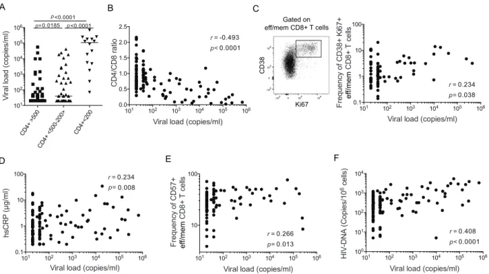
The establishment of a premature immune aging profile in HIV-infected patients has been linked to elevated chronic systemic activation, which results in particular from HIV replication. Unexpectedly, a high proportion (33%) of YAHIC presented with viral replication. These
individuals included in particular patients progressing towards disease (i.e. with CD4+ T-cell counts
below 200 cells/µl), in whom plasma viral load was always detectable (Figure 2A). This was in clear contrast with older HIV-infected patients, who had no detectable viral replication (data not shown). In YAHIC, viral replication was inversely correlated with the CD4/CD8 ratio (Figure 2B),
low values of which are classically associated with immune activation and declining immunity[16].
We found associations between viral replication and the frequency of Ki67+CD38+ memory CD8+
T-cells (Figure 2C), and serum levels of hsCRP (Figure 2D), which are established markers of immune activation and inflammation in HIV-infected patients. High plasma viral load was also
correlated with the frequency of CD57+ CD8+ T-cells (Figure 2E). Last, viral replication correlated
with cell-associated HIV-DNA levels in YAHIC (Figure 2F). Altogether, these data suggest that uncontrolled viral replication in YAHIC is a main cause of disease progression, elevated immune activation, resulting in a premature immune aging profile, as well as seeding of the HIV reservoir.
DISCUSSION
Young adults infected with HIV at birth or during early childhood represent a small yet very particular group of the HIV infected population, due to their young age but long experience of HIV. We report here that YAHIC display a general exhaustion of lymphopoiesis with decreasing counts
of CD34+ hematopoietic progenitors, naïve CD4+ and CD8+ T-cells, B-lymphocytes and NK cells,
and increased frequency of memory senescent T-cells as they progress towards disease. These alterations are similar to those observed in older HIV-infected patients and reminiscent of a premature immune aging process despite their young age. This highlights the potential strong impact of HIV infection on the immune system despite a priori robust immune resources. Immune
aging-like alterations were most obvious in YAHIC with lowest CD4+ T-cell counts, who also had
high viral loads and chronic activation markers, in contrast to older treated HIV-infected patients. While the establishment of a premature immune aging profile in HIV-infected adults with good
viral suppression is likely due to long term exhaustion of primary immune resources[1], our present
data suggest that its primary cause in YAHIC is uncontrolled viral load and related immune activation.
This situation in YAHIC underlines the problem of non-compliance to antiretroviral therapy in this age group. In young HIV-infected patients prescribed anti-HIV drugs, this represents a rather
frequent and particularly sensitive issue[13]. Lack of adherence to antiretroviral treatment not only
results in loss of viral control and increased risk of HIV transmission, but our data indicate that it can also promote HIV reservoir seeding and the establishment of premature immunosenescence in YAHIC, therefore potentially leading to irreversible damage to the lymphopoietic system. It is thus very important to enforce treatment compliance in teenagers and young adults, in order to prevent
the one of elderly people. This is essential not to jeopardize hopes for remission in these young patients. These should serve as strong arguments to convince HIV-infected teenagers and young adults to adhere to antiretroviral therapy recommendations.
AUTHOR CONTRIBUTION
Solène FASTENACKELSperformed the research and analyzed data.
Delphine SAUCE performed the research and analyzed data. Corinne VIGOUROUX designed the research and analyzed data.
Véronique AVETTAND-FÈNOËL performed the research and analyzed data. Jean-Philippe BASTARD performed the research and analyzed data.
Soraya FELLAHI performed the research and analyzed data. Laura NAILLER analyzed clinical data and recruited patients. Elisa AREZES analyzed clinical data and recruited patients.
Christine ROUZIOUXdesigned the research and wrote the paper.
Josiane WARSZAWSKI designed the research and wrote the paper. Jean Paul VIARD designed the research and wrote the paper.
Victor APPAY designed the research, analyzed data and wrote the paper.
ACKNOWLEDGEMENTS
We are very grateful to the patients and staff of the Infectious Diseases and Internal Medicine departments participating in patient recruitment in France. We thank Laura Papagno for technical assistance.
REFERENCES
1. Sauce D, Larsen M, Fastenackels S, Pauchard M, Ait-Mohand H, Schneider L, et al. HIV disease progression despite suppression of viral replication is associated with exhaustion of lymphopoiesis. Blood 2011; 117(19):5142-5151.
2. Menkova-Garnier I, Hocini H, Foucat E, Tisserand P, Bourdery L, Delaugerre C, et al. P2X7 Receptor Inhibition Improves CD34 T-Cell Differentiation in HIV-Infected Immunological Nonresponders on c-ART. PLoS pathogens 2016; 12(4):e1005571.
3. Roederer M, Dubs JG, Anderson MT, Raju PA, Herzenberg LA. CD8 naive T cell counts decrease progressively in HIV-infected adults. J Clin Invest 1995; 95(5):2061-2066.
4. Appay V, Fastenackels S, Katlama C, Ait-Mohand H, Schneider L, Guihot A, et al. Old age and anti-cytomegalovirus immunity are associated with altered T-cell reconstitution in HIV-1-infected patients. Aids 2011; 25(15):1813-1822.
5. Moir S, Malaspina A, Ogwaro KM, Donoghue ET, Hallahan CW, Ehler LA, et al. HIV-1 induces phenotypic and functional perturbations of B cells in chronically infected individuals.
Proc Natl Acad Sci U S A 2001; 98(18):10362-10367.
6. Moir S, Malaspina A, Ho J, Wang W, Dipoto AC, O'Shea MA, et al. Normalization of B cell counts and subpopulations after antiretroviral therapy in chronic HIV disease. J Infect Dis 2008; 197(4):572-579.
7. Alter G, Teigen N, Davis BT, Addo MM, Suscovich TJ, Waring MT, et al. Sequential deregulation of NK cell subset distribution and function starting in acute HIV-1 infection.
Blood 2005; 106(10):3366-3369.
8. Brunetta E, Hudspeth KL, Mavilio D. Pathologic natural killer cell subset redistribution in HIV-1 infection: new insights in pathophysiology and clinical outcomes. J Leukoc Biol 2010; 88(6):1119-1130.
9. Brenchley JM, Karandikar NJ, Betts MR, Ambrozak DR, Hill BJ, Crotty LE, et al. Expression of CD57 defines replicative senescence and antigen-induced apoptotic death of CD8+ T cells.
Blood 2003; 101(7):2711-2720.
10. Papagno L, Spina CA, Marchant A, Salio M, Rufer N, Little S, et al. Immune Activation and CD8(+) T-Cell Differentiation towards Senescence in HIV-1 Infection. PLoS Biol 2004; 2(2):E20.
11. Appay V, Sauce D. Assessing immune aging in HIV-infected patients. Virulence 2017; 8(5):529-538.
12. Blanche S, Scott-Algara D, Le Chenadec J, Didier C, Montange T, Avettand-Fenoel V, et al. Naive T lymphocytes and recent thymic emigrants are associated with HIV-1 disease history
in french adolescents and young adults infected in the perinatal period: the ANRS-EP38-IMMIP study. Clin Infect Dis 2014; 58(4):573-587.
13. Dollfus C, Le Chenadec J, Faye A, Blanche S, Briand N, Rouzioux C, et al. Long-term outcomes in adolescents perinatally infected with HIV-1 and followed up since birth in the French perinatal cohort (EPF/ANRS CO10). Clin Infect Dis 2010; 51(2):214-224.
14. Arrive E, Viard JP, Salanave B, Dollfus C, Matheron S, Reliquet V, et al. Metabolic risk factors in young adults infected with HIV since childhood compared with the general population. PLoS One 2018; 13(11):e0206745.
15. Avettand-Fenoel V, Chaix ML, Blanche S, Burgard M, Floch C, Toure K, et al. LTR real-time PCR for HIV-1 DNA quantitation in blood cells for early diagnosis in infants born to seropositive mothers treated in HAART area (ANRS CO 01). J Med Virol 2009; 81(2):217-223. 16. Serrano-Villar S, Sainz T, Lee SA, Hunt PW, Sinclair E, Shacklett BL, et al. HIV-infected individuals with low CD4/CD8 ratio despite effective antiretroviral therapy exhibit altered T cell subsets, heightened CD8+ T cell activation, and increased risk of non-AIDS morbidity and mortality. PLoS pathogens 2014; 10(5):e1004078.
FIGURES AND LEGENDS
Figure 1. Immune parameters of YAHIC and older HIV infected patients grouped according to CD4 counts
Immunophenotyping of HIV-1-infected patients grouped according to age (young or YAHIC, 18 to 25 years old (black symbols); middle aged, 25 to 55 years old (grey symbols), and old, 65 to 81
years old (white symbols)) and CD4+ T-cell counts: above 500 (square symbols), between 200 and
500 (upward triangle symbols), or below 200 CD4+ T-cells/µl (downward triangle symbols), and
their respective age matched HIV uninfected controls (circle symbols). (A) CD4/CD8 ratio,
absolute counts of B cells, NK cells, CD34+ cells are shown in all groups. Left panels show
A B 0.01 0.1 1 10 0 250 500 750 1000 1250 0 200 400 600 800 1000 200 400 600 800 0 200 400 600 0 10 20 30 40 50 0 20 40 60 80 100 CD4 >50 0 CD4 <500-200 > CD4 < 20 0 CD4 >50 0 CD4 <500-200 > CD4 < 20 0 CD4 >50 0 CD4 < 500-200 >
Uninfected Uninfected Uninfected
Young Middle age Old Young Middle age Old
P<0.0001 P<0.0001 P=0.03 P=0.03 P<0.0001 P=0.001 P=0.0006 P=0.0007 Uninfected CD4 >50 0 CD4 <500-200 > CD4 < 20 0 Uninfected CD4 >50 0 CD4 <500-200 > CD4 < 20 0 Uninfected CD4 >50 0 CD4 < 500-200 > 0.1 1 10 100 P<0.0001 P=0.03 P=0.0007 P=0.01 P<0.0001 P=0.01 P=0.0005 P=0.0003 CD4/CD8 ratio Counts of B cells (cells/µl) Counts of NK cells (cells/µl) Counts of CD34+ cells (cells/µl) Counts of naïve CD8+ T-cells (cells/µl) Percentages of CD57+ f/mem ef CD4+ T-cells Percentages of CD57+ f/mem ef CD8+ T-cells Counts of naïve CD4+ T-cells (cells/µl) D E CD4 CD8 CD3 CD21 CD3 CD56 CD34 CD45 CCR7 CD57 CD27 CD45RA CD27 CD45RA CCR7 CD57 Gated on CD8+ T cells Gated on CD4+ T cells Naïve Naïve eff/mem CD57+ eff/mem CD57+ Gated on 1 Gated on 2 Gated on 1 Gated on 2 1 2 1 2 C
representative flow cytometry stainings. (B, C) Representative flow cytometry stainings and gatings
to identify naïve (i.e. CD45RA+ CCR7+ CD27+ CD57-) or CD57+ effector/memory among total
CD8+ (B) or CD4+ (C) T lymphocytes. (D) Absolute counts of naïve CD8+ and CD4+ T-cells. (E)
Percentages of CD57+ effector/memory CD4+ and CD8+ T-cells. Bars indicate the median.
Statistical differences were calculated using the non-parametric Kruskal Wallis test, which compares together and ranks the four groups of young or middle aged subjects.
Figure 2. Relationship between viral replication, progression, activation and reservoir size in YAHIC
(A) Viral load in YAHIC grouped according to CD4+ T-cell counts. The non-parametric Mann-Whitney test was used to compare groups with one another. Correlations between viral load and (B)
CD4/CD8 ratio, (C) frequency of Ki67+CD38+ memory CD8+ T-cells (the left panel shows a
representative flow cytometry staining), (D) serum hsCRP levels, (E) frequency of CD57+ memory
CD8+ T-cells, (F) cell-associated HIV-DNA in YAHIC. The Spearman’s rank test was used to
determine correlations. 0.013 p= A B CD4+ >50 0 CD4 + <500 -200 > CD4+<20 0 101 102 103 104 105 106 V ira l l oad ( cop ie s/ m l) 101 102 103 104 105 106 0.0 0.5 1.0 1.5 2.0 2.5
Viral load (copies/ml)
CD 4/ CD 8 ra tio 101 102 103 104 105 106 10 100
Viral load (copies/ml)
F requen cy o f CD 57 + ef f/mem CD 8+ T c ell s C 101 102 103 104 105 106 0.1 1 10 100 0.038 0.234 p= r =
Viral load (copies/ml)
F requen cy o f CD 38 + K i67+ ef f/mem CD 8+ T c ell s 101 102 103 104 105 106 100 101 102 103 104
Viral load (copies/ml)
6 c ell s) 0.0001 0.408 p< r = 0.266 r = 0.0001 -0.493 p< r = D <0.0001 <0.0001 0.0185 p p p= 0.008 p= 0.234 r = hsCR P (µg/ml) E F 101 102 103 104 105 106 0.1 1 10 100
Viral load (copies/ml)
HIV-DN A ( C op ie s/ 10 CD38 Ki67 Gated on eff/mem CD8+ T cells