HAL Id: hal-01742935
https://hal.archives-ouvertes.fr/hal-01742935
Submitted on 26 Mar 2018
HAL is a multi-disciplinary open access
archive for the deposit and dissemination of
sci-entific research documents, whether they are
pub-lished or not. The documents may come from
teaching and research institutions in France or
abroad, or from public or private research centers.
L’archive ouverte pluridisciplinaire HAL, est
destinée au dépôt et à la diffusion de documents
scientifiques de niveau recherche, publiés ou non,
émanant des établissements d’enseignement et de
recherche français ou étrangers, des laboratoires
publics ou privés.
Coupling between Inclusions and Membranes at the
Nanoscale
Florent Bories, Doru Constantin, Paolo Galatola, Jean-Baptiste Fournier
To cite this version:
Florent Bories, Doru Constantin, Paolo Galatola, Jean-Baptiste Fournier. Coupling between Inclusions
and Membranes at the Nanoscale. Physical Review Letters, American Physical Society, 2018, 120 (12),
pp.128104. �10.1103/PhysRevLett.120.128104�. �hal-01742935�
Florent Bories,1 Doru Constantin,2, ∗ Paolo Galatola,1, † and Jean-Baptiste Fournier1, ‡
1Laboratoire “Matière et Systèmes Complexes” (MSC), UMR 7057 CNRS,
Université Paris 7 Diderot, 75205 Paris Cedex 13, France
2
Laboratoire de Physique des Solides, CNRS, Univ. Paris-Sud, Université Paris-Saclay, 91405 Orsay Cedex, France
(Dated: March 26, 2018)
The activity of cell membrane inclusions (such as ion channels) is influenced by the host lipid membrane, to which they are elastically coupled. This coupling concerns the hydrophobic thickness of the bilayer (imposed by the length of the channel, as per the hydrophobic matching principle) but also its slope at the boundary of the inclusion. However, this parameter has never been measured so far. We combine small-angle x-ray scattering data and a complete elastic model to measure the slope for the model gramicidin channel and show that it is surprisingly steep, in two membrane systems with very different elastic properties. This conclusion is confirmed and generalized by the comparison with recent results in the simulation literature and with conductivity measurements.
The coupling with the lipid membrane plays an im-portant role in the activity of membrane-bound pro-teins [1, 2], and a sustained research effort aims to de-scribe this interaction, either in the framework of contin-uum theories, or at the microscopic level.
A very reliable conclusion has been that, if the hy-drophobic length of the trans-membrane domain of a pro-tein is different from the hydrophobic thickness of the sur-rounding membrane, the latter is “pinched” or “stretched” to adapt to the (much more rigid) protein [3]. This hy-drophobic matching principle has been very useful in ex-plaining a number of effects (e.g., the transport proper-ties of membrane channels), but is only a partial descrip-tion. Even in the continuum limit, solving the elasticity equation requires a second piece of information, namely the slope imposed to the membrane thickness. The im-portance of the imposed slope is shown, for instance, by its influence on the lifetime of the channel formed by gramicidin, a widely studied antimicrobial peptide [4, 5]. Although most of the experimental techniques (and of the theoretical approaches) consider the membrane in-clusions as isolated objects, the study of dense systems by scattering techniques [6, 7] can yield significant infor-mation at the nanometer scale, impossible to obtain by other means. For instance, using small-angle x-ray scat-tering (SAXS) one can measure the structure factor S(q) of the system as a function of the scattering vector q and determine from it the interaction potential V (d) between two inclusions as a function of the distance d between them [8–12].
We consider the interaction potential between grami-cidin channels inserted in membranes with two compo-sitions: either lipids with a phosphocholine head group, which are major components of biological membranes, or single-chain nonionic surfactants with elastic prop-erties very different from those of the lipids. For our study, we use 1,2-dilauroyl-sn-glycero-3-phosphocholine (DLPC) and pentaethylene glycol monododecyl ether (C12E5), respectively. We calculate V (d) by a continuum
elasticity model [13] in terms of the material parameters for the channel and the membranes (relevant lengths and elastic moduli), and of the coupling parameters. We show that the channels impose to the membrane a pronounced downward slope. We confirm this conclusion by applying our model to other data in the literature.
We consider a tensionless bilayer membrane undergo-ing symmetric thickness variations around a flat midsur-face (see Fig. 1). To second order in the membrane excess thickness u and its gradients, the most general expression for the deformation free-energy density is [13, 14]
f = 1 2u 2+k1 2 (∇u) 2+k2 2 (∇ 2u)2 + a1∇2u + a2∇ · (u∇u) + ¯k det(∇∇u). (1)
This free energy density is normalized by the bilayer com-pressibility modulus Ka [1] and all lengths (including u)
are normalized by the equilibrium thickness d0. The
di-mensionless elastic constants in Eq. (1) are related to the usual elastic constants as follows [13]: k2= κ0/(4Kad20)
is proportional to the monolayer bending rigidity κ0/2;
a1 = κ0c0/(2Kad0) to the monolayer spontaneous
cur-vature c0; a2 = κ0(c0− c00Σ0)/(2Kad0), where c00 is the
derivative of the spontaneous curvature c0with respect to
the molecular area Σ0; ¯k = ¯κ/(4Kad20), where ¯κ is twice
the Gaussian modulus of the monolayer; k1 = Ka0/Ka is
proportional to the tension-like parameter Ka0, which is unknown and in principle non-negligible as it reflects the energy cost associated with the gradients of the area per molecule, not accounted for by the other terms [13].
The cross-sectional area of the gramicidin channel is A0 = 250 Å
2
[7], corresponding to a hard core radius R0 = pA0/π = 8.9 Å. Its thickness is hG = 23 Å [15].
For DLPC (C12:0) membranes we estimate the following values of the elastic parameters: Ka = 0.235 N m−1 and
κ0= 5.6 × 10⁻²⁰J (from the values for C13:0 and C14:0
in Table 1 of Ref. 16), ¯κ = −0.8κ0= −4.48 × 10⁻²⁰J (see
§4 of Ref. 16), d0= 20.8 Å[7], c0= −0.005 Å −1
2
0 Å. The area per lipid molecule is Al = 63.2 Å 2
[18]. The excess hydrophobic thickness U0= hG− d0= 2.2 Å.
For C12E5membranes we use [19, 20]: Ka= 0.25 N m−1,
κ0= 8 × 10⁻²¹J, ¯κ = −3.04 × 10⁻²¹J, c0= 0.0266 Å −1 , c00 ' 0 Å, Al = 42.9 Å 2 . We estimate d0 = 16.9 Å from
the volume of a dodecyl chain compared to the molecular volume of C12E5, yielding U0 = hG− d0 = 6.1 Å. All
measurements were performed at 21◦C.
Because of hydrophobic matching, each inclusion im-poses a fixed excess thickness u(r0, φ) = u0 along its
boundary r = r0, where (r, φ) are polar coordinates
cen-tered on the inclusion. Since the elastic energy includes second-order derivatives, the equilibrium thickness pro-file also depends on the radial derivative of u along the boundary. We therefore assume that the inclusions set a preferred angle via a quadratic boundary potential (per unit length), which can be written in two equivalent forms: g = w 2(∂ru|r0− s) 2= −τ ∂ ru|r0+ w 2(∂ru|r0) 2+ cst. (2)
where w is an anchoring strength, s the tangent of the preferred angle, and τ ≡ ws is the torque exerted by the inclusion when the boundary angle vanishes. According to the magnitude of w, we can distinguish three cases: (i) weak anchoring, where one can set w = 0 without chang-ing substantially the membrane profile u, (ii) strong an-choring, which amounts to letting w → ∞ in (2), and (iii) intermediate strength, where ∂ru|r0 is generally different
from s, but the effect of the anchoring on the profile is considerable. Only cases (i) and (ii) were used in the literature [4, 21, 22], with various values of s.
Calculating the total elastic free energy via multipole expansion [14] we obtain the interaction potential V (d). By Monte Carlo simulation, we follow the positions ri
of N ' 1000 hard-core particles, interacting with the pairwise potential V (d) in a confining circular box of radius rbox = 40. We compute the structure factor
S(q) = N−1hPN
i,j=1J0(q|ri− rj|)i, where J0is the Bessel
function, averaging over all the realizations and all the directions of the wave vector q, for liquidlike ordering.
Among the unknown material constants, a1, a2 and
¯
κ have negligible effect on the interaction potential, as checked by extensive simulations. We adjust the remain-ing parameters k1, w and s by a global fitting procedure
to the set of seven experimental spectra S(q) measured in Ref. 10 for different gramicidin concentrations in the same experimental conditions.
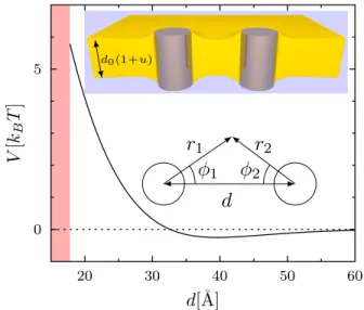
For DLPC, V (d) is shown in Fig. 1. It exhibits a rela-tively short-ranged repulsion of a few kBT close to
con-tact, followed by a shallow attractive well. In the same Fig. 1, we show the calculated shape of the membrane for a distance d = 2d0= 43 Å.
In the outer region the contact angle arctan(∂ru|r0) ≈
∂ru|r0is ' −37
◦and is fixed by the competition between
0 5
V
[k
BT
]
20 30 40 50 60d[˚
A]
d0(1+u)d
r
1φ
1r
2φ
2FIG. 1. Interaction energy V between two gramicidin chan-nels in a DLPC bilayer as a function of their center-to-center distance d. The red shading visualizes the contact between the inclusions. The parameters of the model correspond to our best fit of the experimental data. Upper inset: corre-sponding shape of the membrane for d = 2d0 = 43 Å. The
channels are represented as cylinders. The relative deforma-tion u is magnified by a factor of 3. Lower inset: coordinates for the multipolar expansion.
the torque exerted by the inclusion and the elastic defor-mation induced on the membrane.
Figure 2 displays S(q) as a function of the scattering vector q: experimental data of Ref. 10 (red dots) and corresponding Monte Carlo results, computed for 6 × 10⁶
Monte Carlo steps after thermal equilibration (black curves). A best fit is found for k1 = 5.4(30) × 10−2,
τ = −4.7(4) × 10−2, w < 1 × 10⁻³: see Fig. 2. Assum-ing a constant uncertainty σS = 0.1 for the experimental
points [10], the goodness-of-fit function χ2is of the order
of 2.1 per data point.
We also tested the approximate hypernetted-chain (HNC) solution, shown by dashed blue lines in Fig. 2. The discrepancies with respect to the Monte Carlo re-sults as the surface fraction η increases can be explained by the diffuse character of the repulsion, together with the presence of the attractive well [23].
For C12E5 we used the same global fitting
proce-dure as for DLPC. The experimental spectra S(q) mea-sured in Ref. 10 yield the best fit shown in Fig. 3 for k1 = 1.04(80) × 10−2, w = 0.85(15), s = −1.23(10).
As-suming again a constant uncertainty σS = 0.1 for the
experimental points [10], the goodness-of-fit function χ2
is of the order of 1.3 per data point. The interaction po-tential V (d) is shown in Fig. 4. The equilibrium slope for an isolated inclusion is close to the preferred value s, corresponding to an angle ' −50◦. Although this value is outside the validity range of our linearized model, it clearly indicates a large negative angle.
-5 0
S
0.0 0.1 0.2 0.3 0.4 0.5q [˚
A
−1]
η = 13.66% η = 16.51% η = 24.04% η = 28.34% η = 34.53% η = 39.73% η = 44.17%FIG. 2. Structure factors S as a function of the scattering vector q for gramicidin channels in DLPC bilayers at differ-ent surface fractions η of inclusions. Red dots: experimdiffer-ental data from Ref. 10. Black curves: Monte Carlo simulations for the parameters of the model corresponding to our best fit of the experimental data (see text). Blue dashed lines: HNC approximations. Curves are shifted downwards by unit increments. -2 0 2
S
0.0 0.1 0.2 0.3 0.4 0.5q [˚
A
−1]
η = 7.25% η = 16.50% η = 22.00% η = 28.25%FIG. 3. Experimental structure factors and fits for for gram-icidin channels in C12E5 bilayers. Same notation as in Fig. 2.
The hydrophobic matching principle is very general: due to the high cost of exposing hydrophobic residues to water and to the large difference in compression mod-uli between proteins and membranes [3], the contact hy-drophobic thickness of any bilayer equals that of the em-bedded protein. With the same generality, we assume that the boundary condition for the slope, being set lo-cally at the contact between the protein surface and the hydrophobic/hydrophilic interface of the bilayer, only de-pends on the nature of the inclusion and on the chemical
0 10 20 30 40
V
[k
BT
]
20 30 40 50 60d[˚
A]
0 1 2 20 40 60FIG. 4. Interaction energy V between two gramicidin chan-nels in C12E5bilayers for the model parameters corresponding
to our best fit of the experimental data. Shaded area: contact between the inclusions. Inset: zoom for d > 20Å.
family of the lipids (defined by the nature of the polar head).
Gramicidin must then impose the same torque T = Kad0τ on all lipids with a phosphocholine (PC) head,
ir-respective of the length of the alkyl chain [24], yielding negative equilibrium boundary angles of similar magni-tudes. One counterintuitive consequence is that, for PC membranes with a hydrophobic length larger than that of the gramicidin, the deformation profile should decrease steeply and then increase back to its equilibrium value.
The hydrophobic length of the gramicidin channel is larger than that of DLPC and C12E5 bilayers and it is
not surprising that, in both cases, it imposes a preferen-tial steep negative slope. To validate the generalization above, one would also need to demonstrate a negative contact slope in thicker bilayers. No SAXS results are available for phospholipids with longer chains, but other types of experimental and numerical results are available in the literature. In the following, we show that they support our hypothesis.
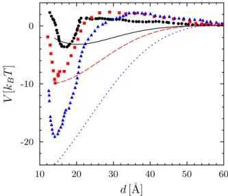
Several authors have simulated gramicidin channels in-serted within thicker lipid bilayers [25–28], and their re-sults could provide evidence as to the membrane profile. Three publications [26, 28, 29] do present such data (see the Supplemental Material [14] for more details) and sup-port our conclusion of a steep decrease in the thickness of phosphocholine membranes at the boundary with the gramicidin channel even for thicker bilayers, resulting in a nonmonotonic profile.
Using coarse-grained molecular dynamics, Yoo and Cui simulated two channels embedded into bilayers with dif-ferent compositions (DMPC, DPPC or DSPC) [30], com-puted the potential of mean force (PMF) and found in each case similar behavior, consisting of a steep
short-4 -20 -10 0
V
[k
BT
]
10 20 30 40 50 60d [˚
A]
FIG. 5. PMF between two gramicidin channels, simulated by Yoo and Cui (symbols; redrawn from Fig. 2A of Ref. 30) and calculated using the present model (lines) in DMPC (black solid dots and solid line), DPPC (red squares and dashed line) and DSPC (blue triangles and dotted line) bilayers, with no adjustable parameters.
range attraction and a long-range repulsion. In Fig. 5 we present their results and compare them to the predic-tions of our elastic model. No fitting is involved: we keep Ka0 and T fixed at the values obtained above for DLPC
bilayers, in agreement with our assumption, and we use literature data [14] for the other material constants of the three lipid systems (notably, the parameters Ka and d0
that appear in the normalized constants k1and w). The
hard core radius is taken as R0= 7 Å, for coherence with
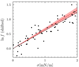
Ref. 30. Our model yields both the attractive and the repulsive part, although shifted further away from con-tact, possibly due to the inherent approximation of the coarse-grained simulation model or to higher-order gra-dient terms. Moreover, the contact value of the predicted potential is in very good agreement with the simulations. Gramicidin is widely used due to its conductivity prop-erties: the channel switches between the open (ion-conducting) and closed states as the monomers dimer-ize and dissociate, respectively. The transition between states can be followed by conductivity measurements [31] as a function of an externally controlled parameter, as shown for gramicidin channels in DOPC under a vari-able applied tension σ [4]. In particular, the formation rate f of the channels can be described, for moderate tension σ < 2 mN/m, as
ln f = C0+ C1σ, (3)
where C0changes with the details of the experimental
sit-uation, while C1only depends on the intrinsic elastic
pa-rameters [14]. Based on our assumption that all PC lipids share the same anchoring properties, we use the parame-ter values obtained by fitting the S(q) data for channels
in DLPC bilayers, which yield for DOPC (after proper normalization with Ka and d0), k1= 4.8 × 10⁻²and τ =
3.6 × 10⁻². Supplemented by literature data [4, 32, 33] for the other material constants of DOPC, these pa-rameters yield a slope C1 = 658(50) m/N, in excellent
agreement with the best fit to the experimental data C1exp = 620 m/N (see Fig. 6). This correspondence is striking since parameters extracted from the SAXS data are used to predict conductivity results, showing that both kinds of measurements are well captured by the continuum elastic model.
A nonmonotonic interface profile close to the inclusion was invoked more than thirty years ago by Huang [21], in order to explain the results of Elliott et al. [15] concerning the lifetime of gramicidin channels in monoacylglycerol bilayers. Although they concern membranes of differ-ent composition, our results indirectly confirm Huang’s insight and emphasize the role of higher-order terms in membrane elasticity.
To summarize, we present a complete elastic model for the membrane-induced interaction between inclusions, comprising a boundary energy associated to the slope of the membrane thickness at its contact with the inclu-sion. By fitting experimental results for the interaction of gramicidin channels in two types of bilayers we obtain the first quantitative measurement of the preferred slope and the associated torque or stiffness constants and show that these parameters are essential for a realistic descrip-tion of the inclusion-membrane interacdescrip-tion in terms of continuum elasticity.
We confirm our results and extend the analysis to phos-pholipids with longer chains by applying it to the poten-tial of mean force between channels obtained by simula-tions [30] and to data on the conductivity of the channel under tension [4]. Surprisingly, even when the hydropho-bic thickness of a phospholipid bilayer is larger than that of the gramicidin channel, the membrane thickness first decreases with the distance from the boundary before increasing to its equilibrium value far away from the in-clusion.
The strongly negative value of the thickness slope s might result from the combination between the specific interactions at the contact of the protein with the hy-drophobic/hydrophilic interface of the bilayer and the conical shape of the gramicidin monomer [34], which gives the channel an hourglass shape rather than a cylin-drical one. If, due to the local interactions, the molecules neighboring the channel tend to be parallel to the sides of the monomers, their axis (and hence the normal to the monolayers) is tilted away from the vertical, resulting in a negative s for a wide variety of membrane components. This could explain the very similar value of s obtained in bilayers formed by C12E5(see above), whose chemical
nature is quite different from that of PC lipids.
We acknowledge financial support from the French Agence Nationale de la Recherche (Contract No.
ANR-0.0 0.5 1.0 1.5
ln
f
(shifted)
0 1 2σ[mN/m]
FIG. 6. Logarithm of the formation rate f of gramicidin chan-nels in DOPC bilayers as a function of the membrane tension σ. Solid dots: experimental data retrieved from Fig. 6b of Ref. 4 (after subtraction of a constant baseline, see Ref 13 for the detailed analysis). Solid red line and red shaded area: our prediction using the elastic model and the parameter values discussed in the text (with no adjustable parameters). Black dashed line: best linear fit.
12-BS04-0023-MEMINT) and useful discussions with Anne-Florence Bitbol. The SAXS experiments were per-formed on beam line D2AM at the European Synchrotron Radiation Facility (ESRF), Grenoble, France. We are grateful to Cyrille Rochas for providing assistance in us-ing beam line D2AM.
∗ [email protected]; www.equipes.lps.u-psud.fr/constantin/ † [email protected]. ‡ [email protected].
[1] T. Gil, J. H. Ipsen, O. G. Mouritsen, M. C. Sabra, M. M. Sperotto, and M. J. Zuckermann, Biochimica et Bio-physica Acta (BBA)-Reviews on Biomembranes 1376, 245 (1998).
[2] A. G. Lee, Molecular BioSystems 1, 203 (2005).
[3] M. Ø. Jensen and O. G. Mouritsen, Biochim. Biophys. Acta-Biomembranes 1666, 205 (2004).
[4] M. Goulian, O. N. Mesquita, D. K. Fygenson, C. Nielsen, O. S. Andersen, and A. Libchaber, Biophys. J. 74, 328 (1998).
[5] C. Nielsen, M. Goulian, and O. S. Andersen, Biophys. J. 74, 1966 (1998).
[6] K. He, S. J. Ludtke, Y. Wu, H. W. Huang, O. S. Ander-sen, D. Greathouse, and R. E. Koeppe II, Biophysical chemistry 49, 83 (1994).
[7] T. A. Harroun, W. T. Heller, T. M. Weiss, L. Yang, and H. W. Huang, Biophys J. 76, 937 (1999).
[8] D. Constantin, G. Brotons, A. Jarre, C. Li, and
T. Salditt, Biophys. J. 92, 3978 (2007).
[9] D. Constantin, B. Pansu, M. Impéror, P. Davidson, and F. Ribot, Phys. Rev. Lett. 101, 098101 (2008).
[10] D. Constantin, Biochimica et Biophysica Acta (BBA) -Biomembranes 1788, 1782 (2009).
[11] D. Constantin, The Journal of Chemical Physics 133, 144901 (2010).
[12] B. Pansu, A. Lecchi, D. Constantin, M. Impéror-Clerc, M. Veber, and I. Dozov, The Journal of Physical Chem-istry C 115, 17682 (2011).
[13] A.-F. Bitbol, D. Constantin, and J.-B. Fournier, PLOS One 7, e48306 (2012).
[14] See Supplemental Material for the general form of the elastic free-energy density, the material constants for the bilayer elasticity used in the model, the multipolar ex-pansion, details on the Monte Carlo simulations and the calculation of the formation rate of the channels under tension. The Supplemental Material includes Refs. [35– 37].
[15] J. R. Elliott, D. Needham, J. P. Dilger, and D. A. Hay-don, Biochim. Biophys. Acta 735, 95 (1983).
[16] D. Marsh, Chem. Phys. Lipids 144, 146 (2006).
[17] R. H. Templer, J. M. Seddon, and N. A. Warrender, Biophysical Chemistry 49, 1 (1994).
[18] N. Kučerka, Y. Liu, N. Chu, H. I. Petrache, S. Tristram-Nagle, and J. F. Nagle, Biophysical Journal 88, 2626 (2005).
[19] T. Sottmann and R. Strey, J. Chem. Phys. 106, 8606 (1997).
[20] E. Kurtisovski, N. Taulier, R. Ober, M. Waks, and W. Urbach, Physical Review Letters 98 (2007).
[21] H. W. Huang, Biophys. J. 50, 1061 (1986).
[22] P. Helfrich and E. Jakobsson, Biophys. J. 57, 1075 (1990). [23] M. Huš, M. Zalar, and T. Urbica, J. Chem. Phys. 138,
224508 (2013).
[24] Which will, however, affect other parameters of the bi-layer, such as the elastic moduli.
[25] T. B. Woolf and B. Roux, Proteins 24, 92 (1996). [26] S.-W. Chiu, S. Subramaniam, and E. Jakobsson,
Bio-physical Journal 76, 1929 (1999).
[27] T. W. Allen, O. S. Andersen, and B. Roux, Proceedings of the National Academy of Sciences 101, 117 (2004). [28] J. Yoo and Q. Cui, Biophysical Journal 104, 117 (2013). [29] A. H. Beaven, A. M. Maer, A. J. Sodt, H. Rui, R. W. Pastor, O. S. Andersen, and W. Im, Biophysical Journal 112, 1185 (2017).
[30] J. Yoo and Q. Cui, Biophysical Journal 104, 128 (2013). [31] T.-C. Hwang, R. E. Koeppe, and O. S. Andersen,
Bio-chemistry 42, 13646 (2003).
[32] W. Rawicz, K. C. Olbrich, T. McIntosh, D. Needham, and E. Evans, Biophys J. 79, 328 (2000).
[33] J. A. Szule, N. L. Fuller, and R. P. Rand, Biophysical Journal 83, 977 (2002).
[34] R. Brasseur, V. Cabiaux, J. Killian, B. de Kruijff, and J. Ruysschaert, Biochimica et Biophysica Acta (BBA) -Biomembranes 855, 317 (1986).
[35] T. Kim, K. I. Lee, P. Morris, R. W. Pastor, O. S. Ander-sen, and W. Im, Biophysical Journal 102, 1551 (2012). [36] I. Basu, A. Chattopadhyay, and C. Mukhopadhyay,
Biochimica et Biophysica Acta (BBA) - Biomembranes 1838, 328 (2014).
[37] L. Yang, T. A. Harroun, W. T. Heller, T. M. Weiss, and H. W. Huang, Biophysical journal 75, 641 (1998).