HAL Id: inserm-00625054
https://www.hal.inserm.fr/inserm-00625054
Submitted on 21 Sep 2011
HAL is a multi-disciplinary open access
archive for the deposit and dissemination of
sci-entific research documents, whether they are
pub-lished or not. The documents may come from
teaching and research institutions in France or
abroad, or from public or private research centers.
L’archive ouverte pluridisciplinaire HAL, est
destinée au dépôt et à la diffusion de documents
scientifiques de niveau recherche, publiés ou non,
émanant des établissements d’enseignement et de
recherche français ou étrangers, des laboratoires
publics ou privés.
spontaneous GABAergic activity.
Olivier Caillard
To cite this version:
Olivier Caillard. Pre & postsynaptic tuning of action potential timing by spontaneous GABAergic
ac-tivity.. PLoS ONE, Public Library of Science, 2011, 6 (7), pp.e22322. �10.1371/journal.pone.0022322�.
�inserm-00625054�
Spontaneous GABAergic Activity
Olivier Caillard1,2*1 INSERM UMR641, IFR Jean-Roche, Marseille, France, 2 Universite´ de la Me´diterrane´e, Marseille, France
Abstract
Frequency and timing of action potential discharge are key elements for coding and transfer of information between neurons. The nature and location of the synaptic contacts, the biophysical parameters of the receptor-operated channels and their kinetics of activation are major determinants of the firing behaviour of each individual neuron. Ultimately the intrinsic excitability of each neuron determines the input-output function. Here we evaluate the influence of spontaneous GABAergic synaptic activity on the timing of action potentials in Layer 2/3 pyramidal neurones in acute brain slices from the somatosensory cortex of young rats. Somatic dynamic current injection to mimic synaptic input events was employed, together with a simple computational model that reproduce subthreshold membrane properties. Besides the well-documented control of neuronal excitability, spontaneous background GABAergic activity has a major detrimental effect on spike timing. In fact, GABAAreceptors tune the relationship between the excitability and fidelity of pyramidal neurons via a
postsynaptic (the reversal potential for GABAA activity) and a presynaptic (the frequency of spontaneous activity)
mechanism. GABAergic activity can decrease or increase the excitability of pyramidal neurones, depending on the difference between the reversal potential for GABAAreceptors and the threshold for action potential. In contrast, spike time
jitter can only be increased proportionally to the difference between these two membrane potentials. Changes in excitability by background GABAergic activity can therefore only be associated with deterioration of the reliability of spike timing.
Citation: Caillard O (2011) Pre & Postsynaptic Tuning of Action Potential Timing by Spontaneous GABAergic Activity. PLoS ONE 6(7): e22322. doi:10.1371/ journal.pone.0022322
Editor: Thierry Ame´de´e, Centre national de la recherche scientifique, University of Bordeaux, France Received November 25, 2010; Accepted June 27, 2011; Published July 15, 2011
Copyright: ß 2011 Olivier Caillard. This is an open-access article distributed under the terms of the Creative Commons Attribution License, which permits unrestricted use, distribution, and reproduction in any medium, provided the original author and source are credited.
Funding: This work was supported by the Institut National de la Sante´ et de la Recherche Me´dicale (INSERM), the Centre National pour la Recherche Scientifique (CNRS), and by a grant from the Fondation pour la Recherche sur l’Epilepsie (FFRE). Part of the equipment used in this study was funded by a European Community grant attributed to D. Debanne (LSHM-CT-2004-511995). The funders had no role in study design, data collection and analysis, decision to publish, or preparation of the manuscript.
Competing Interests: The author has declared that no competing interests exist. * E-mail: [email protected]
Introduction
Variability in neural activity is apparent throughout the central nervous system, in a wide range of electrophysiological signals. Several well recognized sources of noise in cortical neurons contribute to spike train variability, like the probabilistic release of transmitter and the stochastic gating of ion channels [1,2]. Evoked activities in stimuli-driven experiments are always superimposed on ongoing background activity, fluctuations of which contribute to the in vivo trial-to-trial variability [3–6]. Ongoing subthreshold activity of neocortical neurones is characterised by alternating states of low or intense synaptic activity [7,8], supported at least in part by two major neurotransmitters, GABA and glutamate. GABA activates mainly GABAAreceptors, which first reduce the excitability of the
cells by reducing the membrane resistance and second, according to their reversal potential, will bring the membrane potential away or close to the threshold for action potentials [9–11].
Two main functional roles have been established for spontane-ous GABAergic activity. The first relates to the membrane time constant of the cell: if the passive properties of the cell are comparable to a resistor-capacitor circuit, with a capacitance proportional to the surface of plasma membrane, and a resistance inversely proportional to the number of open ion channels, strong activation of GABAA receptors leads to a decrease in the
membrane time constant, which in turn speeds up dV/dt. As a consequence, individual excitatory postsynaptic currents are better resolved in time, and voltage changes are steeper [8,12,13]. The second is attributed to the background noise that creates a dynamic range in the input/output function of a neuron: referred to as stochastic resonance [14,15]: the processing of synaptic events by background activity, according to noise level or variabi-lity, translates the information provided by excitatory events into firing probability, despite the fact that this process is nonlinear [15–17]. Nevertheless, compelling evidence supports the fact that, even in individual cells, not only the average frequency of firing but also the timing of each action potential is relevant for neuronal information coding. Reliability in spike timing is known to be dependent on the structure of the excitatory stimulus [18]. The sequential activation of excitatory and inhibitory inputs in feed-forward networks [19] and the presence of functional GABAergic autapses at some fast-spiking interneurones [20] were shown to be relevant physiological mechanisms that can reinforce spike timing in neuronal networks. In spite of that, since most of the synaptic activity received either in vitro or in vivo appears to be uncorrelated to any specific stimulus, a yet unsolved question is what influence spontaneous GABAergic activity has on the temporal fidelity of pyramidal cell firing, according to the mode of activation of GABAAreceptors [21] or their reversal potential [22].
Here we evaluate and quantify how spontaneous activation of GABAA receptors affects action potential firing and timing,
according to the frequency of action potential discharge, and various modes of GABA release. We also evaluate the relative contribution of shunting inhibition and driving force for GABAA
receptors to neuronal discharge.
Results
Spontaneous GABAergic activity tunes the temporal fidelity of Pyramidal Cell discharge
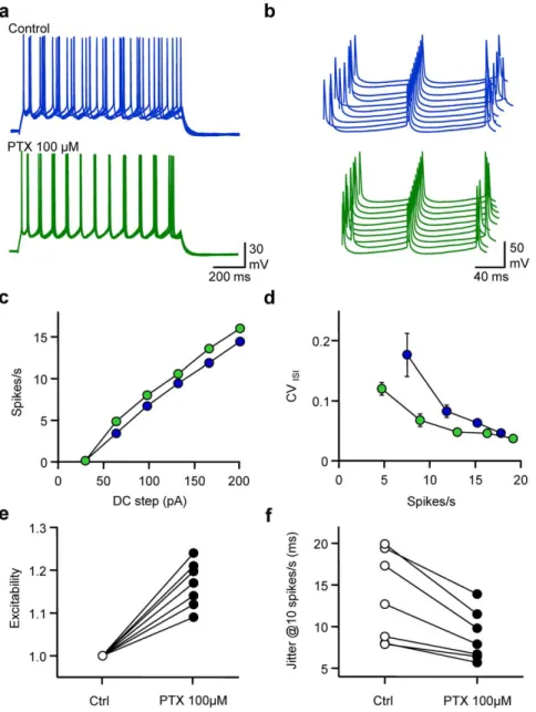
In order to evaluate the impact of spontaneous GABAergic activity received by L2/3 pyramidal neurones on their spike timing, gramicidin-perforated patch-clamp recordings Fig. 1). Although several precautions were taken, the possibility of an
intermediated perforated-patch/whole-cell configuration cannot be completely excluded (see methods). Excitability, defined as the average firing rate, and fidelity measured as the average coefficient of variation of inter-spike intervals along the discharge (CVISI, see
methods) were calculated for various DC steps. The influence of GABAergic activity on these parameters was tested by applica-tion of picrotoxin (PTX, 100mM) a GABAA receptor blocker.
Blockage of GABAA receptors, whilst not affecting resting Vm
(average change 20.260.9 mV, n = 7, P = 0.86) increased the excitability, reflected by a leftward shift of the frequency vs current relationship of recorded cells. Although CVISIwas dependent on
firing frequency in both control conditions and when GABAA
receptors were blocked, the CVISI was reduced at comparable
firing rates in the presence of PTX, especially for low rates of AP firing. When changes in excitability were normalised for each
Figure 1. Spontaneous GABAergic activity affects excitability and spike timing of pyramidal cells. a, Superimposed (5) membrane potential (Vm) fluctuations of a gramicidin-perforated current-clamped L2/3 pyramidal cell in response to a DC step (1 s, 170 pA) before (control, blue) and after extracellular application of picrotoxin (100 mM, PTX, green). The cell was held at around 280 mV in both conditions. Same cell and colour code for b, c and d. b, Waterfall view of Vm when the 5thspike was set as the time reference. c, Mean firing rate vs DC step. d, Coefficient of Variation of the Inter Spike Interval (CVISI) vs firing rate. e, Normalised changes in excitability observed when adding PTX (n = 7). f, Changes in spike
jitter for an interpolated firing rate of 10 spikes/s. doi:10.1371/journal.pone.0022322.g001
individual cell as the average change in spike number at each DC step, a significant increase in intrinsic excitability was observed (11765%, n = 7, P,0.001). Spike time jitter, defined as the standard deviation of the average ISI, was calculated when interpolating CVISIat a firing rate of 10 spikes/s. While spike time
jitter was quite heterogeneous from one cell to another in control conditions (range 6.6–19.9 ms), it was always reduced after PTX application. On average, blocking GABAAreceptors significantly
reduced spike time jitter from 13.465.4 ms to 8.863.0 ms (68610%, n = 7, P,0.05). Thus, GABAergic activity controls both excitability and fidelity of pyramidal cell discharge.
Presynaptic control of temporal fidelity
In order to quantify the contribution of various levels of GABAergic activity to pyramidal cell discharge parameters, currents mimicking spontaneous GABAergic events were injected in whole-cell recorded neurones by means of a dynamic-clamp amplifier in the presence of PTX (100mM). Patterns of spon-taneous GABAergic synaptic conductances were constructed by convoluting the occurrence in time of unitary events with a conductance transient template that had similar characteristics to the average event measured experimentally (Fig. S1). The time of occurrence of each event obeyed a Poisson law and was deter-mined extemporaneously according to the desired target rate of spontaneous activity. In a first set of experiments the reversal potential for GABAAreceptors (EGABA) was set at 270 mV (Fig. 2).
Experiments were performed at rates of GABAergic events going from 0 (control conditions) to 100 events/s. Both excitability and fidelity varied according to the level of GABAA activity. When
changes in excitability were normalised for each recorded neuron, 3-10-33-100 events/s reduced excitability by 0.361% (n = 7, P = 0.085), 862% (n = 9, P,0.001), 1561% (n = 14, P,0.0001) and 4463% (n = 12, P,0.0001) respectively. When spike time jitter at a firing rate of 10 spike/s was normalised in the absence of GABAergic activity for each recorded cell, 3-10-33-100 events/s increased spike time jitter by 148614% (n = 7, P,0.05), 1576 15% (n = 9, P,0.005), 209620% (n = 17, P,0.0001), 316632% (n = 15, P,0.0001) respectively. Thus, spontaneous GABAA
activity not only reduces neuronal excitability, but has a far more significant effect in deteriorating spike time precision.
Fidelity, not excitability depends on the mode of GABAA
activation
In order to verify the conclusions drawn from the experimental data and further analyse the correlation between the rate of the spontaneous activity input, the output firing rate and the fidelity of neuronal discharge, we used a ‘‘leaky integrate and fire’’ with Random action potential Threshold (RT-LIF) model [23,24], in order to focus upon the subthreshold membrane properties according to GABAAactivation while excluding the mechanisms
responsible for action potential triggering. The standard deviation of the threshold was set to 1 mV so that frequency-dependent jittering resembling the experimental data was observed. The model consistently reproduced the firing behaviour of L2/3 pyramidal cells with realistic membrane time constant in the absence of synaptic activity (Fig. S2). Both excitability and fidelity were affected proportionally to the level of GABAAactivity (Fig. 3).
As for dynamic-clamp in vitro experiments, spike time jitter evaluated from RT-LIF was much more prone to changes, according to GABAAactivity and firing rate. Thus for a rate of 10
spikes/s, 8 GABAA events/s were sufficient to increase jitter by
50%, while reducing excitability by only 2%. Both the transient changes in current and membrane resistance associated with GABAAactivity contributed to these effects (Fig. S3) and when one
or the other constituent was annulled, the impact of changes in GABAA activity on excitability and fidelity was reduced. The
charge of the random GABAAconductance transients determined
the impact of random GABAA activity on both excitability and
spike timing. Indeed, the detrimental effects of GABAAactivity
were proportional to the peak GABAA conductance (Fig. S4),
while for a fixed GABAAcharge, slowing or speeding up GABAA
conductance transient kinetics did not affect the impact of GABAA
activity on neither excitability or spike timing (Fig. S5).
The influence of the waveform pattern of GABAAactivity was
then tested on the number and precision of spikes emitted during the injection of various DC steps: the first mimicked the experi-mental spontaneous activity received by pyramidal neurones in vitro (Fig. S1); the second mode corresponded to an invariable pattern of GABAA conductance transients but consistently
re-played, so that these GGABA-Atransients were time-locked during
the different simulations; the third mode corresponded to a con-stant GGABA-A, i.e. tonic activity [21], which was an average over
time of the conductance transients received by the RT-LIF in the previous two modes.
When compared to control conditions, i.e. no GABAergic activity, a rate of 33 randomly occurring GGABA-Atransients per
second notably increased spike time jitter, and both excitability and the fidelity of discharge were reduced (Fig. 4). When GABAA
conductance transients were evoked invariably in time, despite the fact that the excitability was modified to the same extent as with random input, spike time jitter was much less affected. Although CVISIwas larger than in control conditions, especially at low firing
rates, it was smaller than the conditions in which randomly occurring spontaneous activity was generated. Last, the impact of a constant activation of GABAAconductance was tested (i.e. tonic
activation of GABAA receptors [21]). Again, the decrement in
excitability was comparable to other modes of GABAAactivation,
but CVISIvs firing rate relationship was close to control conditions.
In order to get a comprehensive view of the impact of the different modes of GABAAactivation, FI curves were drawn for a range of
0 to 100 events/s in the 3 modes defined above. While FI curves were comparably shifted to the right by GABAA activity in a
frequency dependent manner, whatever the mode, the effect of GABAA activity on CVISI was highly dependent on the mode.
In fact, spike-timing fidelity was noticeably reduced only if the cell, firing at low rate, received a high level of random GABAA
activity.
In order to confirm that GABAA activation modes can affect
spike timing in a different ways, an alternative type of spike timing analysis was performed (Fig. 5). Thus, instead of analysing the variability of the ISI along the discharge, the timing of action potentials initiated over 50 presentations was measured in control conditions and in the 3 different modes of GABAAactivation. The
DC step was adjusted in order to get an average rate of 5 spikes/s in all 4 conditions. Once each AP was detected, the timing data was convolved with a Gaussian waveform, in order to reflect both reliability and temporal precision of spike initiation. In control conditions, temporal organisation faded with time, but was still relatively high at the end of the discharge. In contrast, random patterns (33 events/s) of GABAergic activity rapidly disrupted the temporal organisation of the discharge. When the pattern of GABAergic activity was invariably replayed, peaks of AP initiation probability were higher in number than in control conditions, and varied in amplitude and width such that for specific time intervals AP initiation probability was higher than in control conditions. Last, in the presence of tonic activity, temporal dispersion and variance waveform were close to control conditions until the last third of the discharge.
Thus, in contrast to invariable phasic or tonic GABAAactivity,
randomly occurring spontaneous GABAA activity has a major
detrimental effect on spike timing.
Postsynaptic control of spike timing fidelity
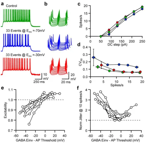
In order to test the influence of EGABA on excitability and
fidelity, dynamic currents mimicking spontaneous GABAergic events were injected in pyramidal neurones at an average rate of
33 events/s in the presence of PTX (100mM). The effects of spontaneous GABAA transients were first compared for E GA-BA= 270 mV and 230 mV (Fig. 6). While a decrease in
excitability and an increase in spike jitter was observed for EGABA= 270 mV, a slight increase in excitability was noted for
EGABA= 230 mV, while the spike jitter was similar to control
conditions. Quantification of changes in excitability at various firing rates showed that the FI curve was shifted either leftward or
Figure 2. Frequency-dependent tuning of pyramidal cell excitability and spike timing. a, From upper to lower, an example of a GABAA
conductance (GGABA-A) pattern of 33 events/s, GABAAcurrent (IGABA-A), Vm and the DC protocol in a dynamic clamp recording of a L2/3 pyramidal cell. b,
Superimposed (5) Vm fluctuations in response to a DC step (1 s, 170 pA) in control (green), in the presence of 33 (blue) or 100 (red) randomly occurring GABAAevents/s dynamically injected with EGABAset at 270 mV. Same cell and colour code for c,d and e. c, Waterfall view of Vm when the 5thspike was
set as the time reference. d, Mean Firing rate vs DC step. e, CVISIvs mean spike firing rate. f, Normalised changes in excitability observed when neurons
receive different levels of randomly occurring GABAAevents/s. g, Normalised changes in spike jitter for an interpolated firing rate of 10 spikes/s.
rightward according to EGABA. While major changes in CVISI
were observed for all firing rates tested at EGABA= 270 mV, they
were negligible at EGABA= 230 mV. A series of experiments were
performed with a broad range of EGABA(from 280 to 0 mV) in
order to establish the relationship between EGABA, excitability and
fidelity. Pyramidal neurone excitability was linearly dependent on EGABA and changes in excitability were abolished when EGABA
was 6.1 mV under the AP threshold (n = 9; 95% confidence interval 10.3/1.9 mV under AP threshold). The relationship
between normalised jitter for an interpolated firing rate of 10 spikes/s was bell-shaped, with a minimum of 1.33 (n = 9; 95% confidence interval 1.12/1.54) for EGABAbeing 2.5 mV under the
AP threshold.
On/Off Control in Spike Timing tuning by GABAAreversal
potential
To further analyze the relationship between the reversal potential for GABAAreceptors, the rate of firing and spike timing, an initial
Figure 3. Modelling frequency-dependent tuning of neuronal excitability and spike timing. a, From upper to lower, an example of G GABA-Apattern of 33 events/s, IGABA-A, Rin, Vm and a DC step (1 s, 120 pA) when running the LIF model. b, Superimposed (5) Vm fluctuations in response to
a DC step (1 s, 160 pA) in control (green), in the presence of 33 (blue) or 100 (red) randomly occurring GABAAevents/s with EGABAset at 270 mV.
Same cell and colour code for c,d and e. c, Waterfall view of Vm when the 5thspike was set as time reference. d, Mean Firing rate vs DC step. e, CVISIvs
firing rate. f, Normalised changes in excitability observed when LIF received 1 to 100 randomly occurring GABAAevents/s. g, Normalised changes in
spike jitter for a firing rate of 10 spikes/s. doi:10.1371/journal.pone.0022322.g003
Figure 4. Randomly occurring GABAAconductance transients underlie the frequency-dependent tuning of pyramidal cell discharge
fidelity. a, Waterfall view of conductance fluctuations injected into the LIF model in control conditions (no conductance, green), when the LIF model received 33 randomly occurring GABAAevents/s (blue), an invariable pattern of 33 GABAAevents/s (red), or when the neuron model received a
constant GGABA-A equivalent to the average conductance for a rate of 33 events/s (orange). Same cell and colour code for b,c,d and e. b,
Superimposed (5) Vm fluctuations of the LIF model in response to a DC step (1 s, 160 pA) in the various conditions depicted in a. c, Waterfall view of Vm when the 5thspike was set as the time reference. d, Mean firing rate vs DC step. e CVISIvs firing rate. f, Mean firing rate displayed on a pseudocolor
scale vs DC step and rate of GABAAactivity when LIF model received random (left), invariable (middle) or constant conductance (right). g, CVISI
displayed on a pseudocolor scale vs firing and GABAAactivity rate. Same conditions as in f.
simulation was performed with RT-LIF receiving an input rate of 33 random GABAAconductance transients per second. EGABAand
DC steps varied from 290 to 0 mV and 50 to 300 pA, respectively (Fig. 7). While an increase in spike jitter, associated with an increase in interspike interval was obvious for EGABA= 270 mV, it was
similar to control conditions for EGABA= 235 mV. Both transient
changes in current and membrane resistance led to changes in excitability and fidelity (Fig. 7). Modifications in excitability were abolished when EGABA was 5 mV above AP threshold. When
considering spike timing, a bell-shaped relationship between CVISI
and EGABAwas observed with a minimum at 10 mV above AP
threshold. When transient changes in membrane resistance were annulled, changes in excitability were abolished if EGABA was
10 mV under AP threshold and the CVISI was minimal when
EGABAwas 5 mV under AP threshold. Thus GABAAcurrents can
almost compensate for the decrease in the fidelity of spike timing and excitability induced by the reduction in membrane resistance when EGABAis slightly above the AP threshold.
In order to get a better view of the impact of spontaneous GABAAactivity on excitability and spike time jitter at different
EGABA, FI curves were drawn for a range of 0 to 100 events/s at 3
different EGABA. FI curves were shifted right and left by GABAA
activity in a frequency dependent manner for EGABA= 270 mV
and EGABA= 0 mV respectively. However at EGABA= 235 mV
increasing GABAAactivity had no influence on the relationship
between DC step and average firing rate. Analysis of the influence of random GABAAactivity on CVISIat these 3 different EGABA
indicated that while the level of GABAA activity had a major
influence on CVISIat EGABA= 270 or 0 mV, it had little effect on
CVISIat EGABA= 235 mV.
Thus, by regulating their GABAAreceptor reversal potential,
i.e. their intracellular chloride concentration [22,25], neurones retain control of how spontaneous GABAergic activity may increases or decrease excitability but also to which level the temporal fidelity of their discharge will be deteriorated.
Discussion
Ongoing background activity has been shown to have an important impact on neuronal dynamics. Changes in the level of
Figure 5. Randomly occurring GABAAconductance transients underlie the frequency-dependent tuning of pyramidal cell discharge
fidelity. a, Superimposed (10) Vm fluctuations of the LIF model in response to a DC step (2 s) that allows a firing rate of 5 spikes/s, in control conditions (green, 95 pA), when the LIF model received 33 randomly occurring GABAAevents/s (blue, 111 pA), an invariable pattern of 33 GABAA
events/s (red, 110 pA), or when the neuron model received a constant GGABA-Aequivalent to the average conductance for a rate of 33 events/s
(orange, 110 pA). b up, Raster plot of spike times collected from 50 consecutive trials in the various conditions depicted in a; down, Spike probability over time for these 50 consecutive trials.
background inhibition alter the electrical compactness and modify the integrative properties [13,26], modulate the gain [27] and thus can dynamically set the range of the input/output function of neurones [15–17]. Here I have emphasized the fact that GABAergic activity not only affects the excitability of pyramidal cells, but also alters spike timing in proportions that can be significantly higher, according to its temporal organisation and EGABA.
Variations in background GABAergic activity
Ongoing subthreshold activity of neocortical neurones is subject to important changes. In the anaesthetised animal, alternating states of low or intense activity can be observed: the down-states during which little or no synaptic background activity is visible and the up-states when the cells are continuously bombarded by spontaneous activity [7,8,26]. The pattern of GABAergic activity that is received by these cells is determined by the number, the variety and activity of presynaptic interneurones [28]. The complexity of the GABAergic system is enhanced by the fact that GABAergic synapses, according to their location, exhibit specific electrophysiological features [29–32]. Additionally, the level of spontaneous GABAergic activity is subject to prominent changes during development [33], short [34,35] or long-term plasticity [36–43] and can be altered in different pathologies [44–47].
In the absence of synaptic activity, the spike generating mechanism shows a certain level of reliability and precision [18]
that can be noticeably reduced by the spontaneous occurrence of few GABAAsynaptic events. Experiments performed in vitro with
endogenous GABAergic activity or computer-modelled back-ground activity injected to the cell by the means of a dynamic-clamp amplifier could be very well predicted with a RT-LIF model. Therefore, while taking into account the fact that the experiments presented here represent only a very limited range of presynaptic activities when compared to what has been reported in vivo during up-states [8], the RT-LIF model appears to be, for this range level of activity, sufficiently complex to capture the essential features of the impact of GABAA activity on excitability and
spike-timing.
Beyond their molecular heterogeneity, GABAAreceptors can be
activated following two different modes, phasic or tonic release. Phasic activation of GABAA receptors relies on synchronous
opening of clustered synaptic receptors, while tonic activation results from the activation of extra synaptic receptors [21]. Results obtained in this study show that tonic and phasic inhibition modify to the same extent the input/output relationship, when a repetitive discharge was induced by step excitation. Thus, the average input/ output relationship appears to be dependent neither on the timing nor the shape of the spontaneous conductance transients but rather on the average conductance over time (Fig. 4). In contrast, major differences between the mode of activation of GABAA
receptors and the fidelity of spike timing were observed. Indeed the increase in variability was dependent on the average GABAA
Figure 6. Reversal potential-dependent tuning of pyramidal cell excitability and discharge fidelity. a, Superimposed (5) Vm fluctuations of a whole-cell L2/3 pyramidal neurone in response to a DC step (1 s, 170 pA) in control conditions (green), when 33 randomly occurring GABAA
events/s were dynamically injected at EGABA= 270 mV (blue) or 230 mV (red). Same cell and colour code for b, c and d. b, Waterfall view of Vm when
the 5thspike was set as the time reference. c, Mean firing rate vs DC step. d, CV
ISIvs firing rate. e, Normalised changes in excitability observed when
neurons received 33 randomly occurring GABAAdynamic currents/s at various differences between EGABAand the calculated threshold for AP in each
individual cell (n = 9). f, Normalised changes in spike jitter observed for a firing rate of 10 spikes/s. Same conditions as in e. doi:10.1371/journal.pone.0022322.g006
conductance; it appeared first that transient changes in inhibitory conductance were more prone to disorganize firing, and second that random trains were more potent than fixed patterns of experimentally derived synaptic conductance waveforms.
In fact, from a control condition where the action potential delay is dependent on the AHP amplitude, the membrane time
constant and the voltage threshold for action potential, transient inhibitory conductances delay Vm from reaching action potential threshold and thus induce both an increase in ISI mean and variability, such that the temporal structure of neuronal discharge is rapidly lost. If now the timing of transient inhibitory con-ductances remains invariable, the probability of firing an action
Figure 7. Frequency and reversal potential-dependent tuning of pyramidal cell excitability and discharge fidelity. a, Superimposed (5) Vm fluctuations of the LIF model in response to a DC step (1 s, 160 pA) in control conditions (green), when the LIF model received 33 randomly occurring GABAAevents/s at EGABA= 270 mV (blue) or 235 mV (red). Same colour code for b, c and d. b, Waterfall view of Vm when the 5thspike was
set as the time reference. c, Mean firing rate vs DC step. d, CVISIvs firing rate. e, Normalised changes in excitability observed when LIF model received
33 randomly occurring GABAAevents/s at various EGABAin control conditions (black), when GABA activity induced only transient changes in IGABA-A
(gray) or when GABA activity induced only transient changes in Rin(light gray). f, Normalised changes in spike jitter observed for a rate of 10 spikes/s.
Same conditions as in e. g, From left to right firing rate displayed on a pseudocolor scale vs DC step and EGABAwhen the LIF model received 33
randomly occurring GABAAevents/s in control conditions (left), when GABAAactivity induced only transient changes in IGABA-A(middle) or when
GABA activity induced only transient changes in Rin(right). h, CVISIdisplayed on a pseudocolor scale vs firing rate and EGABA. Same conditions as in g.
potential is not uniform but varies according to the pattern of inhibitory conductance (Fig. 5). As demonstrated by Mainen and Sejnowski [18], when a DC pattern is invariably replayed, the reliability of the firing pattern increases as a function of the fluctuation amplitude. Tiesinga and Toups [48] later showed that an invariable fluctuating DC pattern uncovers, after a statistical sorting of the different trials, a series of precise spike patterns with different spike times and eventually a different number of spikes [49]. Thus, the variance of inputs uncovers different spike patterns and maintains a high probability of spike timing in narrow win-dows all along the discharge, but increases the variability of the ISI. In contrast tonic inhibition, whatever the analysis method employed (CVISIor firing probability over time) barely
deterio-rates spike timing. Pouille and Scanziani [19] have demonstrated that feed forward inhibition curtailed EPSPs and improved EPSP-spike timing in vitro. In some specific GABAergic interneurones, because autaptic transmission offers a tight temporal link between the spike and GABAergic conductance, the reliability of spike timing is greatly improved by the activation of autaptic GABAA
receptors [20]. Despite the fact that in these two latter examples, where a subpopulation of GABAergic synapses is activated sequentially after some excitatory synaptic inputs or neuronal discharge, the temporal structure of most of the GABAergic activity is often uncorrelated to pre or postsynaptic activity. From the results described here, reducing the fidelity of discharge by adding tonic or phasic inhibition may at first sight appear contradictory with previously published results. However one has to take into account the fact that here GABAAactivity was not
time-locked to patterns of EPSPs and also that fidelity was measured and compared for similar rates of spike discharge. It demonstrates that, once neurones trigger action potentials at a sustained rate, the consequences of a given amount of GABA, whatever its mode of release i.e. phasic random vs phasic in-variable vs tonic, is quantitatively the same on excitability, but appears to be different on spike timing. While tonic activation of GABAAreceptors contributes to the modulation of excitability and
thus may have a key role in epileptogenicity [50], its ability to disorganise firing discharge seems limited. In conclusion, excit-ability and spike timing modulation by GABAAreceptors are not
strictly related, but rely on the mode of GABA release and the eventual temporal correlation between the activation of GABAA
receptors, the excitatory inputs and action potential firing in case of repetitive discharge.
Variations in GABAAreversal potential
GABAA receptors are permeable to chloride and bicarbonate
ions [51]. As bicarbonate ions are much less permeant than chloride, and because the chloride equilibrium potential is usually more negative than the resting membrane potential, activation of GABAAreceptors typically results in a hyperpolarising inhibitory
postsynaptic potential. Nevertheless, the chloride gradient is developmentally regulated and subject to plasticity [25]. A large range of GABAAreversal potentials has been reported in the past
twenty years [52]. Evoked GABAergic activity was shown to affect the timing of spikes evoked by membrane potential oscillations, according to the phase at which GABAAreceptors were activated
and EGABA[53]. According to development, the neural situation,
depolarised EGABA may be relevant and required for proper
network activity, though consideration has to be given that this will modify the temporal organisation of the postsynaptic discharge and the stability of network oscillations [53].
From the present study it appears that intracellular chloride homeostasis has a very important role in defining the changes in excitability and fidelity induced by a given rate of presynaptic
GABA release. Spike timing jitter is closely proportional to the absolute difference between the threshold for action potential and EGABA. From this study it appears that, for a given rate of
GABAergic activity, both the changes in membrane resistance and the currents associated with GABAAconductance affect
excitabil-ity and spike timing. Excitabilexcitabil-ity and spike time jitter are pro-portional to GABAAconductance. Thus, in order to compensate
for changes in excitability induced by GABAA activity and the
associated reduction in membrane resistance, EGABA must be
higher than the threshold for action potential generation. In the case of no change in membrane resistance associated with GABAA
conductance, as is the case in the dynamic-clamp experiments, EGABA appeared lower than the threshold for action potential.
Since GABAA currents tune excitability and spike timing, the
reversal potential where GABAAactivity has the minimal impact
appears to be the potential at which the GABAAcurrents are on
average equal to zero. Thus, the electrophysiological characteris-tics of a given neuron may be associated with a specific EGABA
such that the changes in excitability and reduction in spike timing reliability by GABAergic activity may be alleviated.
Spike-timing and network activity
Neuronal coding of sensory information relies on the mean rate of action potential discharge but also the relative timing between action potentials. Stimulus-dependent changes in spike synchroni-zation have been observed in various cortical areas, and it has been suggested that synchronization of spikes within a precise millisecond range could serve as an efficient mechanism to group neurons into cell assemblies responding to specific stimuli [54,55]. During stimulus processing the recruitment of excitatory neurons and their connections are often the primary requisites for neuronal computation. GABAergic neurones contribute to synchronization. In invertebrates, GABAAreceptors were shown to be of critical
importance as their blockade abolished oscillatory synchronization but not the individual responsiveness of projection neurones [56]. The presence of feed-forward inhibition or autaptic synapses in various areas of the central nervous system may be critical to maintain spike timing and synchrony [19,20,53]. Nevertheless, most of the phasic GABAergic activity appears not to be correlated with sensory stimuli [26], tonic inhibition is present in different brain areas [21] and asynchronous release of GABA is observed in CCK-interneurones in the hippocampus [57–59]. Thus, synaptic and extra-synaptic GABAAreceptors can be activated in various
modes, which share common characteristics but also particular features that may have computational significance for the regu-lation of network activity.
In conclusion, I have emphasized in this study the fact that, in addition to the well-documented modulation of neuronal excit-ability, spontaneous GABAergic activity has a major detrimental impact on spike timing reliability. A future challenge is to under-stand how variability in spike timing contributes to neuronal processing in the brain, in this context the impact of GABA release and chloride homeostasis in deteriorating spike timing should not be neglected.
Materials and Methods Ethics Statement
All rats were maintained on a 12 h light/dark cycle with food and water provided ad libitum. The research involving animals has been approved by the Direction De´partementale des Services Ve´te´rinaires – Pre´fecture des Bouches du Rhoˆne, France (permit number C13-055-8), and the Institut Jean-Roche Animal Care Supervisor (M. Mekahouche, D.V.M Ph.D, permit number 13-122).
All experiments were carried out according to the European and Institutional guidelines for the care and use of laboratory animals (Council Directive 86/609/EEC and French National Research Council).
Cortical slice preparation and electrophysiological recordings
Transverse cortical slices (350–400mm thick) were obtained from 13- to 20-day old Wistar rats as previously described [60]. Experiments were performed at 32uC in ACSF containing the following (in mM): NaCl 125, NaHCO3 26, CaCl2 3, KCl 2.5, MgCl2 2, NaH2PO4 0.8, D-glucose 10, kynurenate (2 mM); and saturated with 95% O2 and 5% CO2. L2/3 pyramidal cells (recorded between 300 and 800mm from the pia) were visualised using an Olympus BX-51 WI microscope equipped with dif-ferential interference contrast optics under infrared illumination and a water immersion lens (X60, 0.9 NA, Olympus). Unless otherwise stated, electrophysiological recordings were performed in whole-cell configuration with a Multiclamp 700B amplifier (Axon Instruments), filtered at 5 kHz and digitized at 20 kHz via a PCI-6220 National Instrument interface controlled by IgorPro (Wavemetrics) and/or Digidata 1322A interface controlled by PClamp software (Axon Instruments). Patch pipettes had a resistance of 3–6 MV when filled with a solution containing (in mM): K-gluconate 120, KCl 20, HEPES 10, EGTA 0.5, Na2ATP 2, NaGTP 0.3, and MgCl2 2, pH 7.4. Cells were recorded if the series resistance, measured throughout experiments, remained stable and ,20 MOhm. In a subset of experiments (Fig. 1, Fig. S1c,d), perforated patch-clamp recordings were performed. Gramicidin (100mg/ml) was added to the intracellular solution. For this recording configuration the tip of the pipette was back-filled with the solution containing no gramicidin to allow a good cell-attached configuration prior to perforation by gramicidin diffusion at the tip of the pipette. Alexa 488 or 568 (Molecular Probes) were added to the intracellular solution in order to confirm the pyramidal cell morphology in some whole-cell recordings. Fluorescence excitation was performed using a PolyV monochro-mator (Till photonics). Fluorescence imaging was performed with a CCD Camera (CoolSnap HQ2). Both excitation patterns and fluorescence acquisition were controlled by Metavue (Molecular Devices) and IgorPro (Wavemetrics) software. Isoguvacine (0.5 mM) was pressure ejected using a pico-Spritzer (TooheySpritzer). Typical pressure and time ejection were 7PSI and 30 ms respectively. Alexa 588 (Molecular Probes) was added to the isoguvacine solution in order to visualise its diffusion when ejected. During the course of the perforated patch-clamp recordings, EGABAwas estimated from the polarity of isoguvacine responses at
different holding membrane potential. This protocol inform me, together with alexa 488 dye imaging that perforated patch did not evolve into whole cell mode during the course of the experiment, Electrophysiological recordings were stopped if a sudden change in EGABAor fluorescence of the soma was observed.
Picrotoxin (100 mM) was prepared in ethanol and stored at 280uC. Kynurenate (200 mM) was prepared in distilled water and stored at 220uC (respectively). Stocks solutions were thawed and diluted into the extracellular solution before use.
Stimuli
Voltage or DC steps, GABAA synaptic conductances and
reversal potentials were constructed, according to experimental needs with Igor Pro software (Wavemetrics), and converted to analog signals via a PCI-6723 National Instrument interface. GABAA like currents were simulated with a dynamic-clamp
amplifier (SM1, Cambridge Conductance), using for inputs the
conductance profile, the reversal potential and the membrane potential of the recorded cell. The calculated current was summed to the DC step and injected into the recorded cell.
Neuronal modelling
GABAergic synaptic events were modelled as a conductance with a dual exponential time course of the form (1-exp(-t/taurise).exp(-t/
taudecay). Spontaneous activity was created by convolution of the
GABAergic event template with the Poisson train occurrence of spontaneous events. The conductance instruction was then either converted to an analog signal to instruct the dynamic clamp amplifier or injected to the computational model. The latter was based on a leaky integrate and fire model (LIF) with random threshold [23,24] unless otherwise stated: Cm.dVi/dt = Sigmai.gi.
(VO2Vi)+Sigmai.I with Cm= 400 pF; Vo= 265 mV. When Vi
reaches a threshold theta, a spike is generated and the membrane potential resets to Vo. Resting membrane resistance was set at
200 MOhm. GABAergic synaptic events, affecting both giand I
were sometimes included, according to the equation IGABA=
gGABA.(Vm2EGABA). EGABAwas set at 270 mV unless otherwise
stated. All modelling was performed with Igor Pro Software.
Analysis
Spontaneous synaptic activity was analyzed with the help of Minianalysis (Synaptosoft, Decatur, GA, USA). Spike analysis was performed with Igor Pro (Wavemetrics, Lake Oswego, OR, USA), Neuromatic functions (Jason Rothman, http://www.neuromatic. thinkrandom.com) were used in addition to homemade functions. 3D graphs were drawn under Origin Pro 8.0 (Origin Lab, Northampton, MA, USA) by smoothing average data with a thin plate spline function.
Spike Threshold was detected when dV/dt exceeded 10 mV/ ms. Measurements of CVISIwere performed by averaging CV for
each sequential interval along the discharge starting from the 2nd spike. With this method, the reproducibility of the full discharge for one trial to another could be evaluated, independently of spike-frequency adaptation that was observed experimentally.
Measurement of spike time jitter @ 10 spikes/s was performed by interpolating the CVISIvs average spike discharge with either
the best linear or exponential fit.
Plots of firing probability (fig. 5) were obtained by convolving the times of spike generation with a Gaussian function with standard deviation equal to the duration of the sweep, divided by the mean number of spikes [61]. Statistical comparisons were made using ANOVA paired tests. Differences were considered significant when P,0.05. Data are reported as mean 6 s.e.m.
Supporting Information
Figure S1 Characteristics of spontaneous GABAAevents
received by L2/3 pyramidal cells. a, Spontaneous occur-rence of GABAergic current transients recorded in a L2/3 pyramidal cell (Vhold= 270 mV). Their frequency was on average
3.861.0 events/s (range 0.9–13.1 events/s, n = 15). b, Average GABAAcurrent transients (from n = 66 events) received by the cell
shown in a. For all cells tested GABAA current transients were
displayed a fast 10–90% rise time (1.560.2 ms, range 0.6–3.7 ms, n = 15); the conductance at the peak of current was 343628 nS (range 146–508 nS, n = 15); their tail was best fitted with a monoexponential decay (time constant 14.761.1 ms, range 7.2– 21.4, n = 15). c, superimposed currents in response to a brief (15 ms) perisomatic pressure ejection of GABAAagonist
isoguva-cine at 4 different Vhold (288,278,268,258 mv) during a
measured at the peak of isoguvacine response vs Vhold. Same cell as
in c. EGABAwas on average 26964 mV (range 282–257 mV;
n = 5). (TIF)
Figure S2 Leaky Integrate and Fire Model with Random Action Potential Threshold. a, Electronic Design of the Leaky Integrate and Fire Model. It is based on an RC circuit with a condition: if Vm crosses the Threshold H, an action potential waveform lasting 10 ms, that peaks at +30 mV and resets at 260 mV, is added. b, H can be either invariable (green line) or random when adding a Gaussian noise to H (red SDV = 1 mV). c, Superimposed (5) Vm fluctuations of the LIF model in response to a DC step (1 s, 160 pA) and Waterfall view of the Vm in order to show the jitter of the previous and following spikes when the 5th spike was set as a the time reference in control (green; SDV H = 0 mV), or when SDV H = 1 mV (red). d,. Mean firing rate vs DC step. Same colour code as in c. e, CVISIvs firing rate when
SDV H = 1 mV. CV remains null if SDV H = 0 mV. (TIF)
Figure S3 Contributions of GABAA current and GABAA
shunt to the frequency-dependent tuning in Excitability and Fidelity. a, From upper to lower, a GGABA-Apattern of 33
events/s, Vm, sum of the DC step (135 pA)+IGABA-A and
membrane input resistance (Rin) fluctuations in the LIF model
when EGABAwas set at 270 mV. b, Same conditions as in a but
Rin remained constant despite transient changes in GGABA-A. c,
Same conditions as in a but IGABA-A remained null during the
simulation despite transient changes in GGABA-A. d, Mean firing
rate displayed on a pseudocolor scale vs DC step and the rate of randomly occurring GABAAactivity in control conditions (left),
when GABAAactivity induces only transient changes in GABAA
currents (middle) and when GABAAactivity induces only transient
changes in Rin(right). e, CVISIdisplayed on a pseudocolor scale vs
firing and GABAAactivity rate. Same conditions as in d.
(TIF)
Figure S4 Amplitude- and Frequency-dependent tuning of pyramidal cell discharge fidelity by randomly occur-ring GABAAconductance transients. a, From upper to lower,
a GGABA-A pattern of 33 events/s, Vm, sum of the DC step
(135 pA)+IGABA-Aand membrane input resistance (Rin)
fluctua-tions in the LIF model when EGABA was set at 270 mV. Peak
GABAAconductance was set at 0.5 nS. b, Same conditions as in a
but peak GABAAconductance was set at 1 nS. c, Same conditions
as in a but peak GABAAconductance was set at 2 nS. d, Mean
firing rate displayed on a pseudocolor scale vs DC step and the rate of randomly occurring GABAA activity when Peak GABAA
conductance was set at 0.5 nS (left), 1 nS (middle) and 2 nS (right). e, CVISIdisplayed on a pseudocolor scale vs firing and GABAA
activity rate. Same conditions as in d. (TIF)
Figure S5 The kinetics of randomly occurring normal-ised GABAA conductance transients do not affect the
frequency-dependent tuning of Excitability and Fidelity. a, From upper to lower, a GGABA-Apattern of 33 events/s, Vm,
sum of the DC step (135 pA)+IGABA-A and membrane input
resistance (Rin) fluctuations in the LIF model when EGABAwas set
at 270 mV. Peak GABAAconductance was set at 2.005 nS, and
GABAAconductance transients had fast kinetics (Taurise= 0.5 ms;
Taudecay= 5 ms). b, Same conditions and GABAAcharge as in a
but GABAAconductance transients had medium kinetics (Peak
GABAAconductance = 1 nS; Taurise= 1 ms; Taudecay= 10 ms). c,
Same conditions as in a but GABAAconductance transients had
slow kinetics (Peak GABAA conductance = 0.2268 nS; Tau ri-se= 10 ms; Taudecay= 30 ms). d, Mean firing rate displayed on a
pseudocolor scale vs DC step and the rate of randomly occurring GABAA activity when GABAA conductance transients had
parameters depicted in a (fast kinetics, left), when GABAA
conductance transients had parameters depicted in b (medium kinetics, middle) and when GABAAconductance transients had
parameters depicted in c (slow kinetics, right). e, CVISIdisplayed
on a pseudocolor scale vs firing and GABAAactivity rate. Same
conditions as in d. (TIF)
Acknowledgments
I wish to thank E.S. Albert and L. Saliba who performed some of the experiments during their Master Diploma Training, K. Lamsa and R. Cudmore for help with perforated patch-clamp recordings and program-ming, respectively. C. Gomez and L. Fronzaroli-Molinieres for technical assistance, N. Ankri, C. Bernard, R. Cudmore, D. Debanne, F. Dubruc, S. Foster, A. Marty, M. Seagar, F. Tell and the members of INSERM UMR 641 for helpful comments and suggestions during completion of this work.
Author Contributions
Conceived and designed the experiments: OC. Performed the experiments: OC. Analyzed the data: OC. Contributed reagents/materials/analysis tools: OC. Wrote the paper: OC.
References
1. Schneidman E, Freedman B, Segev I (1998) Ion channel stochasticity may be critical in determining the reliability and precision of spike timing. Neural Comput 10: 1679–1703.
2. Jacobson GA, Diba K, Yaron-Jakoubovitch A, Oz Y, Koch C, et al. (2005) Subthreshold voltage noise of rat neocortical pyramidal neurones. J Physiol 564: 145–160.
3. Arieli A, Sterkin A, Grinvald A, Aertsen A (1996) Dynamics of ongoing activity: Explanation of the large variability in evoked cortical responses. Science 273: 1868–1871.
4. Azouz R, Gray CM (1999) Cellular mechanisms contributing to response variability of cortical neurons in vivo. J Neurosci 19: 2209–2223.
5. Tsodyks M, Kenet T, Grinvald A, Arieli A (1999) Linking spontaneous activity of single cortical neurons and the underlying functional architecture. Science 286: 1943–1946.
6. Boly M, Balteau E, Schnakers C, Degueldre C, Moonen G, et al. (2007) Baseline brain activity fluctuations predict somatosensory perception in humans. Proc Natl Acad Sci U S A 104: 12187–12192.
7. Brecht M, Roth A, Sakmann B (2003) Dynamic receptive fields of reconstructed pyramidal cells in layers 3 and 2 of rat somatosensory barrel cortex. J Physiol 553: 243–265.
8. Destexhe A, Rudolph M, Pare D (2003) The high-conductance state of neocortical neurons in vivo. Nat Rev Neurosci 4: 739–751.
9. Gulledge AT, Stuart GJ (2003) Action potential initiation and propagation in layer 5 pyramidal neurons of the rat prefrontal cortex: absence of dopamine modulation. J Neurosci 23: 11363–11372.
10. Glickfeld LL, Roberts JD, Somogyi P, Scanziani M (2009) Interneurons hyperpolarize pyramidal cells along their entire somatodendritic axis. Nat Neurosci 12: 21–23.
11. Rheims S, Minlebaev M, Ivanov A, Represa A, Khazipov R, et al. (2008) Excitatory GABA in rodent developing neocortex in vitro. J Neurophysiol 100: 609–619.
12. Pare D, Lang EJ, Destexhe A (1998) Inhibitory control of somatoden-dritic interactions underlying action potentials in neocortical pyramidal neurons in vivo: an intracellular and computational study. Neuroscience 84: 377–402.
13. Destexhe A, Pare D (1999) Impact of network activity on the integrative properties of neocortical pyramidal neurons in vivo. J Neurophysiol 81: 1531–1547.
14. Wiesenfeld K, Moss F (1995) Stochastic resonance and the benefits of noise: from ice ages to crayfish and SQUIDs. Nature 373: 33–36.
15. Wolfart J, Debay D, Le MG, Destexhe A, Bal T (2005) Synaptic background activity controls spike transfer from thalamus to cortex. Nat Neurosci 8: 1760–1767.
16. Aradi I, Santhakumar V, Chen K, Soltesz I (2002) Postsynaptic effects of GABAergic synaptic diversity: regulation of neuronal excitability by changes in IPSC variance. Neuropharmacology 43: 511–522.
17. Aradi I, Santhakumar V, Soltesz I (2004) Impact of heterogeneous perisomatic IPSC populations on pyramidal cell firing rates. J Neurophysiol 91: 2849–2858. 18. Mainen ZF, Sejnowski TJ (1995) Reliability of spike timing in neocortical
neurons. Science 268: 1503–1506.
19. Pouille F, Scanziani M (2001) Enforcement of temporal fidelity in pyramidal cells by somatic feed-forward inhibition. Science 293: 1159–1163.
20. Bacci A, Huguenard JR (2006) Enhancement of spike-timing precision by autaptic transmission in neocortical inhibitory interneurons. Neuron 49: 119–130.
21. Farrant M, Nusser Z (2005) Variations on an inhibitory theme: phasic and tonic activation of GABA(A) receptors. Nat Rev Neurosci 6: 215–229.
22. Kahle KT, Staley KJ, Nahed BV, Gamba G, Hebert SC, et al. (2008) Roles of the cation-chloride cotransporters in neurological disease. Nat Clin Pract Neurol 4: 490–503.
23. Holden AV (1976) Models of stochastic activity of neurones. New York: Springer-Verlag.
24. Manwani A, Steinmetz PN, Koch C (2002) The impact of spike timing variability on the signal-encoding performance of neural spiking models. Neural Comput 14: 347–367.
25. Payne JA, Rivera C, Voipio J, Kaila K (2003) Cation-chloride co-transporters in neuronal communication, development and trauma. Trends Neurosci 26: 199–206.
26. Pare D, Shink E, Gaudreau H, Destexhe A, Lang EJ (1998) Impact of spontaneous synaptic activity on the resting properties of cat neocortical pyramidal neurons In vivo. J Neurophysiol 79: 1450–1460.
27. Chance FS, Abbott LF, Reyes AD (2002) Gain modulation from background synaptic input. Neuron 35: 773–782.
28. Ascoli GA, Alonso-Nanclares L, Anderson SA, Barrionuevo G, Benavides-Piccione R, et al. (2008) Petilla terminology: nomenclature of features of GABAergic interneurons of the cerebral cortex. Nat Rev Neurosci 9: 557–568. 29. Miles R, Toth K, Gulya´s AI, Ha´jos N, Freund TF (1996) Differences betweeen
somatic and dendritic inhibition in the hippocampus. Neuron 16: 815–823. 30. Pearce RA (1993) Physiological evidence for two distinct GABAA responses in
rat hippocampus. Neuron 10: 189–200.
31. Sceniak MP, MacIver MB (2008) Slow GABA(A) mediated synaptic transmission in rat visual cortex. BMC Neurosci 9: 8.
32. Jedlicka P, Deller T, Gutkin BS, Backus KH (2010) Activity-dependent intracellular chloride accumulation and diffusion controls GABA(A) receptor-mediated synaptic transmission. Hippocampus.
33. Colin-Le Brun I, Ferrand N, Caillard O, Tosetti P, Ben Ari Y, et al. (2004) Spontaneous synaptic activity is required for the formation of functional GABAergic synapses in the developing rat hippocampus. J Physiol 559: 129–139.
34. Llano I, Leresche N, Marty A (1991) Calcium entry increases the sensitivity of cerebellar Purkinje cells to applied GABA and decreases inhibitory synaptic currents. Neuron 6: 565–574.
35. Pitler TA, Alger BE (1992) Postsynaptic spike firing reduces synaptic GABAA responses in hippocampal pyramidal cells. J Neurosci 12: 4122–4132. 36. McLean HA, Caillard O, Khazipov R, Ben-Ari Y, Gaı¨arsa J-L (1996)
Spontaneous release of GABA activates GABABreceptors and controls network
activity in the neonatal rat hippocampus. J Neurophysiol 76: 1036–1046. 37. Caillard O, Ben Ari Y, Gaı¨arsa J-L (1999) Mechanisms of induction and
expression of long-term depression at GABAergic synapses in the neonatal rat hippocampus. J Neurosci 19: 7568–7577.
38. Hashimoto T, Ishii T, Ohmori H (1996) Release of Ca2+ is the crucial step for the potentiation of IPSCs in the cultured cerebellar Purkinje cells of the rat. J Physiol 497(Pt 3): 611–627.
39. Rutherford LC, DeWan A, Lauer HM, Turrigiano GG (1997) Brain-derived neurotrophic factor mediates the activity-dependent regulation of inhibition in neocortical cultures. J Neurosci 17: 4527–4535.
40. Kang J, Jiang L, Goldman SA, Nedergaard M (1998) Astrocyte-mediated potentiation of inhibitory synaptic transmission. Nat Neurosci 1: 683–692. 41. Caillard O, Ben Ari Y, Gaı¨arsa J-L (1999) Long-term potentiation of GABAergic
synaptic transmission in neonatal rat hippocampus. J Physiol (Lond) 518: 109–119.
42. Galante M, Nistri A, Ballerini L (2000) Opposite changes in synaptic activity of organotypic rat spinal cord cultures after chronic block of AMPA/kainate or glycine and GABAA receptors. J Physiol 523 Pt 3: 639–651.
43. Ouardouz M, Sastry BR (2000) Mechanisms underlying LTP of inhibitory synaptic transmission in the deep cerebellar nuclei. J Neurophysiol 84: 1414–1421.
44. Hirsch JC, Agassandian C, Merchan-Perez A, Ben-Ari Y, DeFelipe J, et al. (1999) Deficit of quantal release of GABA in experimental models of temporal lobe epilepsy. Nat Neurosci 2: 499–500.
45. Kobayashi M, Wen X, Buckmaster PS (2003) Reduced inhibition and increased output of layer II neurons in the medial entorhinal cortex in a model of temporal lobe epilepsy. J Neurosci 23: 8471–8479.
46. Kobayashi M, Buckmaster PS (2003) Reduced inhibition of dentate granule cells in a model of temporal lobe epilepsy. J Neurosci 23: 2440–2452.
47. El-Hassar L, Milh M, Wendling F, Ferrand N, Esclapez M, et al. (2007) Cell domain-dependent changes in the glutamatergic and GABAergic drives during epileptogenesis in the rat CA1 region. J Physiol 578: 193–211.
48. Tiesinga PH, Toups JV (2005) The possible role of spike patterns in cortical information processing. J Comput Neurosci 18: 275–286.
49. Tiesinga P, Fellous JM, Sejnowski TJ (2008) Regulation of spike timing in visual cortical circuits. Nat Rev Neurosci 9: 97–107.
50. Cope DW, Di GG, Fyson SJ, Orban G, Errington AC, et al. (2009) Enhanced tonic GABAA inhibition in typical absence epilepsy. Nat Med 15: 1392–1398. 51. Kaila K (1994) Ionic basis of GABAAreceptor channel function in the nervous
sytem. Prog Neurobiol 42: 489–537.
52. Marty A, Llano I (2005) Excitatory effects of GABA in established brain networks. Trends Neurosci 28: 284–289.
53. Stiefel KM, Wespatat V, Gutkin B, Tennigkeit F, Singer W (2005) Phase dependent sign changes of GABAergic synaptic input explored silicio and in-vitro. J Comput Neurosci 19: 71–85.
54. Kreiter AK, Singer W (1996) Stimulus-dependent synchronization of neuronal responses in the visual cortex of the awake macaque monkey. J Neurosci 16: 2381–2396.
55. deCharms RC, Merzenich MM (1996) Primary cortical representation of sounds by the coordination of action-potential timing. Nature 381: 610–613. 56. MacLeod K, Laurent G (1996) Distinct mechanisms for synchronization and
temporal patterning of odor-encoding neural assemblies. Science 274: 976–979. 57. Hefft S, Jonas P (2005) Asynchronous GABA release generates long-lasting inhibition at a hippocampal interneuron-principal neuron synapse. Nat Neurosci 8: 1319–1328.
58. Ali AB, Todorova M (2010) Asynchronous release of GABA via tonic cannabinoid receptor activation at identified interneuron synapses in rat CA1. Eur J Neurosci 31: 1196–1207.
59. Daw MI, Tricoire L, Erdelyi F, Szabo G, Mcbain CJ (2009) Asynchronous transmitter release from cholecystokinin-containing inhibitory interneurons is widespread and target-cell independent. J Neurosci 29: 11112–11122. 60. Debanne D, Boudkkazi S, Campanac E, Cudmore RH, Giraud P, et al. (2008)
Paired-recordings from synaptically coupled cortical and hippocampal neurons in acute and cultured brain slices. Nat Protoc 3: 1559–1568.
61. Axmacher N, Miles R (2004) Intrinsic cellular currents and the temporal precision of EPSP-action potential coupling in CA1 pyramidal cells. J Physiol 555: 713–725.