O R I G I N A L A R T I C L E – H E A D A N D N E C K O N C O L O G Y
Is Lymph-node Ratio a Superior Predictor than Lymph Node
Status for Recurrence-free and Overall Survival in Patients with
Head and Neck Squamous Cell Carcinoma?
Sabine Reinisch, MD1, Astrid Kruse, MD, DMD2, Marius Bredell, MD, DMD2, Heinz-Theo Lu¨bbers, MD, DMD2, Thomas Gander, MD, DMD2, and Martin Lanzer, MD, DMD1,2
1Department of General Otorhinolaryngology, Head and Neck Surgery, University Hospital of Graz, Graz, Austria;2Clinic
for Cranio-Maxillofacial Surgery, University Hospital of Zu¨rich, Zu¨rich, Switzerland
ABSTRACT
Introduction. TNM status is questioned as an exact pre-dictor of survival in different tumour entities. Recently, lymph node ratio (LNR) has been described as a predictor of survival in patients with HNSCC. The purpose of this study was to evaluate to which degree LNR could be used as a more accurate predictor than TNM staging?
Methods. A total of 291 patients, with a follow-up of at least 3 years, were analyzed using log-rank statistic, uni-variate and multiuni-variate data analyzes, and p values, for prediction of lymph node ratio on overall and recurrence-free survival.
Results. Survival differed significantly if patients were stratified for LNR. Impact of LNR on survival was sig-nificantly different even in patients with extracapsular spread. Patients with pN0 had no survival benefit compared with patients with pN1 or higher with a LNR lower than 6 %.
Conclusions. LNR is a prognostic tool in patients with a lymph node status pN0–pN2b. LNR remained significant even in patients with extracapsular spread, contrary to TNM status. With LNR, stratification for high-risk patients (higher than 6 % LNR) can be evaluated easily. We would suggest using LNR in the clinical routine.
BACKGROUND
The importance of lymph node status in head and neck squamous cell carcinoma (HNSCC) is well documented and has an important impact on recurrence-free (RFS) and overall survival (OS). The presence of even one metastatic lymph node reduces the outcome dramatically; however, this poorer survival is not expressed when looking at the prognostic value of the current TNM staging system. The search for a prognostic significance within the TNM system along with further prognostic factors is ongoing.1,2
With the introduction of lymph node ratio (LNR)—ratio between number of affected lymph nodes and number of excised lymph nodes—as a diagnostic tool in solid cancers, such as gastric, endometrioid uterine, and colorectal can-cer, interest has turned to the importance of nodal ratio in oral cavity squamous cell carcinoma (OSSCC). Although there is evidence for a prognostic value in oral cavity, other regions in head and neck cancer were not investigated.3–5 In a recently published paper, LNR—besides extracapsular spread—has been the only remaining independent prog-nostic factor in HNSCC in a multivariate analysis.6
Although tumour staging according to the American Joint Committee on Cancer (AJCC/UICC) is widely accepted, therapy regimes based on these classifications alone remain insufficient as the TNM system is based on the simplistic concept of an orderly progression of the tumour within surrounding tissues, then transgressing lymphatic and vascular barriers. Evidence of a more complex system is evolving leading to many other prog-nostic parameters being investigated.1,2Urban et al.7have demonstrated the importance of LNR to estimate the ben-efit of postoperative radiotherapy in oral cavity cancer.
Conduction of Study Patient data derived from patients treated at the University Hospital of Graz (1). The study was approved by the Institutional Ethics Committee (EK-Nr. 21-020 ex 09/10).
Ó Society of Surgical Oncology 2014 First Received: 24 January 2014; Published Online: 21 March 2014 M. Lanzer, MD, DMD
e-mail: [email protected] DOI 10.1245/s10434-014-3634-5
Association between LNR and lymph node staging does currently not exist.
The study purpose is to address the following questions: is LNR among patients with HNSCC, a more valid prognostic factor for recurrence-free and overall survival than TNM staging system; does this evidence valid for other regions than the oral cavity in HNSCC; and to what extend is LNR a tool for treatment decisions? Hypothesis of this study is that LNR, if merged with tumour lymph node staging, offers a more precise predictor for recurrence-free and overall survival, in patients with HNSCC than mere nodal stage as a single factor. To compare the relation between lymph node staging and LNR, we looked at each aspect separately followed by merging of the LNR and lymph node staging to evaluate diversity between those two prognostic factors.
MATERIALS AND METHODS Study Design
We developed a retrospective patient database for patients who underwent primary surgery including neck dissection for head and neck squamous cell carcinoma. Study Sample
The study population was derived from patients who pre-sented at the Department of Otorhinolaryngology and Head and Neck (ENT) at the Medical University Hospital Graz for treatment of HNSCC between 1 January 1999 and 31 December 2009. Of 465 patients, 291 met the criteria. The study was approved by the institutional ethics committee.
Inclusion criteria were a diagnosis of HNSCC and opera-tive treatment at the primary tumor site with or without adjuvant radiotherapy or radiochemotherapy. Subjects were excluded if they had histological findings other than SCC, distant metastasis before neck dissection (ND), or were trea-ted primarily outside the ENT department. Follow-up of patients had to be documented for at least 3 years.
Patients were treated by resection of the primary tumour combined with uni- or bilateral ND. Because this is a retro-spective data collection, we cannot validate the indication of the ND but can characterize them as radical, standardized modified radical, or selective (supraomohyoidal, lateral, posterolateral) as described by the American Head and Neck Society.8 (Starting point of this study was before 2001, therefore the Classification of 1991 was used.)
Study Variables
Variables examined were: age and sex; tumor location, stage, size, and grade; neck lymph node status; histological
factors [vascular—(hemangiosis), lymphatic—(lymphang-iosis), perineural invasion, extracapsular spread of lymph nodes, conglomerate lymph nodes]; resection margin; number of positive lymph nodes; LNR and use of adjuvant therapy (postoperative chemotherapy and/or adjuvant radiotherapy).
Points of Interest
Univariate analysis was used to identify factors signifi-cantly affecting RFS and OS, and multivariate analyses of significant factors was used to identify independent prog-nostic factors.
Data Collection, Management, and Analyses
Data were collected and processed by building a data-base of information about the patient (sex, age), tumour (location, size, lymph node status), surgery (date, type of resection, resection margin, type of ND, number of levels excised, number of lymph nodes excised, number of positive lymph nodes, excision of non lymphatic struc-tures), histopathological diagnostic findings (hemangiosis, lymphangiosis, extracapsular spread, perineural invasion), postoperative therapy, second primary tumours, location and time of recurrence, and OS. Patient data were analyzed using SPSS software (SPSS Inc., Chicago, IL).
Descriptive statistics were computed for each variable. Univariate Cox regression analysis was used for each variable, and odds ratios and p values were calculated. Multivariate Cox regression analysis was used for each predictor variable (p \ 0.05) identified as significant in univariate analysis. We used a forward step-wise (likeli-hood ratio) procedure.
For the analysis of cutoff points for LNR, the ‘‘maxi-mally selected rank statistic’’ method of Lausen and Schumacher9 was used. Cutoff points with the highest diversity of groups were calculated.
RESULTS
In total, 291 patients were included (Table 1). The mean age was 64 (range, 27–87) years. Most (82.8 %) patients were men. The mean follow-up duration was 38 months (maximum, 128 months). Most patients had a tumour in the oropharyngeal region (32 %), followed by the oral cavity (25.4 %), hypopharynx (16.8 %), larynx (15.1 %), unknown primary location (8.6 %), and nasopharynx (2.1 %). Most patients presented with a stage IV tumour (56 %), followed by stage III (19.6 %), stage II (13.7 %), and stage I (10.7 %). Regarding T-status, most patients presented with a T2 tumour (29.4 %). Lymph node status
TABLE 1 Descriptive statistics of 291 patients
Attribute Number Percentage Mean SD Median Minimum Maximum
Age [in years] 291 64.08 10.6 64 27 87
Follow-up [mo] 291 38.39 32.6 27.2 0.1 128.3
Sex Male 241 82.8
Localization Oral cavity 74 25.4
Oropharynx 93 32
Nasopharynx 6 2.1
Hypopharynx 49 16.8
Larynx 44 15.1
Unknown primary 25 8.6
TNM stage Stage I–II 71 24.4
Stage III–IV 220 75.6
Tumor stage T1-2 150 51.6
T3-4 116 39.9
Tx 25 8.6
Lymph node status N0 109 37.5
N1 38 13.1
N2a 28 9.6
N2b 90 30.9
N2c 15 5.2
N3 11 3.8
Tumor grade Well/moderately differentiated 137 47
Poorly/undifferentiated 154 52.9 Perineural invasion No 268 92.1 Yes 23 7.9 Lymphangiosis No 270 92.8 Yes 21 7.2 Hemangiosis No 284 97.6 Yes 7 2.4 Capsule penetration No 237 81.4 Yes 54 18.6
Conglomerate lymph nodes No 261 89.7
Yes 30 10.3
Resection margin Negative 228 78.4
Positive 63 21.6
Number of positive lymph nodes 0 109 37.5
1 77 26.5
2–5 77 26.5
[5 28 9.6
Lymph node ratio 0–6 % 187 64.3
[6–13 % 48 16.5
[13 % 56 19.2
Lymph node ratio (pos/exc) [%] 291 8.04 13.0 3.7 0 100
Adjuvant therapy No 113 38.8
Radiotherapy 140 48.1
Chemotherapy 1 0.3
Radiochemotherapy 37 12.7
was pN0 in 37.5 % of patients, followed by pN2b in nearly 31 % of patients. Because patients with distant metastasis at diagnosis were excluded, all patients were in the M0 state.
Tumour grade was dominated by patients with moder-ately (43.6 %) or poorly (51.2 %) differentiated tumours. Only a few patients presented with well-differentiated or undifferentiated tumours. Perineural invasion was seen in 7.9 % of patients; the percentage of lymphangiosis, he-mangiosis, extracapsular spread, and conglomerate lymph nodes was 7.2, 2.4, 18.6, and 10.3 %, respectively.
Operative success, defined as a negative resection mar-gin by at least 5 mm (R0 resection), was achieved in 78.4 % of patients. R1 resection was achieved in 19.9 % of patients and tumour resection was macroscopically positive (R2 resection) in five (1.7 %) patients. Forty-one (14.1 %) patients underwent radical ND. The largest proportion of patients was treated with modified radical ND (28.9 %), followed by supraomohyoidal (26.3 %), posterolateral (25.1 %), and lateral selective (5.5 %) NDs.
In 66 patients, an additional selective contralateral neck dissection of one level was conducted, if there were sus-pected lymph nodes contralaterally. The mean number of lymph nodes removed was 27 lymph nodes in total. Most patients had a lymph node status higher than N1 (pN2) and a lymph node density of 0–6 % (183 patients).
Results of neck dissections demonstrated a pN0 situa-tion in 37.5 % of patients. One or 2–5 lymph nodes were positive in 26.5 % in both groups. In 9.6 % of patients, more than five lymph nodes were affected. Nearly two-thirds of patients (187) had LNR between 0 and 6 %. A LNR of [6-13 % was documented in 48 patients, and a LNR higher than 13 % in 56 patients. In total, 113 (39.2 %) patients received no adjuvant therapy, 140 (48.1 %) patients received radiotherapy, 37 (12.7 %) received combined radiochemotherapy, and 1 patient received chemotherapy alone.
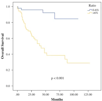
Two-thirds of patients (193) experienced no recurrence during follow-up. Recurrence was observed in 98 (33.7 %) cases; distant metastasis occurred in 34 % of these patients. Both, RFS and OS differed significantly if patients were stratified for LNR. Log-rank test was \0.001 for RFS (Fig.1) and OS (Fig.1), each. RFS and OS remained sig-nificant considering LNR if only patients with a pN2a and pN2b were included in analysis (Fig.2). LNR 0–6 % had a significant favourable prognosis compared to LNR higher than 6 % in those patients, with a p-value of 0.009 and [0.001 for RFS and OS, respectively. Only seven patients with a pN1 had a LNR of higher than 6 %; therefore, comparison of RFS and OS in these patients could not be sufficiently evaluated, and statistics were not valid.
If survival curve of patients with a negative neck were compared to patients with a positive neck and a lymph
node ratio 0–6 %, there was no difference in run of curves. Log-rank test was not significant at p = 0.561 (Fig.3).
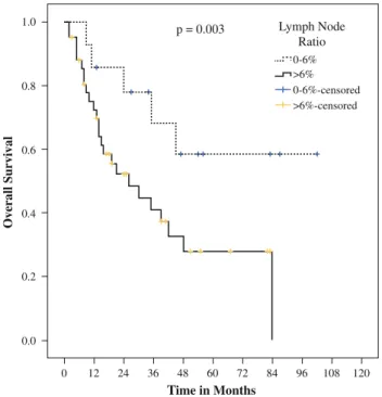
In patients with extracapsular spread, RFS was not significant considering LNR. However, impact of lymph node ratio on OS was significantly different even in patients with extracapsular spread (p = 0.003) (Fig.4). There was no significant difference in RFS and OS of patients without extracapsular spread after data was strat-ified for TNM status.
0 0.0 0.2 0.4 0.6 0.8 1.0 12 24 36 48 60 72 0-6% 6-13% >13% 0-6%-censored 6-13%-censored >13%-censored Time in Months
Recurrence Free Survival
84 96 p = <0.001
Lymph Node Ratio
108 120 132 144
FIG. 1 Recurrence free survival considering lymph node ratio
.00 0.0 0.2 0.4 0.6 25.00 0.8 1.0 50.00 75.00 p < 0.001 Months Overall Survival 100.00 125.00 Ratio 0-6% >6%
FIG. 2 Overall survival depending of lymph node ratio in patients with a pN2a or pN2b status
When the number of positive lymph nodes is considered as sole factor, RFS and OS differed significantly if 0, 1, 2–5, or more than five lymph nodes were affected. P value for RFS and OS was 0.004 and \0.001, respectively. However, if numbers of lymph nodes were stratified with lymph node ratio, numbers of affected lymph nodes were not significant any more.
Lymph node ratio remained a significant factor for RFS and OS even if postoperative radiotherapy was applied (p = 0.005 for each); however, no significance was detected if radiochemotherapy was applied postoperatively as adjuvant therapy.
DISCUSSION
It is advocated that the absence/presence of cervical lymph node metastases is the most important prognostic factor in HNSCC. The challenge is to evaluate accurately the prognostic impact of positive cervical lymph nodes, because N-classification does not predict tumour-free survival.10
Among OSCC patients with positive neck nodes, Shing-aki et al.11 proposed that nodal classification may not necessarily predict prognosis. Descriptive TNM staging cannot be used as survival predictive tool if not combined with other descriptive prognostic factors, such as extracap-sular spread, tumour infiltration, biomarkers to further predict the biologic behaviour of an individual tumour, and last but not least surgical success. In other tumour sites, TNM status also is questioned as an exact predictor of sur-vival. Lymph-node ratio (LNR) has been described as a predictor of survival in patients with head and neck squa-mous cell carcinoma. Furthermore, LNR is described to be superior to TNM nodal status in patients with bladder can-cer.12,13 Ooki et al.14 described the prognostic benefit of LNR over TNM in advanced oesophageal squamous cell carcinoma. In this article, lymph node ratio is evaluated regarding its power for RFS and OS prediction.
Clayman et al.15confounded lymph node levels at highest risk for lymph node metastasis and set recommendations of selective procedure for every tumour site in patients with HNSCC. This hypothesis is supported by Patel et al.16and Shepard et al.17 who could not find a significant benefit of comprehensive neck dissection versus selective neck dissection for locoregional recurrence-free survival. With the knowledge of lymph node levels most likely at risk for each tumour entity, and the reassurance that a radical neck dissection is not superior to selective procedures by the literature mentioned, LNR could be a safety net along the way of selective lymph node surgery. It is suggested that number of lymph nodes differ between tumour patients and healthy people.18Beasley et al.19 dem-onstrated intratumoral lymphogenesis by using a lymphatic endothelial marker, LYVE-1. Others described an increased lymph vessel density in patients with oropharyngeal cancer.20 Hence, it is assumed that lymphatic tumour spread is sup-ported by tumour-associated lymphogenesis and numbers of excised lymph nodes during neck dissection may be of importance to evaluate its potential for lymphogenesis and may be a reflection of tumour biology characteristics.
.00 0.0 0.2 0.4 0.6 0.8 1.0 20.00 40.00 60.00 p = 0.561 Months Overall Survival 80.00 100.00 pN pNO pN+ pNO-censored pN+-censored 120.00
FIG. 3 Overall survival in patients with a pN0 or pN? status if LNR is 0–6 % in patients with pN? 0 0.0 0.2 0.4 0.6 0.8 1.0 12 24 36 48 60 Time in Months Overall Survival 72 p = 0.003 Lymph Node Ratio 0-6% >6% >6%-censored 0-6%-censored 84 96 108 120
Patients with a single positive lymph node (pN1 or pN2a) would benefit from lymph node ratio as a predictive factor, for example in case of a diagnostic lymph node dissection that happens to be one single positive lymph node associated with an unfavourable LNR. According to the LNR the patient may be offered an extension of his ND to improve disease-specific survival. With the use of LNR, underestimation of the neck situation might be prevented. Patients with more than one positive ipsilateral lymph node would benefit from LNR, because locoregional tumour spread could be evaluated. For example, if two lymph nodes are affected and a large number of excised lymph nodes around those affected nodes are tumour-free the possibility of metastasis to other areas is less likely, similarly if metastasis of level 5 is present in absence of involvement of levels 4 or 3.21 On the contrary, if a comprehensive neck dissection delivers wide spread nodal metastasis, distant metastasis is more likely to occur and therefore additional treatment has to be considered.
Two situations should be mentioned where LNR cannot be regarded as a diagnostic tool: (1) in case of lymph node metastasis to the contralateral neck (pN2c); and (2) if one or more lymph nodes are larger than 6 cm (N3) in diam-eter. In case of pN2c, LNR is not valid, due to locoregional tumour spread to the contralateral side; therefore, a bilat-eral comprehensive neck dissection would be necessary to use lymph node ratio for prediction. Likewise, in patients with pN3, clear differentiation between one single lymph node bigger than 6 cm or a conglomerate of smaller lymph nodes often is not possible.
Importance of extracapsular tumour spread in patients with HNSCC is well documented in the literature.22Data of this study could demonstrate the validity of LNR even in patients with extracapsular spread. A ‘‘surrounding wall’’ or effective biologic barrier of not affected lymph nodes seems to play a role in preventing distant metastasis.
According to the presented data—with the limitation of a retrospective data analysis—LNR cannot only be used to evaluate the effectiveness of the surgery, but also implicate further therapies. Importance of LNR for decision making towards adjuvant therapy seems to be a highly accurate question that needs support by further prospective studies on this issue. Unfavourable high lymph node ratio ([6 %) should undergo further comprehensive surgery (if this was not the case in first place) or be treated with adjuvant therapy. The data presented demonstrated the unfavourable prognosis of high lymph node ratio if solely radiotherapy was applied. In combined postoperative treatment (radio-chemotherapy), RFS and OS did not differ. However, the indication that patients with higher LNR should be treated with radiochemotherapy should be taken with precaution, because the number of patients in this subgroup was only 37, thus limiting the statistical significance.
Some patients, especially in case of a pN2a status, might be treated sufficiently by neck dissection only—with the benefit of patient comfort, morbidity, and the chance of radiotherapy in case of a recurrence—because of a benign lymph node ratio.
In conclusion, is the hypothesis of whether LNR is superior than TNM evaluated? In patients with a positive lymph node status, but a lymph node ratio of 0–6 %, we could demonstrate that RFS and OS did not differ comparing to patients with a negative lymph node status. We could demonstrate the significant difference in predicting RFS and OS in patients with a pN2a and pN2b status after stratifying lymph node ratio. Lymph node ratio remained significant in patients with an extracapsular spread—contrary to TNM status. Therefore, we suggest that LNR is used as a predictor for survival in patients with an affected ipsilateral neck. If contralateral neck is affected and/or lymph nodes are a conglomerate, lymph node ratio is not valid.
ACKNOWLEDGEMENT There is no financial disclosure. CONFLICT OF INTEREST Authors declare no conflict of interest.
REFERENCES
1. de Jong RJB, Hermans J, Molenaar J, Briaire JJ, le Cessie S. Prediction of survival in patients with head and neck cancer. Head Neck. 2001;23(9):718–24.
2. Manikantan K, Sayed SI, Syrigos KN, et al. Challenges for the future modifications of the TNM staging system for head and neck cancer: case for a new computational model? Cancer Treatment Rev. 2009;35(7):639–44.
3. Ebrahimi A, Clark JR, Zhang WJ, et al. Lymph node ratio as an independent prognostic factor in oral squamous cell carcinoma. Head Neck. 2011;33(9):1245–51.
4. Gil Z, Carlson DL, Boyle JO, et al. Lymph node density is a significant predictor of outcome in patients with oral cancer. Cancer. 2009;115(24):5700–10.
5. Shrime MG, Bachar G, Lea J, et al. Nodal ratio as an independent predictor of survival in squamous cell carcinoma of the oral cavity. Head Neck. 2009;31(11):1482–88.
6. Lanzer M, Kruse A, Lu¨bbers HT, Zemann W, Reinisch S. Lymph node ratio and capsule penetration as independent risk factors in head and neck squamous cell carcinoma. Head Neck Oncol. 2012;5(4):89–97.
7. Urban D, Gluck I, Pfeffer MR, Symon Z, Lawrence YR. Lymph node ratio predicts the benefit of post-operative radiotherapy in oral cavity cancer. Radiother Oncol. 2013;106(1):74–9. 8. Robbins KT, Clayman G, Levine PA, et al. Neck dissection
classification update—revisions proposed by the American Head and Neck Society and the American Academy of Otolaryngol-ogy-Head and Neck Surgery. Arch Otolaryngol Head Neck Surg. 2002;128(7):751–8.
9. Lausen B, Schumacher M. Maximally selected rank statistics. Biometrics 1992;48(1):73–85.
10. Goldstein DP, Bachar GY, Lea J, et al. Outcomes of squamous cell cancer of the oral tongue managed at the Princess Margaret Hospital. Head Neck. 2013;35(5):632–41.
11. Shingaki S, Takada M, Sasai K, et al. Impact of lymph node metastasis on the pattern of failure and survival in oral carcino-mas. Am J Surg. 2003;185(3):278–84.
12. Svatek R, Shariat SF. Re: lymph node density is superior to TNM nodal status in predicting disease-specific survival after radical cystectomy for bladder cancer: analysis of pooled data from MDACC and MSKCC. Eur Urol. 2008;54(3):690–1.
13. Kassouf W, Agarwal PK, Herr HW, et al. Lymph node density is superior to TNM nodal status in predicting disease-specific survival after radical cystectomy for bladder cancer: analysis of pooled data from MDACC and MSKCC. J Clin Oncol. 2008;26(1):121–6. 14. Ooki A, Yamashita K, Kobayashi N, et al. Lymph node
metas-tasis density and growth pattern as independent prognostic factors in advanced esophageal squamous cell carcinoma. World J Surg. 2007;31(11):2184–91.
15. Clayman GL, Frank DK. Selective neck dissection of anatomi-cally appropriate levels is as efficacious as modified radical neck dissection for elective treatment of the clinically negative neck in patients with squamous cell carcinoma of the upper respiratory and digestive tracts. Arch Otolaryngol Head Neck Surg. 1998;124(3):348–52.
16. Patel RS, Clark JR, Gao K, O’Brien CJ. Effectiveness of selective neck dissection in the treatment of the clinically positive neck. Head Neck. 2008;30(9):1231–6.
17. Shepard PM, Olson J, Harari PM, Leverson G, Hartig GK. Therapeutic selective neck dissection outcomes. Otolaryngol Head Neck Surg. 2010;142(5):741–6.
18. Friedman M, Lim JW, Dickey W et al. Quantification of lymph nodes in selective neck dissection. Laryngoscope. 1999;109(3):368– 70.
19. Beasley NJP, Prevo R, Banerji S, et al. Intratumoral lymphan-giogenesis and lymph node metastasis in head and neck cancer. Cancer Res. 2002;62(5):1315–20.
20. Stacker SA, Achen MG, Jussila L, Baldwin ME, Alitalo K. Lymphangiogenesis and cancer metastasis. Nat Rev Cancer. 2002;2(8):573–83.
21. Dias FL, Lima RA, Kligerman J, et al. Relevance of skip metastases for squamous cell carcinoma of the oral tongue and the floor of the mouth. Otolaryngol Head Neck Surg. 2006;134(3):460–5. 22. Myers JN, Greenberg JS, Mo V, Roberts D. Extracapsular spread:
a significant predictor of treatment failure in patients with squa-mous cell carcinoma of the tongue. Cancer. 2001;92(12):3030–6.