HAL Id: tel-02477360
https://tel.archives-ouvertes.fr/tel-02477360
Role of matrix stiffness in the resistance of squamous
cell carcinoma cells to anticancer therapies
Sanya-Eduarda Kužet
To cite this version:
Sanya-Eduarda Kužet. Role of matrix stiffness in the resistance of squamous cell carcinoma cells to anticancer therapies. Cell Behavior [q-bio.CB]. Université Côte d’Azur, 2019. English. �NNT : 2019AZUR4010�. �tel-02477360�
Etude du role de la rigidité matricielle dans la
résistance des cellules de carcinomes squameux
aux thérapies anti-cancéreuses
Sanya-Eduarda KUŽET
Institut de Recherche sur le Cancer et le Vieillissement, CNRS UMR 7284 - INSERM U1081
Présentée en vue de l’obtention du grade de docteur en Sciences d’Université Côte d’Azur Mention : Interactions moléculaires et cellulaires
Dirigée par Cédric GAGGIOLI
Soutenue le 22 Mars 2019, devant le jury, composé de :
Guerrino MENEGUZZI Directeur de Recherche Emeritus, IRCAN, Nice Président du jury
Danijela VIGNJEVIĆ Directrice de Recherche, INSERM, UPMC, Paris Rapportrice
Céline GONGORA Directrice de Recherche, INSERM, IRCM, Montpellier Rapportrice
Sophie TARTARE-DECKERT Directrice de Recherche, INSERM, C3M, Nice Examinatrice
Frédérique FALLONE Maître de Conference Universitaire MCU, IPBS, Toulouse Examinatrice
Cédric GAGGIOLI Directeur de Recherche, INSERM, IPMC, Nice Directeur de thèse
Summary
Role of matrix stiffness in the resistance of squamous cell carcinoma cells to anti-cancer therapies
Resistance to epidermal growth factor receptor (EGFR) targeted therapy triggered by the tumor niche in head and neck squamous cell carcinoma (HNSCC) represents a challenge in research and in clinics. Despite the fact that over 15% of HNSCC overexpress EGFR, HNSCC are refractory to EGFR Tyrosine Kinase Inhibitors (TKIs) targeted therapy and yet the molecular and cellular mechanisms of EGFR-TKIs resistance in HNSCC are unknown. The tumor niche plays an important role in conventional chemotherapeutic resistance. Cancer associated fibroblasts (CAFs), the most prominent stromal cell in tumor niche, participate in this process. Notably, CAFs are responsible for tumor tissue fibrosis an excessive extracellular matrix (ECM) remodeling that increases matrix stiffness. In carcinoma cells, adhesion to stiff substrate triggers mechano-dependent intracellular signaling pathways that favor tumor resistance to conventional chemotherapies.
My work demonstrates that ECM stiffening is responsible for a significant increase of squamous cell carcinoma (SCC) survival upon the treatment with EGFR TKIs, conventional chemotherapies and combination of both. Over 60% more cells survive treatment with the gefitinib EGFR TKI compared to cells plated on soft matrix. Same effect was observed on matrix derived from CAFs that is known to be stiffer compared to the one derived from fibroblasts isolated from normal skin. Further analysis revealed an induction of partial epidermal-to-mesenchymal transition (EMT) in cells plated on rigid matrices. EMT is know to play a role in resistance of cancer cells to treatments, and I have demonstrated that downregulation of know transcriptional factors involved in EMT leads to an increase of cell susceptibility to EGFR TKI when plated on stiff matrix. To understand in more detail what drives the resistance of SCC cells when plated on stiff we conduced an RNA sequencing.
RNA sequencing of SCC12 cells plated on soft and stiff matrix revealed AXL as main driver of EGFR TKI resistance in HNSCC. I was able to demonstrated that inhibiting AXL in SCC cells, lying on stiff matrices, reverts the EGFR TKI resistance triggered by the tumor niche. Moreover, I show in 3D cell culture the importance of combining AXL and EGFR TKI in treatment of SCCs. Our overall goal was to identify novel therapeutic targets with reduced resistance opportunity.
Finally, research presented in this manuscript carries potential in establishing a prediction biomarker to the response of HNSCCs and other cancers to EGFR TKIs.
Résumé
Etude du role de la rigidité matricielle dans la résistance des cellules de carcinomes squameux aux thérapies anti-cancéreuses
Dans notre modèle de carcinome épidermoïde humain de la tête et du cou (HNSCC), si l’on observe une surexpression du récepteur au facteur de croissance épidermique (EGFR) dans plus de 15 % des cas, les cellules cancéreuses (SCC) sont réfractaires à un traitement par des inhibiteurs de l’activité Tyrosine kinase de l’EGFR (EGFR TKI) tels que le Géfitinib et l’AG1478. A ce jour, les mécanismes moléculaires et cellulaires impliqués dans la résistance des HNSCC aux EGFR TKI sont encore inconnus. On sait aujourd’hui que les niches tumorales jouent un rôle crucial dans la résistance des cellules cancéreuses aux traitements chimiothérapeutiques conventionnels et que les fibroblastes associés au cancer (CAF) présents dans ces niches, participent très certainement à ce processus. Les CAF sont notamment responsables de la fibrose du tissu tumoral et d’un remodelage excessif de la matrice extracellulaire (MEC) aboutissant à une augmentation de sa rigidité. Dans les cellules de carcinome, l'adhésion sur un substrat rigide déclenche des voies de signalisation intracellulaires mécano-dépendantes qui favorisent la résistance des tumeurs aux chimiothérapies conventionnelles.
Mes travaux démontrent que la rigidification de la MEC est responsable d'une augmentation significative de la survie des cellules de carcinome épidermoïde (SCC) en réponse aux traitements par des ITK de l'EGFR, aux chimiothérapies conventionnelles ou à une combinaison des deux. Comparé à des cellules ensemencées sur matrice molle, des cellules cultivées sur matrice rigide survivent mieux (+60%) au traitement avec le Gefitinib (EGFR TKI). Le même effet a été observé sur une matrice dérivée de CAF dont on sait qu'elle est plus rigide que celle dérivée de fibroblastes isolés d'une peau normale. Une analyse plus poussée a révélé une induction partielle de la transition épidermo-mésenchymateuse (EMT) dans des cellules SCC étalées sur des matrices rigides. L’EMT joue un rôle dans la résistance des cellules cancéreuses aux traitements, j’ai ainsi démontré que la régulation négative des facteurs de transcription connus pour être impliqués dans l’EMT conduit, dans le cas de cellules ensemencées sur une matrice rigide, à une augmentation de la sensibilité cellulaire aux EGFR TKI.
Pour comprendre plus en détails ce qui induit la résistance des cellules SCC lorsqu’elles sont étalées sur une plaque rigide, nous avons effectué un séquençage d’ARN. Le séquençage de l'ARN des cellules SCC12 étalées sur une matrice souple et rigide a révélé que la protéine AXL pouvait contribuer à la résistance au Géfitinib des HNSCC. J'ai pu démontrer que l'inhibition de l’expression d'AXL dans les cellules SCC ensemencées sur des matrices rigides, réduit totalement la résistance à l'EGFR TKI induite par la niche tumorale. De plus, je montre, en culture cellulaire 3D, l’importance de combiner AXL et EGFR TKI dans le traitement des SCC.
Notre objectif global était d'identifier de nouvelles cibles thérapeutiques présentant une opportunité de résistance réduite. Les recherches présentées dans ce manuscrit ont le potentiel d’établir un biomarqueur de prédiction de la réponse des HNSCC et d’autres types de cancers aux EGFR TKI.
Acknowledgments
First of all, I would like to thank Dr. Danijela Vignjević and Dr. Celine Gongora for agreeing to review my work. Thank you Dr. Sophie Tartare-Deckert, not only for accepting to be the examiner of my PhD dissertation, but for your kind advices and time as my internal advisor. Thank you Dr. Frederique Fallone for examining my work. Lastly, I would like to thank Professor Emeritus Guerrino Meneguzzi for his support in the beginning of my thesis on the RDEB project and for accepting to be the president of my jury. It is a privilege to have you all in my PhD committee.
I feel tremendous gratitude for my PhD mentor, Dr. Cedric Gaggioli. Thank you for giving me an opportunity to develop my PhD project and for believing in me from the day one. I am a better, more mature person today thanks to your guidance and support. I learned a lot from you not only about science, but also life and myself, and these lessons I will always remember. I am thankful you gave me the opportunity to mentor students and learn by teaching them. Thanks Cedric!
I had happy I worked with amazing people in the lab that pushed me daily to learn and grow. Thank you Heloise for always being there for me and for all your patience and understanding. You made my PhD so much better. You are the best scientist I have met and your work ethic is out of this world! I am excited to see how great you will be! Thank you Isabelle, for literally everything. You were so helpful and kind with all my challenges and French. I cannot thank you enough. I would like to thank Majdi that welcomed me in the lab so generously and Lavinia that was only with us for a short time but left a permanent impact on me. Thank you Stephanie for all the fun that we had. Thomas, I would like to thank you for your scientific advices and jokes we shared. I thank my students Zanna, Cami and Nadia for allowing me to help them. Lastly, I thank two latest members of our team Enora and Joan. Enora, thank you for your support and belief in the project. You made me feel so proud and I cherish that. You are a great, kind-hearted person. Joan, I wish you all the best in the future – you are going to do great.
Thank you Alex for wonderful conversations we had and for caring about me. I felt so connected with you when we first met and it will always stay like that. Lorenzo, thank you for listening to my never-ending complaining. I will miss our coffee breaks.
I would like to thank two strong female role models I had in my scientific career, professor Manuela Baccarini that gave me an opportunity that, at the time, I did not completely deserve. You were the first mentor I had that made me believe in myself and it changed my life. Without your support I would be where I am today. Dr. Josipa Raguž, if I say thank you it cannot even begin to describe what kind of impact you had on me.
My PhD days wouldn’t be the same without my Signalife family. Thank you Gaia, Tomas, Ramona, Paula. Thank you Anthony for making my first days in Nice so easy and joyful.
To my friends, Torsten, Hereroa and Racha.
Torsty, thank you for being such a true, honest friend. We are the same kind of crazy.
Hereroa, you were such an important part of my PhD. Thank you for all the good times we shared and your help with everything. It would be impossible to survive without you.
Thank you Racha! As I once told you – meeting you was worth it all. If I get to keep you in my life, I have succeeded. You are like my twin; we are made out of same material. I never though I will find someone who understands everything about me. I am forever grateful our paths crossed! You are unstoppable and meant for great things. All we do is win win win, no matter what.
To my greatest friends in the whole world – Franka and Zrinka. You were there for me whatever, whenever and that is so rare to find in this world. You are my safe place, place where I can be who I am. Words cannot do you justice and never will, nevertheless I call you my family. You are my sisters and I love you forever no matter what. Franka you accepted me against all odds. Zrinka, you believed in me when it was hard to believe in myself.
Thank you Petra L, for listening to me and for being you – an amazing young woman! I wish we get to spend more unforgettable moments together. Maja, Marina my beautiful, smart girls, thank you for your friendship
Maja Vitas, you were such a great support in my darkest moments and I will appreciate it forever. Thank you for being so proud of me and for your honesty.
I am forever grateful and humbled to have so much love in my life.
I would like to thank all of those who were directly or indirectly part of my PhD journey. I wish to thank random encounters and moments that somehow had a positive impact and drive me to change. To all of those “small” moments that make up life.
To my family! To my Dad! To my Mom!
Thank you Tatinko, Duško Kužet, this is all for you! I love you beyond time and space. Thank you for loving me no matter what, your love was so pure and so liberating. I know you are proud of me, my greatest fan, I miss you everyday more.
To my wonderful Mother whose love is unconditional. Thank you Ilinka Štor, for making me strong and powerful. For being an example of a woman and a mother I wish to be one day. Without you none of this would be possible and I cannot thank you enough for always being on my side. All my success is your success. I love you Mommy.
Lastly, I would like to thank my best friend, my partner, my soul mate, my family – Luka Fatović. I love you with all my heart until the end of time. You showed me how life can be beautiful and how to accept and love myself like you are able to love me. Thank you for being there for me throughout all my PhD and all my ups and downs, so full of patience, understanding and support. I am the luckiest person in the world to have you and you are my greatest accomplishment and the very best thing in my life. I am so excited for our next chapter! Volim te praščiću!
To my Daddy (1957-2016),
FAGUS SYLVATICA
Never wish I be free of pain for you! Sneaking through my day
Following my steps Hidden in the back,
back of every story That you never told Waiting for my night
Sleeping next to me Present in my dreams, dreams I have you back
Just to keep you safe Taking all myself Living just for you Kept forever in the heart,
heart that worries much Memories of truth Pain does not belong to me
I belong to pain Sneaking through my day
Turning into love, Love beyond a life
I am not alone, pain will keep me safe!
List of abbreviations
2D: Two Dimensional
2DRT: Two-Dimensional Radiation Therapy
3D: Three Dimensional
3DCRT: Three-Dimensional Conformal Radiation Therapy
ABL: Abelson Murine Leukemia
ADAMs: A Disintegrin and Metalloproteinases
ADCC: Antibody-Dependent Cellular Cytotoxicity
AJCC: American Joint Committee on Cancer
AML: Acute Myeloid Leukemia
ATP: Adenosine TriPhosphate
AR: Amphiregulin
AXL: Anexelekto
BCL-2: B-cell lymphoma 2
BTC: β-cellulin
CAFs: Cancer Associated Fibroblasts
CAM-DR: Cell Adhesion-Mediated Drug Resistance
CCND1: Cyclin D1
CDKN2A: Cyclin-Dependent Kinase Inhibitor 2A
CTCA: Cancer Treatment Centres of America
DAPI: 4′,6-DiAmidino-2-PhenylIndole
DDR: Discoidin Domain Receptors
DNA: DeoxyriboNucleic Acid
ECM: ExtraCellular Matrix
ECS: ExtraCapsular Spread
EGF: Epidermal Growth Factor
EGFR: Epidermal Growth Factor Receptor
EMT: Epithelial-to-Mesenchymal Transition
EPG: Epigen
EPR: Epiregulin
ERK: Extracellular signal–Regulated Kinase
FAK: Focal Adhesion Kinase
FAP: Fibroblast Activation Protein
FDA: Food and Drug Administration
FGF: Fibroblast growth factor
FGF-BP: Fibroblast growth factor Binding Protein
FRET: Fluorescence Resonance Energy Ttransfer
FSP1: Fibroblast-Specific Protein 1
G3BP2: Ras GTPase-activating protein-Binding Protein 2
GAGs: GlycosAmino Glycans
Gas6: Growth arrest-specific gene 6
Gla: Gamma-carboxyglutamic acid
GPCR: G-Protein Coupled Receptors
HER: Human Epidermal growth factor Receptor
HGF: Hepatocyte Growth Factor
HNSCCs: Head and Neck Squamous Cell Carcinomas
HPV: Human Papilloma Virus
ICAM1: Intercellular Adhesion Molecule 1
IL-1: InterLeukin-1
IL-6: InterLeukin-6
ILK: Integrin-Linked Kinase
IMRT: Intensity-Modulated Radiation Therapy
IRF: Interferon Regulatory Factor
JAK: Janus Kinase
JNK: c-Jun N-terminal kinase
KRAS: Kirsten rat sarcoma 2 viral oncogene homolog
LA-SCCHN: Locally Advanced Squamous Cell Carcinoma of the Head and Neck
LIF: Leukemia Inhibitory Factor
LOX2: Lysil Oxidase-like 2 protein
MIS: Minimally Invasive Surgery
MKK6: Mitogen-activated protein Kinase Kinase 6
MMP: Metalloproteinase
MRI: Magnetic Resonance Imaging
MRTF: Myocardin and Related Transcription Factors
MSCs: Mesenchymal Stem Cells
NF-κB: Nuclear Factor-kappa B
NRG: Neuregulin
NSCLC: Non-Small Cell Lung Carcinoma
NURD: Nucleosome Remodeling Deacetylase
PBRT: Proton-Beam Radiation Therapy
PDAC: Pancreatic Cancer
PDGF: Platelet-Derived Growth Factor
PGs: ProteoGlycans
PI3KCA:
PhosphatIdylinositol-4,5-bisphosphate 3-Kinase Catalytic subunit Alpha
PKB/ AKT: Protein Kinase B
PLCγ: PhosphoLipase C-γ
pRb: Retinoblastoma protein
PTB: Phosphotyrosine binding domain
PTEN: Phosphatase and Tensin homolog
Raf: Rapidly Accelerated Fibrosarcoma
RNA: RiboNucleic Acid
ROCK: Rho-associated protein kinase
RTKs: Receptor Tyrosine Kinases
SCC: Squamous Cell Carcinoma
SDF-1: Stromal cell-derived factor 1
sE-cad: soluble E-cadherin
SHBG: Sex Hormone-Binding Globulin
siRNA: Small Interfering RNA
SPARC: Secreted Protein Acidic and Rich in Cysteine
SRC: Sarcoma
SRF: Serum Response Factor
STAT: Signal Transducer and Activator of Transcription
TAMs: Tumor-Associated Macrophages
TAZ: Transcriptional co-activator with PDZ-binding motif
TFs: Transcription Factors
TGFα: Transforming Growth Factor alpha
TGFβ: Transforming Growth Factor beta
TKIs: Tyrosine Kinase Inhibitors
TLM: Transoral Laser Microsurgery
TME: Tumor microenvironment
TNFα: Tumor necrosis factor alpha
TNFAIP3: Tumor Necrosis Factor α-Induced Protein
TNM: umors tumor-node-metastasis
TORS: TransOral Robotic Surgery
TRAIL: Tumor Necrosis Factor-related Apoptosis-Inducing Ligand
VEGF: Vascular Endothelial Growth Factor
WHO: World Health Organisation
YAP: Yes-Associated Protein
αSMA: alpha Smooth Muscle Actin
List of figures and tables
Figure 1. 5-year Relative Survival of Oral Cavity and Pharynx Cancers depending on the stage ... 29
Figure 2. Head and neck regions affected by the squamous cell carcinomas ... 31
Figure 3. Accumulation of molecular changes in cancerogenesis of head and neck cancers and phenotypical progression ... 35
Figure 4. Composition of tumor microenvironment ... 46
Figure 5. Molecular influence of extracellular matrix on tumor cells ... 49
Figure 6. Difference in rigidity of various tissues measured in pascals, including tumor tissue ... 51
Figure 7. Matrix remodelling impacts cancer cells behaviour on multiple levels ... 53
Figure 8. Schematic representation of mechanotransduction carried out by integrin activation ... 54
Figure 9. Different signaling pathways that are modified as a result of ECM stiffening ... 56
Figure 10. Relationship between stiffness and progression of EMT ... 59
Figure 11. Various mechanisms of fibroblast activation ... 69
Figure 12. Radical neck dissection in patients sufferring from HNSCCs ... 77
Figure 13. Targeted therapies developed against common ErbB receptors for the treatment of HNSCCs and corresponding molecular pathways ... 86
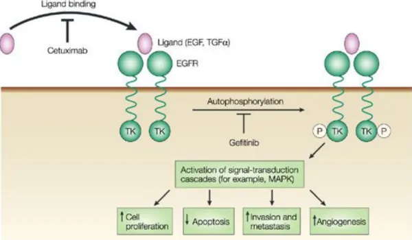
Figure 14.Cetuximab blocks the binding of EGF to its receptor and prevents its activation ... 88
Figure 15. Treatment decision tree for locally advanced squamous cell carcinoma of the head and neck (LA SCCHN) ... 91
Figure 16. CAFs play an important role in chemoresistance through various mechanisms ... 96
Figure 17. Mechanism of resistance to EGFR inhibitors ... 98
Figure 18. Structural differences between three members (TYRO3, AXL and MERTK) of the TAM family receptors ... 101
Figure 19. AXL drives the changes in cellular signaling pathways during EMT ... 104
Figure 20. Gas6/AXL signaling schema ... 105
Figure 21. AXL and EGFR cooperate in cetuximab resistance ... 107
Figure 22. SSC12 cells plated on the stiff matrix are more resistant to treatment with EGFR-TKIs .. 128
Figure 23. SSC12 cells plated on the stiff matrix are more resistant to treatment with EGFR-targeted therapy, monoclonal antibody cetuximab ... 130
Figure 24. SCC12 cells transfected with siRNA against EGFR lose their ability to proliferate on soft but not on stiff matrix ... 130
Figure 25. Stiff matrix protects SCC12 cells from cell death ... 132
Figure 26. Stiffness mediates protection against TKIs gefitinib and AG1478 inCAL27, CAL166 and SCC25 cell lines ... 133
Figure 27. SSC12 cells plated on the 12 and 50kPa are more resistant to treatment with gefitnib .... 134
Figure 28. SCC12 cells plated on matrix produced by nHF+TGFβ are more resistant to treatment with gefitinib ... 135
Figure 29. SSC12 cells plated on the stiff matrix are more resistant to treatment with traditional chemotherapeutic agents, cisplatin, docetaxel and 5-fluorouracil ... 137
Figure 30. SSC12 cells plated on the stiff matrix are more resistant to treatment with traditional chemotherapeutic agents, cisplatin, docetaxel and 5-fluorouracil in combination with gefitinib ... 138
Figure 31.SSC12 cells plated on the stiff matrix are more resistant to treatment with traditional chemotherapeutic agents, cisplatin, docetaxel and 5-fluorouracil in combination with cetuximab ... 138
Figure 32. YAP/TAZ knock down in SCC12 cells leads to an increase in sensitivity to gefitinib when plated on stiff matrix ... 140 Figure 33. SCC12 cells in stiff condition display partial EMT ... 142
Figure 34. Knock out of Zeb1, Zeb2, Snai1, Snai2 and Twist in SCC12 cells leads to an increase in sensitivity to gefitinib when plated on stiff matrix ... 145
Figure 35. RNA sequencing revealed an upregulation of RTK AXL. ... 147 Figure 36. EGFR Tyrosine Kinase inhibitor resistance pathway ... 148 Figure 37. ECM-receptor interactions ... 149 Figure 38. NF-KAPPA B signaling pathway ... 150
Figure 39. RNA sequencing revealed an upregulation of AXL, EGFR, COL4A1, VIM, TNC, FN1, ICAM1, NFKB1, RELA and BCL2 ... 151 Figure 40. AXL is overexpressed in SCC12 cells plated on stiff matrix ... 152 Figure 41. Expression of AXL is under the control of YAP/TAZ co-transcriptional factors. ... 152 Figure 42. Treatment of R428 and gefitinib overrides stiffness mediated resistance in SCC12 cells . 154 Figure 43. SSC12 cells plated on the stiff matrix are not resistant to treatment with AXL inhibitor and EGFR-TKI gefitinib ... 155 Figure 44. R428 and gefitinib induce an increse of cell death in SCC12 spheroids ... 156 Figure 45. SSC12 cells plated on the stiff matrix are not resistant to treatment with traditional chemotherapeutic agents, cisplatin, docetaxel and 5-fluorouracil in combination with R428 ... 157 Figure 46. Expression of AXL is overturned by a combination of EGFR and AXL TKIs ... 158 Figure 47. ICAM1 and A20 are overexpressed in SCC12 cells plated on stiff matrix ... 160
Figure 48. Knock out of A20 in SCC12 cells leads to an increase in sensitivity to gefitinib when plated on stiff matrix ... 161 Figure 49. Oncogenic functions of soluble fragment of E-cadherin ... 182 Figure 50. Stiffness regulates resistance of SCC12 cells to anti-cancer therapies by inducing overexpression of various proteins involved in EMT, inflammation and cell survival ... 188
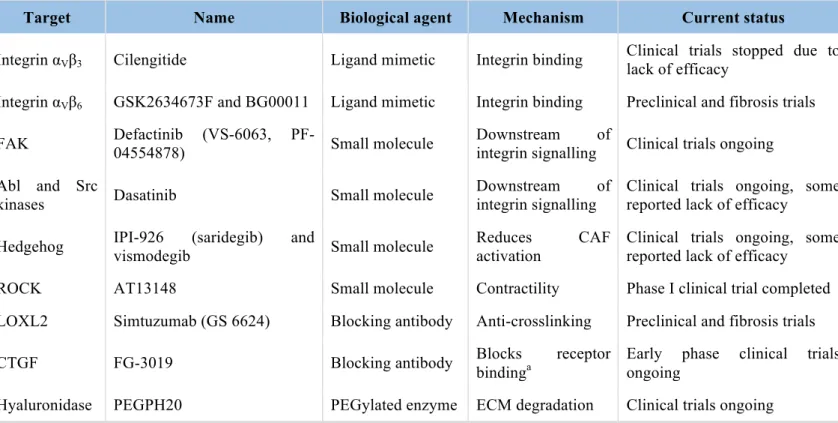
Table 1 Overview of drugs designed to target tumor mechanobiology and their current developing status
... 60 Table 2. TKIs and chemotherapeutic agents used ... 165 Table 3. List of siRNA used ... 166 Table 4. RT-qPCR primers ... 167
Table 5. Primary antibodies used ... 170
Table of Contents
SUMMARY ... 3RÉSUMÉ ... 5
ACKNOWLEDGMENTS ... 7
LIST OF ABBREVIATIONS ... 15
LIST OF FIGURES AND TABLES ... 19
CHAPTER 1: INTRODUCTION ... 27
1.1. HEAD AND NECK SQUAMOUS CELL CARCINOMAS ... 29
1.1.1.
CHARACTERISTICS of HNSCCs ... 31
1.1.2.
ONCOGENIC DRIVERS in HNSCCs ... 35
1.1.3.
EGFR and CANCERS ... 39
1.1.4.
CONCLUSION ... 42
1.2. TUMOR MICROENVIRONEMNT ... 43 1.2.1.
COMPOSITION of TME– GENERAL OVERVIEW ... 45
1.2.2.
EXTRACELLULAR MATRIX FOUND in TUMORS ... 47
1.2.3.
RIGIDITY and REMODELLING of the ECM ... 50
1.2.4.
EPITHELIAL TO MESENCHYMAL TRANSITION and its RELATIONSHIP with STIFF MATRIX………….. ... 58
1.2.5.
STIFF ECM AS A THERAPEUTIC TARGET ... 60
1.2.6.
STROMAL CELLS in TME ... 62
1.2.7.
CONCLUSION ... 73
1.3. TREATMENT OF HNSCCS ... 75 1.3.1.
SURGICAL REMOVAL of HNSCCs ... 77
1.3.2.
RADIOTHERAPY in TREATMENT of HNSCCs ... 79
1.3.3.
CHEMOTHERAPEUTICAL APPROACH in HNSCCs ... 82
1.3.4.
TARGETED THERAPY in HNSCCs ... 86
1.3.5.
CONCLUSION ... 91
1.4. CANCER RESISTANCE ... 93 1.4.1.
CAFs and THEIR ROLE in RESISTANCE of CANCER CELLS to THERAPIES ... 95
1.4.2.
ROLE of ECM in CANCER RESISTANCE ... 97
1.4.3.
RESISTANCE of TUMORS to EGFR TARGETED THERAPIES ... 98
1.4.4.
ROLE of AXL in RESISTANCE to EGFR TARGETED THERAPY ... 101
1.4.5.
ROLE of TNFAIP3 in CANCER RESISTANCE ... 110
1.4.6.
CONCLUSION ... 112
CHAPTER 2: OBJECTIVES ... 115
CHAPTER 3: RESULTS ... 121
I. SUMMARY OF THE RESULTS ... 123 II. RESULTS ... 127 3.1.
PROOF of CONCEPT – STIFFNESS PROMOTES RESISTANCE in HNSCCs ... 129
3.2. WHAT MAKES CELLS on STIFF MATRIX RESISTANT? ... 143
3.3. RNA sequencing ... 147
3.4. MECHANISM BEHIND SCCs RESISTANCE to EGFR INHIBITORS ... 152
3.5. SUPPLEMENTAL DATA ... 160
CHAPTER 4: MATERIALS AND METHODS ... 163
CHAPTER 5: DISCUSSION AND PERSPECTIVES ... 175
5.1. EGFR AND STIFFNESS ... 178
5.2. EMT AND STIFFNESS ... 180
5.3. AXL SIGNALING IN RESISTANCE OF HNSCCS TO EGFR INHIBITORS AND CONVENTIONAL
CHEMOTHERAPY ... 185 5.4. CLINICAL RELEVANCE ... 190
CHAPTER 6: REFERENCES ... 195
Chapter 1:
1.1. HEAD and NECK SQUAMOUS CELL CARCINOMAS
Head and Neck Squamous Cell Carcinomas (HNSCCs) are a heterogenic group of cancers. Not only do they affect the whole head and neck region, driving the heterogeneity of their form (Vokes et al., 1993), but their molecular signature reveals a plethora of genetic changes not yet fully disclosed (Chung et al., 2004).
According to the statists gathered by the World Health Organization (WHO) in 2014, HNSCCs are the 7th most frequent cancer and 9th most frequent cause of death from cancer, killing approximately 400 000 people worldwide (World Health Organization, World Cancer Report 2014). Common cause of HNSCCs are tobacco and alcohol that have a synergistic effect, but other risk factors are reported like human-papilloma virus (HPV) and Epstein-Barr virus (Argiris et al., 2008). As in many other cancers, HNSCC is oftentimes curable if it is diagnosed early, however if it is diagnosed later outcomes can be unfavorable due to the disease reoccurrence and development of metastasis. In developed countries, 5-year survival in average is between 40 to 60% (Figure 1). It is very common that therapies designed for HNSCCs (chemotherapy, radiotherapy, targeted therapy and their combinations) fail in time, and patients become unresponsive to them (Beyzadeoglu et al., 2014, National Cancer Institute, Cancer Stat Facts: Oral Cavity and Pharynx Cancer). In this chapter I will review generalities and the molecular background of HNSCCs, whilst treatments designed for these cancers will be detailed in an individual chapter.
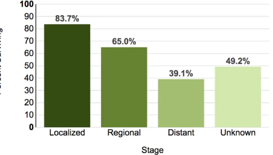
Figure 1. 5-year Relative Survival of Oral Cavity and Pharynx Cancers depending on the stage
If the cancer is found only in one part of the human body it is considered to be localised, and those tumors have the best 5-year prognosis, unfortunately if the cancer has spread regionally or distantly this prognosis is much worse. (Adapted from National Cancer Institute).
1.1.1. CHARACTERISTICS of HNSCCs
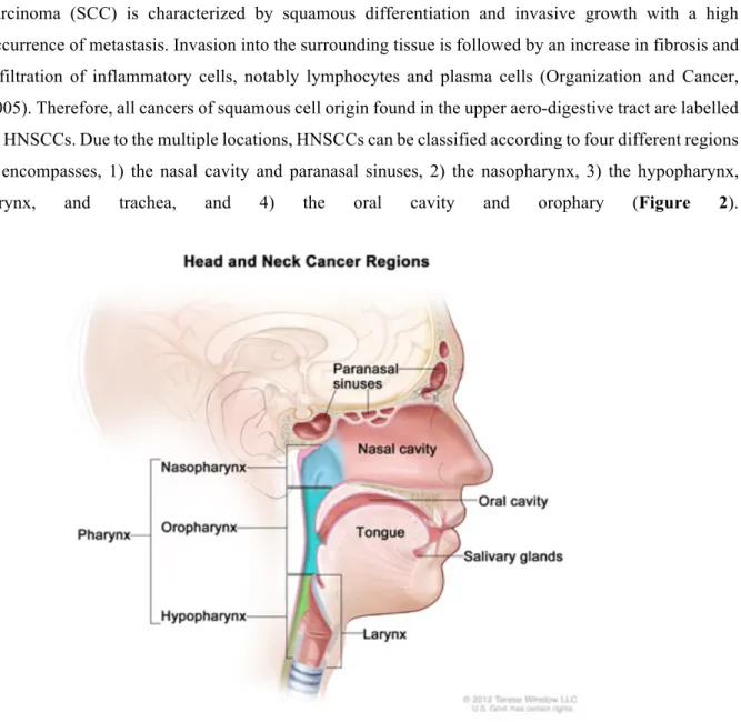
Majority of cancers (up to more than 90%) found in head and neck region are of squamous cell origin (Sanderson and Ironside, 2002), hence the name Head and Neck Squamous Cell Carcinomas (HNSCCs). Squamous cells are found in the outer layer of the skin and in the mucous membranes which are linings of ectodermic origin, covered in epithelium, and are involved in absorption and secretion. Mucous membranes line body cavities such as the airways and intestine (Fry and Vahabi, 2016). Squamous cell carcinoma (SCC) is characterized by squamous differentiation and invasive growth with a high occurrence of metastasis. Invasion into the surrounding tissue is followed by an increase in fibrosis and infiltration of inflammatory cells, notably lymphocytes and plasma cells (Organization and Cancer, 2005). Therefore, all cancers of squamous cell origin found in the upper aero-digestive tract are labelled as HNSCCs. Due to the multiple locations, HNSCCs can be classified according to four different regions it encompasses, 1) the nasal cavity and paranasal sinuses, 2) the nasopharynx, 3) the hypopharynx,
larynx, and trachea, and 4) the oral cavity and orophary (Figure 2).
Figure 2. Head and neck regions affected by the squamous cell carcinomas
This schematic representation describes all possible location of squamous cell carcinoma of head and neck; paranasal sinuses, nasal cavity, oral cavity, tongue, salivary glands, larynx, and pharynx (including the nasopharynx, oropharynx, and hypopharynx). Adapted from National Cancer Institute report on Head and Neck Cancers.
A) HNSCCs epidemiology
In general, HNSCCs are more common in men than in women. A disproportion between two sexes is attributed to the higher exposure to risk factors in men, such as tobacco, alcohol and UV light. In terms of age, HNSCCs are usually diagnosed in patients over the age of 50 (Warnakulasuriya, 2009). Nevertheless, 6% of cancers occur in people younger than 45 years and in counties with a higher incidents disease occurs before the patients turn 40 years old. Worldwide, HNSCCs affects approximately 600 000 people yearly and only 40-50% of patients will survive more than 5 years (Leemans et al., 2011). In European Union there is 67 000 new cases registered each year with highest male incident rates in France and Hungary. Indeed, in France, in 2010, the number of new cases was estimated to be 14 000, the majority of which (75%) were men. Low survival rate can be attributed to the fact that two third of patients are diagnosed in the advanced stage. Stage of the disease is determined by the extent of the tumor, presence of lymph-nodes metastases and distant metastases.
B) Risk factors involved in the development of HNSCCs
Tobacco and alcohol consumption are the two main causes of HNSCCs, and they appear to have a synergistic effect (Blot et al., 1988). Due to the campaigns against smoking in western world, occurrence of HNSCCs in the past decade has been slowly declining. On the other hand, subgroup of HNSCCs (in particular those of oropharynx) caused by the infection with HPV type16 and 18 are becoming more frequent, which could be correlated with in increase of HPV infections. Nowadays, 25% of HNSCCs are positive for the genomic DNA of HPV that exhibit their carcinogenic effects through viral proteins E6 and E7, that inactivate P53 and pRb (retinoblastoma protein) tumor repressor genes (D’Souza et al., 2007; Münger and Howley, 2002). HPV-negative and HPV-positive are usually distinguished by different clinicopathological and molecular characteristics. Interestingly HPV-positive tumors area associated with a favourable prognosis for the patients because of the positive response to the treatment (radiotherapy and chemotherapy). Besides these external factors, some genetic diseases such as Fanconi anemia provide intrinsic susceptibility to HNSCCs (Cloos et al., 1996; Kutler et al., 2003).
C) Molecular classification of HNSCCs
As I mentioned in the very beginning, 90% of head and neck cancers are squamous cells that led researches to believe it is a rather homogenous disease (Pai and Westra, 2009). However, recent observations have revealed that HNSCCs are quite heterogeneous, making it hard to establish accurate prognostics, plan the treatment, and finally identify the genes responsible for cancer development. In 2004 by taking advantage of expression profiling, Chung et al. highlighted the heterogeneity of HNSCCs and sought to identify novel markers for survival predictions. They analysed gene expression patterns of 60 HNSCCs tumors and identified 4 different subtypes with different clinical outcomes. The worst outcome was associates with Group 1 tumors subtype that were characterized by the high expression of Transforming Growth Factor Beta (TGFβ) that is know to correlate with poor clinical outcomes (Endo et al., 2000). Most HNSCCs express Epidermal Growth Factor Receptor (EGFR), but there is an additional evidence that Group 1 cancers have an activation of EGFR pathway. Besides the upregulated genes from EGFR signaling pathway such as Transforming growth factor alpha (TGFα), an EGFR ligand, a kinase downstream of EGFR – Mitogen-activated protein Kinase Kinase 6 (MKK6) and angiogenic switch molecule induced by Epidermal Growth Factor (EGF) - Fibroblast growth factor binding protein (FGF-BP); 15 out of 19 tumors in Group 1 tested by IHC were positive for activated, Tyr-1173 phosphorylated, EGFR. Data suggests that patients in Group 1 should be evaluated for the benefit of EGFR inhibitor treatments. The Group 2 tumors expressed strong mesenchymal cell signature due to the presence of fibroblasts and lack of epithelial characteristics, or cells that may have undergone epithelial to mesenchymal transition (EMT). Tumor organised under Group 3 are characterized by Cytokeratin 14 and 15 expression patterns that are similar to those found in normal tonsil epithelium samples suggesting that there are very few differences between premalignant and malignant state. These tumors showed fewer relapses and therefore best outcomes. The Group 4 tumors had an expression patters very similar to the gene pattern expression induced by cigarette smoke. Since majority of the cohort were heavy smokers, data obtained suggest that there could be variations in the response of smokers to cigarette smoke (Chung et al., 2004). Two years later, same authors went a step further and determined a high-risk signature for HNSCCs using formalin fixed and frozen tumor samples. They showed that genes involved in EMT and nuclear factor-κB (NF-κB) signalling are the most relevant molecular signature of high-risk tumors (Chung et al., 2006). EMT has been associated with late stage tumor progression and metastasis (Boyer et al., 2000) and, in HNSCCs has been previously associated with resistance to EGFR tyrosine kinase inhibitors such as erlotinib (Thomson et al., 2005; Yauch et al., 2005). It is worth to mention that they came up with a 75-gene list of potential prognostic markers some of which are metalloproteinase (MMP) 2, keratin 14, lysil oxidase-like 2 protein (LOX2), stratifin and galectin 1. Interestingly for the research I have done, MMP2, LOX2 and galectin 1 are all proteins involved in tumor microenvironment regulation which could potentially suggest that the HNSCCs tumors with strongly deregulated tumor stroma are the ones classified as high-risk. Nevertheless, data
acquired should be considered preliminary and more efforts are necessary to completely elucidate the HNSCCs subgroups since no clear connection has been established between the genetically identified subgroups and molecular classification. However, it is widely accepted that HPV-positive HNSCCs are a specific subgroup since the tumors are distinctive on a molecular level and have favourable clinical outcomes (Ragin and Taioli, 2007).
1.1.2. ONCOGENIC DRIVERS in HNSCCs
In HNSCCs the most common mutations are in the Transformation related Protein 53 (TP53) gene - 53%, deletions and mutations in the tumor suppressor gene Cyclin Dependent Kinase Inhibitor 2A (CDKN2A) - 58%, amplification of Cyclin D1 (CCND1) - 31%, Phosphatidylinositol 3-kinase (PI3KCA) - 34%, and lastly, EGFR genes - 15% (Argiris et al., 2008), leading to gene instability (Figure 3). Interestingly, EGFR is expressed in 90% of HNSCCs but gene amplification in observed in only 15%, and studies have shown that overexpression of EGFR is associated with poor prognosis (Ang et al.; Grandis and Tweardy, 1993; Rubin Grandis et al., 1998). However, no link has been established between EGFR expression and the effectiveness of treatments, and no other biomarker other than HPV is used for prognosis or treatment establishment.
Figure 3. Accumulation of molecular changes in cancerogenesis of head and neck cancers and phenotypical progression
Heamatoxylin and eosin staining of histological changes in HNSCCs shows the evloution of the tumor in parallel with the accumulation of genetic and epigenetic alterations. (Adaoted from (Argiris et al., 2008).
A) TP53
Malformations of p53 are the most common events in all cancers and HNSCCs are no exception. TP53 tumor suppressor gene is often referred to as a gatekeeper gene due to its role in regulation of the key events in cell, such as cell cycle, metabolism of cancer cells, DNA-repair, apoptosis and senescence. It is located in chromosome 17p13 and can be induced by a plethora of stress-like events like DNA-damage and inflammation (Bensaad and Vousden, 2007; Hussain and Harris, 2006). In HNSCCs point mutations of TP53 are often seen and disruptive mutations have been associated with short survival rate
post surgical removal. According to International Agency for Research on Cancer’s (IARC) TP53
mutation database, codons 238-245 are a hotspot region for mutations in HNSCCs. Somatic mutations are found in 50-80% of HNSCCs
Mutations of TP53 are associated with the use of tobacco (Soussi et al., 2005), although the relationship is not always significant and varies from study to study. Moreover, in the analysis of 420 patients TP53 mutations were more frequent in patients with advanced-stage HNSCCs and showed positive correlation with the poor survival of patients (Poeta et al., 2007). As for HPV-positive HNSCCs in which viral E6 protein binds and inactivates TP53, low frequency of mutations is observed. Regardless of the data, prognostic value of p53 is not yet evident.
B) CDKN2A
CDKN2A is a tumor suppressor gene located on chromosome 9p2 and associated with the cell cycle
progression. CDKN2A codes for 16INK4a protein that plays an important role in cell cycle and senescent through the regulation of cyclin D and cyclin-dependant kinase (CDK) 4/6 (Zhao et al., 2016). In HNSCCs, CDKN2A is often inactivated by mutations or methylations, chromosome loss or homozygous deletion (Reed et al., 1996). In cell cycle, an interaction between cyclins, CDKs and their inhibitors regulates the restriction points, and a stimulation from growth factor is necessary to break G1 cell cycle restriction point in order for cells to enter S phase.
Same as in the case of TP53, tobacco and alcohol consumption correlate with the apparent loss of p16 protein. Methylation in the promoter region, however, did not show any significant correlation with
C) CCND1
CCND1, which encodes for cyclin D1, is located on chromosome 11q13, and is amplified or
overexpressed in more than 30% of HNSCCs cases and even more frequently in HPV-negative tumors (D’Souza et al., 2007; Rothenberg and Ellisen, 2012). Some research shows, that cyclin D is overexpressed in 80% of HNSCCs (Kutler et al., 2003). Main function of cyclin D is a cell cycle regulator, but it can act as a cofactor by binding to transcriptional factors and DNA repair proteins (Poeta et al., 2007). For example, Cyclin D and CDK2 form a complex that phosphorylates Rb important for the S phase progression (Day et al., 2009), therefore these two mutations are not necessarily mutually exclusive explaining the high prevalence of these mutations. Overexpression of cyclin D is a good predictor of patient death caused by tongue cancer and overexpression of cyclin D and loss of P16 expression, together, are an indicator of a worse 5-year survival (Napier and Speight, 2008).
Together with the abrogation of p53, these changes cause cellular immortalization (Smeets et al., 2011).
D) PI3KCA
PI3K gene is located on chromosome 3q26 and represents complex family of lipid kinases that regulate
pivotal cellular processes such as survival, growth, proliferation, motility, cell adhesion, differentiation, morphology, cytoskeletal rearrangement and apoptosis (Murugan et al., 2008). PI3K family is divided into three classes (I, II and III) and different roles have been associated with each three, as well as with different isoforms within each group (Jean and Kiger, 2014) For example, class I consists of two subgroups IA and IB. Interestingly, IA is often recruited and activated at the cell membrane by growth factor receptor tyrosine kinase as EGFR and insulin receptor (Cantley, 2002; Vanhaesebroeck et al., 2001)
In a variety of cancers, notably breast cancer, melanoma and lung squamous cell carcinoma, PIK3CA codes for p110, catalytic subunit of PI3-kinase that is somatically mutated. In HNSCCs, study has been done on 151 tumors sample and around 30% of HNSCCs had a mutation in the PI3K pathway, making it the most mutated oncogenic pathway (Lui et al., 2013). Same was previously observed by others on tumor samples, but as well in HNSCCs cell lines (Murugan et al., 2008). Frequent mutations include E542K, E545K and H1047R hotspots, but others are reported as well. However, mutation responsible for the gain of function of the PIK3CA is still unknown.
What has been established is the correlation with mutations of multiple PI3K pathways and advanced stage of the disease (Lui et al., 2013). It is suggested that PIK3C can be used as a predictive biomarker and a potential target for further treatment development, although it has not yet been used for these purposes.
E) EGFR
When it comes to EGFR things are more complicated than they normally are. In HNSCCs it is believed that EGFR is overexpressed in 90% of the patients, but this data is still not corroborated and different ways of EGFR activation exists.
Besides the “standard” treatment for HNSCCs that includes surgery, radiotherapy, chemotherapy and often combinations of the three (that will be reviewed in more detail later on), EGFR is the only molecular target that has been exploited for treatment of patients. However, the success of the targeted therapy (EGFR-specific antibody) is debatable and more understanding is necessary to unveil the role of EGFR in HNSCCs.
1.1.3. EGFR and CANCERS
EGFR is the most studied, and one of the first characterized cellular oncogenes and the very first identified member of the receptor tyrosine kinase (RTK) family (Burgess et al., 2003; Cohen, 1962). EGFR belongs in the family of EGF receptor or ErbB family that includes EGFR itself, ErbB2 (HER2/Neu), ErbB3 (HER3) and ErbB4. ErbB receptors participate in a variety of cellular functions such as growth, differentiation, proliferation and motility (Ceresa and Peterson, 2014). Consequently, the role of each and every one of them is well established during cancerogenesis, for example in 1987 overexpression of HER2 was associated with poor prognosis in breast cancer (Slamon et al., 1987). EGFR is expressed in the majority of carcinomas. For example, besides in HNSCCs, it is found in 50 to 70% of lung, breast, prostate, kidney, ovarian and colon carcinomas (Normanno et al., 2003; Prenzel et al., 1999; Salomon et al., 1995).
Soon enough, EGFR became an important therapeutic target for many cancer types including HNSCCs. Thus, the concept of targeted therapy, intended to affect only tumor cells to reduce the side effects of conventional treatments, appeared in 1998 with the commercialization of the blocking antibody trastuzumab (Herceptin®) targeting HER2 (Gschwind et al., 2004). Since then, many anti-EGFR therapies have emerged, yet they only benefit a minority of patients because of innate or adaptive resistance. Therefore, understanding the molecular mechanisms implicating EGFR in tumor progression is important for the development of new therapeutic strategies.
A) Activation of ErbB receptors
I) Activation by ligand
ErbB receptors, including EGFR, exist in an inactive monomeric form. Most common way of activating EGFR is by numerous ligands, of which EGF is the most common and studied one (Cohen, 1962; Cohen and Carpenter, 1975). These ligands can be secreted in an autocrine (produced by the cells expressing the receptor), paracrine (secreted by other cells) or juxtacrine manner (Singh and Harris, 2005). Juxtacrine manner is explained as “where the molecule that induces the functional changes in the target cell remains associated with the plasma membrane of the signaling cell, rather than acting in the fluid phase” (Anklesaria et al., 1990).
The binding of a ligand causes a conformational change of the receptor, releasing the domain II responsible for the formation of homo or heterodimers.
Besides EGF, other ErbB receptors ligands can be classified in three groups according to their selectivity for the receptor: EGF, TGFα, amphiregulin (AR) and epigen (EPG) preferentially bind to EGFR; β-cellulin (BTC), heparin binding EGF (HB-EGF) and epiregulin (EPR) specifically activate EGFR and HER4; finally, neuregulins (NRG1 to 4) can bind to HER3 and / or HER4 (Carraway et al., 1997; Chang et al., 1997; Harari et al., 1999; Jura et al., 2009).
Upon activation, ErbB receptors are autophosphorylated on tyrosine residues present in the intracellular domain. Such phosphorylated residues allow anchoring of proteins with SH2 (Src homology) or phosphotyrosine binding (PTB) domains, that build adapter proteins as Shc, Crk, Grb2, Grb7 and Gab1, the Src, Chk and PI3K kinases, and the phosphatases SHP1 and SHP2. Adapter proteins transmit signals by activating various signaling pathways. Besides adapter proteins, ErbB receptors can activate transcription factors, such as proto-oncogenes c-fos, c-jun and c-myc (Yaffe, 2002). Diversity among activated signaling pathways is a consequence of the complexity of the dimers, their ligands and different phosphorylation sites of the receptor. All ErbB receptors and their ligands activate the RAS / RAF / MEK / Erk signaling pathway (Bazley and Gullick, 2005; Carpenter, 2003; Citri et al., 2003) and PI3K pathway, however, the degree of activation and kinetics vary among receptors (Elenius et al., 1999; Fedi et al., 1994; Prigent and Gullick, 1994). Conversely, the activation of the phospholipase PLCγ, Esp15 and Cbl is specific for the EGFR receptor (Chattopadhyay et al., 1999; Levkowitz et al., 1999).
II) Transactivation
ErB receptors can be activated in the absence of ligand by mechanism that are known as transactivation. Janus Kinase 2 (JAK2) can activate (phosphorylate) ErbB receptors via induction by growth factors like cytokines and hormones (prolactin) (Yamauchi et al., 1997). Similarly, Src kinase can activate EGFR by phosphorylating several tyrosine residues (Biscardi et al., 1999). On the other hand, G-protein coupled receptors (GPCRs) can also transactivate ErbB receptors. GPCRs are transmembrane proteins coupled to heterotrimeric G proteins composed of three subunits: Gα, Gβ and Gγ. The binding of a ligand to the GPCR results in the dissociation of the subunits of the G protein into Gα-GTP and Gβγ. Gα-GTP and Gβγ subunits control the activity of several enzymes including kinases, phospholipase C and adenylate cyclase. Daub et al were the first to demonstrate in 1996 the transactivation of EGFR and HER2 by GPCR ligands; lysophosphatidic acid, thrombin and endothelin (Daub et al., 1996). Upon transactivation, proteases from the family of A Disintegrin And Metalloproteinases (ADAMs) are activated and allow the cleavage of EGF family, such as HB-EGF, at the cell surface (Carpenter, 2003; Gschwind et al., 2003; Prenzel et al., 1999). Similarly, another study showed that the activation of the
B) EGFR mutations often found in HNSCCs
In 1986, EGFR was postulated as overexpressed in many cases of HNSCCs (Yamauchi et al., 1997), and later confirmed by different studies (Grandis and Tweardy, 1993; Hama et al., 2009). However, these observations are based on the immuno-stainings of tumor samples that are not always consistent due to the different protocols used and lack of negative controls (normal healthy tissue). Therefore, it is fair to say that EGFR is expressed in majority (90%) of HNSCCs rather than overexpressed, and only 15% of patients have EGFR amplified. Amplification was first reported by Ishitoya et al in 1989 and data about the percentage of tumors being oncogenically activated varies as well (Ishitoya et al., 1989). High-resolution single-nucleotide polymorphism arrays described 31% of cancers carrying the amplification at 7p11. This study was later confirmed on a higher number of samples using in situ hybridisation and immuno-staining (Sheu et al., 2009).
Besides amplification, there are other EGFR activating mutations found in HNSCCs such as point mutations, but also specific mutant forms of EGFR such as EGFRvIII that are found more frequently (Ekstrand et al., 1992). EGFRvIII corresponds to the deletion of exons 2 and 7 of the extracellular part of the receptor and codes for a constitutively active receptor. It has a particular effect on the intracellular signaling pathways causing increase in proliferation, and it has been show to decrease the effect of treatment (Sok et al., 2006).
In the matter of altered signalling, it is interesting to mention that signaling function of EGFR might differ from tumor to tumor. In some cases, EGFR activates Protein Kinase B (PKB) also known as Akt pathway, in others Ras-MAPK pathway, or it can induce CCND1 expression. This kind of multi-faced role can significantly alter the role of EGFR in HNSCCs and influence the outcome of the anti-EGFR treatment. Again, it is not so clear weather the overexpression of EGFR correlates with bad prognosis. For the moment 60% of studies show a correlation and 40% does not (Leemans et al., 2011). Although, for example, early study done by Maurizi et al. showed that the 5-year survival of patients with non-EGFR-expressing tumors was 81% versus 25% in positive patients (Maurizi et al., 1996).
1.1.4. CONCLUSION
HNSCCs are more complicated that it has been described at the early stage of research (Vokes et al., 1993). Nowadays, with the usage of new technologies it has become apparent that the story behind HNSCCs is far more complicated and, despite enormous efforts (Chung et al., 2004, 2006), it is still very difficult to pin-point and identify a specific molecular signature of HNSCCs. It would be of great value to identify prognostic markers based on a molecular signature of the tumor or even its histological transformation (Uramoto et al., 2010) in order to place patients into certain categories and predict the best treatment and, therefore, the outcome.
At the same time, tumor development is not limited to the intrinsic properties of cancer cells only and changes within them (many of which are described in this chapter). It is now widely accepted that microenvironment surrounding cancer cells undergoes changes as well, becoming tumor microenvironment and influencing all stages of cancerogenesis (Hanahan and Coussens, 2012). To fully understand HNSCCs we must look beyond the tumor cells themselves and expand our efforts to the environment they belong to. Several researchers have already took this approach and explored the role of fibroblasts and tumor microenvironment in HNSCCs describing a dynamic relationship (Liu et al., 2011; Sweeny et al., 2012; Tong et al., 2012a).
1.2. TUMOR MICROENVIRONMENT
Tumor development and progression is not solely dependant on the malignancy of cancer cells (Kalluri and Zeisberg, 2006). Cancer epithelial cells are supported by a complex microenvironment referred to as “Tumor MicroEnvironment” (TME) that provides a fertile soil for all stages of tumor advancement (Kuzet and Gaggioli, 2016a). Therefore, it is not surprising that the “seed and soil” hypothesis (Paget, 1889) is still as relevant as it was when it was first published in 1889 and numerous studies have endeavoured in further deciphering the role of TME in such a context. A collaborative nature of cancer cells and the supporting stroma results in chronically proliferative and disseminating organ-like structures characteristic for most human cancers, resulting in characteristic morphological cellular changes (large nucleus, irregular size and shape (Baba and Câtoi, 2007)), invasion and metastases (Hanahan and Coussens, 2012).
Therefore, the role of TME in tumor progression cannot be emphasises enough. It is implicated in all stages of cancer development, from growth, proliferation, invasion, metastasis to chemoresistance (Junttila and de Sauvage, 2013). In this chapter I will further elucidate its composition and role during cancerogenesis.
1.2.1. COMPOSITION of TME– GENERAL OVERVIEW
In the literature there is more than one classification of TME according to the cell origin, cell type, location, immune response-associated gene expression and gene expression.
When it comes to the origin of cells found in TME, we distinguish three mayor parts; cells of hematopoietic origin, cells of mesenchymal origin and non-cellular components. The proportion of these three components vary according to the tumor type and its progression (Egeblad et al., 2010; Quail and Joyce, 2013).
Cells of hematopoietic origin derived from red bone marrow are divided in two groups: (1) cells from lymphoid linage (T cells, B cells and natural killer cells) and (2) cells from myeloid linage (macrophages, neutrophils). These are immune cells found infiltrated within TME as a response to strong induction of inflammation at the tumor site. Besides being an immune response, these cells are implicated in the angiogenesis, tumor growth, migration and metastatic progression (Condeelis and Pollard, 2006; Murdoch et al., 2008).
Cells of mesenchymal origin include fibroblasts, mesenchymal stem cells (MSCs), adipocytes and endothelial cells. These cells participate in the angiogenesis, lymphogenesis, remodelling of extracellular matrix, tumor growth and, as well, in invasion of cancer cells. Bone marrow-derived fibroblasts and MSCs directly support cancer cells creating a favourable environment-specific niche and facilitating tumor progression (Quante et al., 2011). Until recently, adipocytes were considered merely an energy storage cells; however recent studies have revealed the importance of adipocyte-secreted factors (e.g. hepatocyte growth factor, HGF) in the progression of breast cancers (Dirat et al., 2011; Huang et al., 2017). Endothelial cells and pericytes, which form the walls of blood vessels, play a major role in vascular function and angiogenesis, as well as in the regulation of cancer cell diffusion (Butler et al., 2010).
Principal non-cellular component of TME is extracellular matrix (ECM) secreted by the cells of mesenchymal origin. Unlike physiological conditions, tumor ECM is strongly altered. Altered on the level of fibre deposition but as well fibre alignment and increased number of pro-tumorigenic factors. Therefore, tumor ECM influences tumor progression on architectural and biochemical manner (Bissell et al., 1982; Dvorak, 1986)
Further on, the classification of TME on the functional and organisational level is more diverse due to the high complexity of structure and the multiple roles each cell type can play within TME. Some of the authors describe TME as a structure composed of tumor cells and complex surrounding stroma (Ramamonjisoa and Ackerstaff, 2017). Others, in turn, classify it as non-cancerous cells and their stroma (Whiteside, 2008). One of the most common ways of describing TME of developing tumors is that it is an organ-like entity composed of numerous cell types including proliferating tumor cells, the tumor stroma, blood vessels, infiltrating inflammatory cells and variety of associated tissue cells (Whiteside, 2008) (Figure 4). It is important to note that, despite having pro-cancerogenic function, non of cells
within stroma are malignant themselves, but rather are explained as “non-cancerous cells of the tumor organ”. Indeed, many authors compare TME, due to its complexity, to the function of an organ (Egeblad et al., 2010).
Figure 4. Composition of tumor microenvironment
Tumor microenvironment TME is an entity comprised of various cell types including cancer cells, fibroblasts and cancer associated fibroblasts, immune cells such as macrophages, T-cells, neutrophils and blood vessels that “feed” TME. Tumor tissue is embeded in extracellular matrix (ECM) that in tumor tissue is severely remodelled and stiffer.. Adapted from (Junttila and de Sauvage, 2013)).
To follow the context of the research delivered in this manuscript, TME will be explained as a complex tumor stroma. General overview of two stromal mayor components will be presented in the following chapters, extracellular matrix and stromal cells. Higher emphasis will be put on the role of the remodelled ECM and cancer associated fibroblasts (CAFs) that secret it. Nonetheless, due to the
1.2.2. EXTRACELLULAR MATRIX FOUND in TUMORS
The tumour stroma is responsible for tumor initiation, progression and metastasis (Hanahan and Weinberg, 2011). Tumor stroma can be classified as following: the non-cellular connective ECM composed of proteoglycans, hyaluronic acid, fibrous proteins (e.g. fibronectin, collagen and laminin) and cellular components - stromal cells. The stromal cells include mesenchymal supporting cells such as adipocytes, fibroblasts, and CAFs, mesenchymal stromal cells, fibrocytes (Raffaghello and Dazzi, 2015).
Besides this, tumor stroma is abundant in immune and inflammatory cells that will be mentioned briefly in this manuscript but not detailed since they are not in the scope of the research presented.
ECM is an assembly of macromolecules of protein and carbohydrate nature that bind homologous and heterogeneous cells together and organize them into tissues. ECM surrounds cells that synthesise it and, in turn, helps to determine the phenotype by providing not only physical support, but also the biochemical and biomechanical signals necessary for morphogenesis, cell differentiation and tissue homeostasis (Frantz et al., 2010). The macromolecules of the extracellular matrix are grouped into the four categories:
1. Collagens – family of 27 distinct members, grouped into two large subfamily fibrillar collagens and non-fibrillar collagens (Myllyharju and Kivirikko, 2004)
2. ProteoGlycans (PGs) – superfamily containing more than 30 large protein chains often linked to one or more GlycosAminoGlycans (GAGs) chains, such as heparin sulphate (Casu et al., 2010; Iozzo, 1998).
3. Elastin fibres – essential ECM macromolecules comprising an elastin core surrounded by a mantle of fibrillin-rich microfibrils and fibulin (structural glycoproteins) (Kielty et al., 2002) 4. Glycoproteins – proteins which contain short chains of carbohydrates covalently attached to
amino acid side chains (Chothia and Jones, 1997). Some of the members of glycoprotein family are fibronectin, laminin, tenasin-C, integrin, etc.
Microenvironment includes endothelial cells that line the blood and lymphatic vessels, and pericytes that surround these vessels; a wide variety of bone marrow derived cells such as mast cells, macrophages, lymphocytes, neutrophils and immunosuppressive myeloid cells (MICs); MSCs as well as many fibroblasts (Chen et al., 2018a). All these cells are surrounded by a pro-tumor extracellular matrix having, in particular, the characteristic of being more rigid than in healthy tissues (Frantz et al., 2010).
The ECM in not only a network in which cells are integrated, but it also establishes cell-matrix interactions that take place through specialized receptors (adhesion receptors) and allows the communication between the cells and their environment. Among these receptors are integrins, discoidin domain receptors (DDRs) and syndecans (Harburger and Calderwood, 2009; Leitinger and Hohenester,
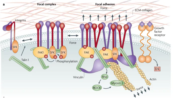
2007; Xian et al., 2010). Integrins, principal transmitters of signals from ECM to the cells are a family of 24 transmembrane heterodimers composed of alpha and beta subunit. Upon their activation and binding to the ECM (through their extracellular domain) (Harburger and Calderwood, 2009) adhesome is recruited to the cytoplasmic tail of an integrin. Adhesome is a protein complex composed of proteins involved in signalling, scaffolding and cytoskeleton (Horton et al., 2015, 2016). These protein complexes, in one hand allow anchoring of the cell cytoskeleton to ECM, and on the other hand support bidirectional transmission of signals between the cell and its environment (signaling controlling migration, survival, proliferation and differentiation) (Ridley et al., 2003). Generally, outside-inside integrin mediated signalling involves the recruitment of the focal adhesion kinase (FAK) and its downstream target Src (Humphries et al., 2006). It is important to stress that, integrins are crucial for mechanical sensing, stiffening and remodelling of ECM, and all fundamental steps in tumor progression including invasion, metastases and drug resistance, which is the main topic of the research presented in this manuscript (Hamidi and Ivaska, 2018).
Besides integrins, various enzymatic and non-enzymatic processes have the power to influence ECM behaviour in terms of structure, remodelling and soluble factors found within. At the same time, molecular components undergo a very large number of post-translational modifications. Under physiological conditions all these processes are tightly regulated and homeostasis is maintained. If homeostasis is disrupted, no control is established over any type of cellular processes including ECM deposition and remodelling. ECM formed under this dis-balance acquires pathological properties that results in fibrosis and, ultimately tumor development (Eckes et al., 1999). ECM found in tumors represents an architectural structure profoundly different from the one of “normal” healthy tissue (Butcher et al., 2009). During tumor progression, there are multiple changes in the production and assembly of ECM molecules secreted an arranged by the different cell populations of the TME (Jinka et al., 2012). Excessive deposition and remodelling of ECM by CAFs is responsible for increased fibrosis that in turn promotes tumor development. (Lu et al., 2012; Samuel et al., 2011). Matrix remodelling is one of the key events during tumor development, and it mostly coordinated by the activity of MMPs and their inhibitors. In cancers, MMPs can be secreted by tumor cells themselves, CAFs and tumor-associated macrophages (TAMs) and they facilitate the invasion and migration of cancer cells by
cysteine (SPARC) protein, periostin, Tenasin C, osteopontin and thrombospondin (Albo et al., 1997; Nong et al., 2015; Wang and Ouyang, 2012)
Tumors, besides being characterized by ECM remodelling, have know to be stiffer than normal tissue as discussed later on.
Simultaneously, various chemokines (CXCL12, CXCL10, CCL21), growth factors (insulin growth factor (IGF), fibroblast growth factor (FGF), hepatocyte growth factor (HGF), angiogenic factors such as vascular endothelial growth factor (VEGF) and TGFβ are released in ECM and bound to it (Gocheva et al., 2006; Kessenbrock et al., 2010; Lerner et al., 2011). Often, growth factors bind to glycosaminoglycan chains attached to ECM and membrane proteins, but recent evidence suggest a specific biding of growth factors to ECM proteins themselves. As an example fibronectin binds HGF and VEGF directly (Rahman et al., 2005; Wijelath et al., 2006).
Given the evidence , the importance of ECM in variety of cellular process (both in physiological and pathological conditions), and its role in the behaviour of epithelial cells is obvious (Pickup et al., 2014; Werb, 1997) (Figure 5) and remains an important aspect to investigate during cancerogenesis, and finally in chemoresistance which is the main subject of my research.
Figure 5. Molecular influence of extracellular matrix on tumor cells
From the initiation of a tumor to the formation of metastases, ECM supports and sustains all hallmarks of cancers. ECM molecules bind to cell surface receptors, activating intracellular signaling pathways. Adherence of cells to ECM induces activation of Erk and PI3K kinases, which promotes autonomic growth of tumor cells. Activation of the FAK kinase inhibits p15 and p21 growth suppressors, and induces apoptosis resistance by p53 inhibition. The molecular components and biophysical properties of ECM promote the induction of epithelio-mesenchymal transition, and increase tumor cell migration, particularly via TGFβ signaling, and RhoA / Rac GTPases. Matrix rigidity also promotes angiogenesis by increasing VEGF signaling in endothelial cells. Adapted from (Pickup et al., 2014).
1.2.3. RIGIDITY and REMODELLING of the ECM
I would like to dedicate this part to elucidating the role of ECM in tumor progression, specifically to the role of ECM remodelling and stiffness. Earlier on, I have mention that composition of tumor ECM is modified compared to normal ECM, and that various players participate in its remodelling leaving architecture of tumor ECM crooked. For example, ECM secreted by CAFs is organised in a more liner manner with increased levels of fibronectin and collagen, notably cross-linked collagen. ECM plays multiple, equally relevant, roles in TME such as facilitating migration and invasion, nevertheless I would like to focus on the fact that ECM found in tumors is significantly stiffer that the one in normal tissue (Butcher et al., 2009) and the fact that stiffness can influence the faith of cancer cells in a process called mechanotransduction.
A) What is stiffness of the ECM?
Cells behaviour does not rely only on biochemical pathways but also on forces applied on them. Cells, including cancer cell, are subjected to isometric nanoscale forces or tensions generated by cell-cell contact or cell-ECM contact. Upon the application of force, cell functions are modified by actomyosin contractility and actin dynamic in collaboration with biomechanical cues ultimately modulating cell behaviour. Process described is mechanotransduction and will be further discussed. Generally, mechanical stress is quantified in Pascals (Pa) and measured as force per unit (N per m2).
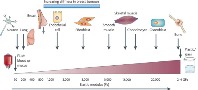
As I have established now, in cancers, homeostasis is lost, thus it is not surprising that the level of stiffness is as well disturbed. Indeed, cancerous tissue can be up to 10 times more rigid than healthy tissue (Figure 6), which is correlated with tumor cell survival and increased proliferation (Jaalouk and Lammerding, 2009; Levental et al., 2009). In addition, it is associated with a higher risk of metastasis, and is a factor of poor prognosis (Schwartz, 2010).
In diagnostic, stiffness of tumors can be exploited for a palpation diagnosis of certain tumors such as breast cancer, magnetic resonance imaging (MRI) or ultrasound that detect cancerous tissue (Poterucha et al., 2015; Zaleska-Dorobisz et al., 2014).