HAL Id: hal-03219798
https://hal.sorbonne-universite.fr/hal-03219798
Submitted on 6 May 2021
HAL is a multi-disciplinary open access
archive for the deposit and dissemination of
sci-entific research documents, whether they are
pub-lished or not. The documents may come from
teaching and research institutions in France or
abroad, or from public or private research centers.
L’archive ouverte pluridisciplinaire HAL, est
destinée au dépôt et à la diffusion de documents
scientifiques de niveau recherche, publiés ou non,
émanant des établissements d’enseignement et de
recherche français ou étrangers, des laboratoires
publics ou privés.
DNA damage response- and JAK-dependent regulation
of PD-L1 expression in head and neck squamous cell
carcinoma (HNSCC) cells exposed to 5-fluorouracil
(5-FU)
Claire Lailler, Michele Lamuraglia, Floriane Racine, Christophe Louandre,
Corinne Godin, Bruno Chauffert, Antoine Galmiche, Zuzana Saidak
To cite this version:
Claire Lailler, Michele Lamuraglia, Floriane Racine, Christophe Louandre, Corinne Godin, et al.. DNA
damage response- and JAK-dependent regulation of PD-L1 expression in head and neck squamous cell
carcinoma (HNSCC) cells exposed to 5-fluorouracil (5-FU). Translational Oncology, Elsevier, 2021, 14
(8), pp.101110. �10.1016/j.tranon.2021.101110�. �hal-03219798�
ContentslistsavailableatScienceDirect
Translational
Oncology
journalhomepage:www.elsevier.com/locate/tranon
DNA
damage
response-
and
JAK-dependent
regulation
of
PD-L1
expression
in
head
and
neck
squamous
cell
carcinoma
(HNSCC)
cells
exposed
to
5-fluorouracil
(5-FU)
Claire
Lailler
a,b,
Michele
Lamuraglia
c,
Floriane
Racine
a,b,
Christophe
Louandre
a,b,
Corinne
Godin
a,b,
Bruno
Chauffert
b,c,
Antoine
Galmiche
a,b,
Zuzana
Saidak
a,b,∗a Laboratoire de Biochimie, Centre de Biologie Humaine (CBH), CHU Sud, Amiens, France b UR7516 “CHIMERE ”, Université de Picardie Jules Verne, Amiens, France
c Laboratoire d’Imagerie Biomédicale (LIB), Sorbonne Université, CNRS, INSERM, Oncologie Médicale, CHU Sud, Amiens, France
a
b
s
t
r
a
c
t
Objectives:TheimmunecheckpointmoleculePD-L1(CD274)isacrucialregulatorofthetumorimmuneresponse.Itsexpressionhasbeenreportedinthetherapeutic contextinHeadandNeckSquamousCellCarcinoma(HNSCC),butitremainsunclearhowtherapeuticallyapprovedmoleculesregulatePD-L1expressioninHNSCC cells.
Materialsandmethods:ThreeHNSCCcelllines(BICR6,PE/CA-PJ34andPE/CA-PJ41)wereusedtoanalyzePD-L1expressionbyimmunoblotting,immunofluorescence andQPCR.Freely-availablesinglecellRNAseqdatafromHNSCCwerealsoused.
Results: 5-Fluorouracil(5-FU)increasedtheexpressionofPD-L1withhighefficacyinHNSCCcells.SinglecellRNAseqdatasuggestedthespecificityoftheregulation ofPD-L1inthiscontext.Theeffectof5-FUonPD-L1expressionwasrelatedtoitsgenotoxiceffectandwaspreventedbyextracellularapplicationofthymidineor usingachemicalinhibitoroftheDNAdamageResponsekinasesATM/ATR.Wefoundthattheeffectof5-FUwasadditiveorsynergisticwithIFN-𝛾,thecanonical inducerofPD-L1inepithelialcells.QPCRanalysisconfirmedthisfindingandidentifiedJAK-dependenttranscriptionalactivationofPD-L1/CD274astheunderlying mechanism.TheinductionofPD-L1by5-FUwaspartiallypreventedbyEpidermalGrowthFactorReceptor(EGFR)inhibitionwithcetuximab.
Conclusion: OurstudyhighlightsthespecificDNADamageResponse-andJAK-dependentinductionofPD-L1by5-FUinHNSCCcells.Thisinductionisregulated bythecytokinecontextandispotentiallytherapeuticallyactionable.
Introduction
HeadandNeckSquamousCellCarcinoma(HNSCC)area heteroge-neousgroupoftumorsthatrequiremultimodaltreatmentwithsurgery andadjuvantradio(chemo)therapy[1,2].Despiteadaptedinitial treat-ments,localrecurrenceandmetastasisremainfrequentandconstitute anindicationforchemotherapyandimmunotherapy[1,2].Therecent introductionofimmunecheckpointblockers(ICB)haschallengedthe medicalpracticeforadvancedstagesofHNSCC.Nivolumaband pem-brolizumab,twomonoclonal antibodiestargetingtheinteraction be-tweenthemoleculePD-L1(ProgrammedcellDeath1-Ligand1,encoded bythegeneCD274)anditsreceptorPD1,areapprovedforthetreatment ofrecurrent/metastatic(R/M)HNSCC[3,4].PD1targetingusedalone isconsideredeffectiveinalmost20%ofR/MHNSCCpatients[3,4].
PD-Listofabbreviations:DEG,differentiallyexpressedgenes;EGFR,EpidermalGrowthFactorReceptor;FDR,falsediscoveryrate;GO,GeneOntology;HNSCC,head andnecksquamouscellcarcinoma;ICB,immunecheckpointblockers;IFN-𝛾,Interferon-𝛾;NGF,NerveGrowthFactor;PD-L1,ProgrammedcellDeath1-Ligand1; R/M,Recurrent/Metastatic;TS,Thymidylatesynthase.
∗Correspondingauthorat:LaboratoiredeBiochimie,centredeBiologieHumaine(CBH),CHUAmiensSud,AvenueLaennec,80054AmiensCedex,France.
E-mailaddress:Saidak.Zuzana@chu-amiens.fr (Z.Saidak).
L1expressionisusuallyanalyzedbyimmunohistochemistryandscored usingtheCPS(CombinedPositiveScore),definedasthesumof PD-L1-positivecancercellsandmonocytes/lymphocytesdividedbythetotal numberoftumorcellsx100[5].TheuseofCPSreflectsthedual expres-sionofPD-L1incancerandtumor-infiltratingimmunecells.Inaddition totheinterestofCPSinpredictingthebenefitofICB,PD-L1 canbe abiomarkerofnegativeorpositiveprognosticvalue,dependingonits expressiononepithelialorimmunecells,respectively[6,7].
Chemotherapyiscurrentlyindicatedforpatientswithadvanced HN-SCC,i.e.thosedirectlypresentingwithR/MHNSCCorthosewithhigh risktumors(nodalextracapsularspreadorinvadedsurgicalmargins). Platinum salts(cisplatin orcarboplatin),taxanes(docetaxelor pacli-taxel), and 5-fluorouracil (5-FU) are used in this setting [8]. After transformationintoFdUMP,5-FUmainlyinterfereswithnucleicacid
https://doi.org/10.1016/j.tranon.2021.101110
Received22January2021;Receivedinrevisedform14April2021;Accepted20April2021
C. Lailler, M. Lamuraglia, F. Racine et al. Translational Oncology 14 (2021) 101110
metabolismbyblockingtheenzymethymidylatesynthase(TS)and in-hibitingdenovopyrimidinesynthesis.Thiseffectof5-FUresultsinan inhibitionofDNAsynthesisandablockincellcycleprogression[9]. AhomeostaticreactioncalledtheDNADamageResponse(DDR)is in-ducedinthiscontextandconstitutesadeterminantofHNSCCsensitivity to5-FU[9].Cetuximab,atargetedtherapydirectedagainstthe Epider-malGrowthFactorReceptor (EGFR)counteractsoncogenic signaling downstreamofEGFRinHNSCCcells[2].
Animmune responsedirectedagainstcancercells isemergingas amechanismthatcontributestotheefficacyoftherapeuticprotocols usedagainstsolidtumors[10,11].Interestingly,arecentstudy examin-ingHNSCCresectedafterneoadjuvantchemotherapyreportedincreased PD-L1expressionincancercellsinthiscontext[12].In71%oftumor samplesfrompatientsthatreceivedinductionchemotherapywithTPF (docetaxel+platinum+5-FU),increasedlevelsofPD-L1anda signifi-cantincreaseinthedensityofCD8+Tcellinfiltrateweredetected[12]. PreviousinvitrostudiesfoundthatcisplatininducedPD-L1expression inHNSCCcells[13,14].Itisunclearwhichchemotherapeuticdrugis themosteffectiveatincreasingPD-L1expressioninHNSCCcellsand howPD-L1expressionisregulatedinthiscontext.
Materialsandmethods
Cellculture.ThecelllinesBICR6,PE/CA-PJ34andPE/CA/PJ-41 aredescribed in detail inthe supplementary Materialsand Methods section.Cells were cultured in Dulbecco’sModified Eagle’s Medium (DMEM)supplementedwith10%fetalcalfserum,2mMglutamine,and penicillin/streptomycin.
Reagentsand chemicals. All chemicalreagents were purchased from Sigma,unless statedotherwise. AfatinibandVE821 were pur-chasedfrom Selleckchem.The JAK inhibitor1(JAKi) was purchased fromCalbiochem(420,099).TheHumanPhospho-RTKArrayKitwas purchasedfromR&DSystems(ProteomeProfilerArray,ARY00B).The antibodiesusedinthisstudyarelistedintheSuppl.Materialsand Meth-ods.
Single cell gene expression analysis. Single cell RNAseq data (5902cellssequencedfrom18 HPV-negativeHNSCC)wereretrieved fromPurametal.(2017)[15](datasetGSE103322).
Geneontology(GO)analysis.WeusedthePANTHERclassification system(http://pantherdb.org/)toperformastatistical overrepresenta-tiontest.TheenrichmentofGeneOntology(GO)termsinourgeneset wascomparedtothewholeHomoSapiensgenome(GObiological pro-cesscomplete),withaFalseDiscoveryRate(FDR)correction[16].
QPCR. Total RNA was extracted and reverse-transcribed using High Capacity cDNAReverse Transcription kit and random hexam-ers(AppliedBiosystems).Amplificationwasperformedwiththe Taq-Man UniversalPCR masterMixon anABI 7900HTSequence Detec-tionSystem (Applied Biosystems).Primersandprobe sets for PD-L1 andGlyceraldehyde-PhosphateDehydrogenase(GAPDH)aredescribed intheSuppl.MaterialandMethods.
Immunoblotting. Complete cell extracts were transferred to nitrocellulose membranes using standard procedures as pre-viously described [17]. The ECL reaction was used to reveal protein. Signal quantifications were performed using ImageJ (https://imagej.nih.gov/ij/download.html).
Immunofluorescence.ImmunofluorescencelabelingofPD-L1was performedonparaformaldehyde-fixedcells,accordingtostandard pro-cedures[17].AdetailedprotocolisgiveninSuppl.Materials&Methods.
Statistical analyses. Analyses were done with R version 4.0.3 (https://www.r-project.org)usingpackages“Hmisc” and“venneuler”. Student’s t-test and ANOVA were used as indicated (GraphPad Prism). The Spearman test was used for gene correlation analyses. p<0.05 was used as the threshold of significance. False discovery rate(FDR)correction wasapplied as indicatedusing theBonferroni method.
Results
5-FUupregulatesPD-L1expressioninHNSCCcells
InordertoexaminetheeffectofchemotherapeuticagentsonHNSCC cells,weusedapanelofthreeHNSCCcelllines(BICR6,PE/CA-PJ34 andPE/CA-PJ41)thatwereexposedton=8chemotherapeuticagents (Fig.1).Allchemotherapeuticagentswereappliedat concentrations corresponding totheirIC50,i.e.in conditionsof comparableefficacy,
for48 h(Suppl.Table 1).Cetuximabwasappliedat aconcentration of 50μg/mL andwas foundtoblockEGFR phosphorylationwithout significantinhibitoryeffectonthegrowthofHNSCCcellsinvitro(data notshown).WethenanalyzedtheproteinexpressionofPD-L1aswell asPD-L2,CD80,CD86,andMHCclassImoleculesbyimmunoblotting (Fig.1).Weobservedthat5-FUincreasedPD-L1expressioninallHNSCC celllines(foldinductionof14.4,3.1and1.7comparedtocontrolfor BICR6,PE/CA-PJ34andPE/CA-PJ41,respectively)(Suppl.Fig.1).No effectof 5-FUwasdetectedontheexpressionofPD-L2,CD80,CD86 andtheMHCclassImolecules(Fig.1).Intwooutofthreecelllines, 5-FUseemedtobethechemotherapeuticagentthatupregulatedPD-L1 expressionthemost.
PD-L1mRNAregulationinsinglecellsfromHPV-negativeHNSCCtumors SinglecellRNAseqdatafromPurametal.[15] wereretrievedin ordertoexaminePD-L1/PD-L2mRNAregulationinHNSCC.High lev-elsofPD-L1mRNAweredetectedindendriticcells,mastcellsandto alowerextentinHNSCCcells(Fig.2A).Indeed,16.4%oftumorcells expressedPD-L1mRNA(Fig.2B).PD-L2wasfoundtobeexpressedin asmallerfractionofthecancercells(6.4%)thatonlyminimally over-lappedwiththepopulationofPD-L1expressingcells(2.1%)(Fig.2B). There wasno correlationbetween PD-L1andPD-L2mRNAlevelsin cancercells(Pearsonr=0.05).Weidentifiedthegeneswhose expres-sionwassignificantlycorrelatedwithPD-L1orPD-L2mRNAincancer cellsfromsinglecellRNAseqdata(Supp.Table2and3,respectively), andcomparedthemwiththegenesthatcorrelatedwithPD-L1mRNA intumorinfiltratingimmunecells(Suppl.Table4).Anumberofgenes weresignificantlyco-expressedwithPD-L1,butnotwithPD-L2in HN-SCCcells(Suppl.Table2and3).Thesenon-overlappinggenecontexts suggesttheexistenceofspecificregulationofPD-L1mRNAinHNSCC cells.AstatisticaloverrepresentationtestofGeneOntologytermswas performedonthepanelofgenesfoundtobecorrelatedwithPD-L1in HNSCCcells,andsuggestedalinkbetweenPD-L1expressionand xeno-biotic/chemotherapeuticmetabolism(Fig.1C).
PD-L1overexpressioninducedby5-FUisrelatedtoitsgenotoxiceffectin HNSCCcells
5-FUcanincorporateintoRNAorblockTSandpreventthesynthesis ofthymidine.Weexaminedtheroleplayedbythesetwomechanismsby analyzingPD-L1expressionincellscultivatedwithextracellularly sup-plieduridineorthymidine(bothataconcentrationof20μM)(Fig.3A). Wefoundthatextracellularthymidine,butnoturidine,wasableto re-vertPD-L1inductionby5-FU(Fig.3A,Suppl.Fig.2A).Wenext envi-sionedthepossibilitythatDNAdamageresponsemightplayarolein PD-L1induction(Fig.3B).WeusedthechemicalinhibitorVE-821, di-rectedagainstthekeykinasesoftheDNADamageResponseATM/ATR, aspreviouslyreportedbyItoetal.[9].WeverifiedthatVE-821 pre-ventedChk1phosphorylationonSer345(asitetargetedbyactivated ATM/ATR)at aconcentrationof10 μM(Fig.3B).Importantly,DNA DamageResponse inhibitionwithVE-821partiallypreventedthe in-ductionofPD-L1by5-FUinthethreeHNSCCcelllinesexaminedinthis study(Fig.3B,Suppl.Fig.2B).WeconcludedthatPD-L1inductionby 5-FUwasrelatedtoitsgenotoxiceffectinHNSCCcells.
Fig.1. ImmunoblotanalysisoftheexpressionofthemainimmunecheckpointmoleculesinHNSCCcellsexposedtochemotherapeuticagents.
ThecelllinesBICR6,PE/CA-PJ34andPE/CA-PJ41wereexposedto5-FU,methotrexate,gemcitabine,paclitaxel,cisplatinatIC50concentrationsfor48h.Cetuximab wasappliedataconcentrationof50μg/mL.Expressionanalysisoftheindicatedmoleculeswasperformedbyimmunoblottingasindicated.Actinimmunolabelling isgivenasloadingcontrol.TheindicatedvaluesarenormalizeddensitometricanalysesofthePD-L1/Actinratio,takingcontrolconditionas1.
Fig.2. CD274/PD-L1expressioninsinglecell RNA-seqdataretrievedfromHPV-negative HN-SCC.
A.AnalysisofCD274/PD-L1mRNAlevelsin singlecellRNA-seqdatabyPurametal.[15] .
n= 2215cancercellsfrom18tumors.B.A VenndiagramofthepercentageofHNSCCcells expressingPD-L1andPD-L2mRNA,andthe overlap.C.AbargraphshowingtheGene On-tology(GO)termsthatwerestatistically over-representedinthetop150genesthatare sig-nificantlycorrelatedwithCD274/PD-L1mRNA incancercells,usingPANTHERGO overrepre-sentationtest.Thefigureshowsthefold enrich-mentofeachGOtermcomparedtowhatwould bestatisticallyexpected.
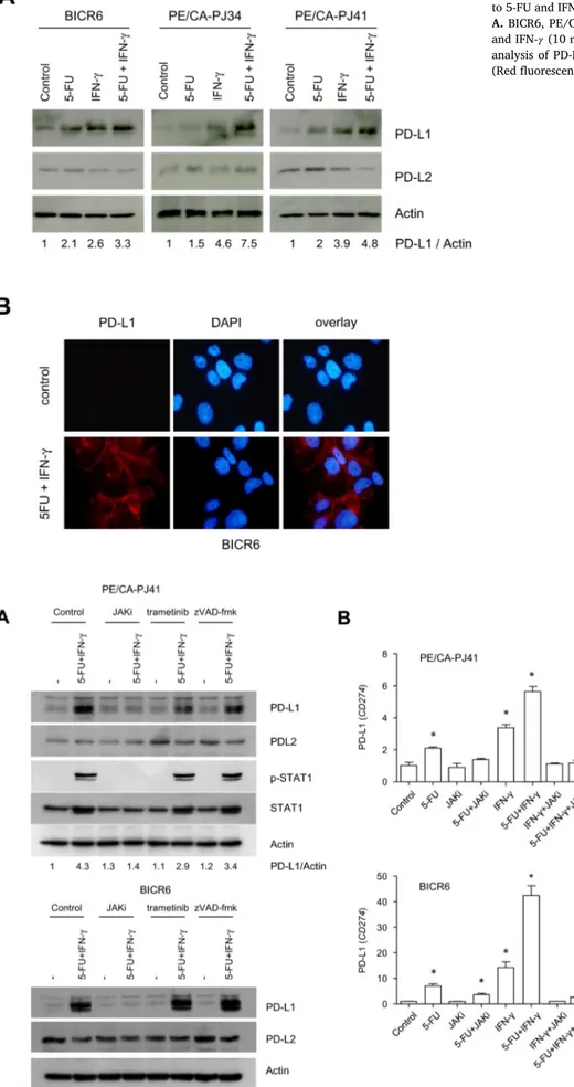
5-FUandIFN-𝛾 interacttoregulatePD-L1mRNAlevelsinHNSCCcells Interferon 𝛾 (IFN-𝛾)is thecanonical inducer of PD-L1 in cancer cells[17].Comparedtovariousothercytokinesorgrowthfactors (IL-6:10 ng/ml,IL-1𝛽: 1ng/ml,TNF-𝛼:25 ng/ml,TGF-𝛽: 5ng/mland NerveGrowthFactor,NGF:40ng/ml),onlyIFN-𝛾 (10ng/ml)robustly inducedPD-L1expressioninourexperimentalconditions(Suppl.Fig. 3).Weexaminedtheinteractionbetween5-FUandIFN-𝛾 inthe regu-lationofPD-L1(Fig.4).Theco-applicationof5-FUwithIFN-𝛾 ledto anadditiveinductionofPD-L1inBICR6andPE/CA-PJ41cells,anda possiblesynergywasobservedin PE/CA-PJ34cells(Fig.4A).A den-sitometricanalysisofthePD-L1/Actinratioindicatedthat5-FUalone inducedPD-L1proteinexpression(foldinductionof 2.1,1.5and2.0 forBICR6,PE/CA-PJ34andPE/CA-PJ41,respectively),butthis
induc-tionwasgreaterwhenthecellsweresimultaneouslyexposedto5-FU and IFN-𝛾 (Foldinduction=3.3, 7.5and4.8 for BICR6,PE/CA-PJ34 andPE/CA-PJ41,respectively)(Suppl.Fig.4).Immunofluorescent mi-croscopydetectedaclearandhomogeneousPD-L1signalonthe sur-faceofculturedBICR6cells,confirmingandextendingourobservations madebyimmunoblotting,suggestingthatPD-L1isfunctionalinHNSCC cellsexposedto5-FU+IFN-𝛾 (Fig.4B).
We further examined the regulation of PD-L1/CD274 in BICR6 andPE/CA-PJ41cells,byperformingimmunoblotandQPCRanalysis (Fig.5A,B).Inordertoexaminetheinteractionobservedbetween5-FU andIFN-𝛾, weused abroad-spectrumchemical inhibitor ofJAK sig-naling,JAKinhibitor-1(JAKi)thatpreventsSTAT1phosphorylationon tyrosine701(Fig.5A).Atthisconcentration,JAKireducedthe clono-genic growthofBICR6 andPE/CA-PJ41cells,eventhough itseffect
C. Lailler, M. Lamuraglia, F. Racine et al. Translational Oncology 14 (2021) 101110
Fig.3.PD-L1inductioninHNSCCcellsisrelatedtothe geno-toxiceffectof5-FU.
A.Thymidine(20μM)anduridine(20μM)wereapplied ex-tracellularlyonBICR6cells±5-FU(IC50)for48h.B.The chemicalinhibitorVE-821wasappliedatafinal concentra-tionof10μMandthecellularextractswereanalyzedby im-munoblottingwiththeindicatedantibodies.
wasnotadditivewiththatof5-FU(Suppl.Fig.5).Importantly,JAKi abrogatedPD-L1proteinexpressioninHNSCCcells(Fig.5A,Suppl.Fig. 6).Meanwhile,wefoundnoeffectoftrametinib,appliedinconditions that radicallyprevented MEK1/2 phosphorylation (data not shown). ThechemicalblockerofcaspaseszVAD-fmk,appliedataconcentration of50μMblockingapoptosis[19],alsohadnoeffectonPD-L1 induc-tionby5-FU+IFN-𝛾 (Fig.5A).SimilarresultswereobtainedbyQPCR (Fig.5B).While5-FUandIFN-𝛾 appliedassingleagentsonlymodestly increasedCD274mRNAlevels,anadditiveeffectwasobservedupon theco-administration ofthetwomolecules(Fig.5B).TheJAKi abro-gatedtheincreasedlevelsofCD274mRNAinducedby5-FU+IFN-𝛾 (Fig.5B).Weconcludedthat5-FUandIFN-𝛾 convergeonJAKsignaling toregulateCD274/PD-L1expressionatthetranscriptionallevel.
EGFRasanactionabletargettopreventPD-L1inductionby5-FUin HNSCCcells
ApreviousstudyreportedtheexistenceofPD-L1regulation down-streamoftheReceptorTyrosineKinase(RTK)EGFR [20]. We there-foreexploredthephosphorylationstatusof49RTKinHNSCCcells.A phospho-RTKarrayindicatedthatBICR6andPE/CA-PJ41mainly ex-pressedphosphorylatedformsofEGFRandHGFR(HepatocyteGrowth FactorReceptor),whiletheotherRTKweredetectableatconsiderably lowerlevels(Fig.6A).Importantly,HNSCCcellsexposedto5-FUatIC50 concentrationfor48hhadincreasedphosphorylationlevelsofEGFR, withsimultaneouslyreducedHGFRphosphorylationlevels(Fig.6A,B). TheincreaseinEGFRphosphorylation wasquantifiedtobe5-foldin BICR6and2.5-foldinPE/CA-PJ41exposedto5-FUfor48h(Fig.6B,
Suppl.Fig.7).WeusedtwodifferentapproachesthatinhibitEGFR ki-naseactivitytoexaminetheroleofEGFRinthissetting:the applica-tionofcetuximab(50μg/ml)orthechemicalinhibitorafatinib(5μM). WhileaslightupregulationofPD-L1wasinducedbyafatinibassingle agentinbasalconditions,bothcetuximabandafatinibinhibitedPD-L1 overexpressioninducedby5-FUtoanextentofaround50%inBICR6 andPE/CA-PJ41(Fig.6C).WeconcludedthatEGFRtargetingpartially preventsPD-L1expressioninHNSCCcellsexposedto5-FU.
Discussion
Inthepresentstudy,weexaminedtheeffectsofvarious chemothera-peuticagentsonHNSCCcellsandobservedthat5-FUrobustlyincreased PD-L1expression.Thisinductionwasobservedinthreeindependent ge-nomiccontexts,suggestingitspotentialbroadrelevance.Aclear inter-action(eitheradditiveorsynergistic,dependingonthecellularcontext) wasobservedbetween5-FUandIFN-𝛾.PD-L1washomogeneously dis-tributed onthesurfaceofthewholepopulation ofHNSCCcells, sug-gestingthatitwasnotaccountedforbyaminorsubpopulationof can-cercellsandthatPD-L1isfunctionalinthiscontext.Theeffectof5-FU onPD-L1expressionappearedtoberelatedtoitsgenotoxiceffectand waspreventedbytheextracellularapplicationofthymidineorbythe DNAdamageResponseinhibitor VE-821,directedagainstthekinases ATM/ATR.Finally,wefoundthatEGFRinhibitionpartiallyprevented PD-L1inductionby5-FU.
RecentstudiespointtotheregulationofPD-L1byoncogenic, inflam-matoryandhypoxicsignalingincancercells[21].Afewstudieshave addressedtheimpactoftherapeuticagentsusedagainstHNSCConthe
Fig.4. ExpressionanalysisofPD-L1uponco-exposureofHNSCCcells to5-FUandIFN-𝛾.
A.BICR6,PE/CA-PJ34andPE/CA-PJ41cellswereexposedto5-FU andIFN-𝛾 (10ng/ml)for24h,asindicated.B.Immunofluorescence analysisofPD-L1onBICR6cellsexposedto5-FU+IFN-𝛾 for24h (Redfluorescence:PD-L1,bluefluorescence:DAPI).
Fig. 5. An immunoblot and QPCR analysis ofCD274/PD-L1mRNAexpressioninHNSCC cellsexposedto5-FUandIFN-𝛾
A.BICR6andPE/CA-PJ41cellswereexposed to5-FU(IC50)andIFN-𝛾 (10ng/ml)for24h. Cellswere preincubated withJAK inhibitor-1 (1 μM), trametinib (1 μM) or zVAD-fmk (50μM)for1hasindicated.Complete cellu-larextractswereusedinordertoperformthe indicatedanalyses.NotethatSTAT1couldnot bedetectedinBICR6cells.B.AQPCRanalysis ofCD274/PD-L1mRNAexpressionin PE/CA-PJ41andBICR6cellsexposedto5-FU,IFN-𝛾
andJAKinhibitor-1(1μM).∗p<0.05with
C. Lailler, M. Lamuraglia, F. Racine et al. Translational Oncology 14 (2021) 101110
Fig.6. EGFRasanactionabletargetforpreventingPD-L1inductionby5-FUinHNSCCcells
A.Aphospho-RTKarraypreformedusingcellularextractspreparedfromBICR6andPE/CA-PJ41cells,eitherincontrolconditionsorafterexposureto5-FU(IC50) for48hB.QuantificationofEGFRandHGFRphosphorylationafter5-FUtreatment,takingcontrolconditionsasreferenceforeachcellline.C.Immunoblotanalysis ofcellularextractspreparedfromBICR6andPE/CA-PJ41cellsexposedto5-FU(IC50),afatinib(5μM)andcetuximab(50μg/ml)for48h,asindicated.Theindicated valuesarenormalizeddensitometricanalysesofthePD-L1/Actinratio,takingcontrolconditionas1.
expressionofPD-L1[13,14,22].Tothebestofourknowledge,our re-portshowsforthefirsttimethat5-FUisaninducerofPD-L1expression inHNSCCcells.Recentstudiesperformedinvarioustypesofprimary tumorspointtosimilareffectsof5-FUindigestivecancers[23,24]. An-otherantimetabolitewitharelatedmodeof action,pemetrexed,was alsorecentlyfoundtoinducePD-L1inNon-SmallCellLungCancercells
[25].OurobservationthattheregulationofPD-L1incancercellsdoes notentirelyoverlapwiththatofimmunecellsconfirmsthedataofChen etal.,althoughthecorrespondingstudydidnotexaminetheeffectsof xenobiotics/chemotherapeutics[18].Weverifiedthattheinductionof PD-L1wasnot accountedforeitherbycellsenescenceorthelossof cancercellviabilityinthis context(datanot shown).Importantly,it waspossibletoabolishtheeffectofthedualexposureofHNSCCcellsto 5-FUandIFN-𝛾 withachemicalinhibitoractiveagainstJAK.JAK sig-nalingincancercellsmightthereforeconstituteapointofconvergence andakeycontrolofthetranscriptionalinductionofPD-L1inHNSCC cellsexposedtochemotherapies.Non-selectivelytargetingJAK signal-ingincancerpatientswouldtargettumorinfiltratingimmunecellsin additiontocancercells[26],anditisthereforedifficulttoanticipate thetherapeuticinterestofthisstrategy.
WeexaminedthepossibilityoftargetingtheinductionofPD-L1 us-ingalreadyapprovedanti-cancerdrugs.WeobservedthatEGFR signal-ingis inducedby5-FUandthatitsinhibitionbycetuximabprevents PD-L1 upregulation.Previous studiesfoundthat oncogenic signaling downstreamofthegrowthfactorreceptors(EGFRandHGFR)positively regulatesPD-L1expressioninHNSCCcells[20,27,28].However,the correspondingstudiesdidnotaddresstheeffectsofchemotherapeutics. Importantly,apreviousstudyevenreportedastrikingconvergenceof EGFRandIFN-𝛾 signalingintheregulationofJAK-STATandPD-L1 ex-pressioninHNSCC[28].Inesophagealcancercellsexposedtoa conven-tionalchemotherapyregimen,aninductionofPD-L1wasalsoobserved thatwaspreventedbyblockingEGFR[29].Whilethisobservationis
reminiscentofourfindings,thecontributionof5-FUwasnotdirectly addressedinthisstudy[29].Importantly,thefactthatcetuximab coun-teractsPD-L1inductionsuggeststhatautocrine/paracrineactivationof EGFR occursinthiscontext. Furtherstudiesaddressingthe composi-tionofthecancercellsecretomeandtheregulationofJAK-STATaxis arewarranted.Anotherkeyquestionworthaddressingistheroleofthe DNAdamageresponseinthiscontext[30].
Activationof anadaptive immune responsedirected against can-cercells isemergingasamechanismthatcontributestotheefficacy of chemotherapeuticprotocols[10,11].Thepresentstudydidnot in-cludeaninvivoexperimentalpartwithanimmunocompetentanimal model.However,basedonpreviousstudiesthatexaminedtheroleof PD-L1incancercellsinhumanHNSCCsamples[6,7],wepostulatethat theinductionofPD-L1by5-FUmightlimittheadaptiveimmune re-sponseagainstcancerinpatientsreceivingradio(chemo)therapy.Our studythereforeraisesinterestingpossibilitiesregardingtheuseofICB inHNSCC.Chemotherapeuticagentsremainanimportanttherapeutic modalityandareoftencombinedwithICBagainstR/MHNSCC[31]. 40%ofHNSCCshow anenrichedinflammatoryresponsewithactive interferon-𝛾 signaling[32].Thetumormicroenvironmentisemergingas akeyplayerintheregulationoftheadaptiveimmuneresponseagainst solidtumors[33].Ourobservationsfurthersuggesttheimportanceof thetumormicroenvironmentinthetherapeuticcontext.Ourstudyalso provides a biologicalrationale fortargetingPD1 in associationwith chemotherapeuticregimen containing5-FU,especiallywhenatumor hasadenseTcellinfiltrate/highlocalproductionofIFN-𝛾.Clinical stud-iesareneededtoexaminethispossibility.
Authorcontributionsstatement
C.L. Conceptualization,Investigation, Writing:Review & Editing;
M.L.Writing:Review&Editing;F.R.Investigation,Writing:Review&
Editing;C.L.:Investigation;C.G.:Investigation;B.C.:Supervision, Fund-ingacquisition, Writing: Review& Editing; A.G.: Conceptualization, Funding acquisition, Project administration, Writing: Original Draft;
Z.S.:Conceptualization,Projectadministration,Writing:OriginalDraft.
DeclarationofCompetingInterest
Theauthorsdeclarethattheyhavenoconflictofinteresttodisclose forthiswork.
Acknowledgments
WethankAmiensUniversityHospital,LiguecontreleCancer,comité delaSomme,andAPOTAC (AssociationPicarde pourl’Optimisation desThérapeutiquesAnti-Cancéreuses)forfinancialsupport.Thefunders hadnoroleinthecollection,analysisandinterpretationofdata,writing ofthereportandinthedecisiontopublish.
Supplementarymaterials
Supplementarymaterialassociatedwiththisarticlecanbefound,in theonlineversion,atdoi:10.1016/j.tranon.2021.101110.
References
[1] L.Q.M. Chow , Head and neck cancer, N. Engl. J. Med. 382 (2020) 60–72 .
[2] D.E. Johnson , B. Burtness , C.R. Leemans , V.W.Y. Lui , J.E. Bauman , J.R. Grandis , Head and neck squamous cell carcinoma, Nat. Rev. Dis. Primers 6 (2020) 92 .
[3] J.D. Cramer , B. Burtness , R.L. Ferris , Immunotherapy for head and neck cancer: recent advances and future directions, Oral Oncol. 99 (2019) 104460 .
[4] J.D. Cramer , B. Burtness , Q.T. Le , R.L. Ferris , The changing therapeutic landscape of head and neck cancer, Nat. Rev. Clin. Oncol. 16 (2019) 669–683 .
[5] E.E.W. Cohen , D. Soulières , C.L. Tourneau , J. Dinis , L. Licitra , M.-.J. Ahn , et al. , Pem- brolizumab versus methotrexate, docetaxel, or cetuximab for recurrent or metastatic head-and-neck squamous cell carcinoma (KEYNOTE-040): a randomised, open-label, phase 3 study, Lancet 393 (2019) 156–167 .
[6] H.R. Kim , S.-.J. Ha , M.H. Hong , S.J. Heo , Y.W. Koh , E.C. Choi , et al. , PD-L1 expres- sion on immune cells, but not on tumor cells, is a favorable prognostic factor for head and neck cancer patients, Sci. Rep. 6 (2016) 36956 .
[7] M. Sanchez-Canteli , R. Granda-Díaz , N. Del Rio-Ibisate , E. Allonca , F. López-Alvarez , J. Agorreta , et al. , PD-L1 expression correlates with tumor-infiltrating lymphocytes and better prognosis in patients with HPV-negative head and neck squamous cell carcinomas, Cancer Immunol. Immunother. 69 (2020) 2089–2100 .
[8] A. Lau , W. Yang , K.-.Y. Li , Y. Su , Systemic therapy in recurrent or metastatic head and neck squamous cell carcinoma- a systematic review and meta-analysis, Crit. Rev. Oncol. Hematol. 153 (2020) 102984 .
[9] S.S. Ito , Y. Nakagawa , M. Matsubayashi , Y.M. Sakaguchi , S. Kobashigawa , T.K. Mat- sui , et al. , Inhibition of the ATR kinase enhances 5-FU sensitivity independently of nonhomologous end-joining and homologous recombination repair pathways, J. Biol. Chem. 295 (2020) 12946–12961 .
[10] W.H. Gmeiner , Fluoropyrimidine modulation of the anti-tumor immune response- prospects for improved colorectal cancer treatment, Cancers (Basel) (2020) 12 .
[11] L. Galluzzi , J. Humeau , A. Buqué, L. Zitvogel , G. Kroemer , Immunostimulation with chemotherapy in the era of immune checkpoint inhibitors, Nat. Rev. Clin. Oncol. 17 (2020) 725–741 .
[12] C. Leduc , J. Adam , E. Louvet , T. Sourisseau , N. Dorvault , M. Bernard , et al. , TPF induction chemotherapy increases PD-L1 expression in tumour cells and immune cells in head and neck squamous cell carcinoma, ESMO Open 3 (2018) e000257 .
[13] C.-.Y. Ock , S. Kim , B. Keam , S. Kim , Y.-.O. Ahn , E.-.J. Chung , et al. , Changes in programmed death-ligand 1 expression during cisplatin treatment in patients with head and neck squamous cell carcinoma, Oncotarget 8 (2017) 97920–97927 .
[14] L. Tran , C.T. Allen , R. Xiao , E. Moore , R. Davis , S.-.J. Park , et al. , Cisplatin alters antitumor immunity and synergizes with PD-1/PD-L1 inhibition in head and neck squamous cell carcinoma, Cancer Immunol. Res. 5 (2017) 1141–1151 .
[15] S.V. Puram , I. Tirosh , A.S. Parikh , A.P. Patel , K. Yizhak , S. Gillespie , et al. , Single-cell transcriptomic analysis of primary and metastatic tumor ecosystems in head and neck cancer, CellCell 171 (2017) 1611–1624 e24 .
[16] H. Mi , A. Muruganujan , J.T. Casagrande , P.D. Thomas , Large-scale gene function analysis with the PANTHER classification system, Nat. Protoc. 8 (2013) 1551–1566 .
[17] A. Galmiche , Z. Ezzoukhry , C. François , C. Louandre , C. Sabbagh , E. Nguyen-Khac , et al. , BAD, a proapoptotic member of the BCL2 family, is a potential therapeutic target in hepatocellular carcinoma, Mol. Cancer Res. 8 (2010) 1116–1125 .
[18] S. Chen , G.A. Crabill , T.S. Pritchard , T.L. McMiller , P. Wei , D.M. Pardoll , et al. , Mech- anisms regulating PD-L1 expression on tumor and immune cells, J. Immunother. Cancer 7 (2019) 305 .
[19] C. Louandre , Z. Ezzoukhry , C. Godin , J.-.C. Barbare , J.-.C. Mazière , B. Chauffert , et al. , Iron-dependent cell death of hepatocellular carcinoma cells exposed to so- rafenib, Int. J. Cancer 133 (2013) 1732–1742 .
[20] C.-.C. Cheng , H.-.C. Lin , K.-.J. Tsai , Y.-.W. Chiang , K.-.H. Lim , C.G.-.S. Chen , et al. , Epidermal growth factor induces STAT1 expression to exacerbate the IFNr-mediated PD-L1 axis in epidermal growth factor receptor-positive cancers, Mol. Carcinog. 57 (2018) 1588–1598 .
[21] J.-.H. Cha , L.-C. Chan , C.-W. Li , J.L. Hsu , M.-C. Hung , Mechanisms controlling PD-L1 expression in Cancer, Mol. Cell 76 (2019) 359–370 .
[22] S.-.H. Kang , B. Keam , Y.-.O. Ahn , H.-.R. Park , M. Kim , T.M. Kim , et al. , Inhibition of MEK with trametinib enhances the efficacy of anti-PD-L1 inhibitor by regulating anti-tumor immunity in head and neck squamous cell carcinoma, Oncoimmunology 8 (2019) e1515057 .
[23] L. Van Der Kraak , G. Goel , K. Ramanan , C. Kaltenmeier , L. Zhang , D.P. Normolle , et al. , 5-Fluorouracil upregulates cell surface B7-H1 (PD-L1) expression in gastroin- testinal cancers, J. Immunother. Cancer 4 (2016) .
[24] T. Doi , T. Ishikawa , T. Okayama , K. Oka , K. Mizushima , T. Yasuda , et al. , The JAK/STAT pathway is involved in the upregulation of PD-L1 expression in pancre- atic cancer cell lines, Oncol. Rep. 37 (2017) 1545–1554 .
[25] C.-.S. Lu , C.-.W. Lin , Y.-.H. Chang , H.-.Y. Chen , W.-.C. Chung , W.-.Y. Lai , et al. , Antimetabolite pemetrexed primes a favorable tumor microenvironment for immune checkpoint blockade therapy, J. Immunother. Cancer 8 (2020) e001392 .
[26] D. Moreira , S. Sampath , H. Won , S.V. White , Y.-.L. Su , M. Alcantara , et al. , Myeloid cell-targeted STAT3 inhibition sensitizes head and neck cancers to radiotherapy and T cell-mediated immunity, J. Clin. Invest. (2021) 131 .
[27] V. Boschert , J. Teusch , A. Aljasem , P. Schmucker , N. Klenk , A. Straub , et al. , HGF-In- duced PD-L1 expression in head and neck cancer: preclinical and clinical findings, Int. J. Mol. Sci. 21 (2020) .
[28] F. Concha-Benavente , R.M. Srivastava , S. Trivedi , Y. Lei , U. Chandran , R.R. Seethala , et al. , Identification of the cell-intrinsic and -extrinsic pathways downstream of EGFR and IFN 𝛾 that induce PD-L1 expression in head and neck cancer, Cancer Res. 76 (2016) 1031–1043 .
[29] H.Y. Ng , J. Li , L. Tao , A.K.-.Y. Lam , K.W. Chan , J.M.Y. Ko , et al. , Chemotherapeu- tic treatments increase PD-L1 expression in esophageal squamous cell carcinoma through EGFR/ERK activation, Transl Oncol 11 (2018) 1323–1333 .
[30] H. Sato , P.A. Jeggo , A. Shibata , Regulation of programmed death-ligand 1 expression in response to DNA damage in cancer cells: implications for precision medicine, Cancer Sci. 110 (2019) 3415–3423 .
[31] P. Szturz , J.B. Vermorken , Management of recurrent and metastatic oral cavity can- cer: raising the bar a step higher, Oral Oncol. 101 (2020) 104492 .
[32] Y.-.P. Chen , Y.-.Q. Wang , J.-.W. Lv , Y.-.Q. Li , M.L.K. Chua , Q.-.T. Le , et al. , Identifica- tion and validation of novel microenvironment-based immune molecular subgroups of head and neck squamous cell carcinoma: implications for immunotherapy, Ann. Oncol. 30 (2019) 68–75 .
[33] X. Jiang , J. Wang , X. Deng , F. Xiong , J. Ge , B. Xiang , et al. , Role of the tumor microenvironment in PD-L1/PD-1-mediated tumor immune escape, Mol. Cancer 18 (2019) 10 .