Université de Montréal
O
6-methylguanine-DNA methyltransferase methylation:
Prevalence and predictive value in Head and Neck squamous
cell carcinoma
Par
Zahi Abou Chacra, MD
Departement de sciences biomédicales Faculté de médecine
Mémoire présenté à la Faculté des études supérieures en vue de l’obtention du grade de
Maîtrise en sciences biomédicales (2-484-1-0)
Décembre 2009
Université de Montréal
Faculté des études supérieures et postdoctorales
Ce mémoire intitulé:
O6-methylguanine-DNA methyltransferase methylation: Prevalence and predictive value in Head and Neck squamous cell carcinoma
présenté par : Zahi Abou Chacra
a été évalué par un jury composé des personnes suivantes :
Richard L Momparler, Ph.D président-rapporteur Denis Soulieres, MD, FRCSC directeur de recherche Louis Guertin, MD, FRCSC codirecteur
Louis A Gaboury, MD, Ph.D, FRCPC, FCAP membre du jury
F
RENCH ABSTRACTIntroduction: Le gène O6-méthylguanine-ADN méthyltransferase (MGMT) code pour une enzyme spécifique réparatrice de l’ADN qui protège les cellules de la toxicité des agents alkylants. Ainsi, l’activité du MGMT est un mécanisme majeur de résistance aux agents alkylants. Il a été démontré qu’une diminution de l’expression du gène MGMT par une hyperméthylation du promoteur résulte en une amélioration de la survie chez les patients avec certains types de tumeurs qui sont traitées avec des agents chimiothérapeuthique alkylants. Objectifs: Déterminer la prévalence de la méthylation du gène MGMT chez des patients avec des cancers épidermoïdes localement avancés de la sphère ORL traités avec chimioradiothérapie et évaluer l’impact de cette méthylation sur la survie. Méthodes: Sur 428 patients consécutifs, traités avec chimioradiothérapie à notre institution et suivis pour un période médiane de 37 mois, 199 spécimens chirurgicaux paraffinés ont été récupérés. L’ADN était extrait et modifié par le traitement au bisulfite. Une réaction en chaîne de la polymérase, spécifique à la méthylation était entreprise pour évaluer l’état de méthylation du promoteur du gène du MGMT. Les résultats de laboratoire étaient corrélés avec la réponse clinique. L’analyse statistique était exécutée à l’aide du test de Fisher pour les données catégoriques et à l’aide des courbes de Kaplan-Meier pour les échecs au traitement. Résultats : Des 199 extraits d’ADN initiaux, 173 (87%) étaient modifiés au bisulfite avec succès. Des ces spécimens modifiés, 71 (41%) ont démontré une hyperméthylation du MGMT. Pour les cas de méthylation et non-méthylation du MGMT, les caractéristiques des patients n’étaient pas
significativement différentes. Les taux de réponse étaient 71 et 73% (p=NS)
respectivement. Le contrôle locorégional était respectivement 87 et 77% (p=0.26), la survie sans maladie était 80 et 60% (p=0.38), la survie sans métastase à distance était 92 et 78% (p=0.08) et la survie globale était 64 et 62% (p=0.99) à 3 ans.
Conclusions : L’état de méthylation du MGMT est fortement prévalent (41%) et semble avoir un possible impact bénéfique sur la survie quand la chimioradiothérapie est administrée aux patients avec des stades avancés de cancers tête et cou.
French key words: MGMT, O6-methylguanine-ADN methyltransferase, cancer tête et cou, carcinome épidermoide tête et cou, hyperméthylation du promoteur.
E
NGLISH ABSTRACTBackground: The O6-methylguanine-DNA methyltransferase (MGMT) gene encodes a specific DNA repair enzyme that protects cells from toxicity of alkylating agents. Thus, MGMT activity is a major mechanism of resistance to alkylating drugs. It has been shown that decreased MGMT gene expression by promoter hypermethylation results in improved survival in patients with certain types of tumors that are treated with alkylating chemotherapeutic agents. Objectives: To determine the prevalence of MGMT methylation in patients with locally advanced Head and Neck Squamous Cell Carcinoma (HNSCC) treated with chemoradiation therapy and to evaluate the impact of this methylation on survival. Methods: Out of 428 consecutive patients treated with chemoradiation therapy at our institution and followed for a median of 37 months, 199 paraffin embedded biopsy or surgical specimens were retrieved. DNA was extracted and subjected to bisulfite treatment. A methylation specific PCR (MSP) was conducted to assess the methylation status of the MGMT gene promoter. Laboratory data was correlated with clinical response. Statistical analysis was
performed using Fisher’s test for categorical data and Kaplan-Meier’s curves and log-rank statistics for failure times. Results: From the initial 199 DNA extracts, 173 (87%) were successfully modified with bisulfite. Out of these, 71 (41%)
demonstrated hypermethylation of MGMT. For MGMT methylated cases and non-methylated cases, patients characteristics were not significantly different. Response rates were 71 and 73% (p=NS), respectively. Local control rate (LCR) was
respectively 87 and 77% (p=0.26), Disease-free survival (DFS) was 80 and 60% (p=0.38), distant metastasis free survival (DMFS) was 92 and 78% (p=0.08) and overall survival (OS) was 64 and 62% (p=0.99) at 3 years respectively. Conclusions: MGMT methylation status is highly prevalent (41%) and seems to have a possible beneficial impact on survival when chemoradiation therapy is given to patients with advanced stage HNSCC.
Key words: MGMT, O6-methylguanine-DNA methyltransferase, head and neck cancer, head and neck squamous cell carcinoma (HNSCC), oral squamous cell carcinoma (OSCC), promoter hypermethylation.
L
IST OF TABLESTable I Common genetic alterations in head and neck carcinogenesis (p.4) Table II Common methylated tumour suppressor genes in head and neck cancer
(p.5)
Table III Baseline characteristics (p.16) Table IV Treatment modalities (p.18)
Table V Prevalence of MGMT methylation (p.19)
Table VI Association of MGMT promoter hypermethylation and histopathologic grade (p.20)
Table VII Effect of MGMT promoter status on patient survival (p.21) Table VIII Reagents (p.35)
Table IX Primer sequences for methylation-specific polymerase chain reaction analysis (p.36)
L
IST OF FIGURESFigure 1 MGMT protein configuration (p.6)
Figure 2 MGMT protein mechanism of action (p.7) Figure 3 Gel electrophoresis (p.13)
Figure 4 Disease free survival (p.22)
A
BBREVIATIONS5-FU: 5-Fluorouracil
ADN: Acide déoxyribonucléique BSA : Bovine serum albumin cGy: Centigray
DFS: Disease-free survival DNA : Deoxyribonucleic acid
EDTA : Ethylenediaminetetraacetic acid DMFS: Distant metastasis free survival dNTP : Deoxy-nucleotide-tri-phosphate H2O : Water
HNSCC: Head and neck squamous cell carcinoma LCR: Local control rate
MgCl2 : Magnesium chloride
MGMT: O6-methylguanine-DNA methyltransferase MSP: Methylation specific polymerase chain reaction NaCl : Sodium chloride
NaOH : Sodium hydroxide NS: Non-significant OS: Overall survival
OSCC: Oral squamous cell carcinoma PCR: Polymerase chain reaction
TABLE OF CONTENTS
1. INTRODUCTION...2
1.1 HNSCC DEMOGRAPHICS, TREATMENT AND PROGNOSIS...2
1.2 GENETIC ALTERATIONS...3
1.3 EPIGENETIC ALTERATIONS...4
1.4 MGMT GENE FUNCTION AND INACTIVATION...6
1.5 INACTIVATION OF MGMT IN HUMAN NEOPLASIA...8
1.6 MGMT METHYLATION IN HNSCC...9
2. MATERIALS AND METHODS ...11
2.1 TISSUE COLLECTION...11
2.2 DNA EXTRACTION...12
2.3 BISULFITE TREATMENT...12
2.4 METHYLATION SPECIFIC POLYMERASE CHAIN REACTION (MSP) ...12
2.5 AGAROSE GEL ELETROPHORESIS...13
2.6 STATISTICAL ANALYSIS...14
3. RESULTS ...15
3.1 BASELINE CHARACTERISTICS OF POPULATION...16
3.2 TREATMENT MODALITIES...17
3.3 MGMT METHYLATION STATUS...18
3.4 MGMT METHYLATION STATUS AND CORRELATION WITH CLINICAL FACTORS...20
3.5 MGMT METHYLATION STATUS AND SURVIVAL...21
4. DISCUSSION ...24
5. CONCLUSION ...30
ANNEX 1 - DNA EXTRACTION FROM PARAFFIN EMBEDDED TISSUE SAMPLES ...31
1. REAGENTS...31
2. PROCESS...31
ANNEX 2 - BISULFITE MODIFICATION OF DNA ...33
1. REAGENTS...33
2. PROCESS...33
ANNEX 3 - METHYLATION SPECIFIC PCR (MSP) ...35
1. REAGENTS...35
2. SETS OF PRIMERS...36
3. PCR PROGRAM...37
ANNEX 4 - AGAROSE GEL ELECTROPHORESIS...38
1.
I
NTRODUCTION1.1 HNSCC demographics, treatment and prognosis
Head and Neck Squamous Cell Carcinoma (HNSCC) is the sixth most common human neoplasm with 50 000 new malignancies diagnosed each year in the United States, representing 5% of all cancer patients. In spite of current available treatment modalities, survival rates remain low around 50% at 5 years.1,2
Initial patient evaluation includes assessment of histology, tumour grading, local invasion, lymph node metastasis status, and presence of distant metastasis. Treatment for these malignancies generally mainly consists of a single modality (surgery or radiation) for early stage disease and bimodality (surgery and radiation or chemotherapy and radiation) for advanced stage disease. Despite the advent of these radical treatments, survival rates for these tumours have not changed significantly over the last 30 years.
Despite common histology and presentation, it remains unclear as to why HNSCC is associated with widely varying clinical behaviours and responses to therapy.3 Thus, clinicians are in need of tools to identify patients at high risk of poor outcome and to ultimately be able to predict response to therapy. Current research efforts focus on improving existing treatment modalities through molecular-based technologies. The goals are maximizing tumour control and minimizing unnecessary
toxicities, while allowing maximal organ preservation. This is better achieved when patient stratification by tumour characteristics is achieved.
1.2 Genetic alterations
It is now well documented that HNSCC is the result of a multi-hit mechanism occurring at different levels in the genes. Progressive accumulation of alterations that lead to activation of oncogenes or inactivation of tumour suppressor genes will trigger tumour development.4 Both genetic as well as epigenetic alterations have been implicated in HNSCC. Genetic alterations are ones that alter the gene sequence, affecting thus its normal expression. Common known genetic alterations in head and neck carcinogenesis are depicted in table I.
Table I – Common genetic alterations in head and neck carcinogenesis
Nagai MA. Genetic alterations in head and neck squamous cell carcinomas. Braz J Med Biol Res. 1999; 32:897-904
1.3 Epigenetic alterations
Epigenetic changes are the ones that alter gene expression without changing the gene sequence itself. A specific change in the promoter region of a gene will affect its expression and is considered as an epigenetic event. The best example is morphogenesis in the mammalian embryo where totipotent stem cells differentiate into organs – that have genetically identical cells.5,6
A well known epigenetic alteration is that of hypermethylation of a gene’s promoter region. When this occurs, there is a change in the 3D configuration of DNA that will hinder gene function, decreasing protein expression. Hypermethylation of CpG islands of a promoter region is a known and frequent event in carcinogenesis.7 Table II depicts common methylated genes in head and neck cancer.
Table II –Common methylated tumour suppressor genes in head and neck cancer
Ha PK, Califano JA. Promoter methylation and inactivation of tumour-suppressor genes in oral squamous-cell carcinoma. Lancet Oncol. 2006; 7(1):77-82
1.4 MGMT gene function and inactivation
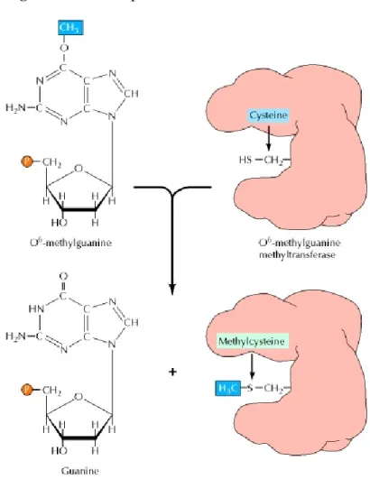
The MGMT gene is located at locus q26, chromosome 10. This gene codes the MGMT protein (see figure 1), a specific DNA repair enzyme that is ubiquitously expressed in normal human tissue. Although its exact physiologic function still to be fully defined, MGMT is known to restore DNA structure to its normal state by transferring aberrantly added methyl or alkyl groups from the O6-position of guanine to the enzyme’s active site. This restores guanine to its normal form preventing DNA strand breaks as shown in figure 2.
Figure 1 – MGMT protein configuration
Figure 2 – MGMT protein mechanism of action
The Cell, fourth edition
However, in tumour cells, this repair mechanism has a protective effect against cell death. In such conditions, the protein repairs DNA alterations incurred from toxicity of alkylating agents (i.e. chemotherapeutic agents) which frequently targets the O6 position of guanine. By rapidly reversing these changes, the MGMT protein prevents thus the formation of lethal cross-links and other mutagenic effects.8,9 In that regards, MGMT activity is a major mechanism of resistance to alkylating drugs.10 Tumours expressing MGMT were found to be 4 to 10 times more resistant to chemotherapeutic drugs.11,12 Inactivation of the MGMT gene is known to occur by hypermethylation of
the promoter region, leading to an under-expression of the protein that would normally repair lethal changes caused by alkylating chemotherapy drugs.
1.5 Inactivation of MGMT in human neoplasia
Recently, the gene encoding for the DNA repair enzyme MGMT has been found to be inactivated in several human cancers. Decreased expression was even observed in the precancerous states, as early as in leukoplakia and hyperplasia.13,14 High rates (27%) of promoter hypermethylation were also found in normal
surrounding mucosa in oral cancer patients.15,16 In vitro experiments showed
decreased MGMT expression in cultured buccal mucosa epithelial cells from healthy individual, when exposed to tobacco extracts.17 In the head and neck literature, the status of MGMT inactivation was analysed in 13 different studies with a prevalence ranging from 18% to 56%.15,18,19,20,21,22,23,24,25,26,27,28,29 It is important to note that all these studies were carried out on a small number of patients (ranging from 30 to 99 patients), except in one study by Dikshit et al. conducted on 235 patients with laryngeal and hypopharyngeal cancer.27
Several studies have established that MGMT promoter hypermethylation and/or loss of MGMT gene expression are predictive of poor survival in cancer patients, namely: hepatocellular, gastric, lung, breast cancer as well as low-grade diffuse astrocystoma.30,31,32,33,34 Thus, it seems that MGMT promoter
biomarker for biologically aggressive diseases in many human tumour types. On the other hand, it has also been shown in multiple studies that MGMT promoter
hypermethylation resulting in a decreased MGMT gene expression improves survival in patients with malignant astrocytoma35, glioblastoma,36 glioma37 and diffuse large B-cell lymphoma,38 who were treated with alkylating chemotherapeutic agents, such as carmustine and temozolomide.
1.6 MGMT methylation in HNSCC
Although 13 cited studies relate the prevalence of MGMT in HNSCC, only 3 correlate methylation status with patient survival. Zuo et al. found that MGMT promoter hypermethylation was a bad prognostic factor in 94 male subjects that were either treated surgically or medically.28 In an Indian study on 51 patients by Puri et al., it was found that promoter hypermethylation of at least 2 different genes
(including MGMT) correlated with increased 2-year disease-free survival.26 A large multicentric case-control study conducted on 235 patients by Dikshit et al. showed that hypermethylation was not a predictor of mortality or second primary cancer.27 However, the 3 above-mentioned studies contained heterogeneous groups of patients specifically in regards to tumour staging and treatment modalities. A more uniform study should generate results that are more representative of a specific group of patients.
We propose here a unique study in that regards: a large number of patients with only advanced stage HNSCC treated homogeneously with similar modalities (chemoradiation) at one single institution. We analysed the promoter
hypermethylation pattern of the human MGMT gene in these tumours and studied the correlation with clinical features, namely, grade, toxicities, recurrences, as well as survival.
2.
M
ATERIALS AND METHODS2.1 Tissue collection
One hundred and ninety nine biopsy samples of primary tumours from paraffin blocks archives were collected from patients with HNSCC at our institution (Notre Dame University Hospital – CHUM, in Montreal, Canada) and from referring hospitals to our center between 1996 and 2002. All patients received treatment with subsequent follow-up at our University Hospital. For every patient, we conducted a retrospective chart review for assessment of patient’s characteristics, tumour staging, treatment modality, complications from treatment, survival and response rates to treatment.
Inclusion criteria: Locally advanced HNSSC, stages III and IV Treatment with chemoradiation only
Minimum follow-up of 3 years
2.2 DNA extraction (Annex 1)
Three to ten 10-µm sections were cut from each paraffin-embedded pathology biopsy block. Paraffin was then washed by xylene and ethanol. Tissue was then degraded by proteinase K.
2.3 Bisulfite treatment (Annex 2)
DNA from tumour was subjected to bisulfite treatment. NaOH was used to denature DNA. Then DNA was modified by bisulfite and hydroquinone as described by Hegi et al.36 Unmethylated cytosine, but not its methylated counterpart, is modified into uracil by this treatment making thus the sequences distinguishable.
2.4 Methylation Specific Polymerase Chain Reaction (MSP) (Annex 3)
MSP was conducted to assess the methylation status of CpG island of MGMT gene promoter as previously described by Esteller et al.20,39
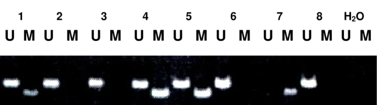
2.5 Agarose gel eletrophoresis (Annex 4)
Amplicons were then migrated on agarose gel. They were then examined under UV light and a Polaroid picture was taken to assess for the presence or absence of the methylation status. See figure 3.
Figure 3 – Gel electrophoresis
U: Unmethylated M: Methylated
U M U M U M U M U M U M U M U M U M
1 2 3 4 5 6 7 8 H2O2.6 Statistical Analysis
Local control rate (LCR) was defined by the time elapsed from initial diagnosis to development of recurrent loco-regional disease. Overall survival (OS) was defined as total deaths by all causes. Disease-free survival (DFS) was the period of time from the initial diagnosis of tumour to the first sign of tumour recurrence. Distant metastasis free survival (DMFS) was the period of time free of distant metastasis. Response rate was defined as satisfactory regression of primary tumour and neck metastasis evaluated clinically and radiologically at 3 months after the end of treatments, which did not require salvage surgical therapy. Survival functions were calculated using the Kaplan-Meier method. Logrank was used to compare the difference of survival curve between groups. Clinical correlation between methylation status and clinicopathological variables was determined using a Chi square test.
3.
R
ESULTSAlmost all tissues were derived from tumours without prior chemoradiation therapy. DNA extraction was performed on 199 tumour specimen. Adequate bisulfite modification was only achieved for 173 of the 199 tumours (87%). This is mainly due to the difficulty and rigorousness of the modification technique. DNA from paraffin-embedded tissues is more fragile than DNA from fresh tissues, explaining its degradation when submitted to bisulfite treatment. Table III depicts baseline patients’ characteristics.
3.1 Baseline characteristics of population
Table III. Baseline characteristics
Characteristic No. of patients Percent Age (years) Mean at diagnosis 56,4 Range 25 - 79
Mean follow-up time (years) 2,5
Sex Male 133 76,9 Female 40 23,1 Tumour site Oropharynx 113 65,3 Larynx 28 16,2 Oral cavity 16 9,2 Hypopharynx 8 4,6 Unknown 5 2,9 Nose 3 1,7 Tumour stage III 30 17,3 IV 135 78,0 Recurrence 8 4,6 Histopathologic grade I (Well differentiated) 26 15,0 II (Moderately differentiated) 87 50,3
III (Poorly differentiated) 51 29,5
IV (Undifferentiated) 0 0,0
Unknown 9 5,2
Of 173 patients, 133 (76.9%) were males and 40 (23.1%) females. Average age at diagnosis was 56.4 years, ranging from 25 to 79 years. The mean follow up time was
37 months. The oropharynx was by far the most common tumour site with 113 patients (65.3%). Other less common sites are larynx and oral cavity, representing 16.2% and 9.2 % of tumours respectively. Stage IV disease was diagnosed in 135 patients, representing 78%, whereas only 17.3% of patients had stage III, leaving the remaining 4.6% of our cohort with recurrent diseases. Staging was performed according to the TNM classification system from clinical and radiological assessment. Eighty seven patients had a moderately differentiated (grade 2)
histopathological tumour grade, representing the majority with 50.3%. Grade 1 was described in 26 patients (15%) and grade 3 in 51 patients (29.5%), leaving 5.2 % of the specimens with an unknown grade.
3.2 Treatment modalities
Treatment modalities are illustrated in table IV. All patients, except one, received both radiation and chemotherapy. This single patient could not sustain the effects of chemotherapy due to weak general medical condition and was thus treated with radiation therapy alone. The usual radiation dose was 7000 cGy over an average period of 48 days and consisted of hyperfractionation. Chemotherapy was
administered concomitantly to radiation therapy, according to 4 different regimens, the most common being carboplatinum with 5-Fluorouracil (5FU) given every 3 weeks in 95 patients (54.9%).The remaining 77 patients (44.5%) received
cisplatinum only given either daily, weekly or every 3 weeks. In 39 treated patients (19.7%) the tumour was found to be a second primary that was managed
independently with similar treatment modalities and conditions. These are the patients that have had HNSCC in the past and subsequently developed a second neoplasia that was not a recurrence.
Table IV - Treatment modalities
Characteristic Details No. of patients Percent
Radiation Therapy 7000 cGy over 48 days 173 100,0
Chemotherapy Concomitant 172 99,4
Carboplatinum & 5FU Every 3 weeks 95 54,9
Cisplatinum Every 3 weeks 42 24,3
Cisplatinum Weekly 11 6,4 Cisplatinum Daily 24 13,9 Second Primary Yes 34 19,7 No 139 80,3 3.3 MGMT methylation status
MSP on 173 DNA samples revealed aberrant hypermethylation of the MGMT promoter in 71 patients (41%) with HNSCC as illustrated in Table V. This subgroup of 71 patients was representative of the overall population with respect to baseline patient characteristics as well as stage of the disease and treatment modalities. Also, positive and negative controls worked appropriately in all rounds of PCR reaction. It is important to note however, that we dealt with substantial sample contamination at the beginning of our experimentation. This mishap was early suspected, as we
collected a strangely very high methylation status (close to 80%) of our specimen and obvious water contamination. The problem was initially seized and appropriate action
was achieved after we have realized that the pipettes were themselves contaminated. After serious repeated cleaning and sterilization of the instrumentation, we were confident in achieving reliable and consistent results that we have used in the current work. Methylation status was calculated following a non-quantitative assessment, i.e., we simply observed for the presence or the absence of the amplicons band in the MGMT MSP. We thus refrained from quantifying the amount of amplicon migration on the gel, since the technique we used is not one that can achieve quantifiable results. Therefore, interpretation of the results did not cause any possible confusion to the reader in almost all of the samples. Nonetheless, a third party was used to proof-read the methylation status. In only 3 specimens, there was a discrepancy between both readers that was resolved by further examination until mutual agreement was reached. These specimens had very small amounts of amplicon that were barely readable on the Polaroid photograph. The final results from this data collection were then categorized as either positive or negative groups.
Table V - Prevalence of MGMT methylation
Status No. of patients Percent
MGMT MSP + 71 41,0
MGMT MSP - 102 59,0
3.4 MGMT methylation status and correlation with clinical factors
MGMT promoter hypermethylation was not found to be correlated with age, sex, tumour site, tumour stage, use of radiation therapy or chemotherapy. We found however a possible significant correlation between the methylated status and tumour grade as illustrated in Table VI. For grade I and II, there was almost equal number of patients in both groups MSP+ and MSP-. On the other hand, for grade III tumours, there were more than double the number of patients in the methylated group, in comparison with the unmethylated group (37 vs. 14 patients respectively), with a p value of 0.06. This finding suggests that poorly differentiated tumours tend to have the MGMT methylation more often that not.
Table VI - Association of MGMT promoter methylation and
histopathological grade
Tumour grade MGMT MSP + MGMT MSP - P value
No. % No. %
I (Well differentiated) 13 7,5 13 7,5 NS
II (Moderately differentiated) 47 27,2 40 23,1 NS III (Poorly differentiated) 37 21,4 14 8,1 0.06
IV (Undifferentiated) 0 0,0 0 0,0 -
Abbreviation: NS, non-signficant
Clinical correlation with complications from toxicities did not reveal any positive significant association with the methylated status. Variables tested included: gastrointestinal toxicities such as mucosititis, nausea, vomiting, weight loss, nasal
tube feeding or gastrostomy tube feeding; haematological and infectious toxicities such as decrease in haemoglobin count, platelet count, white blood cell count, febrile neutropenia or infection; and other toxicities such as need for hospitalisation, tracheostomy or death.
There were no significant differences between response rates to therapy in both groups (71% for the methylated group, vs. 73% for the unmethylated group, p=NS)).
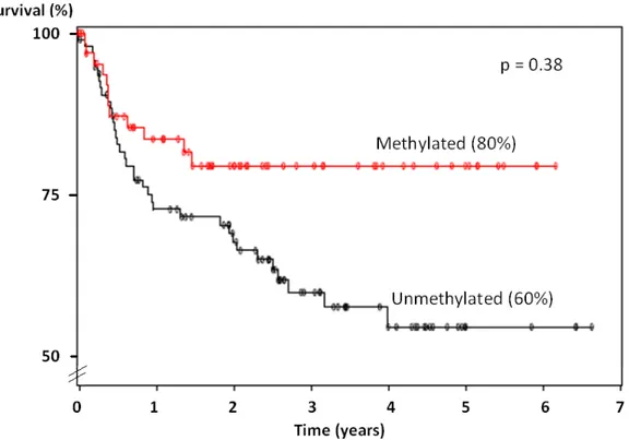
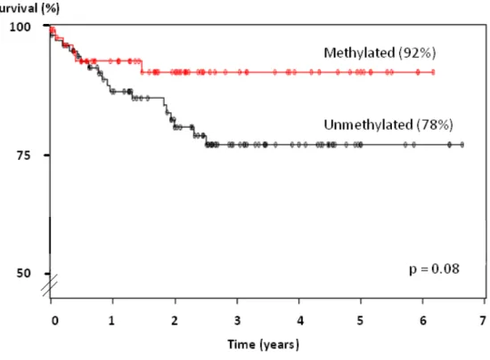
3.5 MGMT methylation status and survival
Since most of HNSCC recurrences occur relatively early in the post treatment period, we used a 3-year survival analysis for our entire patient population. Table VII shows different forms of survival for both methylated and unmethylated groups.
Table VII - Effect of MGMT promoter status on patient survival
Survival at 3 years Methylation status
MGMT MSP + (%) MGMT MSP - (%) P value LCR 87 77 0,26 OS 64 62 0,99 DFS 80 60 0,38 DMFS 92 78 0,08
Abbreviations: LCR, local control rate, OS, overall survival, DFS, disease free survival, DMFS, distant metastasis free survival.
When comparing methylated with unmethylated status, there was no
significant difference for locoregional control at 3 years, representing a LCR of 87% vs. 77% (p=0,26). OS was also similar between both groups with 64% vs. 62% (p=0,99) respectively. There seemed to be a survival advantage of 20% with DFS between both groups, showing 80% vs. 60% respectively, although this was not statistically significant, with a p value of 0,38. (see figure 4) However, a survival advantage of 14% (92% vs 78% respectively) for DMFS has better statistical significance (p=0,08). (see figure 5)
4.
D
ISCUSSIONPredicting response to therapy in patients with HNSCC represents an active area of research in the current century. This study aimed to investigate MGMT hypermethylation status as a potential tumour marker in HNSCC. Methyaltion status was assessed in 173 patients treated with chemoradiation for advanced disease and correlation with clinical outcome was undertaken. Strengths of the current study include a large sample size that is homogeneous in regards to both stage of disease as well as treatment modality.
The frequency of MGMT promoter hypermethylation in this study was 41%, fitting in the published range from 16% to 56% in 13 different reports. The largest study (235 patients) by Dikshit et al. demonstrated a frequency of 27%.27 The second largest (99 patients) is an Indian paper from Viswanathan et al. yielding also 41%. In another report by Zuo et al., hypermethylation was found in 18% of subjects. Most of the remaining articles were conducted on much smaller patient samples.28 Different factors could explain why our results are higher than those obtained by Dikshit and Zuo. First, we have different patient populations, in regards to racial aspects, as well as tumour site. Whereas larynx was the most common tumour for Diskshit, the oral cavity was dominant for Zuo. In our study, it is the oropharynx that represented the majority of cases. Also, different contributing etiologic factors might play a role. In our study, contamination could not have been responsible for the high percentage
observed, since successful controls were used in both bisulfite modification techniques as well as PCR techniques.
Because of the clinical relevance of MGMT gene in oncology, we attempted to determine if MGMT hypermethylation would have an impact on clinical behavior in HNSCC patients. In the current study, 37 out or 51 tumors (72,5%) with a poorly differentiated histological grade expressed MGMT hypermethylation status,
suggesting that high grade HNSCC tend to be rather methylated that not (p=0.06). These results are not sufficient to state that the poorer the tumour differentiation, the likelier the methylated status, since this trend was not observed in the moderately differentiated group. Our findings do not help in indicating neither that patients with a methylated MGMT tend to have a higher tumour grade.
In our cohort of patients, we did not observe any correlation between studied toxicities and hypermethylation status of MGMT. No other reports in the literature studied this correlation for MGMT in HNSSC. It is intuitive for mucosal toxicities to be similar among both groups, since normal non-cancerous mucosa surrounding tumour is similar in both methylated and non-methylated groups.40
Several cited studies herein have published that MGMT is both predictive of good and bad survival for different types of cancer. Only 3 papers discuss survival with MGMT in HNSSC, all with non-concordant results. We found a possible survival advantage for the hypermethylated group in regards to DFS (80% vs. 60%,
p=0,38), and DMFS (92% vs. 78%, p=0,08) . Zuo et al.28 reported a decreased 2-year DFS for the hypermethylated group (38%) vs. 74% for the unmethylated group (p<0.01). Five-year cause specific survival (CSS) and OS were also significantly decreased (p<0.01). However, patient population was inhomogeneous for both patients’ characteristics and treatment modalities. This was also the first report to observe a correlation between MGMT hypermethylation and decreased MGMT protein expression in HNSSC. A study by Puri et al.26 does not support the findings previously described by Zuo et al.. Puri found an increased 2-year DFS (p=0,028) when 2 or 3 genes (p16, MGMT, hMLH1) were simultaneously hypermethylated, but not for patients with hypermethylation of only 1 gene. This paper also lacked
homogeneity and was conducted on 51 samples. The latest report by Dikshit et al.27 is also the largest conducted on 235 patients with laryngeal and hypopharyngeal cancer. This group did not find a correlation between MGMT hypermethylation and survival. Although they were able to achieve better homogeneity in regards to tumour sites, patients with all 4 staged of disease were included and treatment modalities remained thus varied. The possible observed survival advantage in the methylated group undertaking chemoradiation therapy might be exaggerated by the presence of confounding factors, perhaps the concurrent presence of another genetic alteration.
One of the limitations of this study pertains to our relatively small follow-up period. Although most HNSCC tend to recur in the early period following treatment, some will take years before reappearance, whether loco-regionally or distantly.
Following our cohort of patients for longer will allow a more accurate survival analysis, perhaps also helping towards achieving a better statistical significance.
The MSP technique is designed to yield qualitative rather than quantitative methylation results. Absence of the amplicon band at the desired location on the migrated electrophoresis gel easily confirms that the specimen is lacking the
methylated gene, i.e. is unmethylated. On the other hand, a frank white intense band, will confirm the methylated status. There exists, between these clear extremes, a full spectrum of intensities. Thus, with methylation specific PCR, one can only confirm or infirm the presence of methylation, without being able to quantify it. Other techniques of quantitative analysis, such as Real Time PCR have successfully been used to improve level of detection of methylation 41. However, in the current study, a methylated status was attributed to any presence of bands, regardless of intensity. This potentially means that the methylated group is comprised of tumours with different extents of methylation, making it heterogeneous in that regards. This could dilute the impact of the MGMT methylation in our clinical analysis. Also, if fresh instead of paraffin-embedded tissue samples were used, less DNA degradation would have ensued, allowing perhaps improved detection. Furthermore, our inclusion criteria aimed to achieve homogeneity in regards to type of tumour histology,
staging, and modality of treatment. In spite of that, our cohort still contained different sites for HNSCC. However, this should not affect our results since both groups at study were comprised of similar patients’ characteristics.
In order to minimize variation of the quality of our measure and to control for possible ambiguity in interpretation of results, electrophoresis gel readings were done by 2 different independent operators, including one external to the study. Revisions and re-readings were undertaken on challenging instances until consensus was achieved unanimously.
Moreover, provision of our tissue samples came from whole biopsy specimen present on the pathology sections, including thus tumour but also non-cancerous tissue surrounding it. This has additional potential dilutive effect, dampening the clinical outcome accounted by the methylated status. However, published articles advocate that normal tissue surrounding neoplastic tissue potentially harvests pre-cancerous lesions. More interestingly, although this tissue might appear normal on histological exam, it was not on molecular biology testing. Specific molecular changes responsible for carcinogenesis and implicated in the multiple hit theory of cancer development were thus identified. In some of these studies, MGMT
methylation was observed in mucosa with normal histology surrounding cancerous lesions. This contradicts the hypothesis that the concentration of methylated MGMT gene is diluted by normal tissue surrounding the tumour. However other published reports, state the opposite. Righini et al.40 found that 90% of normal mucosa surrounding tumour specimen were free of methylation changes. The lack of consensus in that regards make conclusions difficult to draw.
Throughout our experiments, we encountered technical difficulties that necessitated much time and energy to rectify. Since parts of our experimentation were novel, we had to remodel existing protocols in the literature and create our own by trial and error, especially for bisulfite modification parameters (see Annex 2). The bisulfite modification is very lengthy and technically demanding. Despite meticulous work, it promotes DNA degradation, especially in paraffin-embedded tissue. This explains the non-interpretability of findings in 22 patients. Furthermore, we dealt with significant amounts of contamination during the first batches of bisulfite modification, hinted by aberrantly high (85%) methylation frequency in our
population that was not concordant with the literature. By isolating possible factors, we concluded that contamination originated from pipettes. After extensive cleaning and sterilization, we were able to achieve the current reliable results.
This is the first study to report a possible survival advantage when only MGMT is hypermethylated in HNSCC. Its large sample size and homogeneous characteristics account for its strength. However, the possible observed survival advantage in this study in the methylated group undertaking chemoradiation therapy might have be exaggerated by the presence of confounding factors, perhaps the concurrent presence of another genetic alteration.
5.
C
ONCLUSIONMGMT methylation was a frequent event (41%) in our cohort of HNSSC patients. The presence of MGMT methylation might represent a positive survival advantage in advanced stage HNSSC patients treated with chemoradiation.
Elucidation of genetic mechanisms and their role in development and behaviour of neoplasia will help better isolate individual genetic contributions. On the other hand, large cohorts of clinical studies will enlighten survival analysis pertaining to these genetic contributions. This will help confirming the MGMT methylation as a
predictive factor to treatment. Ensuing patient stratification in regards to genetics will help better target a treatment while maximizing efficacy and minimizing undesirable side effects in HNSCC patients. More precisely, by determining the methylation status as part of a pre-operative patient assessment, physicians will better tailor chemoradiation therapy to patients that will benefit most from it, while reserving perhaps surgical options for those with less than ideal responses to medical treatment.
A
NNEX1
-
DNA
EXTRACTION FROM PARAFFIN EMBEDDED TISSUE SAMPLES1. Reagents
1) Xylene 2) Ethanol 100% 3) Ethanol 95%
3) Proteinase K (20 mg/ml, 20mg proteinase K + 1ml of buffer TEN, aliquot in 20 µl and keep frozen at -20°C)
4) Buffer TEN (10 mM Tris-HCl, pH 8.0, 1 mM EDTA, pH 8.0, 20 mM NaCl)
2. Process
1) Heat 3 to 10 pathology sections (depending on the size of tissue) per patient to 72 °C for 1-3 minutes (Until the paraffin becomes transparent);
2) Wash with xylene for 2 minutes, repeat 4 times; 3) Wash with ethanol 100% for 1 minute, repeat 4 times; 4) Wash with ethanol 95% for 1 minute, repeat 4 times;
5) Prepare the digestion solution 1:100 (1µl of proteinase K in 99µl of buffer TEN);
6) Deposit (100 – 200 µl depending on tissue size) on a pathology section for 3 minutes and scrape the tissue with a plastic instrument;
7) Recover all the scrapings in an Eppendorf tube using a Pasteur pipette and incubate at 52 °C overnight;
8) Inactivate proteinase K at 95 °C for exactly 3 minutes;
9) Centrifuge at 12,000g for 20 minutes at 4 °C and transvase the supernatant in a new Eppendorf tube.
A
NNEX2
-
B
ISULFITE MODIFICATION OFDNA
1. Reagents
1) NaOH 6 M (6g of NaOH (FW 40.0) in 25 mL of deionized H2O, keep at 4°C to cool);
2) NaOH 0.1M (50µl of NaOH 6 M + 2950µl of deionized H2O)(for 8 specimens);
3) Hydroquinone 100 mM (55 mg of hydroquinone (SIGMA#-9003, FW110.11) + 5mL of deionized H2O, freshly prepared and keep at 4°C);
4) Sodium Bisulfite 6.261 M at pH 5 (5.95g of bisulfite (SIGMA#-9000, FW190) + 8mL of deionized H2O, ajust pH with 6M NaOH ~600-700µl , fill to 10 mL with H2O, freshly prepared and keep at 4°C. Heat bisulfite in warm tap water and vortex for 20 minutes).
2. Process
1. Deposit 1.17µl of NaOH 6M in the tube;
2. Add 33.83 µl of extracted DNA from paraffin-embedded tissue; 3. Denature DNA at 37°C for 10 minutes;
4. Spindown the tubes and add 253 µl of bisulfite first and then 72 µl of hydroquinone;
5. Incubate at 55 °C for 5h and brew 3 times during incubation. Then spindown the tubes;
6. Add 150 µl of H2O in each tube;
7. Transfer the diluted solution in a filter tube (Millipore, Genomics, UFC7PCR50) and centrifuge at 1200 xg for 10 minutes at 20°C;
(Starting at this step, always close the tubes in the centrifugal machine to avoid contamination.)
8. Add 350 µl of H2O to the tube, centrifuge at 1200 xg for 10 minutes at 20°C then elutriate the filtrate;
9. Add 350 µl of NaOH 0.1M to the tube, at 1200 xg for 10 minutes at 20°C then elutriate the filtrate;
10. Add 350 µl of H2O to the tube, centrifuge at 1500 xg for 15 minutes;
11. Add 90 µl of H2O to the tube and incubate for 15 minutes at room temperature; 12. Reverse the filter and put it in a new tube to collect the DNA;
13. Centrifuge at 1000 xg for 5 min at 20°C; 14. Keep DNA at - 20°C.
A
NNEX3
-
M
ETHYLATION SPECIFICPCR
(MSP)
MSP is a specific type of Polymerase Chain Reaction that assesses the methylation status of groups of CpG sites within CpG islands
1. Reagents Table VIII
Reagents Quantity/tube Final concentration
Buffer 10 X with MgCl2 15 mM 5 µl 1 X
dNTPs 2mM 5 µl 0.2 mM
MgCl2 25 mM 2 µl 1.8 mM
*Primer MGMT-MF(10x) 5 µl 0.5 µM
*Primer MGMT-MR (10x) 5 µl 0.5 µM
Hotstart TAQ (Qiagen) 0.5 µl 2.5 u
Q solution 10 µl
BSA 1µl
DNA 16.5 µl
* 2 sets of primers are used in the preparation as depicted in table IX, as described previously.42
2. Sets of primers
Table IX - Primer sequences for methylation-specific polymerase chain reaction analysis
Gene Sense primer, 5'→3' Antiense primer, 5'→3'
Product size, bp AT, °C MGMT M TTTCGACGTTCGTAGGTTTTCGC GCACTCTTCCGAAAACGAAACG 80 59 MGMT U TTTGTGTTTTGATGTTTGTAGGTTTTTGT AACTCCACACTCTTCCAAAAACAAAACA 94 59
Abbreviations: Bp indicates base pairs; AT, annealing temperature; M, methylated sequence;
U, unmethylated sequence
3. PCR program 96 °C 5 minutes 94 °C 30 seconds 50 °C 30 seconds X 40 cycles 72 °C 30 seconds 72 °C 10minutes 4°C→ end
A
NNEX4
-
A
GAROSE GEL ELECTROPHORESIS1) Pour 2 grams of agarose powder in 100mL solution of (10mL of buffer Tris-Bromate; EDTA 10X concentrate and 90 mL of H2O);
2) Heat in the microwave until boiling;
3) Add 5µL ethidium bromide to the boiling liquid;
4) Pour in the migration plate and allow 20 minutes for polymerisation;
5) Pour 1L solution of (100mL buffer Tris-Bromate EDTA 10X concentrate and 900mL of H2O) in the electrophoresis machine;
6) Place gel in machine;
7) Charge the slots on the gel with a mix of 15 µL of DNA and 3 µL of loading buffer 6X (Also, use one slot for marker pUC19 MspI);
8) Let migrate at 145 volts for 45 minutes;
9) Examine the gel in the dark under ultraviolet light; 10) Take a picture with the Polaroid.
R
EFERENCES
1
Boring C, Squire TS, Tong T. Cancer statistics. CA – Cancer J Clin 1992;42:19-38 2
Parker SL, Tong T, Bolden S et al. Cancer statistics, 1996. CA Cancer J clin 1996;46:5-27 3
Rodrigo JP, Ferlito A, Suarez C, et al. New molecular diagnostic methods in head and neck cancer. Head & Neck. 2005;27(11):995-1003
4
Weinberg RA. How cancer arises. Sci Am. 1996;275:62-70 5
Reik W. Stability and flexibility of epigenetic gene regulation in mammalian development. Nature. 2007. 24;447(7143):425-32
6
Tycko B. Epigenetic gene silencing in cancer. J Clin Invest. 2000;105(4):401-7. 7
Murakami J, Asaumi J, Maki Y et al. Influence of CpG island methylation status in O6 -methylguanine-DNA methyltransferase expression of oral cancer cell lines. Oncology Reports. 2004;12(2):339-45.
8
Gerson SL. MGMT: its role in cancer aetiology and cancer therapeutics. Nat Rev Cancer. 2004;4(4):296-307.
9
Gerson SL. Clinical relevance of MGMT in the treatment of cancer. J Clin Oncol. 2002;20(9):2388-99.
10
Ludlum DB. DNA alkylation by the haloethylnitrosoureas: nature of modifications prodiced and their enzymatic repair or removal. Mutat Res 1990;233:117-26
11
Yarosh DB. The role of O6-Methylguanine-DNA methyl-transferase in cell survival, mutagenesis, and carcinogenesis. Mutat Res 1985;145:1-16.
12
Schold S, Jr., Brent TP, von Hofe E et al. O6-Alkylguanine-DNA alkyltransferase and sensitivity to procarbazine in human brain-tumour xenografts. J. Neurosurg 1989;70:573-7. 13
Rodriguez MJ. Loss of expression of DNA repair enzyme MGMT in oral leukoplakia and early oral squamous cell carcinoma. A prognostic tool? Cancer Letters. 2007;245:263-268. 14
Sawhney M, Rohatgi N, Kaur J et al. MGMT expression in oral precancerous and cancerous lesions: Correlation with progression, nodal metastasis and poor prognosis. Oral Oncology. 2007;43:515-22.
15
Kato K, Hara A, Kuno T et al. Aberrant promoter hypermethylation of p16 and MGMT genes in oral squamous cell carcinomas and the surrounding normal mucosa. J Cancer Res Clin Oncol. 2006;132:735-43.
16
Zhang S, Guo C, Kong W et al. Promoter hypermethylation of DNA repair gene MGMT in laryngeal squamous cell carcinoma. Medical Sciences. 2006;26(1):101-4.
17
Liu Y, Egyhazi S, Hansson J et al. O6-Methylaguanine-DNA methyltransferase activity in human buccal mucosal tissue and cell cultures. Complex mixtures related to habitual use of tobacco and betel quid inhibit the activity in vitro. Carcinogenesis. 1997;18(10):1889-95. 18
Zhang S, Kong WJ et Liu Z. Promoter hypermethylation of DNA repair gene MGMT in laryngeal squamous cell carcinoma. Medical Sciences. 2006;26(1):101-4
19
De Schutter H, Geeraets H, Verbeken E et al. Promoter methylation of TIMP3 and CDH1 predicts better outcome in head and neck squamous cell carcinoma treated by radiotherapy only. Oncology Reports. 2009;21:507-13.
20
Esteller M, Hamilton SR, Burger PC et al. Inactivation of the DNA repair gene O6
-methylguanine-DNA methyltransferase by promoter hypermethylation is a common event in primary human neoplasia. Cancer Res 1999;59:793-7.
21
Sanchez-Cespedes M, Esteller M, Wu L, et al. Gene promoter hypermethylation in tumors and serum of head and neck cancer patients. Cancer Res 2000;60:892-5.
22
Rosas SLB, Koch W, Carvalho MDC, et al. Promoter hypermethylation patterns of p16,
O6-methylguanine-DNA methyltransferase, and death-associated protein kinase in tumors
and saliva of head and neck cancer patients. Cancer Res 2001;61:939-42. 23
Kwong J, Lo KW, To KF et al. Promoter hypermethylation of multiple genes in nasopharyngeal carcinoma. Clin Cancer Res 2002;8:131-7.
24
Wong TS, Tang KC, Kwong DL, et al. Differential gene methylation in undifferentiated nasopharyngeal carcinoma. Int J Oncol 2003; 22:869-74.
25
Viswanathan M, Tsuchida N, Shanmugam G. Promoter hypermethylation profile of tumor-associated genes p16, p15, hMLH1, MGMT and E-cadherin in oral squamous cell carcinoma. Int J Cancer 2003;105:41-6.
26
Puri SK, Si L, Fan CY et al. Aberrant promoter hypermethylation of multiple genes in head and neck squamous cell carcinoma. Am J Otolaryngol. 2005;26:12-17.
27
Dikshit RP, Gillio-Tos A, Brennan P et al. Hypermethylation, risk factors, clinical characteristics, and survival in 235 patients with laryngeal and hypopharungeal cancers. Cancer. 2007;110(8):1745-51.
28
Zuo C, AI L, Ratliff P et al. O6-methylguanine-DNA methyltransferase Gene: Epigenetic silencing and prognostic value in head and neck squamous cell carcinoma. Cancer Epidemiol Biomarkers Prev 2004;13(6).
29
Maruya S, Issa JP, Weber RS et al. Differential methylation status of tumour associated genes in head and neck squamous cell carcinoma: incidence and potential implications. Clin Cancer Res. 2004;10:3825-30.
30
Matsukura S, Miyazaki K, Yakushiji H et al. Expression and prognostic significance of O6 -methylguanine-DNA methyltransferase in hepatocellular, gastricand breast cancers. Ann Surg Oncol. 2001;8:808-16.
31
Lee HS, Lee HK, Kim HS et al. Tumour suppressor gene expression correlates with gastric cancer prognosis. J Pathol. 2003;200:39-46.
32
Cayre A, Penault-Llorca F, De Latour M et al. O6-methylguanine-DNA methyltransferase
gene expression and prognosis in breast carcinoma. Int J Oncol 2002;21:1125-31. 33
Brabender J, Usadel H, Metzger R et al. Quantitative O6-methylguanine-DNA
methyltransferase methylation analysis in curatively resected non-small cell lung cancer : associations with clinical outcome. Clin Cancer Res. 2003; 9:223-7.
34
Komine C, Watanabe T, Katayama Y et al. Promoter hypermethylation of the DNA repair
gene O6-methylguanine-DNA methyltransferase is an independent predictor of shortened
progression free survival in patients with low-grade diffuse astrocytomas. Brain Pathol. 2003;13:176-84.
35
Jaeckle KA, Eyre HJ, Towns end JJ, et al. Correlation of tumor O6-methylguanine-DNA
methyltransferase levels with survival of malignant astrocytoma patients treated with bis-chloroethylnitrosourea: a Southwest Oncology Group study. J Clin Oncol 1998;16:3310-5 36
Hegi ME, Diserens AC, Gorlia T et al. MGMT gene silencing and benefit from temozolomide in glioblastoma. N Engl J Med. 2005;352(10):997-1003.
37
Esteller M, Garcia-Foncillas J, Andion E, et al. Inactivation of the DNA-repair gene MGMT and the clinical response of gliomas to alkylating agents. N Engl J Med 2000;2000;343:1350-4
38
Esteller M, Gaidano G, Goodman SN, et al. Hypermethylation of the DNA repair gene O6
-methylguanine-DNA methyltransferase and survival of patients with diffuse large B-cell lymphoma. J Natl Cancer Inst 2002;94:26-32
39Esteller M, Hamilton SR, Burger PC, Baylin SB, Herman JG. Inactivation of the DNA
repair gene O6-methylguanine-DNA methyltransferase by promoter hypermethylation is a
common event in primary human neoplasia. Cancer Res 1999;59:793-7.
40Righini CA, de Fraipont F, Timsit JF et al. Tumor-specific methylation in saliva: a
promising biomarker for early detection of head and neck cancer recurrence. Clin Cancer Res 2007, 15;13(4):1179-85.
41
Smiraglia DJ, Smith LT, Lang JC, et al. Differential targets of CpG island
hypermethylation in primary and metastatic head and neck squamous cell carcinoma (HNSCC). JMed Genet 2003;40:25-33.
42Sanchez-Cespedes M, Esteller M, Wu L, et al. Gene promoter hypermethylation in tumors