ORIGINAL ARTICLE
Head and neck squamous cell carcinoma (HNSCC)
–
detection of synchronous primaries with
18
F-FDG-PET/CT
Klaus Strobel&Stephan K. Haerle&Sandro J. Stoeckli&
Madeleine Schrank&Jan D. Soyka&
Patrick Veit-Haibach&Thomas F. Hany
Received: 10 October 2008 / Accepted: 1 January 2009 / Published online: 10 February 2009
# Springer-Verlag 2009 Abstract
Purpose The aim of the study was to evaluate 18
F-FDG-PET/CT for the detection of synchronous primaries at initial staging of patients with head and neck squamous cell carcinoma (HNSCC).
Methods FDG-PET/CT images acquired between March 2001 and October 2007 in 589 consecutive patients (147
women, 442 men; mean age 61.5 years, age range 32–
97 years) with proven HNSCC were reviewed for the presence of synchronous primaries. Cytology, histology and/or clinical and imaging follow-up served as reference standard.
Results FDG-PET/CT showed 69 suspected synchronous primaries in 62 patients of which 56 were finally confirmed in 44 patients. Of the 56 second cancers, 46 (82%) were found in the aerodigestive tract in the following locations: lung (26, 46%), head and neck (15, 17%), oesophagus (5, 9%). Ten second cancers (18%) were located outside the aerodigestive tract (colon, five; stomach, lymphoma, breast, thymus and kidney, one each). Six patients had three synchronous primaries and three patients had four synchro-nous cancers. Nine synchrosynchro-nous cancers were not detected by PET/CT (four head and neck, two lung, two oesopha-geal, one gastric). False-positive PET/CT findings were
mainly related to benign FDG uptake in the intestine due to benign or precancerous polyps or physiological FDG uptake in other head and neck regions. Overall the prevalence of synchronous second primaries according to the reference standard was 9.5%, of which 84% were detected with FDG-PET/CT. In 80% of the patients, therapy was changed because of the detection of a synchronous primary.
Conclusion FDG-PET/CT detects a considerable number of synchronous primaries (8.0% prevalence) at initial staging of patients with HNSCC. Synchronous cancers were predominantly located in the aerodigestive tract, primarily in the lung, head and neck and oesophagus. Detection of second primaries has an important impact on therapy. PET/ CT should be performed before panendoscopy.
Keywords PET oncology . Therapy . PET/CT . PET-CT oncology
Introduction
Due to alcohol and nicotine abuse which is often encountered in patients with head and neck squamous cell carcinoma (HNSCC), they have an elevated risk of developing synchronous and metachronous SCC in other regions of the upper aerodigestive tract. Second cancers predominantly affect the head and neck area, oesophagus or
the lung [1,2]. The prevalence of a second SCC in cadaver
dissection studies ranges between 3.7% and 15.5% [3,4].
For the highest-risk subgroups metachronous SCC occur in
about 4% of patients per year [5]. Panendoscopy studies
have shown the prevalence of synchronous primary SCC to
range from 1.4 to 17% [6]. At our institution a prevalence
of second primary tumours detected with panendoscopy of
K. Strobel (*)
:
M. Schrank:
J. D. Soyka:
P. Veit-Haibach:
T. F. HanyDivision of Nuclear Medicine, Department of Medical Radiology, University Hospital Zurich,
Raemistr. 100,
8091 Zurich, Switzerland e-mail: [email protected] S. K. Haerle
:
S. J. StoeckliDepartment of Otorhinolaryngology, Head and Neck Surgery, University Hospital Zurich,
16.2% has been found, with 6.4% being synchronous and
9.8% being metachronous [6]. 18F-Fluoro-deoxy-glucose
positron emission tomography (FDG-PET/CT) is increas-ingly used for staging of HNSCC with a considerable
impact on treatment decisions [7–9]. In comparison to
morphological imaging methods such as CT or MRI, PET/ CT has the great advantage of assessing the primary tumour, the neck nodes and the potential distant metastases and second primaries in a single examination. Detection and staging of second primaries is of great importance because it has a significant impact on treatment. Second primaries are one reason for the lack of improvement in the overall survival of these patients.
The aim of this study was to evaluate18F-FDG-PET/CT
for the detection of synchronous primaries at the initial staging of patients with HNSCC.
Materials and methods Patients
From the first installation of a combined PET/CT scanner at our institution in March 2001 until October 2007, 620 patients with biopsy-proven HNSCC were investigated with
18
F-FDG-PET/CT for initial staging. Of these patients, 31 were excluded because they were lost to follow-up, or adequate reports were not available. Therefore, the study cohort consisted of 589 patients (147 men, 442 men; mean age 61.5 years, age range 32–97 years). The indication for PET/CT was mainly to rule out distant metastases in patients with advanced HNSCC (large primary tumour and/or clinical suspicion of lymph node metastases) prior to therapy. The study was conducted in accordance with the local guidelines established by the ethics committee for retrospective evaluations. The patient characteristics are
summarized in Table1.
FDG-PET/CT imaging
All the data were acquired on a combined PET/CT in-line system (Discovery LS, Discovery STE, Discovery Rx; GE Health Systems, Milwaukee, WI). These dedicated systems integrate a PET scanner (GE Advance Nxi; GE Health Systems) with a multislice helical CT (LightSpeed plus or LightSpeed 16, LightSpeed VCT; GE Health Systems) and permit the acquisition of coregistered CT and PET images in one session.
Patients fasted for at least 4 h prior to scanning, which started approximately 60 min after injection of a standard
dose of approximately 350 MBq of 18F-FDG. Oral CT
contrast agent (Micropaque Scanner, Guerbet, Aulnay-sous-bois, France, or Gastrografin, Bayer-Schering, Berlin,
Germany) was given 15 min before injection of 18F-FDG.
Patients were examined in the supine position. Initially, the CT scan was acquired starting from the level of the head using the following parameters: 80 mA, 140 kV, 0.5 s per tube rotation, slice thickness 4.25 mm, scan length 867 mm, data acquisition time 12.5–22.5 s. The CT scan was acquired during breath holding in the normal expiratory position.
Immediately following the CT scan, a PET emission scan was acquired with an acquisition time of 3 min per cradle position with a one-slice overlap. The eight to nine cradle positions from the knees to the head resulted in an acquisition time of approximately 24–27 min. The CT data were used for the attenuation correction and the images were reconstructed using a standard iterative algorithm. The acquired images were viewed with software providing multiplanar reformatted images of PET alone, CT alone and fused PET/CT with linked cursors using an Advantage windows workstation (GE Health Systems). Additionally, in 160 patients intravenous contrast agent was given and a diagnostic neck CT scan from the base of the skull to the aortic arch was performed after the PET scan.
PET/CT interpretation
The PET/CT images were retrospectively reviewed by a doubly board-certified radiologist and nuclear medicine physician with 10 years experience in CT reading and
Table 1 Characteristics of 589 patients with NNSCC
Women 147 (25.0%) Men 442 (75.0%) Age (years) Mean 61.5 Range 32–97 Location of primary HNSCC Nasopharynx 29 (4.9%) Oral cavity 226 (38.4%) Oropharynx 149 (25.3%) Hypopharynx 89 (15.1%) Larynx 61 (10.4%) Unknown primary 27 (4.6%) Other 8 (1.4%) Stage of primary NNSCC T-stage Tx 27 (4.6%) T1 85 (14.4%) T2 219 (37.2%) T3 96 (16.3%) T4 162 (27.5%) N-stage N0 137 (23.3%) N1 84 (14.3%) N2 338 (57.4%) N3 30 (5.1%)
4 years experience in reading combined PET/CT in patients with head and neck cancer for the presence of synchronous primary tumours. The reader was blinded regarding the results of clinical investigations including panendoscopy, and other imaging including CT or MRI.
The PET images were analysed for the presence and nature of focal lesions with increased FDG uptake. For all patients, the attenuation-corrected PET images were used for analysis. Lesions were interpreted as second tumours if the uptake was higher than the uptake of the surrounding background tissue so that a lesion was clearly detectable. The morphology of suspicious lesions provided by the low-dose CT part was also used for interpretation: for example, in a spiculated solitary FDG-positive mass in the lung, a second lung primary was diagnosed; in multiple non-spiculated well-demarcated solid nodules in the lung, metastases were diagnosed. FDG uptake typical of physi-ological or benign variants such as in muscle, brown fat or pulmonary infiltration, were not interpreted as second
primaries or distant metastases [10].
Reference standard
In patients with a suspected second primary, additional investigations were performed. In the majority of patients (420) additional panendoscopy was available as the standard of reference. Patients with a suspected lung cancer were referred to pulmonology for flexible bronchoscopy with biopsy. Patients with a suspected oesophageal or colon cancer were referred to gastroenterology for flexible endoscopy with biopsy. In patients without a suspected second primary, imaging (CT and/or PET/CT) and clinical follow-up was performed for at least 6 months (mean
follow-up time 16.2 months, range 6–42 months).
Results
Overall, 56 synchronous tumours in 44 patients out of 589 evaluated patients were confirmed according to the above mentioned reference standard. Thus, the prevalence of synchronous primaries in our cohort according to the standard of reference was 9.5%. Of these, synchronous primaries, 47 (84%) were detected in 41 patients (93%) by FDG-PET/CT.
Location of second primaries
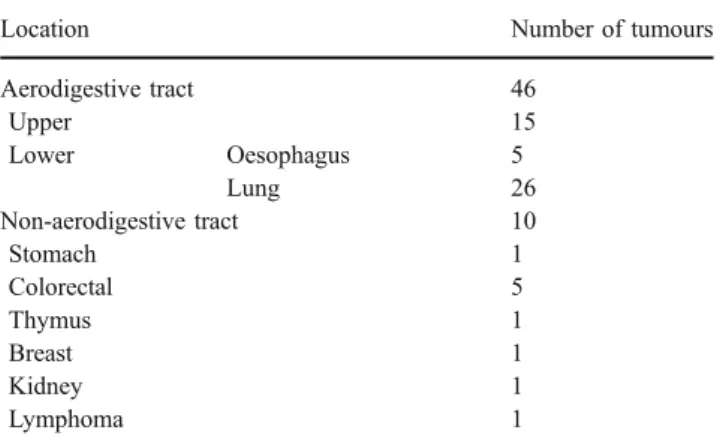
Of the 56 second primary tumours, 46 (82%) were found in the aerodigestive tract in the following locations: lung (26, 46%), upper aerodigestive tract (15, 27%), and oesophagus (5, 9%). Ten second primary tumours (18%) were located outside the aerodigestive tract (five colorectal, one stomach,
one lymphoma, one breast, one thymus, one kidney). The locations of the synchronous second primary tumours are
summarized in Table2. Thirty-five patients had one second
primary (Table 3). Six patients suffered from three
synchronous primary tumours (Table 4; Figs.1 and 2) and
three patients had four synchronous primary tumours
(Table 5).
Extension and histology of the second primaries
Of the 56 synchronous primaries found, 25 (45%) were in advanced stages (>T2 and/or N+ and/or M1), and 31 (55%) were detected in a confined stage (T1/2, N0, M0). In the majority of the second primaries detected a curative treatment approach was possible. Only two patients already showed distant metastases: one patient with an advanced oral cavity SCC and a synchronous advanced non-small-cell lung cancer had liver and bone metastases; and one patient with a tonsillar SCC was diagnosed with an advanced synchronous oesophageal cancer with distant lymph node metastases. Furthermore, a stage IV synchro-nous follicular lymphoma was detected, which required no treatment. Another synchronous small-cell lung cancer was detected with extended disease and was treated with chemotherapy. Of the 56 synchronous malignancies, 38 (68%) were SCCs, and 15 (27%) were adenocarcinomas. The majority of lung cancers were SCCs (77%), and the majority of oesophageal cancers were also SCCs (60%). All synchronous cancers in the colorectal area were adenocar-cinomas. All synchronous cancers in the head and neck region were SCCs.
Second cancers not detected by PET/CT
Nine synchronous cancers were detected by endoscopy and not detected by PET/CT. Among the false-negative PET findings were four small (T1) SCCs in the head and neck area (one oral cavity, two oropharynx, one hypopharynx),
Table 2 Location of 56 confirmed second primaries
Location Number of tumours Aerodigestive tract 46 Upper 15 Lower Oesophagus 5 Lung 26 Non-aerodigestive tract 10 Stomach 1 Colorectal 5 Thymus 1 Breast 1 Kidney 1 Lymphoma 1
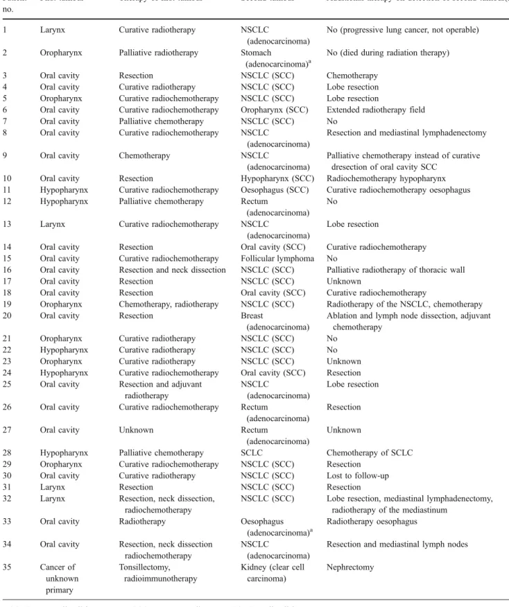
Table 3 Details of 35 patients with two synchronous primaries Patient
no.
First tumour Therapy of first tumour Second tumour Additional therapy on detection of second tumour(s)
1 Larynx Curative radiotherapy NSCLC
(adenocarcinoma)
No (progressive lung cancer, not operable) 2 Oropharynx Palliative radiotherapy Stomach
(adenocarcinoma)a
No (died during radiation therapy) 3 Oral cavity Resection NSCLC (SCC) Chemotherapy
4 Oral cavity Curative radiotherapy NSCLC (SCC) Lobe resection 5 Oropharynx Curative radiochemotherapy NSCLC (SCC) Lobe resection
6 Oral cavity Curative radiochemotherapy Oropharynx (SCC) Extended radiotherapy field 7 Oral cavity Palliative chemotherapy NSCLC (SCC) No
8 Oral cavity Curative radiochemotherapy NSCLC
(adenocarcinoma)
Resection and mediastinal lymphadenectomy 9 Oral cavity Chemotherapy NSCLC
(adenocarcinoma)
Palliative chemotherapy instead of curative dresection of oral cavity SCC
10 Oral cavity Resection Hypopharynx (SCC) Radiochemotherapy hypopharynx 11 Hypopharynx Curative radiochemotherapy Oesophagus (SCC) Curative radiochemotherapy oesophagus 12 Hypopharynx Palliative chemotherapy Rectum
(adenocarcinoma) No 13 Larynx Curative radiochemotherapy NSCLC
(adenocarcinoma)
Lobe resection
14 Oral cavity Resection Oral cavity (SCC) Curative radiochemotherapy 15 Oral cavity Curative radiochemotherapy Follicular lymphoma No
16 Oral cavity Resection and neck dissection NSCLC (SCC) Palliative radiotherapy of thoracic wall 17 Oral cavity Resection NSCLC (SCC) Unknown
18 Oral cavity Resection Oral cavity (SCC) Curative radiochemotherapy
19 Oropharynx Chemotherapy, radiotherapy NSCLC (SCC) Radiotherapy of the NSCLC, chemotherapy 20 Oral cavity Resection Breast
(adenocarcinoma)
Ablation and lymph node dissection, adjuvant chemotherapy
21 Oropharynx Curative radiotherapy NSCLC (SCC) No 22 Hypopharynx Curative radiotherapy NSCLC (SCC) No 23 Oropharynx Curative radiotherapy NSCLC (SCC) Unknown 24 Hypopharynx Curative radiochemotherapy Oral cavity (SCC) Resection 25 Oral cavity Resection and adjuvant
radiotherapy
NSCLC
(adenocarcinoma)
Lobe resection 26 Oral cavity Curative radiochemotherapy Rectum
(adenocarcinoma)
Resection 27 Oral cavity Unknown Rectum
(adenocarcinoma)
Unknown
28 Hypopharynx Palliative chemotherapy SCLC Chemotherapy of SCLC 29 Oropharynx Curative radiochemotherapy NSCLC (SCC) Resection
30 Oral cavity Curative radiotherapy NSCLC (SCC) Lost to follow-up 31 Larynx Resection NSCLC (SCC) Resection 32 Larynx Resection, neck dissection,
radiochemotherapy
NSCLC (SCC) Lobe resection, mediastinal lymphadenectomy, radiotherapy of the mediastinum
33 Oral cavity Radiotherapy Oesophagus (adenocarcinoma)a
Radiotherapy oesophagus 34 Oral cavity Resection, neck dissection
radiochemotherapy
NSCLC
(adenocarcinoma)
Resection and mediastinal lymph nodes 35 Cancer of
unknown primary
Tonsillectomy, radioimmunotherapy
Kidney (clear cell carcinoma)
Nephrectomy
NSCLC non-small-cell lung cancer, SCC squamous cell cancer, SCLC small-cell lung cancer.
which were incidentally seen during panendoscopy. There were two small adenocarcinomas of the oesophagus (T1N0 and T2N0), which were detected by panendoscopy and not detected by PET/CT. One signet ring cell cancer of the stomach, which showed no FDG uptake (T2N0) was incidentally detected by endoscopy during placement of the feeding tube. Additionally, two small bronchogenic
SCCs (both T1N0) were not visible on PET/CT but detected by panendoscopy.
Reasons for false-positive PET/CT findings
False-positive PET/CT findings (22) were mainly related to FDG uptake in the intestine due to benign or precancerous
Table 4 Details of six patients with three synchronous primaries Patient
no.
First tumour
Therapy of first tumour Second tumour Third tumour Additional therapy on detection of second and third tumours 36 Oral cavity Resection Hypopharynx
(SCC)
NSCLC (SCC) Radiotherapy hypopharynx and thorax 37 Oral cavity Rfesection, neck dissection,
radiochemotherapy
Hypopharynx (SCC)
Oesophagus (SCC) Radiochemotherapy hypopharynx, and oesophagus
38 Oropharynx Curative radiotherapy Rectal cancer (adenocarcinoma)
Thymus cancer (SCC)
Resection of rectal and thymus cancer 39 Oral cavity Palliative NSCLC (SCC) Oesophagus
(adenocarcinoma)a No
40 Oropharynx Resection NSCLC (SCC)a NSCLC (SCC)a Bilateral lung lobe resection and mediastinal lymph node resection 41 Oropharynx No therapy Oesophagus (SCC) NSCLC (SCC) No
NSCLC non-small-cell lung cancer, SCC squamous cell cancer.
aMissed by PET.
Fig. 1 A 67-year old male patient with proven oropharyn-geal cancer. FDG-PET/CT images showing the advanced primary tumour (a, b long ar-row) with multiple ipsilateral lymph node metastases (a arrowheads). Focal FDG-posi-tive lesion (a, c short arrow) in the anterior mediastinum, con-firmed as thymus cancer on histology. FDG-positive lesion (a, d wide arrow) in the rectum confirmed as rectal cancer on histology
polyps (eight), which can lead to focal FDG uptake mimicking a small colon cancer. Another reason was physiological FDG uptake in the head and neck area (five). The lymphoid tissue of the tonsils and the musculature of the tongue especially can show asymmetric FDG uptake mimicking small cancers. Other reasons for nonmalignant FDG uptake were physiological uptake in the intestine (two), inflammatory lesion in the lung (four), rectum (one) and oesophagus (one), and FDG uptake in a benign thyroid nodule (one).
Therapeutic changes due to detection of a second primary In 32 of 40 patients (80%) with available follow-up, the therapy was changed because of the detection of a synchronous primary. Of these 32 patients, 15 received additional surgery, 6 additional radiotherapy, 5 addi-tional radiochemotherapy, and 6 addiaddi-tional chemother-apy. Details of the changed therapies are shown in
Table 2.
Fig. 2 A 64-year old patient with proven SCC of the right tongue (a, b long arrow) with-out lymph node metastases. Additional detection of a FDG-positive small mass in the piri-form sinus on the left side (a, c arrowhead), confirmed as syn-chronous hypopharyngeal SCC. FDG-positive tumour (a, d short arrow) in the proximal oesoph-agus, confirmed as another syn-chronous SCC
Table 5 Details of three patients with four synchronous primaries Patient
no.
First tumour Therapy of first tumour
Second tumour
Third tumour Fourth tumour Additional therapy caused by the detection 2..3. and 4. tumours 42 Hypopharynx Curative radiochemotherapy Oral cavity (SCC) Oropharynx (SCC) Rectal cancer (adenocarcinoma)
Radiochemotherapy for oral cavity and oropharynx cancers, neoadjuvant radiotherapy, mesorectal resection, adjuvant chemotherapy 43 Hypopharynx Curative radiotherapy Oral cavity (SCC)a Hypopharynx (SCC)a Hypopharynx (SCC)
More extended radiotherapy 44 Oral cavity Resection Hypopharynx
(SCC) Oropharynx, right side (SCC)a Oropharynx, left side (SCC)a Resection
SCC squamous cell cancer.
a
Discussion
Our study in a large cohort of patients with HNSCC showed that a high number of synchronous cancers can be detected by FDG-PET/CT at initial staging. There are only a few studies concerned with the detection of second primaries with FDG-PET or FDG-PET/CT. Fleming et al. described 123 patients with head and neck tumours initially
staged with FDG-PET/CT [7]. PET/CT indicated ten
suspected synchronous primaries (8.1%). In three of these patients the finding was false-positive caused by inflam-mation, in one patient a false-negative result turned out to be a papillary thyroid carcinoma, and in six patients the suspicious findings were confirmed (one NSCLC, one lymphoma, one breast cancer, one thyroid cancer, one oesophageal cancer, and one prostate cancer). Thus, regarding their reference standard, the prevalence of second cancers in their population was 4.9%, which is lower than in our study with a similar positive predictive value of 60%. Interestingly, only two of the detected second primaries were located in the aerodigestive tract (33.3%), compared with 82% in our population. The studies are difficult to compare, because the patient population in the study by Fleming et al. was much more inhomogeneous than ours: Fleming et al. included about 25% patients without SCC
histology [7].
Schwartz et al. staged 33 HNSCC patients with FDG-PET and found three synchronous cancers (one lung cancer, one oesophageal cancer and one colon cancer), leading to a
prevalence of second cancers of 9.1% [11]. Schmid et al.
performed FDG-PET/CT in 84 patients with HNSCC and found two lesions suspected to be second cancers, one a lung cancer and one a polyp in the colon, leading to a low
prevalence of 1.2% of second primaries [12]. In another
study by the same group, four second primaries (two lung, one prostate, two colon) were detected in 34 patients with oral cavity cancer, resulting in an prevalence of
synchro-nous primaries of 12% [13].
The majority of second primaries in our study were located in the lung, and the predominant histology was SCC. Wax et al. compared FDG-PET imaging with chest radiography and chest CT scans in 59 untreated patients with HNSCC with regard to the presence of synchronous lung lesions. They found 15 patients with suspicious lung lesions, of which ten were further investigated. Two findings turned out to be false-positive, whereas in eight patients, a second primary was confirmed (one small-cell lung cancer, seven NSCLC). Staging using FDG-PET was superior to other imaging modalities and significantly superior to bronchoscopy for the detection of second primaries located in the lung as well as lung metastases
[14–16]. Atabek et al. described a total prevalence of 5.4%
lung cancers in patients with HNSCC [17]. Regarding lung
metastases it has been shown in a prospective multicentre trial in patients with HNSCC with risk factors that pretreatment screening for distant metastases by chest CT
is improved by FDG-PET [18].
The second most encountered synchronous primaries in our study were SCCs located in the head and neck region. The pathogenesis of multiple SCCs in the digestive tract has been well explained by Slaughter et al. with their theory
of field cancerization [19].
As expected we found several focal FDG accumulations in the colon (3%). Incidentally detected focal lesions in the colon should be clarified with endoscopy, and our results confirm this recommendation because invasive cancers were found in five patients and precancerous polyps in another eight patients. These findings are in line with the findings of Kamel et al. who evaluated a large number of PET/CT studies (3,281 patients) and found focal intestinal FDG uptake in 3% (19% malignant, 42% precancerous, 17% inflammatory, 8% benign
tumours, 13%“physiologic”) [20].
In a previous panendoscopy study at our institution most of the synchronous second primaries were found in the
upper aerodigestive tract (60%) [6]. In the present study, the
minority (27%) of the aerodigestive tract cancers were in the upper part. This is explained by the fact that many lung cancers and some oesophageal cancers might be missed by endoscopy due to their peripheral localization or submuco-sal growth, highlighting the additional impact of FDG-PET/ CT in patients who have already undergone panendoscopy. Although FDG-PET/CT is a very accurate imaging modal-ity, the study shows the drawbacks of FDG-PET/CT. Due to the limited spatial resolution, small and superficially growing tumours in the aerodigestive tract can be invisible. Thus FDG-PET/CT will not replace endoscopy in this patient group and these techniques are complementary. There are a variety of malignant tumours which might be FDG-negative and consequently missed by FDG-PET/CT, such as bronchoalveolar carcinomas, neuroendocrine tumours, low-grade sarcomas or signet ring cell
adenocar-cinomas [21–23]. In our experience the CT part of the PET/
CT study can partially help to overcome this limitation by showing suspicious morphological changes.
Furthermore, FDG-PET/CT produces a considerable number of false-positive results, especially in regions were physiological FDG uptake is observed frequently, such as in the upper aerodigestive region and the intestine. These findings might cause additional diagnostic, often invasive and costly procedures, as well as further anxiety to the
patient [10]. In the head and neck area the false-positive
findings can be easily checked by office examination or endoscopy in most patients. We observed a learning curve in reading FDG-PET/CT scans and false-positives can be avoided with increasing experience of the PET/CT reader. Generally, faint symmetric FDG uptake more likely
represents a benign lesion whereas asymmetric high uptake often represents a malignant tumour. Compared to the original reports, our experienced reader were able to reduce the number of false-positives from 25 to 22. The resection of precancerous lesions in the colon, counted as false-positives in this study, is reasonable and helps to prevent the patient from developing a malignant lesion in the colon. Our results show that the majority of the second tumours were detected in an early stage where curative treatment is still an option. The lung cancers especially were curable in many patients. In 80% of the patients, the detection of a second primary had an impact on therapy, as resection of the second primary was performed in the majority of patients.
It is still a matter of controversy as to how FDG-PET/CT should be integrated into the staging algorithm of patients
with HNSCC [24]. We use FDG-PET/CT in patients with
suspected advanced primaries and/or metastases, and we recommend that it should be performed prior to panendo-scopy. FDG-PET/CT seems to be an ideal tool to guide biopsy to the metabolically active lesions, and consequently panendoscopy performs better with the knowledge of the PET results. Furthermore, biopsies might cause false-positive FDG uptake due to inflammation, which can be troublesome for the PET/CT reader. The false-negative findings in this study by panendoscopy were all peripheral lung malignancies, where panendoscopy cannot be used appropriately, or lesions with slow submucosal growth and therefore hardly detectable. Therefore, the combination of FDG-PET/CT and PET/CT-guided endoscopy might be the most sensitive approach to detecting synchronous tumours in an early treatable stage. Whether this approach improves the outcome in terms of overall survival and its cost-effectiveness have to be evaluated in the future.
Our study had limitations. We could not compare the FDG-PET/CT findings with panendoscopy in every patient. Due to the installation of a new scanner during the study, the imaging technique was not exactly the same for all patients. Addition-ally, our head and neck cancer PET/CT protocol changed during the study. A contrast-enhanced neck CT scan was implemented, and this might have increased the accuracy but should not have had too much influence on the detection and characterization of second primaries. Due to the retrospective design we cannot reliably say how many of the detected cancers were already known or suspected clinically before PET/CT imaging. We want to point out that if a lung cancer is known already from panendoscopy a FDG-PET/CT scan is still useful and regularly performed in our institution for staging and treatment guidance.
Conclusion
In conclusion, FDG-PET/CT detects a considerable number of synchronous primaries (8.0% prevalence) at initial
staging of patients with HNSCC. Synchronous cancers were predominantly located in the aerodigestive tract, primarily in the lung, head and neck, and oesophagus. The detection of synchronous primaries has an important therapeutic impact. PET/CT should be performed before panendoscopy.
References
1. Jones AS, Morar P, Phillips DE, Field JK, Husband D, Helliwell TR. Second primary tumors in patients with head and neck squamous cell carcinoma. Cancer 1995;75:1343–53.
2. Hsairi M, Luce D, Point D, Rodriguez J, Brugere J, Leclerc A. Risk factors for simultaneous carcinoma of the head and neck. Head Neck 1989;11:426–30.
3. Ju DM. A study of the behavior of cancer of the head and neck during its late and terminal phases. Am J Surg 1964;108:552–7. 4. Warren S, Gates O. Multiple malignant tumors: a survey of
literature and statistical study. Am J Cancer 1932;51:1358–414. 5. Licciardello JT, Spitz MR, Hong WK. Multiple primary cancer in
patients with cancer of the head and neck: second cancer of the head and neck, esophagus, and lung. Int J Radiat Oncol Biol Phys 1989;17:467–76.
6. Stoeckli SJ, Zimmermann R, Schmid S. Role of routine panendo-scopy in cancer of the upper aerodigestive tract. Otolaryngol Head Neck Surg 2001;124:208–12.
7. Fleming AJ Jr, Smith SP Jr, Paul CM, Hall NC, Daly BT, Agrawal A, et al. Impact of [18F]-2-fluorodeoxyglucose-positron emission tomography/computed tomography on previously untreated head and neck cancer patients. Laryngoscope 2007;117:1173–9. 8. Gordin A, Golz A, Keidar Z, Daitzchman M, Bar-Shalom R,
Israel O. The role of FDG-PET/CT imaging in head and neck malignant conditions: impact on diagnostic accuracy and patient care. Otolaryngol Head Neck Surg 2007;137:130–7.
9. Connell CA, Corry J, Milner AD, Hogg A, Hicks RJ, Rischin D, et al. Clinical impact of, and prognostic stratification by, F-18 FDG PET/CT in head and neck mucosal squamous cell carcinoma. Head Neck 2007;29:986–95.
10. Gorospe L, Raman S, Echeveste J, Avril N, Herrero Y, Herna Ndez S. Whole-body PET/CT: spectrum of physiological variants, artifacts and interpretative pitfalls in cancer patients. Nucl Med Commun 2005;26:671–87.
11. Schwartz DL, Rajendran J, Yueh B, Coltrera M, Anzai Y, Krohn K, et al. Staging of head and neck squamous cell cancer with extended-field FDG-PET. Arch Otolaryngol Head Neck Surg 2003;129:1173–8.
12. Schmid DT, Stoeckli SJ, Bandhauer F, Huguenin P, Schmid S, von Schulthess GK, et al. Impact of positron emission tomography on the initial staging and therapy in locoregional advanced squamous cell carcinoma of the head and neck. Laryngoscope 2003;113:888–91.
13. Goerres GW, Schmid DT, Gratz KW, von Schulthess GK, Eyrich GK. Impact of whole body positron emission tomography on initial staging and therapy in patients with squamous cell carcinoma of the oral cavity. Oral Oncol 2003;39:547–51. 14. Wax MK, Myers LL, Gabalski EC, Husain S, Gona JM, Nabi H.
Positron emission tomography in the evaluation of synchronous lung lesions in patients with untreated head and neck cancer. Arch Otolaryngol Head Neck Surg 2002;128:703–7.
15. Brouwer J, Senft A, de Bree R, Comans EF, Golding RP, Castelijns JA, et al. Screening for distant metastases in patients
with head and neck cancer: is there a role for (18)FDG-PET? Oral Oncol 2006;42:275–80.
16. Ng SH, Chan SC, Liao CT, Chang JT, Ko SF, Wang HM, et al. Distant metastases and synchronous second primary tumors in patients with newly diagnosed oropharyngeal and hypopharyngeal carcinomas: evaluation of (18)F-FDG PET and extended-field multi-detector row CT. Neuroradiology 2008;50:969–79. 17. Atabek U, Mohit-Tabatabai MA, Raina S, Rush BF Jr,
Dasmaha-patra KS. Lung cancer in patients with head and neck cancer. Incidence and long-term survival. Am J Surg 1987;154:434–8. 18. Senft A, de Bree R, Hoekstra OS, Kuik DJ, Golding RP, Oyen
WJ, et al. Screening for distant metastases in head and neck cancer patients by chest CT or whole body FDG-PET: a prospective multicenter trial. Radiother Oncol 2008;87:221–9. 19. Slaughter DP, Southwick HW, Smejkal W. Field cancerization in
oral stratified squamous epithelium; clinical implications of multicentric origin. Cancer 1953;6:963–8.
20. Kamel EM, Thumshirn M, Truninger K, Schiesser M, Fried M, Padberg B, et al. Significance of incidental 18F-FDG
accumu-lations in the gastrointestinal tract in PET/CT: correlation with endoscopic and histopathologic results. J Nucl Med 2004;45:1804–10.
21. Aoki J, Watanabe H, Shinozaki T, Tokunaga M, Inoue T, Endo K. FDG-PET in differential diagnosis and grading of chondrosarco-mas. J Comput Assist Tomogr 1999;23:603–8.
22. Cheran SK, Nielsen ND, Patz EF Jr. False-negative findings for primary lung tumors on FDG positron emission tomography: staging and prognostic implications. AJR Am J Roentgenol 2004;182:1129–32.
23. Yamada A, Oguchi K, Fukushima M, Imai Y, Kadoya M. Evaluation of 2-deoxy-2-[18F]fluoro-D-glucose positron emission tomography in gastric carcinoma: relation to histological sub-types, depth of tumor invasion, and glucose transporter-1 expression. Ann Nucl Med 2006;20:597–604.
24. Stoeckli SJ, Steinert H, Pfaltz M, Schmid S. Is there a role for positron emission tomography with 18F-fluorodeoxyglucose in the initial staging of nodal negative oral and oropharyngeal squamous cell carcinoma. Head Neck 2002;24:345–9.