Abstract Intractable posterior epistaxis (PE) is a frequent emergency for which different treatment modalities are available. While nasal packing causes extreme discomfort and angiography with consecutive selective embolization is not available everywhere, recent studies emphasize the value of sphenopalatine artery (SPA) occlusion by differ-ent techniques and indicate success rates of 13–33%. In our institution, previously endoscopic management of PE con-sisted either of isolated coagulation of an identified bleed-ing source (group A) or cuttbleed-ing and coagulation of arterial branches running through the sphenopalatine foramen (SPF) (group B). According to our neuroradiological and rhino-logical experience we developed a modification of SPA transsection and coagulation following identification of the division in conchal and septal branches of the SPA (group C). During a 26-month period the success rates of these three techniques in 95 patients were compared prospec-tively. The three modalities revealed a re-bleeding rate of 3 out of 21 (21%) in group A, 1 in 6 (16.7%) in group B and 3 in 69 (4.3%) in group C. At the level of the SPF, 36 out of 69 patients had one conchal branch, whereas 30 (43.5%) had two and 3 (4.4%) had three. If SPA transsec-tion and coagulatranssec-tion for intractable PE is adopted the anatomic varieties of the SPA with its division in conchal and septal branches have to be taken into account. According to our experience the septal branch of the SPA plays a ma-jor role in PE. Its occlusion significantly improves the success rate of PE treatment.
Keywords Epistaxis · Sphenopalatine · Artery · Angiography · Surgery
Introduction
Epistaxis most commonly originates from the anterior part of the nose and can be controlled with chemical or electri-cal cautery, anterior packing and/or posterior packing. Pos-terior epistaxis (PE) is potentially life-threatening and of-ten requires transfer to a hospital, where packing, surgery or embolization is performed. Risk factors for epistaxis are history of hypertension, systolic blood pressure >140, al-cohol use, tobacco use, coagulopathy, anticoagulation and non-steroidal anti-inflammatory drugs (NSAIDs). Balrow et al. [1] could demonstrate that such risk factors favor neither a surgical nor a non-surgical treatment. However, posterior site of bleeding, hematocrit <38% and the need for blood transfusion seem to be significant predictors for surgical treatment. Nasal packing is a stressful procedure and is known to cause not only extreme discomfort, but sometimes to lead to skin and mucosal necrosis, breathing impairment and aspiration [11]. From the anatomic [6] and angiographic [19, 23] work-up, we know that the an-terior ethmoidal artery complex can be responsible for bleeding in the antero-superior portion of the nasal cavity, whereas the sphenopalatine artery (SPA) is most important in the posterior part of the nose. Knowledge of the collat-eral blood supply in the nasal cavity is essential in achiev-ing control of the nasal blood circulation. Several neuro-radiological studies pointed out the different anastomoses between the external carotid artery (ECA) and the internal carotid artery (ICA) relevant for the blood supply in the nose [2, 7, 13, 19, 23]. These anastomoses have to be taken in account when surgery or angiography with super-selective embolization is adopted for PE.
During the last 2 decades, angiography and selective embolization gained more impact in the treatment of in-tractable PE [2, 9, 13, 17, 18, 20]. However, the indication of this option still has been biased by the availability of an experienced interventional neuroradiologist. Major com-David Holzmann · Thomas Kaufmann · Paula Pedrini ·
Anton Valavanis
Posterior epistaxis: endonasal exposure and occlusion
of the branches of the sphenopalatine artery
Eur Arch Otorhinolaryngol (2003) 260 : 425–428 DOI 10.1007/s00405-003-0618-7
Received: 23 January 2003 / Accepted: 3 April 2003 / Published online: 29 April 2003 R H I N O L O G Y
For this study, neither financial support nor funding was given. D. Holzmann (✉) · T. Kaufmann · P. Pedrini
Department of Otorhinolaryngology and Head and Neck Surgery, University Hospital of Zürich,
Frauenklinikstr. 24, 8091 Zürich, Switzerland Tel.: +41-1-2555860, Fax: +41-1-2554556, e-mail: [email protected]
A. Valavanis
Department of Neuroradiology, University Hospital of Zürich, Zürich, Switzerland
plications after embolization such as facial nerve paralysis or stroke are rare [5, 10] and depend very much on the ex-perience of the neuroradiologist.
While ligation of the ECA was slowly replaced by sur-gical occlusion of the maxillary artery (MA) as introduced by Chandler and Serrins in 1965 [4], more recent studies recommend a more distal occlusion, i.e., of the spheno-palatine artery (SPA) [12, 16, 22]. The first microsurgical ligature of the SPA was described by Prades in the 1970s [14]. In our experience the endoscopic coagulation of the SPA still had an estimated rebleeding rate of approxi-mately 20%, which is comparable with the recent study performed by Snyderman and colleagues [16]. Rockey and Anand [15] even found in their recently published study a recurrence rate of 33% in their ten operated patients. The goal of this prospective study is to present our surgical technique respecting the particular anatomy of the SPA and to present the results.
Subjects and methods
Between 1 August 1 2000 and 31 October 2002, 245 patients were hospitalized for PE at the Department of Otorhinolaryngology and Head and Neck Surgery at the University Hospital of Zurich. None of these patients had undergone previous hospitalization for epi-staxis or undergone surgery or embolization prior to the actual PE episode. Patients with Osler’s disease were excluded. After initial packing, underlying risk factors potentially responsible for in-tractable PE such as oral anticoagulation and arterial hypertension were treated in all patients. If re-bleeding occurred after removal of nasal packing, patients were considered for endoscopic surgery. A total of 103 patients (42%) underwent surgery for intractable PE. Eight patients were excluded from this study since two suf-fered from severe coagulopathy resulting from progressive hepatic cirrhosis and six further patients were lost to follow-up. Thus, 95
(72 male, 23 female) of the 245 patients (38.8 %) who underwent surgery were included in this study. They were 25.5 to 91.5 years old (mean 62.1 years).
The following surgical concept was applied. After induction of general anesthesia a systematic nasal endoscopy of both nasal cav-ities was performed, first without any decongestion and then after superficial decongestion (cotton swabs in 30 ml oxybuprocain 2% with 30 drops of adrenalin 1:1,000).
If a clear bleeding source was detected, only this bleeding source was coagulated using a monopolar suction-coagulator. All patients treated by this technique were collected in group A.
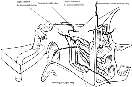
If nasal endoscopy did not reveal any clear bleeding source, the SPA was surgically occluded. This was performed by cutting the posterior end of the middle turbinate to expose the posterior fontanel of the maxillary sinus and the sphenopalatine foramen (SFA). By bluntly penetrating through the posterior fontanel the posterior wall of the maxillary sinus could be exposed. A mucosal flap over the sphenopalatine foramen (SPF) and the ethmoidal crest were elevated and if necessary the ethmoidal crest anterior to the SPF had to be removed to expose the foramen sufficiently. On the level of the SPF all arterial branches (i.e., the conchal branches of SPA) were identified, transsected and coagulated. As a next step the sphenoid sinus was opened by bluntly penetrating through its anterior wall in the sphenoethmoidal recess along the inferior edge of the superior turbinate. The obtained opening was enlarged by down-cutting punches until an arterial branch (i.e., the septal branch of the SPA) started to bleed (Fig. 1). Both ends of it were coagulated. Patients operated on by this technique were collected in group C.
Since not all surgeons were able to identify the septal branch, some patients underwent only surgical occlusion of the conchal branches at the SPA, whereas the septal branch was left untouched (group B) (Table 1).
The nasal cavity of all patients was packed for 24 h at the end of any surgery (groups A, B and C). Based on our experience with angiography in cases of epistaxis recurrence following surgical oc-clusion of the SPA, arteries that have been occluded only by coag-ulation without additional transsection may recanalize within a few days. Thus, the occlusionof an artery in this study meant transsec-tion and coagulating the bleeding ends with a monopolar suctranssec-tion coagulator. No clips were used.
426
Fig. 1 Schematic oblique view to the sphenopalatine artery (SPA)
Results
All 95 surgically treated patients in this study were fol-lowed 2 to 18.3 months (mean 9.3 months). Twenty (21%) patients were treated endoscopically with coagulation of a clear bleeding source (group A), 6 (6.3%) patients were in group B and 69 (72.7%) in group C. Rebleeding occurred in three (21%) in group A, one (16.7%) in group B and three (4.3%) in group C. Recurrence of bleeding was noted in the three group-A patients after 6 and 10 days, respec-tively. They underwent revision surgery according to that used for group C patients and were free of bleeding there-after. The patient in group B whose epistaxis recurred af-ter 10 days underwent revision surgery with occlusion of the septal branch of the SPA as well as the anterior eth-moidal artery complex by an external approach. During follow-up (9 months) she did not have any bleeding again. One patient from group C reported a short episode of bleeding 6.5 months after surgery. The bleeding was mild and neither a consultation of his family doctor nor of an otorhinolaryngologist was necessary. In the remaining two patients of group C with an epistaxis recurrence on the same side within 5 to 8 h, the ethmoidal arteries were ligated through an external approach. They were free of bleeding the following 4 and 5 months, respectively (Table 1).
Complications after surgery were protracted crust for-mation with subjective sensation of dryness in the nose, pain and posterior rhinorrhea. This problem occurred in particular in groups B and C and was rarely seen in patients of group A. Most of these side effects disappeared within 2 to 4 weeks, except for persistent posterior rhinorrhea, which caused only mild discomfort in 12 patients of groups B and C. All patients could leave the hospital in good condition.
In patients where the sphenopalatine foramen was ex-posed, both authors counted the number of arterial branches of the SPA at the level of the SFA. In 69 out of 75 patients the numbers of arterial branches could be noted, 36 out of 69 (52.1%) patients had only one artery, 30 (43.5%) had two and 3 (4.4%) even had three arterial branches. In all patients of group C only one septal branch was identified.
Discussion
The indication to perform surgical treatment for refractory PE cannot be standardized, since too many factors such as age, underlying cardiovascular diseases, courses of failed treatment with packing, etc., may influence the decision
to operate on patients with PE. Investigation of surgical ligation failures with consecutive angiographic investiga-tions revealed that the ethmoidal arteries, internal carotid artery (ICA) branches (i.e., inferolateral trunk, vidian artery) and nasopharyngeal vessels (ascending pharyngeal, de-scending palatine, ade-scending palatine, accessory meningeal artery) may reconstitute flow beyond the proximal occlu-sion and explain early recurrent hemorrhage. The eth-moidal arteries are the collateral pathways to the septal territory. Embolization of the septal branch of the SPA be-yond the site of anastomosis will be curative [2, 19]. Thus, bleeding recurrence can be reduced when the arter-ial blood supply is occluded at its most distal portion [23]. The SPA divides into a medial septal and lateral con-chal branches [6, 19]. This division most frequently is lo-cated proximal to the sphenopalatine foramen (SPF). The septal branch courses along the inferior portion of the an-terior wall of the sphenoid sinus, while the conchal branches most frequently course through the SPF to the middle and inferior turbinate (Fig. 1). In our small series, there were almost no recurrences after occluding all branches of the SPA. As mentioned, prominent ethmoidal arteries may predict further bleeding and a possible need for surgical occlusion.
The failure rate after MA ligation is reported to be be-tween 5–22% [2, 8, 11], which is similar to the SPA clip-ping at the level of the SPF (13–33%) [15, 16]. The latter corresponds to the results of our group-B patients in which the septal branch was not occluded. Our recurrence rate was 16.7%. However, the number of our group-B patients is too small to finally define the treatment efficacy by closing solely the conchal branches in PE. Reviewing the literature on endoscopic SPA occlusion in PE [3, 15, 16, 21], most of the authors describe only one artery at the level of the SPF. Only one study [21] indicates that “some-times” the SPA can already be divided into two branches. Of our patients, 48% (33/69) had more than one arterial branch at the level of the SPF. It might be that one of them represents the anastomosis of the ascending palatine artery with a conchal branch (Fig. 11).
According to our earlier neuroradiological and surgical experience, it is essential not only to coagulate the arterial branches by diathermy, since we could prove a recanal-ization after a few days. Thus, occluding the SPA endo-scopically should mean to expose, transsect and coagulate the conchal branches at the level of the SPF as well as to occlude the septal branch through a sphenoidotomy. If the different anastomoses in the blood supply of the nasal cavity are taken into account, it still remains unclear why treatment like that in group C provides such good results.
Conclusion
At the level of the SPF many patients already have two or sometimes even three (conchal) branches of the SPA. If occlusion of SPA for intractable posterior epistaxis is at-tempted, better results can be achieved by occluding not only all conchal branches, but also the septal branch.
427 Table 1 Surgical treatment of patients with intractable posterior
epistaxis, n =95
Therapy Group A, Group B, Group C,
n =20 n =6 n =69
Local coagulation +
SPA without septal branch +
SPA with septal branch +
References
1. Balrow DW, Deleyiannis WB, Pinczower EF (1997) Effective-ness of surgical management of epistaxis at a tertiary care cen-ter. Laryngoscope 107: 21–24
2. Breda SD, Choi IS, Persky MS, Weiss M (1989) Embolization in the treatment of epistaxis after failure of internal maxillary artery ligation. Laryngoscope 99: 809–813
3. Budrovich M, Saetti R (1992) Microscopic and endoscopic lig-ature of the sphenopalatine artery. Laryngoscope 102: 1390– 1394
4. Chandler JR, Serrins AJ (1965) Transantral ligation of the in-ternal maxillary artery of epistaxis. Laryngoscope 75: 1151– 1159
5. Elden L, Montanera W, TerBrugge K, Willinsky R, Lasjaunias P, Chalres D (1994) Angiographic embolization for the treat-ment of epistaxis: a review of 108 cases. Otolaryngol Head Neck Surg 111: 44–50
6. Lang J (1988). Klinische Anatomie der Nase, Nasenhöhle und Nasennebenhöhlen. Georg Thieme Verlag, Stuttgart, New York, pp 102–105
7. Lasjaunias P, Vignaud J, Hasso AN (1975) Maxillary artery blood supply to the orbit: normal and pathological aspects. Neuroradiology 9: 87–97
8. Mc Donald TJ, Pearson PW (1980) Follow-up on maxillary artery ligation for epistaxis. Arch Otolaryngol 106: 635–638 9. Merland JJ, Melki JP, Chiras J (1982) Place of embolization in
the treatment of severe epistaxis. J Otolaryngol 11: 271–274 10. Metson R, Hanson DG (1983) Bilateral facial nerve paralysis
following arterial embolization for epistaxis. Otolaryngol Head Neck Surg 91: 299–303
11. Montgomery WW, Reardon EJ (1980) Early vessel ligation for control of severe epistaxis. In: Snow JB Jr (ed) Controversies in otolaryngology. WB Saunders, Philadelphia, pp 315–319
12. O’Flynn PE, Shadaba A (2000) Management of posterior epi-staxis by endoscoic clipping of the sphenopalatine artery. Clin Otolaryngol 25: 374–377
13. Parnes LS, Heeneman H, Vinuela F (1987) Percutaneous em-bolization for control of nasal blood circulation. Laryngoscope 97: 1312–1315
14. Prades J (1976) Abord endonasal de la fosse pterygo-maxil-laire. LXXIII Cong Franc Compt Rendus des Seance: 290–296 15. Rockey JG, Anand R (2002) A critical audit of the surgical management of intractable epistaxis using sphenopalatine artery ligation/diathermy. Rhinology 40: 147–149
16. Snyderman CH, Goldman SA, Carrau RL, Ferguson BJ Gran-dis JR (1999) Endoscopic sphenopalatine artery ligation is an effective method of treatment for posterior epistaxis. Am J Rhi-nol 13: 137–140
17. Sokoloff J, Wickbom I, McDonald D (1974) Therapeutic per-cutaneous embolization in intractable epistaxis. Radiology 111: 285–287
18. Strutz J Schumacher M (1990) Uncontrollable epistaxis. Arch Otolaryngol Head Neck Surg 116: 697–699
19. Valavanis A, Setton A (1993) Embolization of epistaxis. In: Valavanis A (ed) Interventional neuroradiolergy. Spinger, Ber-lin, Heidelberg, New York, pp 55–61
20. Vitek JJ (1991) Idiopathic intractable epistaxis: endovascular therapy. Radiology 181: 113–116
21. Voegels RL, Thomé DC, Iturralde PPV, Butugan OB (2001) Endoscopic ligature of the sphenopalatine artery for severe posterior epistaxis. Otolaryngol Head Neck Surg 124: 464–467 22. Weaver EM, Chaloupka JC, Putman CM, Roth TC, Horky JK, Sasaki CT (1999) Effect of internal maxillary arterial occlusion on nasal blood flow in swine. Laryngoscope 109: 8–14 23. Welsh LW, Welsh JJ, Scogna JE, Gregor FA (1990) Role of
angiography in the management of refractory epistaxis. Ann Otol Rhinol Laryngol 99: 69–73