INFLAMMATORY DISORDERS
Five-year outcome in immune-mediated scleritis
Wolfgang Bernauer&Beat Pleisch&Matthias Brunner
Received: 26 March 2014 / Revised: 11 June 2014 / Accepted: 12 June 2014 / Published online: 10 July 2014 # Springer-Verlag Berlin Heidelberg 2014
Abstract
Background Immune-mediated scleritis is a rare condition, and the information on the clinical course and complications is scarce. The aim of this study was to identify prognostic factors, complications, and therapeutic effects in patients with immune-mediated scleritis.
Methods Patients with diagnosis of scleritis and a follow-up time of 5 years were identified. Systemic disease, laboratory investigations, type of scleritis, disease activity, therapy, and complications were recorded. The study design was a retro-spective, non-comparative, interventional case series. Results Systemic disease was identified in 15 (37 %) patients at presentation and in 18 (45 %) after 5 years. Rheumatoid arthritis (15 %), granulomatosis with polyangiitis (7.5 %), and polychondritis (7.5 %) were the most predominant disorders. Persistent scleritis (>5 years) was associated with systemic disease (66 vs. 6 %; p<0.05) and positive auto-antibodies (48 vs. 23 %; p = 0.18). Control of ocular inflammation was achieved in 38 of 40 (95 %). Prednisone (14 patients) and/or methotrexate (8) were the predominant drugs to control per-sistent disease. Complications included interstitial keratitis (2), inflammatory astigmatism (2), corneal melt (3), macular edema (6), and severe systemic disease (5).
Conclusion The presence of systemic disease and positive auto-antibodies are associated with persistent scleritis. Immu-nosuppressive agents allow control of scleritis, but may con-tribute to severe systemic complications.
Keywords Scleritis . Cornea . Keratitis . Outcome . Inflammation . Immunopressive therapy . Steroids
Introduction
Scleritis often occurs as a chronic inflammatory process. Although in some instances it may be caused directly by an infective agent, the vast majority of cases present as an immune-mediated disorder [1–3]. Immune-mediated scleritis may be associated with systemic disease. Usually systemic therapy is required to control inflammation. Morphologic criteria are used to classify scleral inflammation clinically into anterior and posterior types [3]. Anterior scleritis is subclassified into diffuse, nodular, necrotising with inflamma-tion, and necrotising without inflammation (scleromalacia).
Little information on the clinical course and complications of scleritis is available [4–7]. This study aims to gain knowl-edge on the long-term outcome of scleritis, and to identify prognostic factors. This was done by studying systemic dis-ease and laboratory findings, and by the assessment of com-plications and therapeutic effects.
Patients and methods
In 2010 we reviewed the records of more than 100 patients who were referred to our institution with the diagnosis "scleral inflammation". Forty patients and 56 affected eyes with diag-nosis of immune-mediated scleritis and the possibility to be followed by our team for the next 5 years were identified and investigated. After having obtained informed consent, these patients were included in the study. The demographic data are shown in Table1. Immune-mediated scleritis was diagnosed on clinical grounds (constant and nocturnal pain with spread-ing to the periorbital region, scleral edema, tenderness of the
This work has not previously been presented. W. Bernauer (*)
:
B. Pleisch:
M. BrunnerOMMA Eye Center, Theaterstrasse 2, 8001 Zürich, Switzerland e-mail: [email protected]
W. Bernauer
Department of Ophthalmology, University Hospital of Zürich, Frauenklinikstrasse 24, 8091 Zürich, Switzerland
globe, presence of nodules, or signs of scleral necrosis), and was supported in two cases by tissue examination (absence of infective agents). Patients with superficial inflammation (rath-er episcl(rath-eritis than scl(rath-eritis), infectious scl(rath-eritis, neoplastic lesions, and/or incomplete data were excluded. A standardised protocol was used to record systemic disease, laboratory in-vestigations, type of scleritis, disease activity, therapy, and complications (Table 2). The duration of scleritis was deter-mined as abscence of clinical inflammation for more than 6 months after stopping therapy (local and systemic).
For the treatment of scleritis, an anti-inflammatory strategy was applied as published elsewhere [8]. For non-necrotising scleritis oral nonsteroidal anti-inflammatory drugs such as flurbiprofen (100–300 mg/24 h), sometimes in combination with steroids, were used to induce remission. For consolida-tion, methotrexate, azathioprine, anti-TNF-alpha blockers or cyclosporine were applied. Necrotising scleritis was treated with high-dose oral prednisone or intravenous methylprednis-olone in combination with cyclophosphamide. Cyclophospha-mide was given parenterally in an initial phase, later per os. Posterior scleritis was treated in this series solely with oral prednisone. All medication was given according to the guide-lines of the Rheumatology Department, University Hospital of Zürich (Head Prof. Beat Michel) and supervised by experi-enced rheumatologists. Successful medical control of disease was defined as absence of symptoms, clinical inflammation and, in necrotising disease, halt of further tissue destruction.
The contingency table (see Table6) was analysed using Fisher’s exact test. Patients with persistent scleritis (more than
5 years of ongoing scleral inflammation) were compared with patients who showed a shorter course of scleral inflammation.
Results
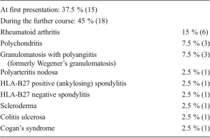
The median age at first presentation was 51 years, and 65 % were females. In 37.5 % (15 patients), a systemic disorder was already known at presentation; in the further course the figure raised to 45 % (18). Rheumatoid arthritis was the most fre-quent systemic disease (15 % or six patients), followed by polychondritis in three, and granulomatosis with polyangiitis (GPA) in three patients (Table 3). The type of scleritis at presentation is shown in Table 4: Non-necrotising anterior scleritis was the most frequent type (77.5 %, 31 eyes). Necrotising anterior scleritis was seen in five and posterior inflammation in six eyes. Two eyes had both inflammation of the anterior and posterior segments. In 26 out of 40 patients, the scleritis stayed unilateral. In four patients, however, scleral inflammation became bilateral (Table5). During follow-up, the type of scleritis changed in four patients; one patient who had initially presented with anterior disease developed poste-rior scleritis. One patient with initial posteposte-rior disease
Table 1 Demographic data
Age at presentation (median) 51 years (range 31–74) Females/males 65 %/35 %
Relative and absolute number of patients at follow-up (months)
100 % (40) at 12 95 % (38) at 36 95 % (38) at 60 55 % (18) at 120 months Deceased (relative and absolute number
of cases at follow-up in months)
5 % (2) 12–60 17.5 % (7) 61-120
Table 2 Protocol for the investigation of scleritis patients Systemic disease
Laboratory investigations (serum rheumatoid factor, ANA, ANCA) Type of scleritis
Disease activity at presentation, 12, 36, 60, and 120 months Therapy
Complications
Table 3 Association of scleritis with systemic disease At first presentation: 37.5 % (15)
During the further course: 45 % (18)
Rheumatoid arthritis 15 % (6)
Polychondritis 7.5 % (3)
Granulomatosis with polyangiitis (formerly Wegener’s granulomatosis)
7.5 % (3) Polyarteritis nodosa 2.5 % (1) HLA-B27 positive (ankylosing) spondylitis 2.5 % (1) HLA-B27 negative spondylitis 2.5 % (1)
Scleroderma 2.5 % (1)
Colitis ulcerosa 2.5 % (1) Cogan’s syndrome 2.5 % (1)
Table 4 Type of scleritis at presentation
Unilateral Bilateral Diffuse/nodular 77.5 % (31) 45 % (18) 32.5 % (13) Necrotising 12.5 % (5) 12.5 % (5) 0 with inflammation 7.5 % (3) 7.5 % (3) without inflammation 5 % (2) 5 % (2) Posterior 15 % (6) 7.5 % (3) 7.5 % (3) Totala 62 % (26)a 38 % (16)a a
developed non-necrotising anterior scleritis 2 years later. Two patients with non-necrotising inflammation at first visit devel-oped ischemic areas, thus necrotising scleritis (Fig.1).
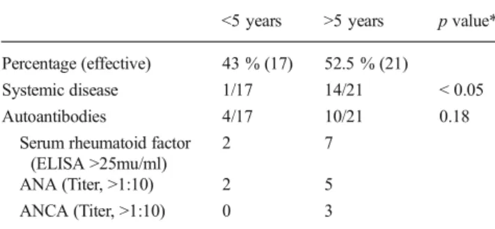
In 21 out of 40 patients (52 %) scleral inflammation lasted more than 5 years. This persistant scleritis (more than 5 years ongoing scleral inflammation) was associated with systemic disease (14/21 in comparison to 1/17 patients with shorter inflammation episodes). This association was statistically sig-nificant when analysed by Fisher’s exact test. The presence of positive circulating antibodies (10/21 versus 4/17) showed a trend to favour persistent inflammation. This is shown in Table6.
Information on the medical control of scleral inflammation is given in Tables7and8. Control of scleritis was achieved in 38 out of 40 patients. In two patients with necrotising scleritis, the loss of scleral tissue could not be halted. One of these patients, a 75-year-old male, developed stomach carcinoma and therapy with cyclophosphamide was subsequently stopped. This patient died 18 months after first presentation. The other patient, a 39-year-old female with severe rheuma-toid arthritis, developed ongoing scleromalacia despite immu-nosuppression with oral steroids, cyclophosphamide and TNF-alpha-blockade (Figs.2and3).
The complications of scleral disease are listed in Table9. Corneal complications included interstitial keratitis (two eyes), peripheral corneal melts (three eyes) and scleritis-induced astigmatism (two eyes) (Fig.4, Table10). Six patients devel-oped cystoid macular edema. In two patients the scleritis had led to persistent structural damages and functional loss (long-standing retinal detachments without light perception). Several patients developed severe systemic disease: myocardial
infarction at age 44 and IgA-nephropathy, pericarditis with valvular and coronary heart disease (possibly associated with the polyarteritis nodosa), and two patients died within the 5-year follow-up period (Table11).
Discussion
Information on the outcome of scleritis is scarce. To date, there are case reports and the Moorfields, Boston, and Baltimore cohorts [1,3,6] that help to understand the nature of scleral inflammation. Our study sets the focus on the long-term course of scleritis and, in this way, teaches us about the outcome of immune-mediated scleritis.
Scleritis is a potentially long-lasting problem. Fifty-two percent of our patients showed a disease duration of more than 5 years. Although this figure may be biased by the fact that we see patients as tertiary referrals, we were surprised by this high number. An underlying systemic disease and positive autoantibodies were identified as risk factors for such a long disease duration.
Melts, interstitial keratitis, and peripheral ulcerative kerati-tis are well known corneal complications of sclerikerati-tis [4–7]. Inflammatory astigmatism due to scleritis was previously described [9,10], but none of those patients had such a high cylinder measurement as our patient with 8.5 diopters (Ta-ble10). A second patient of our series had a cylinder mea-surement of 2.5 diopters. In both patients it was reversible after remission of scleritis. We feel that it is the increase in
Table 5 Scleritis course
(0–60 months) Unilateral→ bilateral 4 Changing type of scleritis 4 Anterior→ Posterior 1 Posterior→ Anterior 1 Nodular/diffuse→ Necrotising 2
Fig. 1 patient with non-necrotising at first visit developed ischemic areas, thus necrotising scleritis
Table 6 Duration of scleritis
<5 years >5 years p value* Percentage (effective) 43 % (17) 52.5 % (21)
Systemic disease 1/17 14/21 < 0.05 Autoantibodies 4/17 10/21 0.18
Serum rheumatoid factor (ELISA >25mu/ml)
2 7
ANA (Titer, >1:10) 2 5 ANCA (Titer, >1:10) 0 3
No clinical inflammation for >6 months after stopping therapy (local and systemic)
*p by Fisher’s exact test
Table 7 Medical control of disease activity
Yes No
Control of inflammation 38 2a
No discomfort, no further destruction
a
Fig. 4 A 73-year-old female who developed anterior and posterior non-necrotizing scleritis. The scleral nodule in the upper and nasal quadrants resulted in a corneal astigmatism of−8.50 in 117°. The course of the corneal curvature is shown in Table10
Table 10 Corneal astigmatism before and after onset of anterior non-necrotizing scleritis which responded to treatment with oral steroids. The affected eye is shown in Fig.4
Follow-up time Autorefraction (NIDEK ARK-700A) Pre-inflammatory refraction −3.50 −1.25 110° Day 1 a−8.50 117° Day 14 −1.00 −6.00 110° Day 28 −1.75 −4.50 116° Day 42 −1.50 −4.25 113° Day 90 −0.75 −3.75 111° Day 120 −1.00 −3.75 111° a
Sphere not measurable
Table 9 Complications (months 0–60)
Interstitial keratitis 2 Scleritis-induced astigmatism 2 Peripheral corneal melt 3
Cystoid macular edema 6
Persistent structural damage→ enucleation 2 Severe systemic disease 5 Myocardial infarction at age 44, IgA-nephropathy 1 Pericarditis, valvular/coronary heart disease 1 Deceased (patients #5 and #6)* 2 *For details of these patients see Table11
Fig. 3 The same eye as in Fig.2. The progression over a period of 5 years is obvious
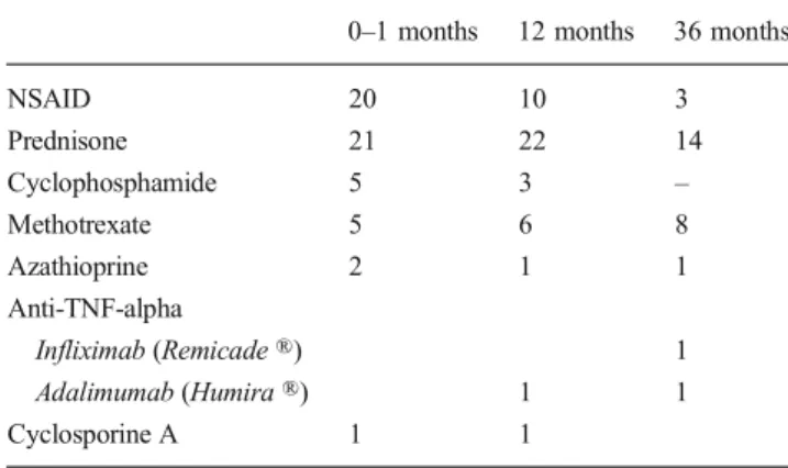
Fig. 2 Scleromalacia in a 39-year-old female with severe rheumatoid arthritis. There is progressive scleral destruction despite immunosuppres-sion with oral steroids, cyclophosphamide, and TNF-alpha blockade Table 8 Systemic medication in 40 scleritis patients
0–1 months 12 months 36 months
NSAID 20 10 3 Prednisone 21 22 14 Cyclophosphamide 5 3 – Methotrexate 5 6 8 Azathioprine 2 1 1 Anti-TNF-alpha Infliximab (Remicade ®) 1 Adalimumab (Humira ®) 1 1 Cyclosporine A 1 1
volume of the inflamed sclera that leads to changes of scleral rigidity with subsequent instability of limbal contour and corneal steepening in the affected quadrant.
Macular edema was observed in 11 % of our patients. When detected at an early stage it was fully reversible after high-dose oral steroids. One of our patients presented with posterior scleritis only and developed anterior scleritis years later in the same eye, and three other eyes changed the type of scleritis (Table5). This is in contrast to Tuft’s study [11] who found that the majority of patients remain in the same clinical category. It seems that a change in the scleritis pattern is not unusual when patients are available for long-term follow-up.
The evaluation of the best medication for the treatment of scleritis was beyond the scope of this study. With the correct application of traditional and new generation immunosup-pressive agents, preferably in collaboration with a rheuma-tologist, disease control is possible in most cases. In our series, remission was achieved in 95 %. In one patient with long-standing rheumatoid arthritis and scleromalacia (Figs.2
and3), tissue loss could not be halted despite application of TNF-alpha blockers and, at a later stage, cyclophosphamide in combination with oral steroids. This demonstrates again that this vasculitic complication is particularly difficult to tackle [1,6,7].
Our patients showed a considerable morbidity and a high number of lethal complications. Five percent of our patients died within 5 years and 17.5 % within 10 years of follow-up time. Three of the deaths were older than 75 years and pre-sumably died of natural causes. Of concern are the two pa-tients who died with aplastic anemia and septicemia. This demonstrates that the immunosuppressive therapies have the potential to cause lethal complications, although the systemic disease itself may be life-threatening [1,2]. New agents allow
control of scleritis, but may also contribute to systemic complications.
Proprietary interest None.
References
1. Akpek EK, Thorne JE, Qazi FA, Do DV, Jabs DA (2004) Evaluation of patients with scleritis for systemic disease. Ophthalmology 111: 501–506
2. Sainz de la Maza M, Foster CS, Jabbur NS (1995) Scleritis associated with systemic vasculitic diseases. Ophthalmology 102:687–692 3. Watson PG, Heyreh SS (1976) Scleritis and episcleritis. Br J
Ophthalmol 60:163–191
4. Bernauer W, Ficker LA, Watson PG, Dart JKG (1995) The manage-ment of corneal perforations associated with rheumatoid arthritis. Ophthalmology 102:1325–1337
5. Sainz del la Maza, Jabbur NS, Foster CS (1993) An analysis of therapeutic decisions for scleritis. Ophthalmology 100:1372–1376 6. Sainz de la Maza M, Molina N, Gonzalez-Gonzalez LA, Doctor PP,
Tauber J, Foster CS (2012) Clinical characteristics of a large cohort of patients with scleritis and episcleritis. Ophthalmology 119:43.50 7. Wieringa WG, Sieringa JE, Dam-van Loon NH, Los LI (2013) Visual
outcome, treatment results, and prognostic factors in patients with scleritis. Ophthalmology 120:379–386
8. McCluskey PJ, Wakefield D (1996) Scleritis and Episcleritis. In: Pepose JS, Holland GN, Wilhelmus KR (eds) Ocular infection and immunity, 1st edn. Mosby, St. Louis, pp 642–662
9. Arellanes-Garcia L, del Carmen Preciado-Delgadillo M, Graza-Leon M (2011) Refractive changes in patients with autoimmune scleritis. J Ophthalmol Inflamm Infect 1:173–175
10. Meller D, Böker T (1997) Complications of misdiagnosed and inad-equately treated necrotizing scleritis studied by ultrasound biomicroscopy and computerized corneal topography. Int Ophthalmol 21(1):35–37
11. Tuft SJ, Watson PG (1991) Progression of scleral disease. Ophthalmology 98:467–471
Table 11 Deaths (0–120 months)
Patient Cause of death Type and duration of scleritis (months)
Systemic
inflammatory disease
Systemic medication
C.A.,♂, age 76 years, # 5 Stomach carcinoma Necrotising, 9 Steroids, cyclophosphamide E.M.,♀, age 68 years, #6 Not known Anterior and posterior,> 60 Steroids, azathioprine E.W.,♂, age 72 years, # 7 Aplastic anemia Necrotising, 38 Rheumatoid arthritis Steroids, methotrexate,
cyclophosphamide, later azathioprine H.HR.,♂, age 45 years, # 12 Sepsis (immunosuppression?) Diffuse anterior, 120 Polychondritis Steroids, infliximab K. H.,♂, age 70 years, #13 Renal failure Diffuse anterior, 21 Polychondritis Steoids, cyclosporine L. M.,♀, age 80 years, # 17 Heart failure Diffuse anterior, 15 Cogan’s syndrome Steroids, cyclophosphamide S.F.,♂, age 79 years, # 26 Aortic aneurysm Necrotising, 9 Steroids, cyclophosphamide