THE CHARACTERIZATION OF HUMAN yD-CRYSTALLIN MUTANTS AND THEIR DIFFERENTIAL INTERACTIONS
WITH THE LENS CHAPERONE aB-CRYSTALLIN By
Kate L. Moreau
B.A. Molecular Biology and Biochemistry Rutgers University, 2004
SUBMITTED TO THE DEPARTMENT OF BIOLOGY IN PARTIAL FULFILLMENT OF THE REQUIREMENTS
FOR THE DEGREE OF DOCTOR OF PHILOSOPHY
AT THE
MASSACHUSETTS INSTITUTE OF TECHNOLOGY FEBRUARY 2011
C 2011 Massachusetts Institute of Technology. All rights reserved.
'is
Signature of Author IASSACHUSETTS INSTITUTEOCT
2
7
2C
0
LIBRARIES
ARCH!Es
A ~-N A Department of Biology October 2010 Certified by Accepted b Co-Chair, Department Professor of Jonathan A. King Molecular Biology Thesis Supervisor Stephen P. Bell Committee on Graduate StudentsTHE CHARACTERIZATION OF HUMAN yD-CRYSTALLIN MUTANTS AND THEIR DIFFERENTIAL INTERACTIONS
WITH THE LENS CHAPERONE aB-CRYSTALLIN by
Kate L. Moreau
Submitted to the Department of Biology on October 13, 2010 in Partial Fulfillment of the
Requirements for the Degree of Doctor of Philosophy in Biology
ABSTRACT
Cataract, the leading cause of blindness worldwide, is the opacification of the eye lens. In.age-related cataract, as well as roughly one half of congenital cataract cases, aggregation or precipitation of crystallin proteins results in the formation of large structures that scatter light, preventing the pinpoint focusing on the retina normally accomplished by the lens.
The human eye lens is composed of fiber cells packed with crystallins up to 450 mg/ml. Human yD-crystallin (HyD-Crys) is a monomeric, two-domain protein,
predominantly localized to the lens nucleus. Both domains of this long-lived protein have double Greek key
p-sheet
folds with well-packed hydrophobic cores. Three mutations resulting in amino acid substitutions in the y-crystallin buried cores-two in the N-terminal domain (N-td), and one in the C-terminal domain (C-td)-causeearly-onset cataract in mice. It has not been possible to identify the aggregating precursors within lens tissues, and the question persists as to the nature of structure and stability changes in the crystallins leading to their increased propensity for aggregation and cataract formation within the lens environment. To compare in vivo cataract-forming phenotypes with in vitro unfolding and aggregation of y-crystallins, mouse mutant
substitutions were introduced into HyD-Crys.
WT HyD-Crys unfolds in vitro through a three-state pathway, exhibiting an intermediate with the N-td unfolded, and the C-td native-like. L5S and V75D also displayed three-state unfolding pathways, with the first transition, unfolding of the N-td,
shifted to significantly lower denaturant concentrations. 190F was globally destabilized and the overall unfolding transition was shifted to lower denaturant concentrations. During thermal denaturation, the mutant proteins exhibited lowered thermal stability compared with WT. Kinetic unfolding experiments further confirmed this
The cataract phenotype of increased protein aggregation could be a direct
property of the mutation, or could reflect an inability to be recognized by the a-crystallin chaperone. We have therefore carried out experiments comparing the interactions of WT HyD-Crys, V75D and 190F with a major component of the lenticular chaperone system, the small heat shock protein human aB-crystallin. Suppression levels of the aggregation that competes with the refolding pathway after dilution from GdnHCl were
indistinguishable between WT and mutant proteins. However, dramatic differences among the mutants were observed under different conditions, including physiological conditions in the absence of denaturant and partially destabilizing acidic conditions such as those that may exist in the vicinity of degrading lysosomes in maturing lens fiber cells. In particular, incubation under physiological conditions highlighted differential
interactions between the lens chaperone and the mutant HyD-Crys proteins themselves. Destabilized precursors to the cataractous state may populate various non-native
structural conformations that, although aggregation-prone, elude recognition by a-crystallin and efficiently continue along the aggregation pathway.
Coupled with the observation of different unfolding pathways for the mutant crystallins, these results support the existence of multiple pathways for cataract formation in which partially unfolded species are differentially recognized by the passive lens chaperone system. While these observations were made utilizing destabilized mutant proteins, the same mechanisms may underlie the formation of age-related cataractous aggregates induced in vivo by covalent modification of the lens proteins.
Thesis Supervisor: Jonathan A. King Title: Professor of Molecular Biology
ACKNOWLEDGEMENTS
None of this would have been possible without the assistance, encouragement and support of many people. First, I would like to thank my advisor, Professor Jonathan King, for giving me a place in his lab. You have been not only a scientific advisor, but you've shared your perspectives on life and responsibilities inside and outside of the lab. Your passion for science and education is inspiring. While offering your opinions, you have been supportive of my decisions, or sometimes the lack thereof. Thank you for your encouragement and confidence building-I'm working on that.
I would also like to thank my thesis committee members. Professors Bob Sauer and Amy Keating, thank you for giving your perspectives and suggestions, as well as your time, over the past several years. Professor Thomas Schwartz and Dr. Caroline Shamu, thank you for taking the time to read my thesis and serve on my defense committee.
To the entire King lab, thank you for creating such an interesting, fun and memorable working environment! Thank you to Dr. Kelly Knee for being a most excellent bay-mate. You are always ready to listen, whether I need to work through an experiment, vent my frustrations or share a gross story. I hope that we can continue to share our brand of humor and our love of the Clover truck. To Dr. Ligia Acosta-Sampson, thank you for sharing your vast a-crystallin knowledge. You are always ready to answer questions or help troubleshoot and I certainly would not have completed the experiments in this thesis without your advice. To Dessy Raytcheva, good luck as you approach the end of your graduate career! Your upbeat outlook is inspiring and your ability to TA every semester amazes me. You have such a great perspective on graduate school and science, and I enjoy our overlapping tea breaks during the day. To Fanrong Kong, good luck with all of your mutants! You are the next bird on the crystallin tree. To Nathaniel
Schafheimer and Oksana Sergeeva, good luck as you both progress through your graduate studies. Your enthusiasm is refreshing! To Drs. Ishara Mills-Henry, Jiejin Chen and Yongting Wang, thank you for all your help as I started in the King lab. You all have answered my many questions with patience and excellent advice. Thank you to Dan Goulet for being the lab socialite and getting us all out to see The Maquis. Thank you to Cammie Haase-Pettingell for answering countless random questions and encouraging our lab outings. You are always ready to lend a hand. Thank you to all other members of the King lab, including Jeannie Chew, Cindy Woolley, Ginger Yang, Althea Hill, Dr. Takumi Takata, Professor Jacqueline Piret, and Lisa Guisbond, for creating such a fun and unique lab!
I cannot ever express the importance of the friendships I have made at MIT. As a first year student, I quickly bonded with my classmates Crystal Lee, Jamie Newman and Gina Lafkas. Thank you for the (in)sanity that you provided, the laughter we have shared (oh my!) and the amazing support system that you have been throughout my time here. Crystal, I don't know how you fit it all into a 24-hour day, but your hard work and perseverance are inspiring. To top it off, you are an excellent baker! Thank you for all the laughs we've shared, for making sure I made it home through all of our first year and
for listening to my wacky, often winding rants. Jamie, you are an amazing listener. You are always there for a much needed break with words of encouragement. I will never forget you laughing yourself into tears, and I hope we can continue to do so (just minus the stress of graduate school).
I must also thank my friends back in New York and New Jersey. Shana Dworken, you have been my PR person and my cheerleader since high school! I cannot thank you enough for supporting my decisions, giving thoughtful advice and encouragement and being such a fun roommate. Amy Chiaravallo, I'm so glad we became close friends, even if it didn't start out that way. Thank you for being there through thick and thin, you have truly seen my ups, my downs and the messes in between, and have always been there with advice and humor (and probably a much needed beer). Marisa Montemorano, thank you for being such a supportive friend, keeping me on my toes with your pranks and introducing me to new music and films.
I absolutely must thank my family for their unwavering support. To my mom, Diane Drahos, thank you for never pressuring me, but allowing me to follow my own path. I am so happy we have become friends as well as family. We certainly share the same
sense of humor! To my sister, Colleen Drahos, thank you for your support and encouragement. We have become much closer over the past few years and I hope we continue to do so. Thank you to my father, Steve Drahos, for encouraging me to take the harder math class. Although my math usage is rather minimal now, thank you for
pushing me. Finally, to my Nana, the funniest 90 year old ever (who does not have cataracts). You have been a constant presence in my life, always encouraging, always supportive, and always truthful. I am incredibly thankful for you.
Last, but certainly not least, to my husband Mark Moreau. Thank you for moving to Massachusetts. Thank you for believing in me (and jet packs), for making me laugh and for being there every day. I know I have your confidence when my own falters. I love you.
This research was supported by National Institutes of Health grants EYO 15834 and GM17980 awarded to Jonathan A. King.
BIOGRAPHICAL NOTE
Education
Ph.D. Massachusetts Institute of Technology, Department of Biology Expected 2011 Cambridge, MA
B.A. Rutgers University, Department of Molecular Biology and June 2004 Biochemistry, New Brunswick, NJ
Major: Molecular Biology and Biochemistry, with Highest Honors
Research and Professional Experience
2006-2010 Graduate Research Assistant in the laboratory of Professor Jonathan King, MIT Department of Biology, Cambridge, MA 2004-2005 Proteomics Research Associate, Alfa Wassermann Proteomic
Technologies, LLC, West Caldwell, NJ
Summer 2003 Research Intern in the Department of Ion Channels Merck and Co., Inc., Rahway, NJ
Summer 2002 Research Intern, Provid Pharmaceuticals, North Brunswick, NJ 2001-2004 Undergraduate Research Assistant in the laboratory of Professor
Gaetano Montelione, Rutgers University, New Brunswick, NJ
Thesis: Protein Expression and Folding Optimization for High-Throughput Proteomics
Publications
Moreau, K.L. and King, J. 2009. Hydrophobic Core Mutations Associated with Cataract Development in Mice Destabilize Human yD-Crystallin. J Biol. Chem. 284(48): 33285-33295.
TABLE OF CONTENTS
PREFATORY MATERIAL
T itle P age ... 1
A b stract...3
A cknow ledgem ents...5
Biographical Note...7 Table of Contents... 9 L ist of F igures...12 L ist of T ables...14 List of Abbreviations...15 CHAPTER 1: INTRODUCTION A. The Human Eye...18
B. Lens Development, Structure and Transparency...18
C . The Lens C rystallins...21
1. The Small Heat Shock Protein a-Crystallin...22
a. a-Crystallin Structure...22
b. a-Crystallin Function...24
2. The @y-C rystallins...29
a. yD-Crystallin...30
b. yC- and yS-Crystallins...35
c. The @-C rystallins...37
D. Cataract Disease...39
1. Age-Related Cataract...42
2. Congenital and Juvenile Cataract...43
E. Cataract Questions and Answers: The Biological Context of This Thesis ... 45
F. Protein Folding, Misfolding and Aggregation...48
1.
p-Sheet
Protein Folding...492. Determinants of Protein Stability and the Effects of Mutations...51
3. Protein Misfolding, Aggregation and Disease...53
a. The Amyloidoses and Cross-@ Structure Formation...54
b. Domain Swapping...57
c. The Serpinopathies and Loop-Sheet Insertion...61
d. Native State Polymerization...63
CHAPTER 2: HYDROPHOBIC CORE MUTATIONS ASSOCIATED WITH CATARACT DEVELOPMENT IN MICE DESTABILIZE HUMAN yD-CRYSTALLIN A . A bstract... 68
C. Experimental Procedures...73
1. Mutagenesis, Expression, and Purification of Recombinant H yD -C rys...73
2. Circular Dichroism Spectroscopy...74
3. Fluorescence Emission Spectroscopy...74
4. Equilibrium Unfolding/Refolding...74
5. Thermal Denaturation... 75
6. Unfolding Kinetics... 75
7. Refolding and Aggregation Turbidity Measurements...76
D . R esults... . 76
1. Protein Purification and Structural Characterization...76
2. Equilibrium Unfolding and Refolding of Wild-type and Mutant Human yD-Crystallins... 80
3. Thermal Unfolding of Wild-type and Mutant Human yD-Crystallins... 83
4. Kinetic Unfolding of Wild-type and Mutant Human yD-Crystallins...87
5. Refolding and Aggregation of Partially Unfolded Wild-type and Mutant HyD-Crys... 91
E . D iscussion ... 94
CHAPTER 3: DIFFERENTIAL INTERACTIONS OF HUMAN aB-CRYSTALLIN WITH HUMAN yD-aB-CRYSTALLIN CONGENITAL CATARACT MUTANTS A . Introduction ... 102
B. Experimental Procedures...105
1. Cloning, Protein Expression and Purification...105
2. Aggregation and Aggregation Suppression Assays...106
3. Native Mixing and Interaction Assay...107
4. pH Dependence of Trp Fluorescence and Tertiary Structure...107
5. HyD-Crys Fibril Formation and Inhibition of Fibril Formation by a B at pH 3 ... 108
6. Thioflavin T Fluorescence Assay...109
7. Endpoint Solution Turbidity Measurements...109
C . R esu lts...109
1. WT and Mutant HyD Proteins Aggregate to Similar Extents...109
2. aB Suppressed WT and Mutant Protein Aggregation to Similar E xtents...111
3. ctB Interacted Differentially with HyD-Crys Proteins Initially in their N ative State...115
4. aB Slowed Fibrillation Kinetics of WT and Mutant HyD-Crys P rotein s...122
5. Both HyD-Crys and aB Underwent Structural Changes Under Acidic C onditions...130
D . D iscussion ... 132 1. The Differential Aggregation Propensities of HyD-Crys and Its
Interaction with the Chaperone aB...134
2. V75D and 190F Mutants Exhibit a Range of Aggregation Patterns and are Differentially Recognized by cB...135
3. Relevance of Amyloid Formation by HyD-Crys and Interference by aB Under Acidic Conditions...137
4. Are These Aggregation-Prone Intermediates Similar to One Another?...139
5. There are Varied Pathways to Cataract...139
CHAPTER 4: FINAL DISCUSSION AND FUTURE DIRECTIONS A . Final D iscussion...142
B . Future D irections...144
CHAPTER 5: REFERENCES...147
CHAPTER 6: APPENDICES A. NMR Analysis of the Populated Folding Intermediate of V 75D H yD -Crys...170
1. Statement of Collaboration...170
2. B ackground...170
3. Preliminary Results and Discussion...171
B. pKa Alteration in the Buried Core of HyS-Crys...179
1. Statement of Collaboration...179
2. B ackground...179
3. Preliminary Results and Discussion...180
C. Appendix References...185
D. Analysis of Equilibrium Unfolding/Refolding Data...186
1. Calculating GdnHCl Concentration...186
2. Calculating Urea Concentration...186
3. Two-State Equilibrium Unfolding/Refolding...187
4. Three-State Equilibrium Unfolding/Refolding...189
E. Analysis of Kinetic Unfolding Data...191
1. Two-State Kinetics...191
LIST OF FIGURES CHAPTER 1
Figure 1-1: Schematic diagram of the human eye...19
Figure 1-2: The stages of lens development...20
Figure 1-3: Three-dimensional structures of cB... 23
Figure 1-4: Three-dimensional structure and topological diagram of H yD -C rys... 31
Figure 1-5: HyD-Crys tryptophan fluorescence and equilibrium unfolding pathw ay...33
Figure 1-6:
p-Crystallin
oligomeric assemblies...38Figure Figure Figure Figure Figure Figure 1-7: Gross cataract and protein aggregate phenotypes...40
1-8: HyD-Crys amino acid substitutions studied in this thesis...47
1-9: Amyloid structural models...56
1-10: Domain swapping and aggregate formation...60
1-11: The serpins and loop-sheet insertion...62
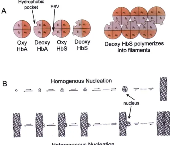
1-12: Native state polymerization of mutant hemoglobin...65
CHAPTER 2 Figure 2-1: Far-UV CD spectra of native WT and mutant HyD-Crys...78
Figure 2-2: Fluorescence spectra of native WT and mutant H yD -C rys... 79
Figure 2-3: Equilibrium unfolding/refolding of WT and mutant H yD -C rys... 82
Figure 2-4: Thermal unfolding of WT and mutant HyD-Crys...85
Figure 2-5: Kinetic unfolding of WT and mutant HyD-Crys...89
Figure 2-6: Refolding aggregation of WT and mutant HyD-Crys...92
Figure 2-7: Aggregation of 190F upon incubation at intermediate denaturant concentrations...93
CHAPTER 3 Figure 3-1: Aggregation reactions for WT and mutant HyD-Crys...112
Figure 3-2: Aggregation suppression reactions for WT and mutant HyD-Crys in the presence of aB...113
Figure 3-3: Size exclusion chromatography of aggregation and aggregation suppression samples...116
Figure 3-4: Quantification of aggregation suppression levels...117
Figure 3-5: Size exclusion chromatography time courses on native mixing single protein controls...119
Figure 3-6: Size exclusion chromatography time courses on native mixtures containing aB and HyD-Crys...120 Figure 3-7: Western Blot analysis of 190F + aB native mixture
Figure 3-8: Figure 3-9: Figure 3-10 Figure 3-11 Figure Figure 3-12 3-13
ThT fluorescence measurements for WT and mutant
H yD -Crys at pH 3...124 Endpoint turbidity measurements for WT and mutant
H yD -C rys at pH 3...125 : ThT fluorescence measurements of WT and mutant
HyD-Crys in the presence of ctB at pH 3...127 : Endpoint turbidity measurements for WT and mutant
HyD-Crys in the presence of aB at pH 3...128 : The pH dependence of HyD-Crys and ctB tryptophan fluorescence ... 131 : Models of y-crystallin aggregation...133
CHAPTER 4
Figure 4-1: Model for the molecular mechanism of cataract formation...146
CHAPTER 6
Figure 6-1: Urea equilibrium unfolding/refolding of V75D HyD-Crys...172 Figure 6-2: Comparison of WT and V75D HSQCs under native conditions...174 Figure 6-3: Comparison of V75D HSQCs under native and
partially denaturing conditions...175 Figure 6-4: Comparison of HSQCs: V75D under partially denaturing
conditions and WT isolated C-td under native conditions...176 Figure 6-5: Residual dipolar couplings for V75D in 3.6 M urea...177 Figure 6-6: Urea equilibrium unfolding/refolding of WT and Q148E
HyS-Crys from pH 4-8...182 Figure 6-7: Comparison of Cm and AG'N-U values for WT and Q148E
LIST OF TABLES
CHAPTER 2
Table 2-1: Equilibrium unfolding/refolding parameters for WT
and mutant HyD-Crys at pH 7.0 and 37*C...84 Table 2-2: Thermal unfolding parameters for WT and mutant
HyD-Crys at pH 7.0... 86 Table 2-3: Kinetic unfolding parameters for WT and mutant
HyD-Crys at pH 7.0 and 18'C...90
CHAPTER 3
Table 3-1: Solution turbidity measurements for WT and mutant
HyD-Crys in the absence and presence of aB...114 Table 3-2: ThT fluorescence parameters and endpoint turbidity of WT
and mutant HyD-Crys at pH 3 in the absence and
presence of cB ... 129
CHAPTER 6
Table 6-1: Equilibrium unfolding parameters for WT and Q148E
LIST OF ABBREVIATIONS
(in alphabetical order)
aA: human aA-crystallin axB: human aB-crystallin AD: Alzheimer's disease AFM: atomic force microscopy
bis-ANS: 4,4'-bis(1-anilinonaphthalene 8-sulfonate) CD: circular dichroism
CRABP 1: cellular retinoic acid binding protein 1 CR: Congo Red
C-td: C-terminal domain CV: column volume DTT: dithiothreitol
EDTA: ethylenediaminetetraacetic acid FPLC: fast protein liquid chromatography FRET: fluorescence resonance energy transfer GdnHCl: guanidinium hydrochloride
HbS: sickle cell form of human hemoglobin (containing E6V substitution) HyC-Crys: human yC-crystallin
HyD-Crys: human yD-crystallin HyS-Crys: human yS-crystallin
HSQC: heteronuclear single quantum coherence Ig: immunoglobulin
IPTG: isopropyl-fp-thiogalactoside MyB-Crys: murine yB-crystallin MyD-Crys: murine yD-crystallin MyS-Crys: murine yS-crystallin NaPi: sodium phosphate
NMR: nuclear magnetic resonance N-td: N-terminal domain
PrPc: human prion protein cellular form
PrPsc: human prion protein infectious scrapie form RDC: residual dipolar coupling
SDS-PAGE: sodium dodecyl sulfate polyacrylamide gel electrophoresis SEC: size exclusion chromatography
sHSP: small heat shock protein
TEM: transmission electron microscopy ThT: Thioflavin T
Tris: Tris(hydroxymethyl)-amino methane TTR: Transthyretin
UV: ultraviolet WT: wild type
CHAPTER 1:
A. The Human Eye
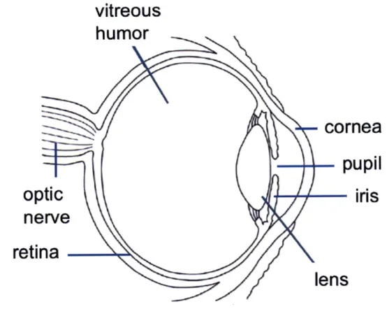
The human eye is a chambered eye that uses refraction to focus light. Light first passes through the cornea, the outer transparent layer of the eye where about two-thirds of refraction takes place (Oyster 1999). The cornea maintains its transparency by way of an ordered arrangement of collagen fibers (Oyster 1999). Light next enters the pupil, whose size is controlled by contraction and expansion of the surrounding iris, thereby controlling the amount of light that enters. The transparent lens sits behind the cornea and pupil and accounts for the remaining one-third of the eye's refractive power (Oyster 1999). The lens is responsible for fine-focusing of light onto the retina and its
transparency is vital for visual acuity. Its transparency is maintained in a different manner than that of the cornea and is discussed further in the following section. Light exits the lens, passes through the vitreous humor and is focused on photoreceptor cells of the retina (Oyster 1999). Activation of these cells results in signal relay through the optic nerve and processing in the brain, resulting in our visual perception. Figure 1-1 is a schematic diagram of the eye. Its major structures are denoted.
B. Lens Development, Structure and Transparency
Lens development occurs in humans around gestational day 28. It begins with formation of the lens placode from surface ectoderm and its subsequent invagination to form the lens vesicle (Figure 1-2) (Augusteyn 2010). The anterior of the lens vesicle is lined with a single layer of epithelial cells and the remainder is composed of elongated primary fiber cells, known as the embryonic nucleus. The epithelial cells may be thought of as lens stem cells, both maintaining the anterior epithelial layer and undergoing mitosis to produce daughter cells that migrate to the lens equator where they begin to
differentiate into fiber cells (Figure 1-2) (Augusteyn 2010). The differentiating cells elongate to form long, thin ribbon-like structures that surround the embryonic nucleus. At this time, major intracellular changes occur, including high-level expression of the soluble crystallin proteins followed by degradation of all organelles, including the nucleus and ribosomes (Augusteyn 2010). Epithelial cells continue to differentiate
vitreous
humor
optic
nerve
retina
-Figure 1-1: Schematic diagram of the human eye
cornea
-
pupil
Early optic cup
If-i
Presumptive lens vesicle Anterior lens - epithelium Vesicle lens Lens epithelium nucleusregion Cortex
Cor'
Seco ary
N
r cells Lens capsule
Figure 1-2: The stages of lens development. The first row depicts initial formation of the lens vesicle beginning with the lens placode. The second and third rows depict the continued formation of the lens.
Optic vesicle
throughout life, layering over existing fiber cells in shells similar to the layers of an onion. The embryonic nucleus and the surrounding layers formed in utero, as well as secondary fiber cells formed early after birth are collectively known as the lens nucleus. The cortex layer surrounds the nucleus and the cells in these outer layers maintain some level of protein synthesis and metabolic activity. The entire lens is surrounded by a collagenous lens capsule, making it a truly isolated tissue that is completely lacking in both arterial and venous blood flow (Figure 1-2) (Oyster 1999). The development and structure of the lens is such that it contains some of the oldest cells and proteins in the entire body that must maintain their molecular structure and organization over the lifetime of the individual.
The lens must remain transparent to visible light and numerous strategies are employed to reduce or remove light-scattering structures from the tissue. Fiber cells comprising the nucleus are compacted as new layers are added, thinning the interdigitated cell membranes and diminishing intercellular space to maintain a refractive index similar to the cytoplasm, thus reducing light scatter (Michael et al. 2003). In the cortex, fiber cells have a regular hexagonal shape to allow tight packing that achieves the same goal (Michael et al. 2003). Coordinated organelle degradation is initiated during fiber cell maturation to remove these large structures, which have a higher refractive index than the cytoplasm (Bassnett 2002). Crystallin protein expression is highly upregulated during fiber cell differentiation as they comprise 90% of proteins in the mature lens. Short-range ordered packing of the crystallins at concentrations of 250-450 mg/ml results in soluble fiber cell interiors with uniform density. This short-range order is responsible for transparency of the concentrated solution and a polydisperse mixture of crystallins avoids crystallization within the lens (Delaye and Tardieu 1983; Oyster 1999).
C. The Lens Crystallins
The crystallins are the major soluble proteins of the human lens, and they play an indispensible role in maintaining lens transparency. Human lenses, as well as those of all mammals, contain the ubiquitous a-,
P-
and y-crystallins. Because the lens is formed inrequirement of the crystallins is superior solubility and stability of their native conformations.
1. The Small Heat Shock Protein a-Crystallin
a-Crystallin is a member of the small heat shock protein family of chaperones. Two different genes encode the homologous proteins aA- and cB-crystallin (herein cA
and cfB), which share 57% identity. Both monomers are approximately 20 kDa and together they form polydisperse hetero-oligomeric complexes ranging in size from 300
-1000 kDa (Reddy et al. 2006). Isolated recombinant aA and aB formed homo-oligomers that have chaperone activity in vitro (Sun et al. 1997). aA is lens-specific while aB is expressed widely in both mouse and rat, in especially high levels in the heart and skeletal muscle (Dubin et al. 1989; Iwaki et al. 1990). Both proteins are found in the lens
epithelium and their expression is highly upregulated in fiber cells during differentiation (McAvoy 1978; Van Leen et al. 1987).
a. a-Crystallin Structure
The small heat shock proteins (sHSPs) comprise a large family of 12-43 kDa proteins that form highly varied oligomers. They are present across all archea, eubacteria and metazoans with few exceptions (Narberhaus 2002), and in higher organisms multiple genes encode a range of sHSPs, including 10 in humans (Haslbeck et al. 2005). All
sHSPs possess unstructured N-terminal domains (N-td) whose sequences are not well conserved and they share a common structural feature known as the a-crystallin domain. This domain has an immunoglobulin (Ig)-like,
p-sandwich
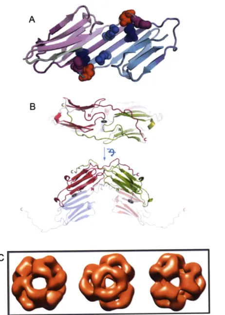
structure (Bagndris et al.2009). Crystal structures of the isolated a-crystallin domains of both rat Hsp20 and human aB revealed that a-crystallin domains formed dimers by aligning their
s6/7
strands to form an extended, continuous sheet (Figure 1-3A). This dimeric assembly also created a groove lined with non-polar residues and the authors suggested that the N-tds (truncated in these constructs to allow for crystallization) would fill these grooves
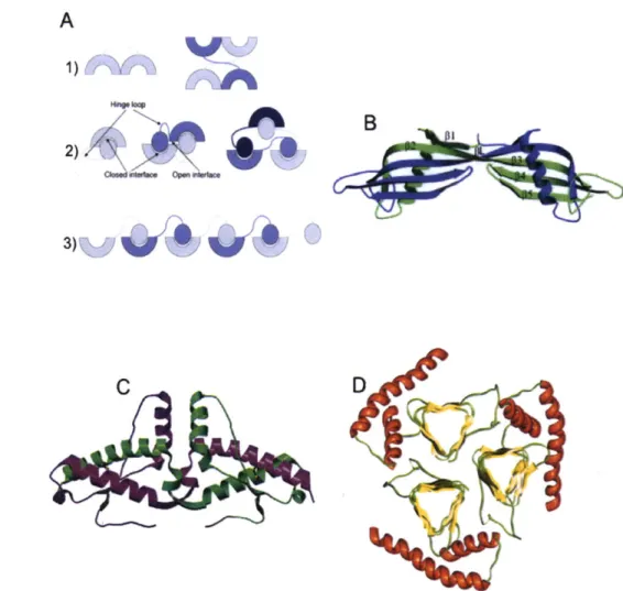
B
Figure 1-3: Structures of human aB-crystallin. (A) Crystal structure of truncated a-crystallin domain dimer highlighting the antiparellel
p-sheet interface. Specific residues are shown in spacefill. RI 16 (light blue) and R120 (deep blue) are residues that are substituted in congenital cataract and cardiomyopathies. D109 (red) forms an ion pair with R120. Reprinted and modified with permission from Elsevier (Bagneris et al. 2009). (B) Crystal structure of dimers including the interactions of the C-terminal extensions. Reprinted and modified with permission from Laganowsky et al. (2010). (C) Cryo-EM reconstruction of a 24 subunit complex modified from Peschek et al.
(2009).
(Bagndris et al. 2009). The overall structure was the same for both proteins, although the
s-strands
from each subunit aligned in different registers for the rat and human proteins, resulting in a more compact structure for human aB (Bagneris et al. 2009). Further structural analysis of human aA and aB constructs that included portions of the C-terminal tail identified varied conformations of a flexible hinge loop and the palindromic sequence E-R-T-I-X-I/V-T-R-E, where the middle region I-X-I/V (the identity of X often being proline) is particularly well conserved (Laganowsky et al. 2010). These physical properties of the protein contribute to the inherent polydispersity of c-crystallin (Figure1-3B).
Crystal structures of monodisperse sHSPs have been determined for wheat
Hsp16.9 (van Montfort et al. 2001), which formed a dodecamer composed of two stacked discs, and Methanococcusjannaschii Hsp16.5 (Kim et al. 1998), whose structure was different, forming a 24-subunit hollow sphere. Unfortunately, the polydisperse nature of a-crystallin has long hindered crystallization of the assembled complex. Transmission electron microscopy (TEM) of recombinant aA and cB confirmed that even the isolated subunits form polydisperse mixtures. Cryo-electron microscopy reconstruction was possible for aB and the complex was roughly spherical and composed of 24 subunits (Figure 1-3C) (Peschek et al. 2009).
b. a-Crystallin Function
Murine Hsp25 and bovine a-crystallin were the first sHSPs shown to exhibit chaperone activity by suppressing the thermal aggregation of a number of model
substrates (Horwitz 1992; Jakob et al. 1993). a-Crystallin suppresses aggregation in an ATP-independent manner and it does not appear to refold its bound substrates.
Substrates remain bound to the chaperone, sequestered from other aggregating molecules, although in some cases these interactions are thought to be transient (Hatters et al. 2001). There is in vitro evidence that sHSPs maintain bound substrates in a folding-competent state and shuttle them to ATP-dependent chaperone systems where refolding is initiated (Lee et al. 1997; Tanksale et al. 2002). Raman et al. described the temperature
unable to suppress aggregation of insulin B chain at a 1:1 ratio, but at 40.6 C aggregation was completely suppressed. Along with an increase in activity, changes in quaternary structure were observed by an increased binding of the hydrophobic probe 4,4'-bis(1-anilinonaphthalene 8-sulfonate) (bis-ANS) at temperatures above 30'C, indicating greater exposure of hydrophobic surfaces (Raman et al. 1995). An increase in chaperone
function was accompanied by irreversible structural changes identified by increased binding of bis-ANS at temperatures over 50'C. These changes were maintained upon
cooling to physiological temperature or below (Das and Surewicz 1995; Kumar et al.
2005). Further analysis of the individual subunits aA and aB showed that these changes
occurred specifically in aA and the chaperone function of aA exceeded that of aB after heating (Reddy et al. 2000; Kumar et al. 2005). Although cA expression is no longer heat-inducible within the lens environment, it has maintained the structural plasticity associated with heat stress.
As briefly described in the preceding section, a-crystallin and all sHSPs can be divided into three regions of primary sequence: (1) an N-terminal region, consisting of residues 1-67 of aB, that is unstructured and poorly conserved; (2) the conserved a-crystallin domain, which spans residues 68-148 of aB and is the defining feature of all sHSPs; and (3) a flexible C-terminal extension (residues 149-175 of aB). Upon first glance, the conservation of the a-crystallin domain suggests that this region is the site of chaperone function, binding aggregation-prone intermediates. However, several studies have shown that various functions are shared throughout the three regions of sHSPs.
Dynamic structures that readily undergo subunit exchange are a hallmark of the sHSPs. Fluorescence resonance energy transfer (FRET) experiments using two
populations of aA demonstrated that reversible subunit exchange occurred between oligomers (Bova et al. 1997). The rate of exchange was positively correlated with temperature (Sun et al. 1998). The presence of substrate reduced subunit exchange in a
size-dependent manner, with larger substrate proteins having a more profound effect (Bova et al. 1997). Wheat Hsp 16.9 and M jannaschii Hsp 16.5 also readily underwent subunit exchange (Bova et al. 2002; Sobott et al. 2002) making this a property of both mono- and polydisperse sHSPs.
Subunit-subunit interactions are crucial for the proper assembly and subunit exchange of sHSPs. The N-terminal domains of both aA and aB are implicated in subunit interactions. Truncation of the first 19 residues of aA had no effect on complex assembly, while removal of the following 36 residues resulted in improper assembly, and the dominant species consisted of trimers and tetramers that did not undergo exchange
(Bova et al. 2000). Pasta et al. identified a narrow region of the N-td in both aA
(residues 20-28) and aB (residues 21-29) that, when deleted, resulted in the formation of smaller oligomers that exhibited faster subunit exchange (Pasta et al. 2003). These oligomers were larger than those observed by Bova and colleagues and still underwent exchange, suggesting that residues of the N-td outside of the deleted sequence
specifically mediated this function. Various truncations of the final nine residues of the C-terminal extension of aA resulted in the formation of somewhat smaller sHSP
complexes with greatly reduced rates of subunit exchange with either WT cA or aB (Aquilina et al. 2005; Kallur et al. 2008). The C-terminal extension is also indicated in increasing solubility of the c-crystallin complex (Smulders et al. 1996). Slight decreases in subunit exchange were observed for the congenital cataract mutants RI 16C CA and R120G aB (both found within the a-crystallin domain); these findings may be due to the location of these residues at the interface of the dimeric structure rather than direct contribution of this domain to subunit exchange (Cobb and Petrash 2000; Liang and Liu 2006). Destabilization of the dimeric structure would likely affect subunit exchange and
oligomerization (Michiel et al. 2009). These processes are clearly driven by a combination of factors, and regions in the N-terminal domain modulate subunit exchange.
In terms of substrate binding, mutagenesis studies implicate the hydrophobic, phenylalanine-rich N-terminal domain as critical for binding. Amino acid substitutions that altered selected N-terminal phenylalanine residues completely abolished the
chaperone activity of murine aB (Plater et al. 1996). More direct binding studies using a heterobifunctional cross-linker identified two regions of aB, 57-69 in the N-td (just overlapping with the c-crystallin domain) and 93-107 in the a-crystallin domain, that are in close proximity to the bound substrate protein, alcohol dehydrogenase in this case (Sharma et al. 1997). Further binding studies utilizing protein pin arrays identified seven
regions important for substrate binding, two regions corresponding to the N-td, four from the a-crystallin domain and a single region from the C-terminal extension (Ghosh et al. 2005). Proteolytic protection assays also confirmed that four sites within the N-td and one region in the C-terminal extension of cB were shielded from cleavage in the
presence of bound reduced a-lactalbumin (Aquilina and Watt 2007). Further support for the role of the N-td in substrate binding comes from studies on a variety of other sHSPs
including wheat Hsp16.9, M jannaschii Hsp16.5 and PsHsp18.1 from the pea plant. Cross-linking of substrate with PsHsp18.1 revealed several bound sites in the N-terminal arm of this protein (Jaya et al. 2009). Chimeras with exchanged N-terminal regions between wheat Hsp 16.9 and PsHsp18.1 exhibited differential aggregation suppression abilities against a set of substrate proteins, suggesting that this domain plays a major role in substrate binding (Basha et al. 2006). These results indicate that substrate-interacting regions span the entire protein but are concentrated in the N-terminal domain.
Detailed studies of the binding site(s) on a-crystallin were undertaken by McHaourab and colleagues. Using a series of increasingly destabilized T4 lysozyme mutants, two different "binding modes" were identified, the first corresponding to a high-affinity/low capacity state and the second to a low-affinity/high capacity state
(McHaourab et al. 2002; Koteiche and McHaourab 2003). They later demonstrated that the complexed T4 lysozyme molecules were mostly unfolded and bound in a distinct manner where the C-terminal domain was protected from solvent while the N-terminal domain was exposed (Claxton et al. 2008). Experiments with the lenticular substrates
pB
1- ands3B2-crystallin
demonstrated that a-crystallin recognized and bound monomeric forms of both proteins and promoted their dissociation and unfolding (Sathish et al. 2004; McHaourab et al. 2007). In addition, these studies showed that the two binding modes are in fact distinct sites, and both have the ability to bind the predominantly unfoldedconformation of T4 lysozyme (Claxton et al. 2008).
The substrate binding sites of a-crystallin recognize a wide range of proteins. This begs the question: what substrate features are being recognized? Like Trigger factor and the ATP-dependent Hsp70/40 system and the chaperonins GroEL/S and TriC, the sHSPs including a-crystallin recognize and bind exposed hydrophobic regions of partially unfolded and destabilized proteins (Bloemendal et al. 2004; Hartl and
Hayer-Hartl 2009). Studies using the substrate a-lactalbumin showed that a-crystallin only binds to intermediates or molten globule structures that are unstable in solution and aggregation-prone. No significant binding was observed for stable molten globule structures, such as the partially reduced species of c-lactalbumin (Lindner et al. 1997). Carver et al. demonstrated that soluble, stable, but unfolded proteins did not interact with
a-crystallin. Both c-casein and a-ovalbumin bound bis-ANS, indicating exposure of hydrophobic patches, but neither of these proteins interacted with a-crystallin, nor did they aggregate in solution (Carver et al. 1995).
Despite the general characterization of structural requirements for the substrate, identification of specific interacting regions on substrates is lacking. Studies using bovine c-crystallin cross-linked to bound yeast alcohol dehydrogenase identified two distinct peptides as probable substrate binding sites, indicating that upon destabilization and partial unfolding specific protein regions are recognized (Santhoshkumar and Sharma 2002). A short peptide derived from @A1/A3-crystallin (residues 102-117) bound a-crystallin and inhibited chaperone function (Rao et al. 2008), which suggests that this region may be a recognition site in the full-length protein. The identification of specific binding regions, in particular those of the physiological lenticular substrates, are of great interest as this information could potentially aid in the development of preventive cataract medication.
a-Crystallin is the sole chaperone system of lens fiber cells. Due to the lack of protein synthesis machinery, the supply of a-crystallin is finite. Its increased localization to the water-insoluble fraction of aged normal and cataractous lenses (Harrington et al. 2004; Harrington et al. 2007) suggests that with age, its bound substrate load becomes
saturating and the chaperone-substrate complex aggregates and precipitates within fiber cells. aA knockout mice displayed a cataract phenotype with inclusions containing both
cB- and y-crystallins (Brady et al. 1997); however, aB knockouts were afflicted with skeletal muscle degeneration without cataract and suffered from dramatically shortened lifespan (Brady et al. 2001). It was therefore impossible to examine the importance of cB in the aging lens and it may be critical for maintenance of long-term fiber cell structure and lens transparency.
In addition to its chaperone role, a-crystallin is also a structural protein in the lens, interacting with plasma membranes and the lens-specific cytoskeletal proteins BFSP1 (filensin) and BFSP2 (CP49). The lens-specific beaded filaments are
intermediate filament-like structures composed of BFSP1 and 2; they are heavily
decorated with "beads" which have been identified as a-crystallin oligomers (Carter et al. 1995). a-Crystallin is required for the in vitro formation of native-like beaded filaments (Carter et al. 1995). In non-lens cells, intermediate filaments are associated with a host of proteins including chaperones and the proteasome, and they are crucial to plasma
membrane stability (Song et al. 2009). They no doubt play a role in maintaining the unique structure of fiber cells in the lens nucleus and cortex. Although the precise role of the a-crystallin interaction with BFSP1 and 2 is unclear, both components are crucial for proper lens function.
It is well documented that proteins of the aging human lens are significantly modified through covalent damages including oxidation, deamidation and truncation. These destabilizing modifications result in partial unfolding and population of
aggregation-prone intermediates that, as discussed in this section, are prime targets for sequestration by a-crystallin. As the fixed amount of free a-crystallin is diminished, the buffer capacity of the lens to prevent protein aggregation is reduced and there may be a threshold above which protein aggregation dominates. Adequate functional c-crystallin is therefore critical for proper maintenance of lens transparency and refraction.
2. The /3y-Crystallins
The y-crystallins comprise the second major group of crystallins in human lenses. Both are mainly structural proteins whose high solubility and dramatic stability are well suited to the environment of lens fiber cells. Both
s-
and y-crystallins arecomposed of two domains, although interdomain interactions and oligomeric states differ between the two (Bloemendal et al. 2004). Several different
P-
and y-crystallins, whichare homologous but contain many sequence variations amongst one another, are major contributors to lens transparency. They are, by and large, lens-specific with small amounts of P1B2-crystallin expressed in the retina, brain and testis (Magabo et al. 2000).
Recent proteomic analyses identified y-crystallins in the retina of diabetic mice, although the implications of this finding are unknown (Fort et al. 2009).
The y-crystallins are monomeric proteins found in terminally differentiated lens fiber cells. In humans, there are seven genes encoding yA-F and yS-crystallin (CR
YGA-F; S). CRYGA-F are located in a cluster on chromosome 2q33-36 (Shiloh et al. 1986)
while CRYGS is spatially distinct, found on chromosome 3q26.3 (Wijnen et al. 1989). The major y-crystallins expressed in the human lens are yC, yD, and yS, while yE and yF are pseudogenes (Brakenhoff et al. 1990). They are the final crystallins expressed in the developing lens. All y-crystallins are composed of two homologous domains, an N-terminal domain (N-td) and a C-N-terminal domain (C-td) that arose from a gene
duplication event (Shimeld et al. 2005). Each domain is constructed from eight
p-strands
that form two intercalated Greek Key motifs. The domains interact by way of well-conserved interface residues, suggesting that the domain interface plays a major role in the maintenance of protein structure.a. yD-Crystallin
Human yD-crystallin (HyD-Crys) is one of the two major y-crystallins found in the lens nucleus, comprising up to 8% of total soluble lens protein (Robinson et al. 2006). Its expression is upregulated during the last stages of fiber cell differentiation, following that of both the a- and
p-crystallins.
As one of the major structural proteins in the lens, it is important for formation of the polydisperse, yet regular arrangement of crystallins that results in lens transparency. It is particularly enriched in the oldest region of the lens, requiring this protein to be unusually long-lived.HyD-Crys is a 20.6 kDa, 173 residue protein. Its crystal structure has been solved to 1.25
A (Basak et al. 2003). As with other y-crystallin structures, each Greek Key motif
exchanges its third strand with the facing motif (Figure 1-4B). The protein is rich in aromatic residues, including several aromatic pairs, that make substantial contributions to its stability and folding (F. Kong, personal communication). Each domain contains a highly stabilizing tyrosine corner, in which the Tyr side chain is hydrogen bonded to the peptide backbone (Hemmingsen et al. 1994). Determination of the high resolution
N
0:002000k52
N-terminal
B
Motif
1
Motif 2
N-terminal domain
Motif 3
C-terminal domain
Motif 4
C-terminal domain
Figure 1-4: (A) Crystal structure of human yD-crystallin. PDB ID
1HKO.
The four tryptophan residues are shown in space fill. (B) Topology diagram of HyD-Crys. Note the intercalated nature of the Greek Key motifs. The tyrosine corners are denoted as well. (See Introduction section F.1) Y62 N Y151--
C
... . .. ...... ... ...structure of HyD-Crys has enabled extensive biochemical and biophysical study of this protein.
HyD-Crys contains four highly conserved buried tryptophan residues, two in each domain (W42 and W68 in the N-td; W130 and W156 in the C-td; Figure 1-4A), and in the native state this protein exhibits anomalous Trp fluorescence quenching
(Kosinski-Collins and King 2003). Extensive studies of this phenomenon have determined that one Trp (W42 and W130) in each domain is highly fluorescent and upon excitation, transfers energy to the second Trp residue (W68 and W156) in the same domain. The unique backbone conformation, in combination with specifically oriented water molecules in the vicinity of W68 and W 156, allow fast electron transfer to the protein backbone (Chen et al. 2006). Fluorescence anisotropy demonstrated the rigid nature of the Trp
environments, which suggests that the hydrophobic core of HyD-Crys is tightly packed (Chen et al. 2008). Upon protein unfolding, quenching is relieved due to loss of these stringent structural requirements. The large change in Trp fluorescence upon unfolding has been utilized in a range of studies, including those documented in this thesis, to elucidate determinants of folding and stability for HyD-Crys (Figure 1-5A).
The long lifetime of HyD-Crys in the lens requires superior stability. It can be recombinantly expressed in E. coli and purified from the soluble fraction of the cell
lysate. It was resistant to denaturation in 8 M urea (Kosinski-Collins and King 2003) and had a melting temperature above 80*C (Flaugh et al. 2006). Equilibrium
unfolding/refolding experiments determined a three-state in vitro unfolding pathway (Flaugh et al. 2005a) with the first transition at 2.2 M GdnHCl corresponding to unfolding of the N-td. The second transition, with a midpoint at 2.8 M GdnHCl,
corresponds to unfolding of the C-td. These measurements yielded an apparent total AG = 16.6 kcal/mol for the WT protein (Figure 1-5B) (Kosinski-Collins et al. 2004; Flaugh et al. 2005a). A three-state unfolding pathway implies the presence of a populated partially-folded intermediate. Experiments with triple Trp mutants showed that this intermediate,
A
1000 800 400 200 320 340 360 380 Wavelength (nm)B
Native Intermediate UnfoldedFigure 1-5: HyD-Crys Trp fluorescence and equilibrium unfolding pathway. (A) Trp emission fluorescence spectra for native and unfolded HyD-Crys. Native state fluorescence is quenched compared to the unfolded state. (B) Equilibrium unfolding/refolding of HyD-Crys highlights the population of a partially folded intermediate. Upon dilution from high GdnHCl concentrations
an off-pathway aggregation reaction competes with productive refolding.
400
populated around 2.5 M GdnHCl, had a folded C-td and a fully unfolded N-td (Kosinski-Collins et al. 2004). Differential domain stability has been documented for other
crystallins including bovine yB-crystallin. Although structurally homologous to HyD-Crys, the domain stabilities were reversed, with the N-td being more stable than the C-td. Likewise, the unfolding intermediate had a native N-td and a completely unfolded C-td (Rudolph et al. 1990; Mayr et al. 1997).
Kinetic stability is an equally, if not more important factor in the maintenance of HyD-Crys native tertiary structure for upwards of 80 years. Initial equilibrium
unfolding/refolding experiments identified a hysteresis when the protein was incubated for six hours at 37'C. The hysteresis was more pronounced at 25'C, even after a 24-hour incubation period (Kosinski-Collins 2004; Kosinski-Collins et al. 2004; Mills-Henry 2007). This suggests a kinetic barrier to unfolding, possibly in the N-terminal domain. Mills-Henry used half-chevron plots to extrapolate the unfolding half-time of HyD-Crys in the absence of denaturant. The half-time for unfolding of the N-td was calculated to be
19 years, while that of the C-td (both in the context of the full-length protein) was a more modest 129 days (Mills-Henry 2007). This is an exceptionally long life, even in the dilute context of kinetic fluorescence experiments. A high activation energy for
unfolding (24 kcal/mol) was also determined for bovine yF-crystallin, supporting the role of kinetic stability in the long-term maintenance of y-crystallin structure (Das and Liang 1998). Excluded volume effects due to macromolecular crowding, which may also increase thermodynamic stability of the native state, probably serve to increase the effective lifetime of HyD-Crys and other crystallin proteins (Bloemendal et al. 2004).
The domain interface of HyD-Crys consists of a hydrophobic patch capped above and below by pairs of polar residues (Basak et al. 2003). Alanine mutagenesis showed that this hydrophobic cluster was important for both native state stability and folding of the N-td (Flaugh et al. 2005a). Substitution of the polar residue pairs with alanine destabilized the protein in a similar manner, specifically affecting the N-td (Flaugh et al. 2005b). One polar pair consists of two glutamine residues (Q54/Q143) and substitution of either or both with glutamate to model deamidation, one of the most common covalent damages to proteins of the aging lens (Wilmarth et al. 2006), lowered the kinetic barrier
to unfolding (Flaugh et al. 2006). The domain interface contributed to the high AG by specifically stabilizing the N-td (Mills et al. 2007). A complete description of the folding pathway was given by Flaugh et al. (Flaugh et al. 2006). The authors proposed that
folding begins with motif 4 in the C-td to form the interface Greek Key, and is followed by motif 3 to form the complete C-td. Motif 2 in the N-td is then formed using the interface as a template and motif 1 folds in the final stages.
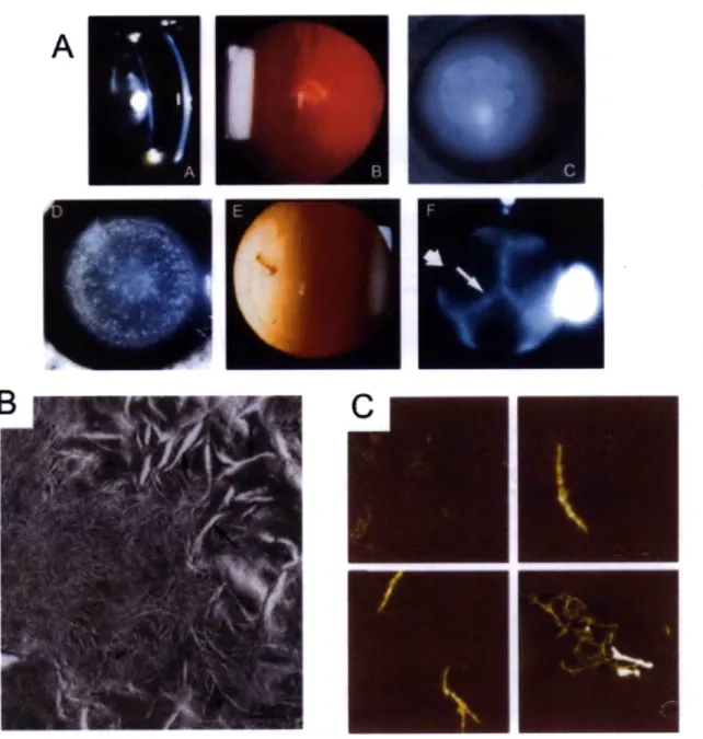
Upon dilution from high denaturant concentrations, a robust aggregation reaction competes with productive refolding of HyD-Crys (Kosinski-Collins and King 2003). The aggregates, visualized by atomic force microscopy (AFM), were filamentous, although not amyloid in nature (Figure 1-7C). Subsequent analysis of this aggregation pathway by Acosta-Sampson and King found that the C-td is the likely region involved in the
initiation of aggregation (Acosta-Sampson and King 2010) .
b. yC- and yS-Crystallins
Human yC-crystallin (HyC-Crys) is the other major y-crystallin of the lens
nucleus, totaling 12-16% of soluble protein (Robinson et al. 2006). It is 173 residues and 20.7 kDa, and 71% identical to HyD-Crys. Although the crystal structure of the human protein has not been determined, a structure for the homologous murine yC-crystallin is available (Purkiss et al. 2007). HyC-Crys is resistant to heat-induced denaturation and aggregation, and required temperatures above 60'C for protein aggregation and precipitation to occur (Fu and Liang 2001). Equilibrium unfolding/refolding at room temperature indicated unfolding transitions above 2 M GdnHCl and global analysis of
absorbance, Trp fluorescence and circular dichroism data found that HyC-Crys also unfolded by way of a populated intermediate, with a total AG = 8.7 kcal/mol (Fu and Liang 2002a). High thermodynamic stability is a shared property of HyD- and HyC-Crys.
On the contrary, HyC-Crys is much less soluble than its counterpart. Purkiss et al. suggest that this is at least in part due to the presence of a cysteine residue at position 79. Substitution of an arginine at this position, as found in HyD-Crys, resulted in a dramatic solubility increase, from < 1 mg/ml for WT HyC-Crys to 90 mg/ml for the C79R mutant (Purkiss et al. 2007). However, C79R was not the only substitution that increased
solubility, indicating that this position is not the sole determinant of differential solubility between these proteins. Regardless, it seems that HyC-Crys evolved under selective pressure for high stability rather than high solubility. This underscores the importance of maintaining a range of polydisperse crystallin proteins in the lens.
The third major y-crystallin of the lens is human yS-crystallin (HyS-Crys). This protein is 177 amino acid residues in length and 20.7 kDa. Although evolutionarily more divergent that HyD and HyC, HyS-Crys maintains ~50% identity with both of them. No crystal structure exists for the full-length human protein, although an NMR solution structure of murine yS-crystallin and a crystal structure of the C-terminal domain from HyS-Crys support its high structural similarity to other y-crystallins (Purkiss et al. 2002; Wu et al. 2005). This protein has a short N-terminal extension of four residues before the start of the first Greek Key motif. N- and C-terminal extensions are more commonly found in the -crystallins (discussed in more detail below). In addition to these
differences, HyS-Crys exhibits differential expression patterns from other y-crystallins. It is highly expressed in the lens cortex as opposed to the lens nucleus and cDNA
transcripts have been identified in 40-year-old human lenses (Wistow et al. 2002). HyS-Crys stability, while overall great, is less than that of HyD-Crys. In
equilibrium unfolding/refolding experiments, it reversibly unfolded through a cooperative two-state mechanism with a transition midpoint of 2.3 M GdnHCl (Mills et al. 2007). Its melting temperature was 74'C. Like HyD, the kinetic stability of HyS-Crys was
measured by half-Chevron plot analysis. In contrast to equilibrium experiments, kinetic unfolding detected the presence of a populated unfolding intermediate, suggested to have
an unfolded N-td and a folded C-td. Extrapolated half-times of unfolding for each
domain in the context of the full-length protein were 1.6 years for the N-td and 2 days for the C-td (Mills-Henry 2007). Again, although the N-td is the first to unfold and is less
stable in isolation, its high kinetic stability creates a significant barrier not necessarily expected from thermodynamic analysis alone. It is important to note the ~10-fold difference in unfolding half-times between HyD- and HyS-Crys. Because of the later expression of HyS-Crys in the adult lens cortex, the need for kinetic stability may not be quite as great as in the lens nucleus (Mills-Henry 2007). Therefore, selective pressure to maintain this feature was not as strong as for HyD-Crys.
c. The /-Crystallins
The -crystallins are the third class of ubiquitous crystallin proteins found in the human lens. Like y-crystallins, they are predominantly structural proteins, and their expression is upregulated during fiber cell differentiation. Upregulation occurs after that of the a-crystallins and before that of the y-crystallins (Aarts et al. 1989). There are seven
p-crystallin
genes in the human genome and their products are categorized as either acidic (1A1, 1A2, 1A3 andpA4)
or basic (pB1, 1B2 and 1B3) (Lampi et al. 1997).PAl
andpA3
are different proteins arising from different start codons in the same transcript (Hogg et al. 1986), withpA3
being larger. The @B1- andpB3-crystallins
are expressed especially early and they are found in the lens nucleus, while @B2 is prominentthroughout all lens fiber cells (Aarts et al. 1989; Lampi et al. 1998). The acidic
p-crystallins are widely expressed in both the lens cortex and nucleus, although
pA2-crystallin protein is not detected (Lampi et al. 1997; Lampi et al. 1998). Unlike the y-crystallins,
p-crystallins
form a range of oligomers and separation of lens-derivedcrystallins by size exclusion chromatography identified two populations: the
pH fraction,
composed of hexamers and octamers, is more prominent in the lens nucleus; and thePL
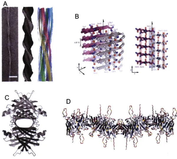
fraction, comprising smaller oligomers such as dimers and tetramers, dominates in the cortex (Zigler et al. 1980).Crystal structures are available for three human P-crystallins:
PA4,
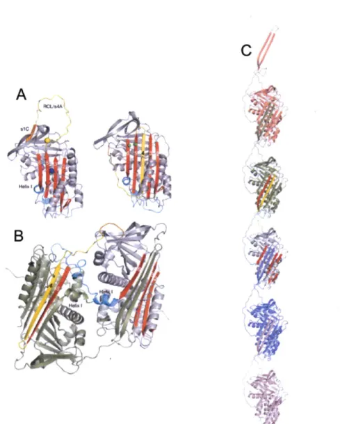
PB2 and a truncated form of @B1 (Figure 1-6) (Bax et al. 1990; Van Montfort et al. 2003; Chaikuad et al. 2010). @-Crystallins share their overall tertiary structure with the y-crystallins but differ in important ways. First, allp-crystallins
possess N-terminal extensions and the basicp-crystallins
have C-terminal extensions as well. These extensions are flexible and do not contribute to the Greek Key motifs or participate in domain interface interactions. Second, the linker region connecting the two domains varies considerably from that of the y-crystallins. While the y-crystallin linker is flexible and allows for intramolecular domain pairing, the@-crystallin
linker can remain extended so that intermolecular pairing is sterically allowed as well. This is clearly evident in the.-
B
C
Figure 1-6: The
p-crystallins
form varied oligomers. (A) The crystal structure ofPA4-crystallin
is a dimer. Each monomer forms an intramolecular domain interface like that of HyD-Crys and the dimer forms from monomers packing against each other and rotating roughly 900 relative to one another. One monomer is blue and the other is teal. (B) The crystal structure of @B2-crystallin. In contrast to the previous example, this dimer is formed by domain swapping that generates intermolecular domain interfaces. One monomer is blue and the other is teal. (C) The crystal structure of @B1-crystallin is similar to that of @A4-crystallin. One monomer is blue and the other is teal.38
structure of
pfB2-crystallin,
which forms a dimer by domain swapping (Bax et al. 1990). In contrast, the crystal structure of truncatedpB
1 -crystallin revealed a domain interface formed by intramolecular pairing as in the y-crystallins and the dimer was formed by the pairing of monomers adjacent to one another (Van Montfort et al. 2003). Very recently, a structure for A4-crystallin was deposited in the Protein Data Bank (PDB ID: 31wk). This dimer is formed from two molecules with y-crystallin-like bent linkers and an interface similar to that formed bypB1-crystallin
(Chaikuad et al. 2010). Higher order oligomerization in thep-crystallins
is not completely understood and in vitro conditions have not yet replicated the larger oligomers observed in the solublepH
lens protein fraction (Bloemendal et al. 2004).In general
p-crystallins
are less stable than the y-crystallins. Dimeric rat PfB2-crystallin can be completely unfolded in 5 M urea through a three-state mechanism and the populated intermediate is a partially unfolded monomer with an unfolded N-td and a native C-td (Wieligmann et al. 1999). Human P3B2-crystallin equilibrium unfolding wasdescribed by a three-state transition with a total AG ~ 11.8 kcal/mol (Fu and Liang 2002a). The acidic -crystallins are well-known to hetero-oligomerize and it was shown that hetero-oligomerization of
PAl
andp1B1
enhanced stability (Bateman et al. 2003).This mechanism may be utilized within the crowded lens environment.
D. Cataract Disease
Cataract disease is the leading cause of blindness in the world, affecting over 22 million Americans age 40 and older (Prevent Blindness America 2008). Cataract is any opacification of the lens and in general is a disease directly related with aging. These opacities are the result of the scattering of light as it passes through the lens (Figure 1-7A,B). There are currently no treatments to prevent cataract or delay its onset. The only "'cure" is surgical removal of the opaque lens and replacement with an artificial
intraocular lens implant. The implant is placed inside the natural lens capsule, which is not removed during surgery. This procedure, although lauded by optometrists and ophthalmologists, is not without risk. In up to 40% of patients who have undergone cataract surgery, a secondary opacity develops known as posterior capsular opacification
A
Figure 1-7: Gross cataract and protein aggregate phenotypes. (A) A variety of cataracts presented by patients afflicted with congenital cataracts. Reprinted with permission from Elsevier (Hejtmancik 2009). (B) Fibril-like structures observed in thin sections of the lens from the OXYS rat, a strain with increased oxidative stress. Reprinted with permission from Elsevier (Marsili et al. 2004). (C) Time course of AFM images of fibrillar HyD-Crys aggregates produced in vitro upon dilution from high concentrations of denaturant. Reprinted and modified with permission from Kosinski-Collins and King (2003).