Cross-regulation and interaction between
eukaryotic gene regulatory processes
Noah Spies
Bachelor of Arts in Mathematics Cornell University, 2006
Submitted to the Department of Biology in partial fulfillment of the requirements for the degree of Doctor of Philosophy at the Massachusetts Institute of Technology
June 2012
c 2012 Massachusetts Institute of Technology. All rights reserved.
Noah Spies
Department of Biology May 1, 2012
Author
David P Bartel
Professor of Biology, MIT Thesis Advisor
Christopher B Burge Professor of Biology, MIT Thesis Advisor
Stephen P Bell
Professor of Biology, MIT
Cross-regulation and interaction between eukaryotic gene regulatory
processes
by Noah Spies
Submitted to the Department of Biology on May 1, 2012 in partial fulfillment of the requirements for the degree of Doctor of Philosophy in Biology
Thesis Supervisors: David P Bartel and Christopher B Burge, Professors of Biology
Abstract
Regulation of genes is fundamental to all living processes and can be exerted at many sequential steps. We studied several eukaryotic gene regulatory mechanisms with an emphasis on understanding the interplay between regulatory processes on a genome-wide scale.
Gene splicing involves the joining of exonic RNA stretches from within a precursor messenger RNA (mRNA). Splicing typically occurs co-transcriptionally as the pre-mRNA is being produced from the DNA. We explored the relationship between the chromatin state of the gene-encoding DNA and the splicing machinery. We found a marked enrichment for nucleosomes at exonic DNA in human T cells, as compared to surrounding introns, an e↵ect mostly explained by the biased nucleotide content of exons. The use of nucleosome positioning information improved splicing simulation models, suggesting nucleosome positioning may help determine cellular splicing patterns. Additionally, we found several histone marks enriched or depleted at exons compared to the background nucleosome levels, indicative of a histone code for splicing. These results connect the chromatin regulation and mRNA splicing processes in a genome-wide fashion.
Another pre-mRNA processing step is cleavage and polyadenylation, which determines the 30 end of the mature mRNA. We found that 3P-Seq was able to quantify the levels of 30 end
isoforms, in addition to the method’s previous use for annotating mRNA 30 ends. Using 3P-Seq
and a transcriptional shuto↵ experiment in mouse fibroblasts, we investigated the e↵ect of nuclear alternative 30 end formation on mRNA stability, typically regulated in the cytoplasm. In genes
with multiple, tandem 30 untranslated regions (30 UTRs) produced by alternative cleavage and
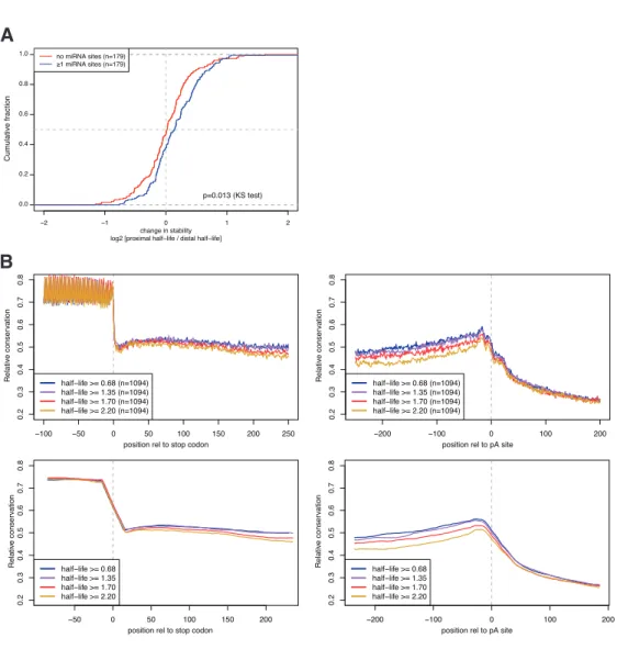
polyadenylation, we found the shorter UTRs were significantly more stable in general than the longer isoforms. This di↵erence was in part explained by the loss of cis-regulatory motifs, such as microRNA targets and PUF-binding sites, between the proximal and distal isoforms.
Finally, we characterized the small interfering RNAs (siRNAs) produced from heterochromatic, silenced genomic regions in fission yeast. We observed a considerable bias for siRNAs with a 50 U, and used this bias to infer patterns of siRNA biogenesis. Furthermore, comparisons with between wild-type and the Cid14 non-canonical poly(A) polymerase mutant demonstrated that the exosome, the nuclear surveillance and processing complex, is required for RNA homeostasis. In the absence of a fully functional exosome complex, siRNAs are produced to normal exosome targets, including ribosomal and transfer RNAs, indicating these processes may compete for substrates and underscoring the interconnectedness of gene regulatory systems.
Acknowledgments
I am indebted to the excellent mentoring I have received both formally and informally in the course of my studies. First and foremost, I could not have completed this work without the direction and guidance of my two PhD advisors, Dave Bartel and Chris Burge. Chris and Dave have always been available to consult about the minutiae as well as the big picture of my work, and I feel I have learned a tremendous amount from my personal interactions with both of them. They have also fostered incredibly supportive lab environments, and I must thank everyone the both labs for scientific and non-scientific input, collaboration and friendship. In particular, I must thank Graham Ruby for early mentoring on the computational side and Calvin Jan for teaching me the ins and outs of wet lab experimentation.
Thanks to Jess Hurt for being an excellent bay mate and also Jess Hurt and Vincent Butty for discussion of the content of this thesis.
I would also like to thank my thesis committee, Phil Sharp and Aviv Regev, not only for the many productive discussions we have had about my projects, but also the encouragement they have provided me through my graduate studies. While I have had a broad network between my two labs and the biology community, it has always been good to know that I have the support of my thesis committee. Thanks also to Angela DePace for joining us for my thesis defense.
Finally, I would like to thank my friends and family. It has been great fun moving to Cambridge and making new friends. It would not have been worth it if I didn’t have the love and friendship of Torrey, who has been supportive and interested in my work. And finally, I have to thank my parents, Wendy and Rupert, who have supported me at every step, and instilled a sense of curiosity in me from an early age. Who knew that would lead me down the road of the scientist?
Contents
1 Introduction 9
1.1 The importance of gene regulation . . . 9
1.2 Chromatin . . . 10
1.3 Transcription . . . 11
1.4 Messenger RNA Maturation . . . 14
1.5 Post-transcriptional regulation of gene expression . . . 17
1.6 RNA-induced transcriptional silencing in fission yeast . . . 20
1.7 References . . . 21
2 Chromatin signatures and RNA processing 31 2.1 Introduction . . . 33
2.2 Results . . . 33
2.3 Discussion . . . 40
2.4 Experimental Procedures . . . 43
2.5 References . . . 45
3 Stability regulation through alternative polyadenylation 51 3.1 Introduction . . . 53
3.2 Results . . . 55
3.3 Discussion . . . 61
3.4 Methods . . . 62
3.5 References . . . 63
4 Competition between fission yeast RNAi and degradation pathways 67 4.1 Introduction . . . 69
4.2 Results . . . 70
5 Conclusion 87
5.1 Summary and Progress . . . 88
5.2 Themes and Perspectives . . . 88
5.3 Future directions . . . 89
5.4 References . . . 90
A Supplementary information for Chapter 2 91
B Supplementary information for Chapter 4 106
Chapter 1
Introduction
1.1
The importance of gene regulation
The genome is life’s blueprint. Contained within is the complete information required to produce all the proteins used to piece together the cell’s structures and all the enzymes required to per-form nearly all biological processes. The DNA-encoded genomes of thousands of organisms are now completely sequenced, but we are only be-ginning to understand how the di↵erent genetic sequences are combined to produce the complex cellular actions our bodies perform every living minute. Key to our understanding of molecu-lar biology are two things: (1) the knowledge of what each gene in the genome actually does and (2) an understanding of under what conditions those genes are activated or deactivated. This thesis focuses on the latter of these two: gene regulation.
The central dogma of biology provides a sim-ple framework for understanding gene regulation. A gene is first turned on by transcribing its DNA into messenger RNA. The mRNA is then pro-cessed and exported from the nucleus to the cyto-plasm where it is translated into a protein. The protein folds into a three-dimensional structure capable of performing its cellular role until it is finally degraded. Each of these steps is poten-tially the target of gene regulatory mechanisms. This chapter describes the mRNA regulatory sys-tems. In the interest of clarity this introduction
will focus on gene regulation in mammals but most of these fundamental processes are under-stood through research performed in more basal organisms such as yeast. Most of the regulatory systems are described here from a mechanistic perspective. However, the biological role of regu-lation cannot be understated and will be touched upon in specific cases relevant to the research described in subsequent chapters.
An important theme of this introduction is the cross-talk between gene regulatory systems. We will see examples of how transcription can a↵ect splicing through modulating the rate of the transcribing polymerase; how 30 end processing factors are recruited at transcription initiation; how splicing places an exon-junction complex on an mRNA, enabling translation-dependent quality control of splicing; and how chromatin-modifying enzymes are recruited to the fission yeast centromeres co-transcriptionally via siR-NAs.
This thesis explores several topics related to the interaction between regulatory processes. The second chapter examines the interplay between co-transcriptional gene splicing and the chromatin state of that gene. The third chapter explores how regulation of a nuclear process, cleavage and polyadenylation, creates isoform variants that are di↵erentially regulated by various degradation
pathways in the cytoplasm. Finally, the fourth chapter characterizes the fission yeast small RNAs involved in co-transcriptional silencing of cen-tromeres. The particular fission yeast mutants
studied demonstrate that the nuclear exosome degradation pathway can compete with RNAi components for substrates.
1.2
Chromatin
Packaging of DNA into chromatin DNA
is stored in the nucleus in a compact form we know as chromatin, so-called because of its abil-ity to be stained and viewed under a microscope (Flemming 1882). The protein constituents of chromatin are the histones, which were identified early on but it wasn’t until much later that it was understood how chromatin formed around them. Nearly 100 years after the discovery of chromatin, the histone octamer, or nucleosome, was identified as the core repeating protein struc-ture around which DNA was wrapped (Kornberg 1974; Olins and Olins 1974). The nucleosome is made up of four subunits, histones H2A, H2B, H3 and H4. An (H3–H4)2 tetramer forms the center
of the nucleosome, with two H2A–H2B dimers bound to either side (Luger et al. 1997). A fifth histone, H1, serves as a linker between nucleo-somes. Together, these histones are responsible for packing over a meter worth of DNA, end-to-end, into a nucleus 1/10,000 that size (Woodcock
and Ghosh 2010).
The nucleosome not only acts to compact DNA but also performs a vital role in regulating the activity of the genes it packages. The his-tone subunits contain flexible tails that can be modified so as to mark the DNA regions that are wrapped around them, thereby helping recruit regulatory factors (Brownell et al. 1996). In the extreme case, these factors can tightly condense the DNA into what is called heterochromatin, blocking access to RNA transcriptional machin-ery and leading to nearly complete gene silencing (Trojer and Reinberg 2007).
Post-translational modification of histones Histones in the vicinity of genes are generally post-translationally modified in a manner indicative
of the gene’s activity level. These modifications typically involve the covalent addition of acetyl, methyl, or ubiquitin groups to lysine and argi-nine residues on the N-terminal “tail” of each of the histones, although there is a large number of possible modifications (Suganuma and Workman 2011). Promoters of silent genes are enriched for H3K9me3 (tri-methylation on lysine 9 of histone H3) and H3K27me3, whereas active genes typi-cally show enrichment for H3K4me3 and various acetylations (Zhou et al. 2011). The body of tran-scribed regions is generally high for H3K36me3 and H3K79me2, marks established by the elon-gating polymerase II complex.
The locations of these modified histones can be assayed using a method called chromatin im-munoprecipitation, or ChIP (Solomon et al. 1988). Proteins are first cross-linked to DNA using a chemical, frequently formaldehyde. Following purification of chromatin from the cell, antibod-ies specific for certain histone tail modifications can be used to immunoprecipitate DNA regions bound by modified histones (or, depending on the choice of antibody, other chromatin-bound factors). The DNA can be digested or fragmented resulting in⇠146 bp fragments, the length of DNA bound and protected by a single nucleosome. The cross-links can be reversed, and the bound DNA regions can be interrogated by high-throughput methods such as micro-array (Blat and Kleckner 1999; Ren et al. 2000), known as ChIP-Chip, or high-throughput sequencing, known as ChIP-Seq (Robertson et al. 2007; Johnson et al. 2007; Barski et al. 2007). These methods have immensely in-creased our understanding of the global distribu-tion of transcripdistribu-tion factors and modified histones. However, these results are frequently difficult to translate into understanding of cause and e↵ect
Introduction 1.3. Transcription
because of the global nature of perturbations and the impracticality of genetically modifying his-tone genes, which are highly duplicated in the genome (Heniko↵ and Shilatifard 2011).
Nucleosome positioning Active genes are typically characterized by an entirely nucleosome free region surrounding the transcription start site (TSS), allowing easy access to transcription factors and the RNA polymerase II (Yuan et al. 2005). Genes that are active in one cell type lose this nucleosome-free region (perhaps more accurately, but less commonly, known as the nucleosome-depleted region) when silenced in a di↵erent tissue, suggesting an active process can regulate the openness of this chromatin stretch (Ozsolak et al. 2007). It remains unclear whether the nucleosome-free region is established prior to transcription, or as a side-e↵ect of recruit-ing general transcription factors and the poly-merase complex to the DNA. Maintenance of the nucleosome-free region at active promoters pushes neighboring nucleosomes into well-defined positions just downstream of the TSS (the +1 nu-cleosome) and upstream of the promoter (alterna-tively called the 1 or 2 nucleosome, depending on species).
Nucleosomes preferentially bind to some DNA sequences. This inherent sequence bias can be understood at the structural level: the nucleo-some induces significant bending of bound DNA, and this bending is achieved more readily for
some sequences (Luger et al. 1997). For exam-ple, G·C base pairs are preferred when the major groove faces in toward the nucleosome, and in-deed these sequences are preferred every ⇠10 nu-cleotides (which coincides with a full twist of DNA wrapped around the nucleosome) (Kaplan et al. 2009). While the nucleosome contacts DNA pri-marily through the sequence-independent phos-phate backbone, some additional amino acid-base contacts also increase nucleosome affinity for A·T base pairs in the minor groove.
These sequence preferences lead to markedly lower affinities for nucleosomes in the nucleosome-free region at the TSS as well as near the tran-scription termination site (Kaplan et al. 2009). While the nucleosome-free region is regulated between cell types in mammals, in vitro experi-ments mixing yeast histones and genomic DNA recapitulate a nucleosome-free region, indicating promoter sequences are inherently unfavorable to nucleosome binding (Kaplan et al. 2009). Re-cent results suggest ATP-dependent chromatin remodelers are required for specific placement of the +1 and subsequent nucleosomes near the 50 end of the transcript (Zhang et al. 2011). It remains an open question how much DNA se-quence a↵ects nucleosome positioning outside of these highly stereotyped regions and in species other than yeast, where most of these studies have been performed (Zhang et al. 2009; Kaplan et al. 2009).
1.3
Transcription
Polymerase II transcription initiation and
elongation The DNA-dependent RNA
poly-merase II (pol II) transcribes protein-coding genes into messenger RNA. Early studies of partially purified polymerases demonstrated separate ac-tivities for three individual enzyme complexes, named pol I, pol II and pol III based on the purification scheme used (Roeder and Rutter 1969). While work initially focused on pol III, researchers in the late 1970s and early 1980s
pu-rified a set of basal transcription factors which assist in recruiting the 12 subunit pol II to DNA, converting it into an elongation-competent form (Thomas and Chiang 2006).
However, pol II transcription is a far more complex process in the context of a living eukary-ote. In vivo transcription begins with the binding of transcription factors to regulatory elements in the core promoter regions near the transcription start site and to enhancer elements which can
be many hundreds of kilobases distant from the TSS (Visel et al. 2009). These transcription fac-tors recruit chromatin-modifying enzymes, such as histone acetylases, which open the chromatin around the TSS. The open chromatin allows basal transcription factors to bind core promoter ele-ments such as the TATA box, recruiting pol II to the DNA (Lee and Young 2000).
Once recruited to the DNA, pol II transcribes a short distance, clearing the core promoter. In many genes, the polymerase stalls a short dis-tance into the transcript. This paused pol II may in part be responsible for positioning the +1 nu-cleosome immediately downstream (Valouev et al. 2011). Binding of transcription factors to the promoter region, and the action of the positive transcription elongation factor B (P-TEFb) ki-nase, may release the polymerase from this pause into an elongating form (Rahl et al. 2010).
While pol II and the basal transcription fac-tors TFIIB/D/E/F/H together can transcribe naked DNA in vitro, these factors are insuffi-cient for effiinsuffi-cient transcription elongation along a nucleosome-bound DNA-template. Pol II has some ability to transcribe through a nucleosome-bound region, although this activity requires the DNA sequence to have a relatively low inherent affinity for nucleosomes (Bondarenko et al. 2006).
There are several distinct but non-exclusive models describing how pol II may transcribe past nucleosomes in vivo. Biochemical comple-mentation assays identified an additional fac-tor, named FACT (facilitates chromatin tran-scription), which enables pol II to elongate ef-ficiently along a chromatinized DNA template (Orphanides et al. 1998). FACT acts as a his-tone chaperone, likely by removing one of the outer histone H2A–H2B dimers while leaving the core (H3–H4)2 tetramer intact (Selth et al. 2010).
Removal of the H2A–H2B dimer appears to be sufficient to allow pol II to transcribe through the nucleosome, although it is also possible that H2A–H2B dimer displacement is merely a side-e↵ect of increased DNA accessibility caused by FACT activity (Winkler and Luger 2011). In a
second model, the entire nucleosome is evicted from the DNA by a histone chaperone. Under a third model, post-translational modification of the histone tails – in particular, acetylation – can reduce nucleosome affinity for DNA, potentially enhancing pol II’s inherent ability to move past nucleosome-bound DNA (Selth et al. 2010).
Co-transcriptional regulation through the pol II CTD A number of kinases are involved in transcription by polymerase II, often acting through the carboxy-terminal domain (CTD) re-peats of the largest pol II subunit. The consensus repeat amino acids Tyr-Ser-Pro-Thr-Ser-Pro-Ser appear 52 times in the human pol II, and provide a target for these kinases (Lee and Young 2000). Although fewer repeats exist in other organisms such as yeast, the consensus sequence remains the same. Early during transcription, the CTD becomes phosphorylated at Ser5 of these repeats. Following pause release of pol II, Ser2 becomes phosphorylated. By using phospho-specific an-tibodies, Ser2 phosphorylation can be used to distinguish the fully elongation-competent form of pol II from the initiating or early elongating forms.
Phosphorylation of the pol II CTD is impor-tant in regulating not only the action of the poly-merase itself but also the post-transcriptional modifications of the nascent mRNA. Following the first CTD phosphorylation event on Ser5, capping enzymes are recruited to the polymerase. This ensures that the 50end of the mRNA receives a 7meG cap immediately upon exit from the poly-merase complex and thus protects the message from degradation by 50! 30 exonucleases (Moore
and Proudfoot 2009).
The pol II CTD also recruits the U1 snRNP splicing complex to the nascent transcript as it is being produced, a step that appears to be necessary for efficient pre-mRNA splicing to oc-cur (Das et al. 2006; Das et al. 2007). Splicing reactions can occur even as pol II continues to transcribe. Splicing reactions can finish in 5–10 min and transcription elongation has been
mea-Introduction 1.3. Transcription
sured at approximately 4kb/min. Therefore, in moderately-sized (> 20kb) genes, earlier introns are likely to complete splicing prior to transcrip-tion terminatranscrip-tion (Singh and Padgett 2009).
It has been suggested that the pol II elon-gation rate itself may be a determinant in the efficiency of co-transcriptional gene modifications. To test this hypothesis, de la Mata et al. (2003) blocked the endogenous pol II complex using the transcription-inhibiting drug ↵-amanitin, and re-placed its action with an elongation-impaired (and ↵-amanitin-resistant) pol II mutant. The authors found that an alternatively skipped exon in their reporter gene was included at a higher fre-quency in the final transcript when transcription was switched to the slow pol II mutant. Further-more, a genome-wide survey of splicing found increased levels of exon inclusion when using the slow polymerase mutant or after partially inhibit-ing pol II elongation usinhibit-ing drugs (Ip et al. 2011). These studies support the hypothesis that slower transcription elongation allows trans factors more time to be recruited to the pre-mRNA, enhancing the recognition of splicing cis-regulatory motifs. However, the pleiotropic e↵ects of genome-wide pol II inhibition are likely to be marked and it is possible that the observed increase in splicing efficiency is a secondary e↵ect of aberrant levels of the trans-factors. Additionally, the biological relevance of pol II elongation rate on splicing has yet to be demonstrated, in large part due to the difficulty in directly measuring the rate at higher resolution. Similar work has suggested a kinetic model may also regulate poly(A) site selection (Pinto et al. 2011).
Transcription termination While the 30 end of the mRNA transcript is determined by cleav-age and subsequent mRNA polyadenylation, pol II transcription often continues several kb down-stream of the cleavage and polyadenylation site (Ford and Hsu 1978; Nevins and Darnell 1978).
Proper transcription termination is likely to be important not only for avoiding transcription of downstream genes and recycling pol II, but also
for reinitiation of pol II. It has been suggested that chromatin forms loops between the initiating and terminating regions of a gene (Richard and Manley 2009), and a recent study showed that failure to terminate transcription led to down-regulation of transcription initiation of the same gene (Mapendano et al. 2010).
Pol II termination and release from DNA have been the subject of many research studies, but the actual molecular mechanisms governing these steps are still poorly understood. Two predomi-nant models have been put forth, and recent work has suggested these mechanisms may work either in parallel or even in concert.
The first model for pol II transcription termi-nation involves co-transcriptional degradation of the pol II-associated RNA following cleavage and polyadenylation of the mRNA. Under this model, the nuclear 50 ! 30 exonuclease Xrn2 (known as Rat1 in yeast) accesses the free 50 end of the downstream RNA cleavage product, and degra-dation proceeds quickly enough that the Xrn2 is able to catch up to the pol II, “torpedoing” it and causing it to terminate transcription (West et al. 2004; Kim et al. 2004).
In contrast to the torpedo model is the so-called allosteric model, in which the cleavage and polyadenylation process directly a↵ects the elon-gating pol II complex, reducing its stability and leading to drop-o↵ at some point downstream of the cleavage site. In support of this model, it appears that transcription termination can oc-cur prior to cleavage and polyadenylation in vivo in some cases (Rosonina et al. 2006), implying that termination would precede Xrn2 binding and degradation.
A combined termination model has also been proposed. Replacement of the nuclear-localized Xrn2 by its cytoplasmic counterpart Xrn1 led to degradation of the downstream cleavage prod-uct following cleavage and polyadenylation, but importantly termination was impaired (Luo et al. 2006). This suggests that Xrn2 plays an im-portant role in transcription termination that is separate from its exonuclease activity. Xrn2
ap-pears to recruit a number of other factors which could potentially mediate this activity (Richard and Manley 2009). The details of such a hybrid model remain to be elucidated, and the
mecha-nisms involved are likely to require a number of factors which may vary from gene to gene and possibly from cell type to cell type.
1.4
Messenger RNA Maturation
50 end capping Prior to translation into pro-tein, an mRNA must undergo several maturation steps. The first modification is a capping of the 50 end of the mRNA, which occurs shortly after tran-scription initiation. The cap was originally discov-ered in 1974 as a methylated nucleotide in bulk mRNA, after it became possible to cleanly sep-arate poly(A)-containing messenger RNA from ribosomal and transfer RNA (Desrosiers et al. 1974; Perry and Kelley 1974)⇤. The structure of this methylated nucleotide was soon to be deter-mined as a 7meG, linked by a 50–50 triphosphate bridge. Addition of the 7meG cap serves not only to protect the message from degradation by 50 ! 30 exonucleases but is also required for
translation (Muthukrishnan et al. 1975).
Splicing Soon after the characterization of mRNA capping came the observation that nu-clear mRNA is much longer than cytoplasmic mRNA, leading to the discovery that the mature cytoplasmic mRNA has removed portions of the DNA-encoded gene (Berget et al. 1977; Chow et al. 1977).
It was suggested and subsequently confirmed that, through base-complementarity, the snRNP U1 ribonucleo-protein complex recognizes a GU-containing consensus at the 50 end of the intron being spliced out (Lerner et al. 1980; Rogers and Wall 1980). The AG-containing 30 splice site se-quence as well as an upstream pyrimidine-rich tract is bound by the U2 auxiliary factor U2AF and a further upstream branch point A is coop-eratively bound by mBBP†(Wahl et al. 2009). The U2 snRNP displaces SF1/mBBP, leading to
an intron bound at both ends by snRNP com-plexes. Subsequent to U1 and U2 binding, the pre-assembled tri-snRNP U4/U6·U5 is recruited, eventually displacing U1 from the 50 splice site.
Under the guidance of the spliceosome, the 20-hydroxyl of the branch point adenosine attacks the 50 splice site phosphodiester bond, freeing the 30 end of the upstream exon. The free 30 hydroxyl then attacks the phosphodiester bond at the 30 splice site, splicing together the upstream and downstream exons and releasing the noose-shaped intron lariat.
Pre-mRNA splicing is a highly dynamic pro-cess involving well over 100 factors (Wahl et al. 2009). Recognition of the correct splice site se-quences is aided by several mechanisms. First, as was previously mentioned, splicing factors are recruited by the transcribing pol II complex via its CTD, allowing efficient loading of U1 onto the nascent transcript. In another example of cross-talk between gene regulatory stages, U1 re-cruitment may also be enhanced by binding to the 50 cap of the nascent mRNA (Izaurralde et al. 1994; Konarska et al. 1984). Second, stepwise binding to the branch point A, polypyrimidine tract and the splice sites enables independent recognition and verification of the correct splic-ing sites by multiple factors. Finally, numerous auxiliary cis-regulatory splicing elements (splic-ing enhancers and silencers) are bound by serine-arginine repeat SR-proteins and interact with the core spliceosomal machinery to enhance recogni-tion of correct splice sites and prevent splicing at incorrect locations (Matlin et al. 2005).
It is thought that splicing occurs with
ex-⇤Perry and Kelley (1974) was in the inaugural edition of the journal Cell, originally published by the MIT Press.
†Also known, rather unimaginatively, as splicing factor 1, or SF1, not to be confused with the steroidogenic factor
Introduction 1.4. Messenger RNA Maturation
tremely high fidelity (Wang and Burge 2008). An ongoing challenge has been to understand the nature of this high precision given that modern splicing simulations poorly distinguish true splice sites from decoys, even when modeling all known cis-regulatory sequences.‡
Most mammalian genes contain multiple ex-ons, with an average of more than 8 exons per gene in mouse and humans (Roy and Gilbert 2006). The process of splicing is important in joining the coding exons together, revealing the correct open reading frame for subsequent trans-lation. Additionally, these exons can be joined in di↵erent patterns, in a process commonly re-ferred to as alternative splicing, producing varia-tion in the resulting proteins as well as varying non-coding regulatory portions of the messages. Recent advances in high throughput sequenc-ing have enabled genome-wide identification of new splicing isoforms as well as quantification of tissue- and treatment-specific splicing patterns (Wang et al. 2008). It was recently estimated that about 90% of mammalian multi-exon genes un-dergo some sort of alternative splicing (Wang et al. 2008), underscoring the integral role splicing plays in contributing to genome complexity. A holy grail for the splicing field is to use sequence features to predict changes in splicing between tissues. A recent proof-of-principle study was able to accurately predict the direction of change for many alternative splicing events (Barash et al. 2010), although predicting the magnitude of such changes is still difficult.
Discovery of the poly(A) tail A flurry of ac-tivity in the late 1960s and ’70s demonstrated the importance of the poly(A) tail. First came the observation by Edmonds and Caramela in 1969 of long homopolymeric stretches of adenine in nuclear RNA and the subsequent realization that these polyadenylated RNAs might be precursors to cytoplasmic messenger RNAs (Edmonds et
al. 1971). Further characterization demonstrated that these poly(A) sequences came at the 30 end of mRNA (Molloy et al. 1972).
In 1971, Darnell et al. made several key obser-vations. First, by using a very short time-course, they were able to show that the amount of poly(A) incorporation following actinomycin D treatment significantly exceeded the amount of transcrip-tion, indicating that the poly(A) tail is added post-transcriptionally. Secondly, because nuclear RNA becomes polyadenylated prior to the appear-ance of polysomal, translating, polyadenylated mRNA, they suggested that the nuclear RNA was a precursor of the cytoplasmic mRNA.
Finally, the critical importance of the poly(A) tail was suggested by experiments in which the drug cordycepin was added to cells. Cordycepin is a modified form of adenine which, because it lacks a hydroxyl group its 30end, terminates RNA synthesis at A residues. Upon drug treatment, poly(A) tail formation was almost completely ab-rogated and newly synthesized RNA no longer appeared on polyribosomes. As a result of these experiments, it became clear that the poly(A) tail is an integral step in the maturation of messenger RNA and it was suggested that blocking poly(A) tail addition prevented transport of mRNA from the nucleus to the cytoplasm (Darnell et al. 1973), a suggestion that was ultimately shown to be cor-rect.
Work later that decade explored how this poly(A) tail came to terminate mRNA. Pulse-labelling experiments showed longer transcription products that hybridized to their viral template DNA downstream of the poly(A) site, demon-strating that transcription proceeds beyond the polyadenylation site and that cleavage of the nascent mRNA transcript precedes polyadeny-lation (Ford and Hsu 1978; Nevins and Darnell 1978). (This was a key observation in understand-ing the process of transcription termination, dis-cussed on p. 13.)
‡It should be noted that we are currently unable to identify the branch point site, let alone predict the positive
regulatory e↵ect of having a good or poor branch point sequence. These simulations can only model the efficacy of the splice site sequences and intronic and exonic splicing elements.
Sequence determinants of cleavage and polyadenylation Early sequencing at the 30 end of mRNA’s yielded the AAUAAA consensus poly(A) motif (Proudfoot and Brownlee 1976) and subsequent works deleting or mutating this sequence demonstrated its key role in cleavage and polyadenylation (Fitzgerald and Shenk 1981; Wickens and Stephenson 1984). However, the mere presence of the poly(A) signal motif was not sufficient in some cases for efficient cleav-age and polyadenylation (Simonsen and Levinson 1983). Mutagenesis of the region downstream of the poly(A) signal of the well-studied simian virus SV40 polyadenylation site suggested the existence of an auxiliary downstream U-rich motif impor-tant in poly(A) site recognition (McDevitt et al. 1986).
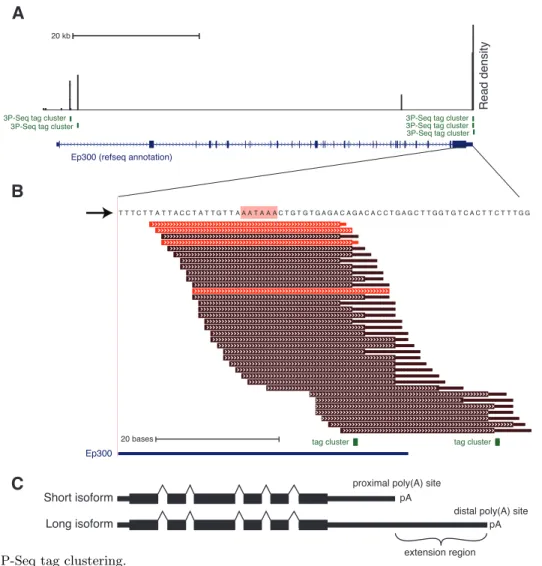
Recent surveys of expressed sequence tags (ESTs) and cDNA sequences available in
pub-lic databases gave a genome-wide view of the poly(A) signal sequences in mouse and human (Legendre and Gautheret 2003; Tian et al. 2005). Tian et al. (2005) identified 29,283 poly(A) sites in human and 31,179 poly(A) sites in mouse; of these, over 70% contained either the aforemen-tioned AAUAA motif, or the closely related AUUAAA hexamer. The other minor-frequency poly(A) sig-nal motifs were A-rich and all but one contained a third-position U. Cleavage and polyadenylation occurred on average at least 21bp downstream of this motif, although there was considerable heterogeneity in the exact cleavage site. The fre-quent presence of a U-rich downstream sequence element (DSE) 15–30bp downstream of the cleav-age site was confirmed, as well as a less common U-rich upstream sequence element (USE) 50 of the poly(A) site (Legendre and Gautheret 2003). A previously reported GU-rich DSE appears to be used infrequently (Cheng et al. 2006b).
Protein factors involved in poly(A) site recognition and cleavage and polyadenyla-tion Determination of an mRNA’s 30 end is a multi-step process. First, the region must be transcribed by pol II, revealing the RNA cis
mo-tifs involved in poly(A) site recognition. Sec-ond, the 30 end processing complex must be re-cruited to the nascent mRNA, where it cleaves the RNA. Finally, the upstream cleavage prod-uct – the mRNA – receives a poly(A) tail. The cleavage and polyadenylation process is mediated by a large protein complex, involving more than 14 core components in mammals (Mandel et al. 2008).
A number of protein subcomplexes are in-volved in poly(A) site recognition, including CPSF, CstF and cleavage factors CF Im and CF
IIm. CPSF is first to arrive, at transcription
initi-ation, recruited by the basal transcription factor TFIID to pol II (McCracken et al. 1997; Dan-tonel et al. 1997). Mammalian CPSF, short for cleavage and polyadenylation specificity factor, is composed of five subunits, which together bind to the AAUAAA (or similar) poly(A) signal once it is transcribed. The cleavage stimulation factor, or CstF, comprised of three subunits, binds the U-rich (or GU-rich) DSE downstream element, imparting additional specificity in poly(A) site selection (Takagaki and Manley 1997). CF Imalso
increases specificity for the complex by binding the USE upstream element (Mandel et al. 2008). Despite the detailed dissection of these protein factors, it wasn’t until very recently that it was directly shown that the actual endonuclease com-ponent is CPSF (Mandel et al. 2006; Takagaki and Manley 1997).
Also integral to the 30 end complex are the poly(A) polymerase PAP and the nuclear poly(A) binding protein PABPN. Although these two fac-tors are required for the cleavage activity, their main role is in creating the mRNA poly(A) fol-lowing endonucleolytic cleavage at the poly(A) site. In vitro, PAP is able to add a poly(A) tail to an RNA molecule, but the CPSF complex as well as PABPN are required to specify the correct length of the poly(A) tail (Mandel et al. 2006). The poly(A) polymerase PAP is converted from a non-processive to processive form by associa-tion with CPSF and the poly(A) binding protein PABPN. Association of PABPN to the nascent
Introduction 1.5. Post-transcriptional regulation of gene expression
poly(A) tail (positioned one PABPN every 11–14 nucleotides of poly(A) tail) enables the processive polyadenylation reaction only to a final length of 250–300 bp, thereby ensuring a uniform poly(A) tail length (K¨uhn et al. 2009).
Nearly all pol II transcripts are internally cleaved and polyadenylated by the canonical path-way discussed above. A major exception to this is the replication-dependent histone genes, in-cluding H2A, H2B, H3, H4 as well as the linker histone H1 (Marzlu↵ et al. 2008). During S phase, the cellular histone content must double, and this is achieved by a rapid induction of histone gene transcription, followed by sudden degradation of these transcripts at the end of S phase. This sud-den degradation is mediated through a stem-loop structure immediately downstream of the histone stop codon, which is recognized in the nucleus by the SLBP stem-loop binding complex (includ-ing some canonical cleavage and polyadenylation factors). The histone pre-mRNA is cleaved down-stream of the stem-loop, and the bound SLBP performs many of the functions of a canonical poly(A) tail, including protecting the message from degradation and enhancing efficient trans-lation (Marzlu↵ et al. 2008). The sudden degra-dation of histone mRNAs following S phase is mediated by this structure as well (Pandey and Marzlu↵ 1987).
Regulated cleavage and polyadenylation The number of potential poly(A) sites in the genome far exceeds the number of genes (Tian et al. 2005). At least some of these alternative poly(A) sites are used in a regulated fashion.
There are several types of alternative cleavage and polyadenylation. First, alternative last exons in-clude two poly(A) sites, one in an intron and one at the end of the longest isoform. A competition between splicing of the intron and cleavage and polyadenylation of the intronic poly(A) site deter-mines whether the internal or final poly(A) site is used. Second, tandem UTRs include at least two poly(A) sites within the final UTR region. In this case, competition between the poly(A) sites determines which gets used. Lastly, regulation of the poly(A) tail length is also known to occur.
The accuracy of the cleavage and polyadeny-lation is important, as exemplified by biological regulation and mis-regulation of 30 end forma-tion. Mutation of a sub-optimal early poly(A) signal to a higher efficiency form upregulates the prothrombin gene, leading to a hereditary high risk for thrombosis (Danckwardt et al. 2008). Use of proximal tandem 30 UTR isoforms is associ-ated with increased proliferation (Sandberg et al. 2008) and oncogenic transformation (Mayr and Bartel 2009). Finally, the U1A gene component of the U1 snRNP provides an example of regulated length of poly(A) tail formation. Outside of the context of the full U1 snRNP complex, the U1A protein can bind to its own 30UTR prior to cleav-age, where it represses the action of the poly(A) polymerase, resulting in a short poly(A) tail and lower expression of the U1A gene (Gunderson et al. 1994; Boelens et al. 1993). This is an elegant self-regulatory mechanism which allows U1A to downregulate its own expression when it is in excess over the other U1 snRNP components.
1.5
Post-transcriptional regulation of gene expression
Export Because a failure to correctly splice to-gether the coding exons could lead to aberrant translation of the intronic sequence, quality con-trol mechanisms exist to ensure that only spliced messages are efficiently translated. A critical bar-rier to translating mis-processed mRNAs is the nuclear membrane. There is evidence suggesting
involvement of each of the nuclear mRNA process-ing steps in regulatprocess-ing nucleo-cytoplasmic export: capping, splicing and cleavage and polyadenyla-tion.
The 7meG cap at the 50 end of mRNAs binds
to the aptly-named cap-binding complex in the nucleus, and this complex helps recruit export
machinery to the mRNA in a splicing-dependent manner (Cheng et al. 2006a). While unspliced messages, most notably the histone genes, are able to be exported, in general, splicing does en-hance mRNA export (Luo and Reed 1999; Valen-cia et al. 2008). Export factors are also recruited to the poly(A) tail, and it is likely that not only cleavage (to release the mRNA from chromatin) but also polyadenylation are required for most mRNAs to become fully export-competent. How-ever since capping, splicing and cleavage and polyadenylation are interdependent processes, it is difficult to tease out direct from indirect e↵ects on export (Bird et al. 2005).
As replication-dependent histones are not spliced nor cleaved as normal mRNAs, and don’t receive a poly(A) tail, their export is most likely regulated by export sequences within the mRNA (Erkmann et al. 2005).
Translation and translational control An mRNA is prepared for translation in the cyto-plasm by binding of a number of eukaryotic initi-ation factors (eIFs) to the 50 cap and the poly(A) tail. Current models suggest these eIFs bridge the 50 and 30 ends of the mRNA, circularizing the message (Sonenberg and Hinnebusch 2009). The 40S small ribosomal subunit is recruited to the 50 untranslated region, where it scans until it finds an AUG start codon. The 60S large riboso-mal subunit joins with the 40S subunit to form the initiation complex, which can begin translat-ing the mRNA into a polypeptide. Translation continues until a stop codon is reached, which is recognized by a eukaryotic release factor (Amrani et al. 2006).
Translation is a tightly regulated process, including via global mechanisms a↵ecting bulk translation. For example, translation is globally downregulated under stress conditions. Trans-lation of specific messages is also regulated, for example, by upstream open reading frames, or uORFs, which place a stop codon upstream of the true start codon, thereby inhibiting translation initiation of the main ORF.
Nonsense-mediated decay is another impor-tant example of interplay between gene regula-tory steps. Following splicing in the nucleus, a protein complex called the exon junction com-plex, or EJC, is placed about 20 bp upstream of the exon-exon junction. Once in the cytoplasm, EJCs are displaced by the ribosome during the pioneer round of translation, but if an error in splicing occurs, placing an in-frame stop codon far enough upstream of an EJC, the message will be recognized as aberrant and it will be degraded (Amrani et al. 2006). This mechanism is an exam-ple of how the translation machinery can correct for errors that occurred in the nucleus despite the physical separation of the processes.
Localization and compartmentalization of mRNAs The location of an mRNA within the cytoplasm can play an important regulatory role. Localization of specific messages is determined by binding of trans-factors to localization elements, typically found in the mRNA’s 30 UTR (Martin and Ephrussi 2009). For example, the -actin mRNA is targeted to sites of active actin poly-merization via binding of the zipcode binding protein ZBP1 to the -actin 30 UTR (Martin and Ephrussi 2009). ZBP1 enables the active translo-cation via binding to myosin motors, causing the mRNA to be pulled along the cytoskeleton to the leading edge of migrating cells (Oleynikov and Singer 2003).
In addition to targeted subcellular localiza-tion of individual messages, bulk mRNAs are delivered to two types of cytoplasmic foci, stress granules and P bodies. Stress granules compart-mentalize mRNAs trapped in translation initia-tion, and may lead to di↵erences in local concen-trations of the translation machinery within the cytoplasm (Buchan and Parker 2009). mRNAs targeted for degradation may become aggregated into a di↵erent structure, called a P body. Within the P body, a degradation complex-associated mRNA may be stored or actively degraded. Tar-geting to P bodies can be modulated by processes such as nonsense mediated decay or
stability-Introduction 1.5. Post-transcriptional regulation of gene expression
regulating factors such as AU-rich elements and microRNAs (see below) (Parker and Sheth 2007). It remains to be shown conclusively that target-ing of messages to stress granules or P bodies is causative of translation inhibition or degradation, rather than a reaction to these processes.
Death of an mRNA mRNA degradation
typ-ically begins by degradation of the poly(A) tail (Garneau et al. 2007). Two poly(A) nucleases are involved sequentially in the degradation of the poly(A) tail (Yamashita et al. 2005). First, the PAN2/3 nucleases are responsible for steadily shortening the poly(A) tail from its starting length of ⇠250 bp to around 110 bp. At this point, the CCR4 complex performs a sudden fur-ther shortening of the poly(A) tail to a point where the mRNA itself becomes destabilized and undergoes 30 ! 50degradation by the cytoplasmic exosome or decapping and 50 ! 30degradation by
Xrn1, or likely a combination of both (Garneau et al. 2007). As the poly(A) tail and 50 cap are required for efficient translation, deadenylation and decapping are associated with translational inhibition. In certain cases, degradation can be-gin by a targeted endonucleolytic cleavage event, such as mediated by an siRNA (see below), al-lowing the exonucleases to access the mRNA and degrade each cleavage fragment.
AU-Rich elements and other stability-regulating elements Because the translocat-ing ribosome is able to displace most RNA-binding trans factors from the open reading frame, the 30 untranslated region (30 UTR) plays an in-tegral role in regulation of cytoplasmic mRNAs.
AU-rich elements (AREs) are an important class of 30 UTR regulatory sequences. Approx-imately 20 di↵erent RNA-binding factors bind AREs, including AUF1/hnRNP D, the Hu fam-ily proteins, and tristetraprolin (or TTP). Some of these factors, such as AUF1, tend to destabi-lize ARE-containing mRNAs, while others, such as HuR, are thought to antagonize these desta-bilizing e↵ects through competitive binding to
the AREs (Barreau et al. 2005). While AREs are characterized by the occurrence of an AUUUA motif, this consensus sequence is not generally sufficient to change a message’s stability. Func-tional AREs are usually found within the context of a larger U or A/U rich region of the 30 UTR, but the degeneracy of these motifs makes it dif-ficult to predict from sequence alone functional sites (Barreau et al. 2005).
To identify global binding preferences for RNA-binding factors, a method called CLIP-Seq may be used. CLIP-Seq, short for cross-linking, immunoprecipitation and high-throughput se-quencing (the RNA-binding protein analogue to ChIP-Seq) uses UV light to directly cross-link protein to bound RNA, enabling stringent im-munoprecipitation conditions to isolate bound RNA (Licatalosi et al. 2008). CLIP-Seq has re-cently been used to elucidate the binding pat-terns of HuR (Mukherjee et al. 2011; Lebedeva et al. 2011), and the application of CLIP-Seq or other genome-wide approaches to additional ARE-binding proteins may soon provide a more complete understanding of the ARE motifs and interactions between the various factors binding them.
microRNAs and siRNAs microRNAs are a
class of ⇠22 nt long RNAs which mediate mRNA regulation, primarily through the 30 UTR. Mi-croRNAs, or miRNAs, are transcribed into a primary microRNA transcript by pol II. miRNAs fold back into a hairpin structure which is excised from the pri-miRNA by the nuclear RNase III endonuclease Drosha (Lee et al. 2003). The re-sulting pre-miRNA is exported to the cytoplasm where another RNase III enzyme, Dicer, cleaves the loop o↵ the pre-miRNA hairpin, liberating the two strands (Bartel 2004). One of the strands, the mature miRNA, is loaded into the Argonaute protein. There are four Argonautes in mammals, Ago1–Ago4 (Filipowicz et al. 2008).
The miRNA recruits the ago-containing si-lencing complex to an mRNA with complemen-tarity to the so-called miRNA seed sequence, the
2–8 most 50 nucleotides of the miRNA (Bartel 2009). This silencing complex, known as the RNA-induced silencing complex, or RISC, speeds degra-dation of targeted messages (Lim et al. 2005) and can also additionally reduce translation (Guo et al. 2010; Hendrickson et al. 2009; Selbach et al. 2008; Baek et al. 2008). The destabilizing e↵ect is generally mediated through enhanced deadeny-lation followed by decapping and degradation by an exonuclease (Fabian et al. 2010). However, like the intended targets of synthetic siRNAs, miRNA targets with near-perfect complementarity can be endonucleolytically cleaved by Ago2 (Filipowicz et al. 2008).
Importantly, efficacious microRNA target sites can be predicted accurately from the mRNA sequence alone. The accuracy of early prediction methods relied heavily on the level of conser-vation (Enright et al. 2003; Stark et al. 2003) or conservation above background (Lewis et al. 2003) of sequences complementary to the miRNA seed sequence. While hexamers complementary to nucleotides 2–7 of the microRNA show a small conservation signal and are slightly e↵ective in
downregulating the host mRNAs, a full 7mer match to nucleotides 2–8 is much more e↵ective. In mammals, rather than direct sequence com-plementarity to the first base of the microRNA, an A opposite this position further improves mi-croRNA targeting (Lewis et al. 2005). A number of additional features, such as being in an A/U rich region, improve the prediction of microRNA target sites such that they may be identified and ranked according to efficacy without the need for conservation (Nielsen et al. 2007; Grimson et al. 2007).
Small interfering RNAs, or siRNAs, are a class of short regulatory RNAs related to microRNAs. siRNAs are distinguished by their biogenesis from dsRNA precursors, which are cleaved directly by Dicer (rather than from hairpins which require Drosha cleavage first) and loading into RISC. Once in RISC, siRNAs and miRNAs are function-ally equivalent, though because of their origins, siRNAs tend to show extensive complementar-ity to their targets, leading to endonucleolytic cleavage (Bartel 2004).
1.6
RNA-induced transcriptional silencing in fission yeast
An interesting merging of regulatory mecha-nisms occurs at the centromeres of the fission yeast Schizosaccharomyces pombe, where the RNA-interference machinery is used to recruit chromatin-modifying enzymes, leading to hete-rochromatinization of the centromeric DNA (Cam et al. 2009; Moazed 2009). Fission yeast is an ex-cellent model organism for studying the e↵ects of RNAi as each enzyme in that pathway is found in single copy in the genome, enabling simple ge-netic manipulation. Each of the aforementioned proteins is non-essential for viability, but RNAi mutants are unable to form heterochromatin at the centromeres and exhibit chromosome segre-gation defects (Volpe et al. 2003).
The process of silencing centromeres begins with transcription of the dg and dh repeats in the outer regions of each centromere (Cam et al. 2009;
Moazed 2009). These repeat transcripts recruit the RNA-induced transcriptional silencing com-plex known as RITS. RITS includes the fission yeast homologs of the canonical RNA interfer-ence pathway, including Argonaute and Dicer, as well as the RNA-dependent RNA polymerase, an RNA-binding non-canonical poly(A) polymerase and several chromatin-modifying enzymes includ-ing most notably an H3K9 methyltransferase. The Dicer enzyme processes duplex repeat RNA into siRNAs that are loaded into Argonaute. The siRNA-containing RITS complex is then recruited to nascent transcripts as they are produced co-transcriptionally, bringing the H3K9 methyltrans-ferase into proximity of the chromatin. Spread-ing of H3K9me along the DNA establishes hete-rochromatin throughout the centromere, up until boundary elements demarcated by tRNA genes
Introduction 1.7. References
or other unique sequences (Cam et al. 2005). An apparent paradox is how transcription of a silenced, heterochromatic region can be respon-sible for establishing the heterochromatin itself. Recent work showed that transcription is tightly regulated by the cell cycle (Gullerova and Proud-foot 2008; Kloc et al. 2008; Chen et al. 2008). During S phase, pol II is permitted access to
transcribe the dg and dh repeats, presumably loading the RITS complex with ample siRNAs for an entire cell cycle including an extended G2 phase. However, what event actually prompts the centromeres to be recognized for heterochro-matinization is still unclear (Lejeune and Allshire 2011).
1.7
References
Amrani, N, MS Sachs, and A Jacobson (June 2006). “Early nonsense: mRNA decay solves a translational problem.” In: Nat Rev Mol Cell Biol 7.6, pp. 415–25. doi: 10.1038/nrm1942 (cit. on p. 18).
Baek, D, J Vill´en, C Shin, FD Camargo, SP Gygi, and DP Bartel (Sept. 2008). “The impact of microRNAs on protein output.” In: Nature 455.7209, pp. 64–71. doi: 10.1038/nature07242 (cit. on p. 20).
Barash, Y, JA Calarco, W Gao, Q Pan, X Wang, O Shai, BJ Blencowe, and BJ Frey (May 2010). “Deciphering the splicing code.” In: Nature 465.7294, pp. 53–9. doi: 10.1038/nature09000
(cit. on p. 15).
Barreau, C, L Paillard, and HB Osborne (2005). “AU-rich elements and associated factors: are there unifying principles?” In: Nucleic Acids Res 33.22, pp. 7138–50. doi: 10.1093/nar/gki1012 (cit. on p. 19).
Barski, A, S Cuddapah, K Cui, TY Roh, DE Schones, Z Wang, G Wei, I Chepelev, and K Zhao (May 2007). “High-resolution profiling of histone methylations in the human genome.” In: Cell
129.4, pp. 823–37. doi: 10.1016/j.cell.2007.05.009 (cit. on p. 10).
Bartel, DP (Jan. 2004). “MicroRNAs: genomics, biogenesis, mechanism, and function.” In: Cell 116.2, pp. 281–97 (cit. on pp. 19, 20).
Bartel, DP (Jan. 2009). “MicroRNAs: target recognition and regulatory functions.” In: Cell 136.2, pp. 215–33. doi: 10.1016/j.cell.2009.01.002 (cit. on p. 20).
Berget, SM, C Moore, and PA Sharp (Aug. 1977). “Spliced segments at the 50 terminus of adenovirus 2 late mRNA.” In: Proc Natl Acad Sci U S A 74.8, pp. 3171–5 (cit. on p. 14).
Bird, G, N Fong, JC Gatlin, S Farabaugh, and DL Bentley (Dec. 2005). “Ribozyme cleavage reveals connections between mRNA release from the site of transcription and pre-mRNA processing.” In: Mol Cell 20.5, pp. 747–58. doi: 10.1016/j.molcel.2005.11.009 (cit. on p. 18).
Blat, Y and N Kleckner (July 1999). “Cohesins bind to preferential sites along yeast chromosome III, with di↵erential regulation along arms versus the centric region.” In: Cell 98.2, pp. 249–59 (cit. on p. 10).
Boelens, WC, EJ Jansen, WJ van Venrooij, R Stripecke, IW Mattaj, and SI Gunderson (Mar. 1993). “The human U1 snRNP-specific U1A protein inhibits polyadenylation of its own pre-mRNA.” In:
Cell 72.6, pp. 881–92 (cit. on p. 17).
Bondarenko, VA, LM Steele, A Ujv´ari, DA Gaykalova, OI Kulaeva, YS Polikanov, DS Luse, and VM Studitsky (Nov. 2006). “Nucleosomes can form a polar barrier to transcript elongation by RNA polymerase II.” In: Mol Cell 24.3, pp. 469–79. doi: 10.1016/j.molcel.2006.09.009 (cit. on p. 12).
Brownell, JE, J Zhou, T Ranalli, R Kobayashi, DG Edmondson, SY Roth, and CD Allis (Mar. 1996). “Tetrahymena histone acetyltransferase A: a homolog to yeast Gcn5p linking histone acetylation to gene activation.” In: Cell 84.6, pp. 843–51 (cit. on p. 10).
Buchan, JR and R Parker (Dec. 2009). “Eukaryotic stress granules: the ins and outs of translation.” In: Mol Cell 36.6, pp. 932–41. doi: 10.1016/j.molcel.2009.11.020 (cit. on p. 18).
Cam, HP, T Sugiyama, ES Chen, X Chen, PC FitzGerald, and SIS Grewal (Aug. 2005). “Compre-hensive analysis of heterochromatin- and RNAi-mediated epigenetic control of the fission yeast genome.” In: Nat Genet 37.8, pp. 809–19. doi: 10.1038/ng1602 (cit. on p. 21).
Cam, HP, ES Chen, and SIS Grewal (Feb. 2009). “Transcriptional sca↵olds for heterochromatin assembly.” In: Cell 136.4, pp. 610–4. doi: 10.1016/j.cell.2009.02.004 (cit. on p. 20). Chen, ES, K Zhang, E Nicolas, HP Cam, M Zofall, and SIS Grewal (Feb. 2008). “Cell cycle control of
centromeric repeat transcription and heterochromatin assembly.” In: Nature 451.7179, pp. 734–7. doi: 10.1038/nature06561 (cit. on p. 21).
Cheng, H, K Dufu, CS Lee, JL Hsu, A Dias, and R Reed (Dec. 2006a). “Human mRNA export machinery recruited to the 5’ end of mRNA.” In: Cell 127.7, pp. 1389–400. doi: 10.1016/j. cell.2006.10.044 (cit. on p. 18).
Cheng, Y, RM Miura, and B Tian (Oct. 2006b). “Prediction of mRNA polyadenylation sites by sup-port vector machine.” eng. In: Bioinformatics 22.19, pp. 2320–5. doi: 10.1093/bioinformatics/ btl394(cit. on p. 16).
Chow, LT, RE Gelinas, TR Broker, and RJ Roberts (Sept. 1977). “An amazing sequence arrangement at the 50 ends of adenovirus 2 messenger RNA.” In: Cell 12.1, pp. 1–8 (cit. on p. 14).
Danckwardt, S, MW Hentze, and AE Kulozik (Feb. 2008). “3’ end mRNA processing: molecular mechanisms and implications for health and disease.” In: EMBO J 27.3, pp. 482–98. doi: 10.1038/sj.emboj.7601932 (cit. on p. 17).
Dantonel, JC, KG Murthy, JL Manley, and L Tora (Sept. 1997). “Transcription factor TFIID recruits factor CPSF for formation of 30 end of mRNA.” In: Nature 389.6649, pp. 399–402. doi: 10.1038/38763(cit. on p. 16).
Darnell, JE, L Philipson, R Wall, and M Adesnik (Oct. 1971). “Polyadenylic acid sequences: role in conversion of nuclear RNA into messenger RNA.” eng. In: Science 174.8, pp. 507–10 (cit. on p. 15).
Darnell, JE, WR Jelinek, and GR Molloy (Sept. 1973). “Biogenesis of mRNA: genetic regulation in mammalian cells.” eng. In: Science 181.106. review covers early poly(A) developments, pp. 1215– 21 (cit. on p. 15).
Introduction 1.7. References
Das, R, K Dufu, B Romney, M Feldt, M Elenko, and R Reed (May 2006). “Functional coupling of RNAP II transcription to spliceosome assembly.” In: Genes Dev 20.9, pp. 1100–9. doi: 10.1101/gad.1397406 (cit. on p. 12).
Das, R, J Yu, Z Zhang, MP Gygi, AR Krainer, SP Gygi, and R Reed (June 2007). “SR proteins function in coupling RNAP II transcription to pre-mRNA splicing.” In: Mol Cell 26.6, pp. 867–81. doi: 10.1016/j.molcel.2007.05.036 (cit. on p. 12).
de la Mata, M, CR Alonso, S Kadener, JP Fededa, M Blaustein, F Pelisch, P Cramer, D Bentley, and AR Kornblihtt (Aug. 2003). “A slow RNA polymerase II a↵ects alternative splicing in vivo.” In: Mol Cell 12.2, pp. 525–32 (cit. on p. 13).
Desrosiers, R, K Friderici, and F Rottman (Oct. 1974). “Identification of methylated nucleosides in messenger RNA from Noviko↵ hepatoma cells.” In: Proc Natl Acad Sci U S A 71.10, pp. 3971–5 (cit. on p. 14).
Edmonds, M and MG Caramela (Mar. 1969). “The isolation and characterization of adenosine monophosphate-rich polynucleotides synthesized by Ehrlich ascites cells.” eng. In: J Biol Chem 244.5, pp. 1314–24 (cit. on p. 15).
Edmonds, M, MH Vaughan, and H Nakazato (June 1971). “Polyadenylic acid sequences in the heterogeneous nuclear RNA and rapidly-labeled polyribosomal RNA of HeLa cells: possible evidence for a precursor relationship.” eng. In: Proc Natl Acad Sci USA 68.6, pp. 1336–40 (cit. on p. 15).
Enright, AJ, B John, U Gaul, T Tuschl, C Sander, and DS Marks (2003). “MicroRNA targets in Drosophila.” In: Genome Biol 5.1, R1. doi: 10.1186/gb-2003-5-1-r1 (cit. on p. 20).
Erkmann, JA, R S`anchez, N Treichel, WF Marzlu↵, and U Kutay (Jan. 2005). “Nuclear export of metazoan replication-dependent histone mRNAs is dependent on RNA length and is mediated by TAP.” In: RNA 11.1, pp. 45–58. doi: 10.1261/rna.7189205 (cit. on p. 18).
Fabian, MR, N Sonenberg, and W Filipowicz (2010). “Regulation of mRNA translation and stability by microRNAs.” In: Annu Rev Biochem 79, pp. 351–79. doi: 10.1146/annurev- biochem-060308-103103 (cit. on p. 20).
Filipowicz, W, SN Bhattacharyya, and N Sonenberg (Feb. 2008). “Mechanisms of post-transcriptional regulation by microRNAs: are the answers in sight?” In: Nat Rev Genet 9.2, pp. 102–14. doi: 10.1038/nrg2290(cit. on pp. 19, 20).
Fitzgerald, M and T Shenk (Apr. 1981). “The sequence 50-AAUAAA-30forms parts of the recognition site for polyadenylation of late SV40 mRNAs.” In: Cell 24.1, pp. 251–60 (cit. on p. 16). Flemming, W (1882). Zellsubstanz, Kern und Zelltheilung. Verlag von FCW Vogel (cit. on p. 10). Ford, JP and MT Hsu (Dec. 1978). “Transcription pattern of in vivo-labeled late simian virus 40
RNA: equimolar transcription beyond the mRNA 30 terminus.” eng. In: J Virol 28.3, pp. 795–801 (cit. on pp. 13, 15).
Garneau, NL, J Wilusz, and CJ Wilusz (Feb. 2007). “The highways and byways of mRNA decay.” In: Nat Rev Mol Cell Biol 8.2, pp. 113–26. doi: 10.1038/nrm2104 (cit. on p. 19).
Grimson, A, KKH Farh, WK Johnston, P Garrett-Engele, LP Lim, and DP Bartel (July 2007). “MicroRNA targeting specificity in mammals: determinants beyond seed pairing.” In: Mol Cell
27.1, pp. 91–105. doi: 10.1016/j.molcel.2007.06.017 (cit. on p. 20).
Gullerova, M and NJ Proudfoot (Mar. 2008). “Cohesin complex promotes transcriptional termination between convergent genes in S. pombe.” In: Cell 132.6, pp. 983–95. doi: 10.1016/j.cell.2008. 02.040(cit. on p. 21).
Gunderson, SI, K Beyer, G Martin, W Keller, WC Boelens, and LW Mattaj (Feb. 1994). “The human U1A snRNP protein regulates polyadenylation via a direct interaction with poly(A) polymerase.” In: Cell 76.3, pp. 531–41 (cit. on p. 17).
Guo, H, NT Ingolia, JS Weissman, and DP Bartel (Aug. 2010). “Mammalian microRNAs predomi-nantly act to decrease target mRNA levels.” In: Nature 466.7308, pp. 835–40. doi: 10.1038/ nature09267(cit. on p. 20).
Hendrickson, DG, DJ Hogan, HL McCullough, JW Myers, D Herschlag, JE Ferrell, and PO Brown (Nov. 2009). “Concordant regulation of translation and mRNA abundance for hundreds of targets of a human microRNA.” In: PLoS Biol 7.11, e1000238. doi: 10.1371/journal.pbio.1000238 (cit. on p. 20).
Heniko↵, S and A Shilatifard (July 2011). “Histone modification: cause or cog?” In: Trends Genet. doi: 10.1016/j.tig.2011.06.006 (cit. on p. 11).
Ip, JY, D Schmidt, Q Pan, AK Ramani, AG Fraser, DT Odom, and BJ Blencowe (Mar. 2011). “Global impact of RNA polymerase II elongation inhibition on alternative splicing regulation.”
In: Genome Res 21.3, pp. 390–401. doi: 10.1101/gr.111070.110 (cit. on p. 13).
Izaurralde, E, J Lewis, C McGuigan, M Jankowska, E Darzynkiewicz, and IW Mattaj (Aug. 1994). “A nuclear cap binding protein complex involved in pre-mRNA splicing.” In: Cell 78.4, pp. 657–68
(cit. on p. 14).
Johnson, DS, A Mortazavi, RM Myers, and B Wold (June 2007). “Genome-wide mapping of in vivo protein-DNA interactions.” In: Science 316.5830, pp. 1497–502. doi: 10.1126/science.1141319 (cit. on p. 10).
Kaplan, N, IK Moore, Y Fondufe-Mittendorf, AJ Gossett, D Tillo, Y Field, EM LeProust, TR Hughes, JD Lieb, J Widom, and E Segal (Mar. 2009). “The DNA-encoded nucleosome organization of a eukaryotic genome.” In: Nature 458.7236, pp. 362–6. doi: 10.1038/nature07667 (cit. on p. 11). Kim, M, NJ Krogan, L Vasiljeva, OJ Rando, E Nedea, JF Greenblatt, and S Buratowski (Nov. 2004). “The yeast Rat1 exonuclease promotes transcription termination by RNA polymerase II.” In: Nature 432.7016, pp. 517–22. doi: 10.1038/nature03041 (cit. on p. 13).
Kloc, A, M Zaratiegui, E Nora, and R Martienssen (Apr. 2008). “RNA interference guides histone modification during the S phase of chromosomal replication.” In: Curr Biol 18.7, pp. 490–5. doi: 10.1016/j.cub.2008.03.016(cit. on p. 21).
Konarska, MM, RA Padgett, and PA Sharp (Oct. 1984). “Recognition of cap structure in splicing in vitro of mRNA precursors.” In: Cell 38.3, pp. 731–6 (cit. on p. 14).
Kornberg, RD (May 1974). “Chromatin structure: a repeating unit of histones and DNA.” In: Science 184.139, pp. 868–71 (cit. on p. 10).
Introduction 1.7. References
K¨uhn, U, M G¨undel, A Knoth, Y Kerwitz, S R¨udel, and E Wahle (Aug. 2009). “Poly(A) tail length is controlled by the nuclear poly(A)-binding protein regulating the interaction between poly(A) polymerase and the cleavage and polyadenylation specificity factor.” In: J Biol Chem 284.34, pp. 22803–14. doi: 10.1074/jbc.M109.018226 (cit. on p. 17).
Lebedeva, S, M Jens, K Theil, B Schwanh¨ausser, M Selbach, M Landthaler, and N Rajewsky (Aug. 2011). “Transcriptome-wide Analysis of Regulatory Interactions of the RNA-Binding Protein HuR.” In: Mol Cell 43.3, pp. 340–52. doi: 10.1016/j.molcel.2011.06.008 (cit. on p. 19). Lee, TI and RA Young (2000). “Transcription of eukaryotic protein-coding genes.” In: Annu Rev
Genet 34, pp. 77–137. doi: 10.1146/annurev.genet.34.1.77 (cit. on p. 12).
Lee, Y, C Ahn, J Han, H Choi, J Kim, J Yim, J Lee, P Provost, O R˚admark, S Kim, and VN Kim (Sept. 2003). “The nuclear RNase III Drosha initiates microRNA processing.” In: Nature 425.6956, pp. 415–9. doi: 10.1038/nature01957 (cit. on p. 19).
Legendre, M and D Gautheret (Feb. 2003). “Sequence determinants in human polyadenylation site selection.” In: BMC Genomics 4.1, p. 7 (cit. on p. 16).
Lejeune, E and RC Allshire (June 2011). “Common ground: small RNA programming and chromatin modifications.” In: Curr Opin Cell Biol 23.3, pp. 258–65. doi: 10.1016/j.ceb.2011.03.005 (cit. on p. 21).
Lerner, MR, JA Boyle, SM Mount, SL Wolin, and JA Steitz (Jan. 1980). “Are snRNPs involved in splicing?” In: Nature 283.5743, pp. 220–4 (cit. on p. 14).
Lewis, BP, Ih Shih, MW Jones-Rhoades, DP Bartel, and CB Burge (Dec. 2003). “Prediction of mammalian microRNA targets.” In: Cell 115.7, pp. 787–98 (cit. on p. 20).
Lewis, BP, CB Burge, and DP Bartel (Jan. 2005). “Conserved seed pairing, often flanked by adenosines, indicates that thousands of human genes are microRNA targets.” In: Cell 120.1, pp. 15–20. doi: 10.1016/j.cell.2004.12.035 (cit. on p. 20).
Licatalosi, DD, A Mele, JJ Fak, J Ule, M Kayikci, SW Chi, TA Clark, AC Schweitzer, JE Blume, X Wang, JC Darnell, and RB Darnell (Nov. 2008). “HITS-CLIP yields genome-wide insights into brain alternative RNA processing.” In: Nature 456.7221, pp. 464–9. doi: 10.1038/nature07488 (cit. on p. 19).
Lim, LP, NC Lau, P Garrett-Engele, A Grimson, JM Schelter, J Castle, DP Bartel, PS Linsley, and JM Johnson (Feb. 2005). “Microarray analysis shows that some microRNAs downregulate large numbers of target mRNAs.” In: Nature 433.7027, pp. 769–73. doi: 10.1038/nature03315 (cit. on p. 20).
Luger, K, AW M¨ader, RK Richmond, DF Sargent, and TJ Richmond (Sept. 1997). “Crystal structure of the nucleosome core particle at 2.8 A resolution.” In: Nature 389.6648, pp. 251–60. doi: 10.1038/38444 (cit. on pp. 10, 11).
Luo, MJ and R Reed (Dec. 1999). “Splicing is required for rapid and efficient mRNA export in metazoans.” In: Proc Natl Acad Sci U S A 96.26, pp. 14937–42 (cit. on p. 18).
Luo, W, AW Johnson, and DL Bentley (Apr. 2006). “The role of Rat1 in coupling mRNA 30-end processing to transcription termination: implications for a unified allosteric-torpedo model.” In: Genes Dev 20.8, pp. 954–65. doi: 10.1101/gad.1409106 (cit. on p. 13).
Mandel, CR, S Kaneko, H Zhang, D Gebauer, V Vethantham, JL Manley, and L Tong (Dec. 2006). “Polyadenylation factor CPSF-73 is the pre-mRNA 30-end-processing endonuclease.” In: Nature
444.7121, pp. 953–6. doi: 10.1038/nature05363 (cit. on p. 16).
Mandel, CR, Y Bai, and L Tong (Apr. 2008). “Protein factors in pre-mRNA 30-end processing.” In: Cell Mol Life Sci 65.7-8, pp. 1099–122. doi: 10.1007/s00018-007-7474-3 (cit. on p. 16). Mapendano, CK, S Lykke-Andersen, J Kjems, E Bertrand, and TH Jensen (Nov. 2010). “Crosstalk
between mRNA 30 end processing and transcription initiation.” In: Mol Cell 40.3, pp. 410–22. doi: 10.1016/j.molcel.2010.10.012 (cit. on p. 13).
Martin, KC and A Ephrussi (Feb. 2009). “mRNA localization: gene expression in the spatial dimension.” In: Cell 136.4, pp. 719–30. doi: 10.1016/j.cell.2009.01.044 (cit. on p. 18). Marzlu↵, WF, EJ Wagner, and RJ Duronio (Nov. 2008). “Metabolism and regulation of canonical
histone mRNAs: life without a poly(A) tail.” In: Nat Rev Genet 9.11, pp. 843–54. doi: 10.1038/ nrg2438 (cit. on p. 17).
Matlin, AJ, F Clark, and CWJ Smith (May 2005). “Understanding alternative splicing: towards a cellular code.” In: Nat Rev Mol Cell Biol 6.5, pp. 386–98. doi: 10.1038/nrm1645 (cit. on p. 14). Mayr, C and DP Bartel (Aug. 2009). “Widespread shortening of 30UTRs by alternative cleavage
and polyadenylation activates oncogenes in cancer cells.” In: Cell 138.4, pp. 673–84. doi: 10.1016/j.cell.2009.06.016 (cit. on p. 17).
McCracken, S, N Fong, K Yankulov, S Ballantyne, G Pan, J Greenblatt, SD Patterson, M Wickens, and DL Bentley (Jan. 1997). “The C-terminal domain of RNA polymerase II couples mRNA processing to transcription.” In: Nature 385.6614, pp. 357–61. doi: 10.1038/385357a0 (cit. on p. 16).
McDevitt, MA, RP Hart, WW Wong, and JR Nevins (Nov. 1986). “Sequences capable of restoring poly(A) site function define two distinct downstream elements.” In: EMBO J 5.11, pp. 2907–13 (cit. on p. 16).
Moazed, D (Jan. 2009). “Small RNAs in transcriptional gene silencing and genome defence.” In: Nature 457.7228, pp. 413–20. doi: 10.1038/nature07756 (cit. on p. 20).
Molloy, GR, MB Sporn, DE Kelley, and RP Perry (Aug. 1972). “Localization of polyadenylic acid sequences in messenger ribonucleic acid of mammalian cells.” eng. In: Biochemistry 11.17, pp. 3256–60 (cit. on p. 15).
Moore, MJ and NJ Proudfoot (Feb. 2009). “Pre-mRNA processing reaches back to transcription and ahead to translation.” In: Cell 136.4, pp. 688–700. doi: 10.1016/j.cell.2009.02.001 (cit. on p. 12).
Mukherjee, N, DL Corcoran, JD Nusbaum, DW Reid, S Georgiev, M Hafner, M Ascano Jr, T Tuschl, U Ohler, and JD Keene (Aug. 2011). “Integrative Regulatory Mapping Indicates that the RNA-Binding Protein HuR Couples Pre-mRNA Processing and mRNA Stability.” In: Mol Cell 43.3, pp. 327–39. doi: 10.1016/j.molcel.2011.06.007 (cit. on p. 19).
Muthukrishnan, S, GW Both, Y Furuichi, and AJ Shatkin (May 1975). “50-Terminal 7-methylguanosine in eukaryotic mRNA is required for translation.” In: Nature 255.5503, pp. 33–7 (cit. on p. 14).