Analysis of Peripapillary Atrophy Using Spectral
Domain Optical Coherence Tomography
The MIT Faculty has made this article openly available.
Please share
how this access benefits you. Your story matters.
Citation
Manjunath, Varsha, Heeral Shah, James G. Fujimoto, and Jay S.
Duker. “Analysis of Peripapillary Atrophy Using Spectral Domain
Optical Coherence Tomography.” Ophthalmology 118, no. 3 (March
2011): 531–536.
As Published
http://dx.doi.org/10.1016/j.ophtha.2010.07.013
Publisher
Elsevier
Version
Author's final manuscript
Citable link
http://hdl.handle.net/1721.1/98863
Terms of Use
Creative Commons Attribution-Noncommercial-NoDerivatives
Analysis of Peripapillary Atrophy using Spectral Domain Optical
Coherence Tomography
Varsha Manjunath, BS1, Heeral Shah, MD1, James G. Fujimoto, PhD2, and Jay S. Duker, MD1
1 New England Eye Center, Tufts Medical Center, Boston, MA
2 Dept of Electrical Engineering and Computer Science, Research Laboratory of Electronics,
Massachusetts Institute of Technology, Cambridge, MA
Abstract
Objective—To study retinal morphological changes around the optic disc in patients with
peripapillary atrophy (PPA) with high-resolution spectral domain optical coherence tomography (SD OCT).
Design—Cross-sectional, retrospective analysis
Participants—One hundred and three eyes of 73 patients with PPA and 21 eyes of 12 normal
patients seen at the New England Eye Center, Tufts Medical Center between January 2007 and August 2009.
Methods—SD OCT images taken through the region of PPA were quantitatively and
qualitatively analyzed. Inclusion criteria included eyes with at least 300 μm of temporal PPA as detected on color fundus photographs. The study population was divided into subgroups according to the following clinical diagnoses: glaucoma (n=13), age-related macular degeneration (n=11), high myopia (n=11), glaucoma and high myopia (n=3), and optic neuropathy (n=11). Fifty-four patients were classified with other diagnoses. Using OCT software, retinal thickness and retinal nerve fiber layer thickness (RNFL) were both manually measured perpendicular to the internal limiting membrane and retinal pigment epithelium (RPE) 300 μm temporal to the optic disc, within the region of peripapillary atrophy. Qualitative analysis for morphological changes in the atrophic area was also performed.
Main outcome measures—Qualitative assessment and quantitative measures of retinal and
retinal nerve fiber layer thickness in PPA.
Results—The study group was categorized by 6 characteristics demonstrated in the area of PPA
by SD OCT: RPE loss with accompanying photoreceptor loss, RPE disruption, RNFL thickening with plaque-like formation, intraretinal cystic changes, inner and outer retinal thinning, and abnormal retinal sloping. Statistical analysis of measurements revealed a statistically significant difference in the total retinal thickness between normal eyes and eyes with PPA (p = 0.0005), with
Corresponding Author/Reprints Jay S. Duker, MD, Department of Ophthalmology, Chairman, Tufts Medical Center, 800 Washington St., Box #450, Boston, MA 02111, Tel: 617-636-4677, Fax: 617-636-4866, JDuker@tuftsmedicalcenter.org.
Conflict of Interest
Authors with financial/conflicting interests are listed after references.
Publisher's Disclaimer: This is a PDF file of an unedited manuscript that has been accepted for publication. As a service to our customers we are providing this early version of the manuscript. The manuscript will undergo copyediting, typesetting, and review of
NIH Public Access
Author Manuscript
Ophthalmology. Author manuscript; available in PMC 2012 March 1.
Published in final edited form as:
Ophthalmology. 2011 March ; 118(3): 531–536. doi:10.1016/j.ophtha.2010.07.013.
NIH-PA Author Manuscript
NIH-PA Author Manuscript
normals 15% thicker than the PPA group; however, the RNFL thickness was not significantly different between the normal and PPA group (p = 0.05).
Conclusion—Eyes with peripapillary atrophy manifest characteristic retinal changes that can be
described via SD OCT.
Peripapillary atrophy (PPA) is a clinical finding associated with chorioretinal thinning and disruption of the retinal pigment epithelium (RPE) in the area surrounding the optic disc. It is non-specific and can occur in both benign and pathologic conditions, including glaucoma
1 and high myopia 2.
The normal cross-sectional anatomic structure of the peripapillary region of the eye is arranged with all the retinal cell layers organized in a parallel fashion, ending at the edge of the optic disc, with the topmost retinal nerve fiber layer increasing in thickness before diving into the disc. This normal architecture is disrupted with the development of peripapillary atrophy. Classification of peripapillary atrophy into two zones was first proposed by Jonas,
et al 3: zone beta, a central zone of chorioretinal atrophy with visible large choroidal vessels
and sclera, and zone alpha, a more peripheral area of irregular hypo- and
hyper-pigmentation, with both types often present side by side in the same eye 3. Histological
investigations of peripapillary atrophy describe changes in beta zone atrophy as a complete loss of retinal pigment epithelium cells and photoreceptors, and thinning of the choroid in combination with an almost complete collapse of the choriocapillaris. Alpha zone atrophy has been described as having structural irregularities in the retinal pigment epithelium cells
3–7.
Optical coherence tomography (OCT) can perform noninvasive, real time imaging of the
retina and anterior eye 8–10. OCT is a technology that provides high-resolution
measurements and cross-sectional imaging of the retina and retinal nerve fiber layer (RNFL). The technology is analogous to ultrasound B-mode imaging, measuring the echo time delay of back-reflected light rather than sound from structures at different longitudinal
distances8–10. Successive axial scans at different transverse points are used to generate
cross-sectional images. The axial resolution in OCT imaging is inversely proportional to the bandwidth of the light source. Spectral domain (SD) OCT devices utilize an interferometer with a spectrometer and a high speed camera to measure echoes of light in the Fourier domain. Standard commercial OCT instruments use a superluminescent diode light source at a near infrared wavelength of 820 nm with a bandwidth of 50 nm, achieving 5–8 μm axial resolution in tissue.
Usually a nonspecific finding, PPA has also been closely correlated with glaucoma and high myopia. However, the way that the peripapillary region is affected in these diseases is not well understood. Closer examination of PPA in vivo using SD OCT was of interest to gain a better understanding of the changes occurring in this region. Although the clinical and histological findings in peripapillary atrophy have been described, the OCT correlate of peripapillary atrophy has not been assessed in detail. Therefore, the purpose of this study is to examine OCT changes in the region of peripapillary atrophy.
Patients and Methods
A retrospective review of 103 eyes (73 patients) with peripapillary atrophy was undertaken. Each patient underwent complete ophthalmic examination and Cirrus HD-OCT (software version 4.0, Carl Zeiss Meditec, Inc., Dublin, CA, USA) imaging in the ophthalmology department of the New England Eye Center, Tufts Medical Center, Boston, Massachusetts between January 2007 and August 2009. Examination included best corrected visual acuity, slit-lamp examination, fundus biomicroscopy, fundus photography, and OCT examination.
NIH-PA Author Manuscript
NIH-PA Author Manuscript
This study protocol was approved by the Institutional Review Board (IRB)/Ethics Committee of the Tufts Medical Center and is adherent to the tenets of the Declaration of Helsinki.
Inclusion for the study required the presence at least 300 μm of clinically apparent temporal peripapillary atrophy visible on fundus imaging. Both eyes were included if they met the criteria for atrophy. Five line raster scans at a ± 7 degree angle from the horizontal through the center of the fovea and the optic nerve were analyzed. A 5-line raster scan consists of 5 horizontal lines, each 6 mm long and spaced 0.25 mm apart. Each high definition line is comprised of 4096 A-scans, with a transverse resolution of ~15–20 μm. The raster scan closest to the fovea was selected for measurements. Only good quality images with an intensity of 6 out of 10 or greater were used in this study.
Cirrus HD-OCT software provides a measurement tool to draw straight lines. One observer (VM) manually measured two parameters on the raster images: (1) total retinal thickness, which was measured from the internal limiting membrane to Bruch membrane, and (2) RNFL thickness which was also measured perpendicular to the retinal pigment epithelium. All measurements were performed 300 μm temporal to the edge of the RPE, which this
study defined as the edge of the optic disc.11 Bruch’s membrane may be seen extending
further than the RPE in histological studies12, however SD OCT does not allow for that level
of cellular resolution. Automated RNFL thickness measurements generated along a standard 3.4 mm circle centered on the optic disc were not utilized in this study as the measured area of thickness would have been beyond the region of PPA in most cases.
In addition to linear thickness measurements, angle measurements were performed on the OCT images to measure the angle between the RPE and the vertical edge of the disc in both normal eyes and those with PPA. Cirrus software does not provide a tool for angle
measurements, therefore the images were exported to publicly available research analysis software ImageJ (National Institutes of Health, USA, http://rsbweb.nih.gov/ij/, accessed December 20, 2009). The vertical to horizontal aspect ratio is distorted in OCT images to facilitate better visualization of retinal details and measurements of distance or angle must account for this. Accordingly, before angles were measured using ImageJ software, the scale was set by measuring a horizontal line of a known length, the pixel aspect ratio was altered to 1/3 to account for the difference in horizontal to vertical ratio, and the units were changed to micrometers. All angle measurements were verified for accuracy by drawing a right angle triangle using the Cirrus line tool and calculating the angle using trigonometry. All
measurements were also performed on 21 eyes of 12 normal patients lacking PPA for comparison.
The images were analyzed for morphological changes in the retina in the area of peripapillary atrophy. When the area of atrophy on the Cirrus generated en face fundus image was measured using Cirrus software, the same distance could be measured on the corresponding OCT image, confirming that the area of observed morphological changes were in the same location on both the fundus image and OCT scan.
Retinal thickness and retinal nerve fiber layer thickness measurements were compared between the normal and PPA groups using the Student t-test with statistical significance defined as a P value of less than 0.05.
NIH-PA Author Manuscript
NIH-PA Author Manuscript
Results
Baseline Characteristics
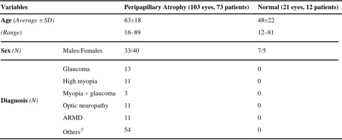
The results were assessed for 103 eyes of 73 patients with peripapillary atrophy and 21 eyes of 12 normal patients. The PPA group consisted of 33 males and 40 females, with a mean age of 63 (SD +/− 18 years, range, 16 to 89 years). In the normal group, there were 7 males and 5 females, with a mean age of 48 (SD +/− 22 years, range, 12 to 81). Forty-three eyes from the PPA group and 3 eyes from the normal group were excluded from the study because the raster scan image quality was less than 6 out of 10 in intensity, both eyes were not scanned, or at least 300 μm of peripapillary atrophy was not present as measured on the color photograph (PPA group). Of the 103 eyes included in the PPA group, 13 (12.6%) eyes were clinically classified as having glaucoma, 11 (10.7%) had age-related macular
degeneration (ARMD), 11 (10.7%) had high myopia, 3 (2.9%) had glaucoma and high myopia, 11 (10.7%) had optic neuropathy, and 54 (52%) were classified under other diagnoses (Table 1). Other diagnoses included brain tumors, cataracts, cystoid macular edema, epiretinal membrane, and diabetic retinopathy. Within the clinical subgroups, there were ten eyes with primary open angle glaucoma and three eyes with normal tension glaucoma, three eyes with myopic degeneration, and eyes with both wet and dry ARMD. These groups were not analyzed separately from the main clinical subgroup they fell under.
Qualitative Analysis
Qualitative analysis of the Cirrus HD-OCT raster scans of eyes with PPA assisted in classification into 6 groups: RPE and partial photoreceptor loss, RPE disruption (Figure 1), RNFL thickening with plaque-like formation (Figure 2), intraretinal cystic changes (Figure 3), inner and outer retinal thinning, and abnormal retinal sloping (Figure 4). The occurrence of each of these findings within each of the six clinical subgroups was quantified (Table 2). The findings of RPE disruption and loss, photoreceptor loss, and retinal thinning were consistent with changes previously noted on histological sections of retinas having
peripapillary atrophy 4–7.
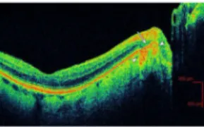
In 30 eyes (29%), RPE and photoreceptor loss was seen and 84 eyes (82%) demonstrated RPE disruption. The axial resolution of the SD OCT images allowed for the detection of RPE loss with photoreceptor loss, which was determined by disturbances in the RPE along with denuded photoreceptor layer above the RPE. Analysis of the SD OCT images also demonstrated the subtleties of RPE disruption which was assessed on OCT scans by increased signal penetration into the choroid beyond the hyper-reflective melanin layer (Figure 1).
Abnormal structure of the RNFL in eyes with PPA, referred to as RNFL “plaques” in this paper were noted in 46 eyes (45%). These abnormal formations were defined as those nerve fiber layers measuring greater than 90 μm, which is one standard deviation above the mean RNFL thickness of the normal group in this study (Figure 2).
Retinal thinning was seen 20 eyes (19%). The OCT raster images were taken in such a manner as to include both the macula as well as the optic disc. The retina was analyzed from the fovea to the edge of the disc and reduction in inner and outer retinal thickness proximal to the disc was considered under the category of retinal thinning. Retinal thickness was also measured 300 μm temporal to the edge of the optic disc for separate quantitative analysis.
Intraretinal cystic changes were seen within the peripapillary region in 2 eyes (2%) (Figure 3). These observed cystic changes have not been previously reported with relation to peripapillary atrophy. The cystic changes were not believed to be cross-sectional profiles of blood vessels based on the location within the retinal layers as well as the irregular shape.
NIH-PA Author Manuscript
NIH-PA Author Manuscript
Furthermore, registration of the OCT B scans with the rendered OCT fundus image, failed to show that any blood vessels aligned with the position of the cysts.
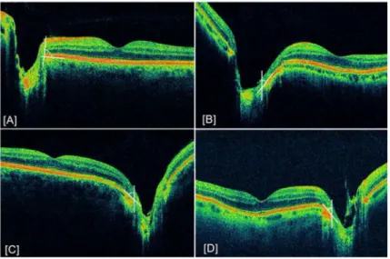
Abnormal retinal sloping was noted in 24 eyes (23%). In OCT scans of normal eyes, it was noted that the retina takes a perpendicular approaches toward the nerve head, with all retinal layers present to the very edge of the disc then sharply dropping off (Figure 4A). A vertical
line placed at the edge of the RPE, also the presumed edge of the optic disc in this study11,
is at a 90º angle to the RPE layer. In contrast, in eyes with PPA, the retina appears to have an abnormal sloping appearance while approaching the disc (Figure 4B, C, and D). An abnormal retinal slope in this study was defined as an angle less than 90º between the RPE and a vertical line placed at the edge of the optic disc. As described above, angle
measurements were performed on exported Cirrus images using research analysis software, ImageJ. The pixel aspect ratio of the OCT images was factored into the angle measurements for accuracy.
Quantitative Analysis
Quantitative analysis was done comparing the total retinal thickness in eyes with PPA compared to normal retinas using t-tests, with P value less than 0.05 considered significant. A preliminary test for the equality of variances indicated that the variances of the two groups were not significantly different F = 0.594, P = .09. Therefore, a two-sample t-test was performed that assumed equal variances. Retinal thickness measurements obtained from Cirrus raster scans in the normal group (Mean = 281.76 μm, SD = 43.53, N =21) were significantly thicker (P = 0.0005) than retinal thickness in eyes with PPA (Mean = 240.25 μm, SD = 56.48, N = 103). The normal eyes were approximately 15% thicker than the PPA study eyes. Retinal thickness was compared between each group of patients (glaucoma, high myopia, high myopia and glaucoma, ARMD, optic neuropathy, and the other diagnosis group) to the group of normal eyes. Normal eyes were found to have significantly greater total retinal thickness than eyes with glaucoma, ARMD, optic neuropathy, and the group of other diagnoses (P = 0.02, P = 0.04, P = 0.002, P = 0.005 respectively).
A two-sample t-test was performed that assumed unequal variances (F = 0.145, P = 0.000066) was performed comparing RNFL thickness measurements between the group of normal eyes and PPA. Comparison between the normal (M = 76.24 μm, SD = 13.73, N = 17) and PPA (M = 88.87 μm, SD = 36.02, N = 45) group, did not reveal a significant difference in thickness (P = 0.05).
Discussion
Peripapillary atrophy is seen in a number of different disease states, but is also considered a
normal aging phenomenon 12, 13. As illustrated in this study, examination of the
peripapillary region in cross-section by SD OCT facilitated the characterization of
alterations in retinal architecture. OCT imaging is analogous to a near “optical biopsy”8, and
in that sense, a literature review of histological descriptions of peripapillary atrophy demonstrated good correlation with OCT findings in this study.
RPE and photoreceptor disruption
Disruption of normal retinal pigment epithelium architecture is the hallmark feature of peripapillary atrophy. Overall, RPE and photoreceptor loss, which is the histological correlate of beta zone atrophy, was observed in 28% of the images. Within the clinical subgroups, RPE and photoreceptor loss were the most common OCT findings in the eyes with glaucoma (54%) and high myopia (82%). Beta zone atrophy in glaucoma and myopic crescents in highly myopic eyes, which fulfill the criteria of B zone, are associated with an
NIH-PA Author Manuscript
NIH-PA Author Manuscript
absolute scotoma, which is consistent with findings of photoreceptor and RPE loss as seen
on OCT 14,15. Structural disruption of the RPE in the alpha zone has been demonstrated to
cause a relative scotoma in this region 14,15.
RPE irregularities, in contrast to complete loss, were observed in 80% of images across all groups; its occurrence was noted most commonly in glaucoma (77%), optic neuropathy (82%), and other diagnosis (100%) subgroups. In a study of peripapillary atrophy in normal
eyes with a mean age of 53 +/− 16 years, Lee, et al 16 noted an occurrence of 100% alpha
and 75% beta atrophy. If one considers that there were 12 patients with PPA but no active retinal disease within the group of patients categorized under “other diagnosis” in this study, the findings are consistent with Lee et al, also demonstrating alpha atrophy is 100 % of these eyes.
RNFL plaque
In this study, manual measurement of the retinal nerve fiber layer thickness within the region of PPA, 300 μm from the edge of the optic disc, did not reveal a significant difference between normal eyes and those with PPA (P = 0.05). In eyes with PPA, loss of normal RNFL architecture with the presence of a thickened or plaque-like formation was observed in 45% of eyes (Figure 2). The OCT of these “plaques” showed an increased area of homogenous reflectivity in the area of RNFL. RNFL “plaques” were the most common finding in high myopia (82%), ARMD (82%), and “other diagnosis” group (41%). The histology of the peripapillary region of highly myopic eyes with glaucoma has suggested that only sclera and retinal nerve fiber layer are present in the peripapillary region, without
interposed retinal layers 7. The appearance is consistent with many of the eyes that were
observed to have plaques (Figure 2). However, deposition of material or abnormal findings within the RNFL in peripapillary atrophy has not been previously reported. The etiology of this observed thickening is unknown and further histological investigation is necessary to better characterize the origin of these RNFL plaque-like changes.
Cystic changes
An intraretinal cystic change was observed in two eyes that were studied, with occurrence in one patient with high myopia (Figure 3A) and one patient with ARMD (Figure 3B). There have been no histological reports concerning intraretinal cystic changes related to
peripapillary atrophy. In an OCT study by Shimada, et al 17 peripapillary changes observed
in Japanese patients with high myopia included peripapillary detachments in eyes with pathologic myopia (PDPM), pitlike structures, microfolds, and retinoschisis. PDPM was noted to appear as intrachoridal and subretinal cystic spaces in 11% of high myopes that were examined. Although the cystic changes described here may represent PDPM, the orange fundus lesions related to PDPM as documented by Shimada, were not observed in the fundus images of the eyes in this study. Alternatively, the finding by Shimada described as retinoschisis may be another explanation of the cystic changes seen in on the images this study. The small number of eyes observed to have cystic changes on OCT scans may be low in this study considering the small number of eyes with high myopia, however it is possible that the development of peripapillary atrophy represents a later or different change than those eyes examined by Shimada. Alternatively, the changes observed by Shimada may represent different changes seen in high myopes or possibly that Japanese high myopes present with unique morphological features.
Retinal thinning
Based on qualitative assessment of the OCT images, 85% of images from the PPA group with glaucoma demonstrated thin inner and outer retinal appearance, which was the most frequent occurrence compared to all other groups. As with retinal nerve fiber measurements,
NIH-PA Author Manuscript
NIH-PA Author Manuscript
total retinal thickness was measured 300 μm from the edge of the optic disc within the region of peripapillary atrophy utilizing Cirrus software, which demonstrated that total retinal thickness was statistically significantly thicker in normal eyes compared with eyes with PPA. When each clinical group was analyzed individually, normal eyes were significantly thicker than the PPA groups with glaucoma, ARMD, optic neuropathy, and other diagnosis. Histological studies previously showed inner and outer retinal layers to be
thinner in glaucomatous eyes 5, but it is interesting that the retina is also thinner within the
area of atrophy in these other disease states. Of note, the atrophic fundus appearance of PPA does not necessarily signify the complete absence of retinal tissue in the region.
Abnormal Retinal Sloping
Through qualitative analysis, it was observed that there was a difference in the angle of the slope of the retina approaching the optic nerve in eyes with PPA. This sloping pattern appeared most often within the glaucoma subgroup, occurring in 85% of images. Lee, et al
16, observed tapering of the ganglion cell, inner and outer plexiform layers at the edge of the
optic disc while the RNFL continued into the optic cup in PPA. Caprioli, et al 18 studied the
changes in the RNFL slope in glaucomatous eyes, and observed that the peripapillary nerve fiber layer had a more level approach toward the optic nerve head in normal individuals, and a declining approach toward the optic in glaucoma patients. They inferred that the net effect on the slope could be a combination of PPA and loss of NFL. It is important to take into consideration that the angles seen on OCT images do not necessary directly reflect the true physical angles in the eye due to image distortion, as well as motion artifact. This study is noting a common pattern of retinal sloping observed on OCT images that were seen in this group of study patients compared to normal eyes.
In conclusion, this study describes retinal changes associated with peripapillary atrophy using high definition OCT. The qualitative findings include RPE and photoreceptor loss, RPE disruption, inner/outer retinal thinning, which have been described previously through histological studies, as well as the novel findings of RNFL thickening and plaque-like formation, intraretinal cystic changes, and abnormal changes in retinal sloping.
Acknowledgments
Financial SupportThis work was supported in part by a Research to Prevent Blindness Challenge grant to the New England Eye Center/Department of Ophthalmology -Tufts University School of Medicine, NIH contracts R01-EY11289-24, R01-EY13178-10, R01-EY013516-07, Air Force Office of Scientific Research FA9550-07-1-0101,
FA9550-07-1-0014, and Massachusetts Lions Eye Research Fund.
The sponsor or funding organization had no role in the design or conduct of this research.
References
1. Jonas JB, Fernandez MC, Naumann GO. Glaucomatous parapapillary atrophy: occurrence and correlations. Arch Ophthalmol 1992;110:214–22. [PubMed: 1736871]
2. Xu L, Li Y, Wang S, et al. Characteristics of highly myopic eyes: the Beijing Eye Study. Ophthalmology 2007;114:121–6. [PubMed: 17070594]
3. Jonas JB, Nguyen XN, Gusek GC, Naumann GO. Parapapillary chorioretinal atrophy in normal and glaucoma eyes. I. Morphometric data. Invest Ophthalmol Vis Sci 1989;30:908–18. [PubMed: 2722447]
4. Fantes FE, Anderson DR. Clinical histologic correlation of human peripapillary anatomy. Ophthalmology 1989;96:20–5. [PubMed: 2919048]
NIH-PA Author Manuscript
NIH-PA Author Manuscript
5. Jonas JB, Konigsreuther KA, Naumann GO. Optic disc histomorphometry in normal eyes and eyes with secondary angle-closure glaucoma. II. Parapapillary region. Graefes Arch Clin Exp
Ophthalmol 1992;230:134–9. [PubMed: 1577293]
6. Kubota T, Jonas JB, Naumann GO. Direct clinico-histological correlation of parapapillary chorioretinal atrophy. Br J Ophthalmol 1993;77:103–6. [PubMed: 8435408]
7. Dichtl A, Jonas JB, Naumann GO. Histomorphometry of the optic disc in highly myopic eyes with absolute secondary angle closure glaucoma. Br J Ophthalmol 1998;82:286–9. [PubMed: 9602626] 8. Huang D, Swanson EA, Lin CP, et al. Optical coherence tomography. Science 1991;254:1178–81.
[PubMed: 1957169]
9. Hee MR, Izatt JA, Swanson EA, et al. Optical coherence tomography of the human retina. Arch Ophthalmol 1995;113:325–32. [PubMed: 7887846]
10. Puliafito CA, Hee MR, Lin CP, et al. Imaging of macular diseases with optical coherence tomography. Ophthalmology 1995;102:217–29. [PubMed: 7862410]
11. Lai E, Wollstein G, Price LL, et al. Optical coherence tomography disc assessment in optic nerves with peripapillary atrophy. Ophthalmic Surg Lasers Imaging 2003;34:498–504. [PubMed: 14620759]
12. Curcio CA, Saunders PL, Younger PW, Malek G. Peripapillary chorioretinal atrophy: Bruch’s membrane changes and photoreceptor loss. Ophthalmology 2000;107:334–43. [PubMed: 10690836]
13. Spaide RF. Age-related choroidal atrophy. Am J Ophthalmol 2009;147:801–10. [PubMed: 19232561]
14. Rensch F, Jonas JB. Direct microperimetry of alpha zone and beta zone parapapillary atrophy. Br J Ophthalmol 2008;92:1617–9. [PubMed: 18782800]
15. Jonas JB, Gusek GC, Fernandez MC. Correlation of the blind spot size to the area of the optic disk and parapapillary atrophy. Am J Ophthalmol 1991;111:559–65. [PubMed: 2021162]
16. Lee KY, Tomidokoro A, Sakata R, et al. Cross-sectional anatomic configurations of peripapillary atrophy evaluated with spectral domain-optical coherence tomography. Invest Ophthalmol Vis Sci 2010;51:666–71. [PubMed: 19850838]
17. Shimada N, Ohno-Matsui K, Nishimuta A, et al. Peripapillary changes detected by optical coherence tomography in eyes with high myopia. Ophthalmology 2007;114:2070–6. [PubMed: 17543388]
18. Caprioli J, Park HJ, Ugurlu S, Hoffman D. Slope of the peripapillary nerve fiber layer surface in glaucoma. Invest Ophthalmol Vis Sci 1998;39:2321–8. [PubMed: 9804140]
NIH-PA Author Manuscript
NIH-PA Author Manuscript
Figure 1. Example of retinal pigment epithelium (RPE) disruption in peripapillary atrophy in an eye with age-related macular degeneration (ARMD)
The white arrow delineates a section of RPE disruption within the region of peripapillary atrophy. White arrowheads indicate increased signal penetration beyond hyper-reflective RPE.
NIH-PA Author Manuscript
NIH-PA Author Manuscript
Figure 2. Example of retinal nerve fiber layer plaques
[A] Right and [B] left eye of patient with high myopia demonstrating retinal nerve fiber layer (RNFL) thickening and plaque-like changes measured to be 192 μm and 132 μm respectively, 300 μm temporal from the edge of the optic disc (indicated by white arrows). These images also demonstrate the retinal nerve fiber layer in the peripapillary region to be resting on the sclera without interposed retinal layers.
NIH-PA Author Manuscript
NIH-PA Author Manuscript
Figure 3. Example of intraretinal cystic changes
Intraretinal cystic changes indicated by white arrows. [A] Intraretinal cyst at nasal edge of the disc in a patient with high myopia. [B] Small cystic changes at the temporal edge of the optic disc in a patient with age-related macular degeneration (ARMD).
NIH-PA Author Manuscript
NIH-PA Author Manuscript
Figure 4. Example of abnormal retinal sloping
White lines delineate the angle between the optic disc edge and retinal pigment epithelium (RPE) cell layer. [A] Normal eye demonstrating parallel approach of all retinal layers to the optic disc and a 90º angle between optic disc edge and RPE [B] Sloping appearance of retina in a glaucomatous eye with a 68º angle between disc edge and RPE [C] 71º angle in a patient with optic neuropathy [D] 66º angle in a patient with glaucoma.
NIH-PA Author Manuscript
NIH-PA Author Manuscript
NIH-PA Author Manuscript
NIH-PA Author Manuscript
NIH-PA Author Manuscript
Table 1 Demographics of normal eyes and eyes with peripapillary atrophy
Variables Peripapillary Atrophy (103 eyes, 73 patients) Normal (21 eyes, 12 patients)
Age (Average ± SD) 63±18 48±22 (Range) 16–89 12–81 Sex (N) Males/Females 33/40 7/5 Diagnosis (N) Glaucoma 13 0 High myopia 11 0 Myopia + glaucoma 3 0 Optic neuropathy 11 0 ARMD 11 0 Others† 54 0
N = number of patients, SD = Standard deviation, ARMD = age-related macular degeneration
†
NIH-PA Author Manuscript
NIH-PA Author Manuscript
NIH-PA Author Manuscript
Table 2
Occurrence of optical coherence tomography defined characteristics within each clinical subgroup
ARMD Glaucoma High myopia Myopia/Glaucoma Optic neuropathy Other † Total Subjects ( N ) 11 (10.7%) 13 (12.6%) 11 (10.7%) 3 (2.9%) 11 (10.7%) 54 (52%) 103
RPE and photoreceptor loss
2 7 9 1 5 6 30 RPE disruption 5 10 4 2 9 54 84 RNFL plaque 9 4 9 0 2 22 46 Retinal thinning 1 11 1 2 3 2 20 Cystic changes 1 0 1 0 0 0 2
Abnormal retinal sloping
1 11 3 1 3 5 24


