HAL Id: tel-03198814
https://tel.archives-ouvertes.fr/tel-03198814
Submitted on 15 Apr 2021
HAL is a multi-disciplinary open access archive for the deposit and dissemination of sci-entific research documents, whether they are pub-lished or not. The documents may come from teaching and research institutions in France or abroad, or from public or private research centers.
L’archive ouverte pluridisciplinaire HAL, est destinée au dépôt et à la diffusion de documents scientifiques de niveau recherche, publiés ou non, émanant des établissements d’enseignement et de recherche français ou étrangers, des laboratoires publics ou privés.
Huntingtin functions in the regulation of axonal
transport : consequences on neuronal network
homeostasis and behavior, in health and disease
Helène Vitet
To cite this version:
Helène Vitet. Huntingtin functions in the regulation of axonal transport : consequences on neuronal network homeostasis and behavior, in health and disease. Neurobiology. Université Grenoble Alpes [2020-..], 2020. English. �NNT : 2020GRALV038�. �tel-03198814�
THÈSE
Pour obtenir le grade de
DOCTEUR DE L’UNIVERSITÉ GRENOBLE ALPES
Spécialité : Neurosciences - Neurobiologie
Arrêté ministériel : 25 mai 2016 Présentée par
Hélène VITET
Thèse dirigée par Frederic SAUDOU
et co-encadrée par Chiara SCARAMUZZINO, Université Grenoble-Alpes
préparée au sein du Laboratoire Grenoble Institut des Neurosciences
dans l'École Doctorale Chimie et Sciences du Vivant
Conséquences de la régulation du transport
axonal par la
Huntingtine sur l'homéostasie
de réseaux neuronaux et sur le
comportement, en conditions saine et
pathologique
Huntingtin functions in the regulation of
axonal transport: consequences on neuronal
network homeostasis and behavior, in health
and disease
Thèse soutenue publiquement le 11 décembre 2020, devant le jury composé de :
Monsieur FREDERIC SAUDOU
PROFESSEUR DES UNIVERSITES - PRATICIEN HOSPITALIER, UNIVERSITE GRENOBLE ALPES, Directeur de thèse
Madame KARINE MERIENNE
DIRECTRICE DE RECHERCHE, CNRS DELEGATION ALSACE, Rapporteure
Madame CORALIE FASSIER
DIRECTRICE DE RECHERCHE, INSERM DELEGATION PARIS, Rapporteure
Monsieur ALAIN MARTY
DIRECTEUR DE RECHERCHE, CNRS DELEGATION ILE-DE-FRANCE VILLEJUIF, Examinateur
Monsieur ALAIN BUISSON
PROFESSEUR DES UNIVERSITES, UNIVERSITE GRENOBLE ALPES, Président
3
À mon grand-père, dans l’espoir qu’il soit en paix avec
5
Preface
L’état d’équilibre des déséquilibres
Avant de rentrer dans les profondeurs et méandres de la régulation du transport axonal, arrêtons-nous sur l’importance de l’homéostasie, l’état d’équilibre, de la cellule ou de tout autre système. La définition de l’homéostasie est fidèlement explicitée par son étymologie grecque ; « homéostasie » est composée de hómoios, « similaire », et de stásis, « stabilité ». Cette « stabilité similaire », du fait de sa capacité à s’autoentretenir, permet l’établissement d’un équilibre capable de répondre à des perturbations au sein d’un système. Ces perturbations peuvent être transitoires ou permanentes et impactantes menant soit à un rapide retour à un autre état d’équilibre soit à un emballement du système. Ces conséquences sont visibles au sein d’une cellule telle qu’un neurone régulant le transport d’une vésicule tout comme au sein d’une planète régulant sa température. Dans les deux cas, une perturbation importante, et donc une perte de l’homéostasie, amène à la neurodégénérescence ou au réchauffement climatique dont nous sommes aujourd’hui les témoins. Ainsi, sans profonde perturbations menant à l’emballement du système, l’essence même d’un état d’équilibre serait d’être perturbé, déséquilibré, modifié, mais jamais perdu ; l’équilibre sera toujours trouvé. Le chimiste trouve l’équilibre chimique d’une réaction en perturbant l’équilibre de plusieurs espèces par leur mélange. Le dramaturge se joue de l’élément perturbateur initial pour mettre en scène le chemin menant au prochain équilibre. Le musicien produit des mélodies en contrôlant les perturbations et le retour à l’équilibre de l’air en le faisant vibrer avec une corde.
L’équilibre, la physiologie, voilà ce que le chercheur étudie pour comprendre le déséquilibre, la pathologie. En pratique, c’est généralement l’observation de la pathologie qui arrive en premier. Claude Bernard a par exemple étudié l’effet du curare qui paralysait les animaux les proies des indiens d’Amazonie pour une meilleure compréhension du fonctionnement de la jonction neuromusculaire. De même pour la maladie d’Huntington, de nombreuses études se sont en premier lieu penchées sur les conséquences de l’induction d’effets toxiques provoqués par la maladie plutôt que la perte de fonctions physiologiques. Ainsi, c’est en investissant dans la recherche fondamentale centrée sur les phénomènes physiologiques que la science fondamentale permet l’application à la pathologie. En effet, en comprenant l’équilibre d’un système, il est possible de le rétablir lorsqu’il est perturbé ; la médecine aujourd’hui, réussit à perturber l’équilibre du système malade pour soigner, s’il ne s’est pas encore emballé.
Les déséquilibres apparaissent comme problématiques mais ne nous sont-ils pas également nécessaires ? L’équilibre apparaitrait donc comme nécessaire aux déséquilibres et à leurs effets. En effet, les déséquilibres ne font-ils pas de nous des Hommes et non pas des machines, pour le pire et le
6 meilleur ? Sans déséquilibres, et sans sa capacité d’adaptation due à l’homéostasie, l’Homme n’aurait sûrement pas évolué de la façon dont il l’a fait. De plus, nos avancées scientifiques auraient été limitées car le chercheur a besoin de perturber l’équilibre pour le comprendre. Ainsi, ne faudrait-il donc pas atteindre un état d’équilibre des déséquilibres ? En effet, trop peu de perturbations de l’équilibre nous aurait peut-être empêché d’évoluer en tant qu’Hommes et scientifiques et a contrario, trop de perturbations peuvent mettre à mal l’homéostasie et créer de nouvelles conditions (climatiques par exemple) non compatibles avec la vie humaine. Enfin, la fin d’un sous-système par perte homéostasique peut également être considéré comme un mécanisme de préservation de l’homéostasie d’un système supérieur atteignant alors un nouvel état d’équilibre. Ceci ajoute finalement un caractère relatif à la notion d’équilibre.
7
Title
Huntingtin functions in the regulation of axonal transport: consequences on neuronal network homeostasis and behavior in health and disease
Abstract
Neuronal circuits are at the basis of behaviors such as motor coordination or learning and memory. As being part of a network, neurons communicate at synapses through finely tuned molecular and cellular processes. One key mechanism regulating synapse homeostasis involves the transport of vesicles within axons and dendrites, which is dysregulated in many neurological disorders such as Rett syndrome, Alzheimer’s (AD) and Huntington’s diseases (HD). Thus, deciphering the regulation of vesicular transport within neurites in physiological context is crucial to understand, and potentially restore, the consequences of these dysregulations in pathological contexts.
Huntingtin (HTT) protein, known for its devastating role in HD when mutated, is a key actor of axonal transport. It promotes and regulates vesicular transport in neurites by scaffolding adaptors and molecular motors. Particularly, HTT phosphorylation status at S421 regulates the directionality of BDNF, APP and VAMP-7 vesicles within neurites in cultured and transfected neurons. However, several questions remain to be elucidated regarding the mechanisms and the consequences of this HTT-dependent regulation of vesicular transport such as the neuritic specificity (axons or dendrites) and the behavioral consequences of such modification. Finally, we do not know whether transport regulation can be influenced in pathological conditions to restore disease-associated phenotypes in
vivo.
This thesis aims at characterizing in vivo the mechanisms and the consequences of axonal transport regulation of three different types of vesicles through the phosphorylation of Huntingtin at S421 and to investigate its propensity to restore disease-associated phenotypes in mouse models of human neurological disorders.
In order to reproduce in vitro the in vivo networks associated with neurological disorders we used microfluidic devices that allow the reconstitution of neuronal networks in vitro. We investigated the transport of Amyloid Precursor Protein (APP) vesicles, precursors of synaptic vesicles (SVPs) or dense-core vesicles (DCVs) in neurons in which the HTT phosphorylation status was modified. These neurons came from mice in which Serine 421 has been replaced by an aspartic acid to mimic the
8 phosphorylated form of HTT (HTTS421D) or by an alanine to mimic the unphosphorylatable form of HTT (HTTS421A).
APP homeostasis is impaired in AD. We investigated APP transport and accumulation at corticocortical synapses. We found that Akt-mediated HTT phosphorylation at S421 regulates the directionality of APP containing vesicles in axons but not in dendrites: the unphosphorylatable form of HTT decreases axonal anterograde flux of APP and reduces its levels at presynaptic zones both in
vitro and in vivo. Reducing anterograde flux of APP in familial AD mouse model restored synapse
homeostasis in vivo and memory deficits (Publication 1; Bruyere*, Abada*, Vitet* et al., eLife, 2020). BDNF transport within DCVs is dysregulated in the corticostriatal network of Rett syndrome’s patients. We found that endogenous HTT phosphorylation at S421 or a chemical inhibitor of calcineurin (FK506) rescue BDNF transport in the corticostriatal network, neuronal communication, and behaviors of Rett syndrome model mice (Publication 2; Ehinger et al., Embo Mol Med, 2020).
Finally, it has been shown that SVP axonal transport regulates the number of SVs at the synapse, which, within a corticostriatal synapse, is essential for motor skill learning. We found that HTT phosphorylation increases the recruitment of the molecular motor KIF1A on SVPs, thus promoting anterograde transport and the probability of release. Silencing KIF1A in the corticostriatal network of HTTS421D mice, we found that pHTTS421 increases the number of SVs at the synapse and impairs procedural memory through a specific HTT-KIF1A dependent mechanism. This study defines a pathway by which axonal transport of SVP impact the behavioral phenotype (Publication 3; Vitet et al., in prep).
Keywords
Huntingtin, phosphorylation, serine 421, axonal transport, Brain-Derived-Neurotrophic-Factor, Amyloïd Precursor Protein, Synaptic Vesicle Precursor, Alzheimer’s disease, Rett syndrome, memory, microfluidics, cortical networks
9
Titre
Conséquences de la régulation du transport axonal par la Huntingtine sur l'homéostasie de réseaux neuronaux et sur le comportement, en conditions saine et pathologique
Résumé
Les circuits neuronaux régissent les comportements tels que la coordination motrice ou la mémoire et l’apprentissage. Au sein d’un réseau, les neurones communiquent par des processus moléculaires et cellulaires finement réglés à la synapse. Un des mécanismes régulant l’homéostasie synaptique, le transport de vésicules dans les neurones, est dérégulé dans les maladies neurologiques telles que le syndrome de Rett, la maladie d’Alzheimer et la maladie d’Huntington. Ainsi, investiguer la régulation du transport de vésicules dans les neurites dans un contexte physiologique est important pour comprendre, et potentiellement rétablir, les conséquences de ces dérégulations pathologiques. La protéine Huntingtine (HTT), connue pour son implication dans la maladie d’Huntington, est un acteur clé du transport axonal. Elle promeut et influence le transport des vésicules en favorisant le recrutement des adaptateurs et des moteurs moléculaires. Sa phosphorylation à la sérine 421 (pHTTS421) régule la directionnalité des vésicules de BDNF, d’APP et de VAMP-7 dans des neurones transfectés in vitro. Cependant, les mécanismes et les conséquences de la régulation du transport par HTT, comme la spécificité neuritique et les conséquences comportementales, restent peu connues. Enfin, nous ignorons si la régulation du transport peut être influencée dans des conditions pathologiques afin de restaurer les phénotypes in vivo.
Ce projet de thèse vise à caractériser les mécanismes et les conséquences de la régulation du transport axonal de trois types de vésicules par pHTTS421 et d’investiguer sa propension à restaurer les phénotypes associés à des maladies neurologiques dans des modèles murins.
Dans le but de reproduire in vitro les réseaux associés à des maladies neurologiques, nous avons utilisé des chambres microfluidiques. Nous avons étudié le transport des vésicules d’APP, des précurseurs des vésicules synaptiques (PVSs) ou des vésicules à cœur dense (VCDs) contenant BDNF au sein d’un réseau neuronal dans lequel pHTTS421 a été modifiée. Ces neurones sont issus de souris pour lesquelles la sérine 421 a été remplacée par un acide aspartique ou par une alanine pour mimer respectivement l’état phosphorylé (HTTS421D) ou non phosphorylable (HTTS421A) de la HTT.
10 Dans la maladie d’Alzheimer, l’homéostasie d’APP est dérégulée. Nous avons donc étudié son transport et son accumulation synaptique dans un circuit corticocortical. Nous avons trouvé que la phosphorylation de la sérine S421 par Akt régule la directionnalité des vésicules d’APP uniquement dans les axones : HTTS421A diminue le flux antérograde axonal d’APP ainsi que ses niveaux à la synapse in vitro et in vivo. Réduire le flux antérograde d’APP dans un modèle murin d’Alzheimer restaure
l’homéostasie synaptique in vivo et les déficits de mémoire associés (publication 1 ; Bruyère*, Abada*, Vitet* et al., eLife, 2020).
Le transport de BDNF est dérégulé dans le réseau corticostriatal des jeunes filles atteintes du syndrome de Rett. Nous avons observé que l’expression endogène de pHTTS421 ou l’injection d’un composé inhibant la calcineurine (FK506) restaure le transport de BDNF dans un réseau corticostriatal, la communication neuronale et les symptômes associés chez les souris modèles du syndrome de Rett (Publication 2 ; Ehinger et al., Embo Mol Med, 2020).
Enfin, Le transport axonal des PVSs régule le nombre de vésicules synaptiques (VSs), ce qui, dans un réseau corticostriatal, est essentiel à l’apprentissage de compétences motrices. Nous avons montré que pHTTS421 augmente le recrutement de la kinésine KIF1A sur les PVSs, augmentant le transport antérograde et la probabilité d’exocytose. En réduisant les niveaux de KIF1A dans le réseau corticostriatal des souris HTTS421D, nous avons trouvé que pHTTS421 augmente le nombre de VSs et altère la mémoire procédurale. Cette étude décrit comment le transport axonal des PVSs impacte les phénotypes comportementaux (publication 3 ; Vitet et al., in prep).
Mots clés
Huntingtine, phosphorylation, sérine 421, transport axonal, BDNF, APP, Précurseur des Vésicules Synaptiques, maladie d’Alzheimer, syndrome de Rett, mémoire, microfluidique, réseaux corticaux
Laboratory
Grenoble Institut des Neurosciences, INSERM U1216, Bâtiment Edmond J. Safra, Chemin Fortuné Ferrini, 38700 La Tronche
11
Remerciements
L’ensemble de ce travail n’aurait pas été possible sans la volonté de certaines personnes de repousser les frontières de notre savoir, de créer une bonne atmosphère de travail ou de veiller à mon équilibre. Par ces quelques lignes, je voudrais leur rendre hommage et les remercier.
Mes premiers remerciements vont aux membres du jury, Alain Buisson, Alain Marty, et tout particulièrement à Karine Merienne et Coralie Fassier qui ont pris le temps de lire ce long manuscrit, pour avoir accepté de rejoindre mon jury de thèse. Par votre présence et votre appréciation, vous me permettez de repousser mes limites et celles de la science. Je tenais également à souligner le rôle important d’Alain Marty, Alain Buisson, Sébastien Carnicella, Laurent Venance et Sandrine Humbert pour leur participation à nos réflexions concernant mes projets lors de mes Comité de suivi de thèse. Fred, je te remercie de m’avoir fait confiance tout au long de cette thèse et particulièrement dès le début où tu as pris le risque de commencer une thèse avec moi sans me connaître. Tu as été le seul directeur de recherche à le faire et j’en suis très reconnaissante. Grâce à toi j’ai beaucoup appris, notamment concernant l’écriture et le monde de la recherche en général. Merci de m’avoir fait confiance pour la rédaction de la revue, de la révision du papier APP mais aussi pour t’avoir secondé pendant le congrès de Montpellier. Merci également de m’avoir donné cette chance de pouvoir travailler sans me soucier de problèmes matériels ; c’est une chose rare que j’ai su apprécier.
Sandrine, je te remercie d’avoir considéré ma candidature spontanée que tu as reçu il y a maintenant plus de quatre ans. Ton accompagnement et tes conseils tout au long de mes projets ont également été très appréciés.
Je voulais également remercier ces gens de l’ombre, qu’on ne voit pas et qui ont pourtant été essentiels. Ces personnes m’ont permis, par le biais de leurs dons à la fondation pour la recherche médicale ou l’association Huntington France, de réaliser cette thèse et d’accroître nos connaissances pour trouver des solutions aux défis de santé actuels.
Jérôme, merci de m’avoir accueillie dans ton laboratoire alors que je ne savais ni pipeter ni parler anglais décemment. Mon séjour au Texas m’a profondément marquée et changée, tant personnellement que professionnellement. Tant et si bien que j’irai jusqu’à dire qu’il m’a été essentiel pour la réalisation de ma thèse.
Mes chers collègues qui, soyons honnêtes, sont également mes amis, je vous remercie d’avoir contribué à mon équilibre pendant ces quatre années.
12 Chiara, non so come ringraziarti per esserci stata quando ne avevo più bisogno. Non hai esitato un attimo a prendere il posto di Julie su un argomento che conoscevi solo superficialmente. Mi hai sostenuta quando ne avevo bisogno, sei stata la mia protezione, sei sempre stata lì per me. Mi ricorderò delle pause caffè che prendevamo sulla terrazza per parlare entrambe. Grazie Chiara. Julie, bien plus qu’une encadrante, tu es devenue une vraie amie. Tu m’as tout appris, tes protocoles, tes petites manies, ta passion, j’ai tout pris. Si bien qu’on a fini par se comprendre sans même se parler, aujourd’hui encore. Ton enthousiasme était le mien. Tu m’as manquée mais on se voit bientôt à Tahiti ! Anne-Sophie, tu le sais, j’ai été très heureuse de travailler avec toi. Les bases de statistiques que nous avons établies ensemble au tout début m’ont suivi toute ma thèse. J’apprécie ta rigueur et je te souhaite sincèrement le meilleur dans tes nouveaux projets. Merci également d’avoir été présente tout au long de ma thèse pour répondre à chacune de mes questions.
Elo je te remercie pour ta disponibilité et l’intérêt désintéressé que tu portes aux gens. Merci de m’avoir aidé à démêler les profondeurs du striatum et adoucir les aspérités de l’électrophy.
Bobinette, que serais-je devenue sans ton soutien permanent et tes soirées crêpes mémorables ? Dès mes premiers pas au labo tu as su me mettre à l’aise. Puis se sont succédés les sorties au ski, en rando, le bloc, la bastille à 21h sur un coup de tête, les confidences dans ta voiture, les concerts d’électro plus ou moins appréciés, la soirée mémorable à Montpellier, la sortie roller à Aix les bains, ect…. Bref, merci d’avoir été toi pendant ces quatre années, tu as rendu cette thèse plus douce et plus facile.
Caro, nous avons peu travaillé ensemble mais j’ai apprécié ces moments. Tu m’as transmis tout ce que je sais de l’étude du comportement, merci. Je me souviendrai également de ce petit cocktail que nous avons partagé toutes les deux entre deux confidences, à refaire quand tu veux !
Les co-bureau, merci pour ces bonnes doses de rire que vous m’avez procurées. Max, merci de poser des questions naïves et improbables auxquelles personne ne pense. Mais merci aussi pour ta sensibilité et ta générosité dans ta manière d’être. Nagham, merci pour ces moments tard au bureau, ces moments ont été précieux parce que très simples et sincères. Peut-être que maintenant on arrivera à trouver du temps pour faire un time story ;). Eve, tu savais animer ce bureau à coups d’affaires sensibles et de sujets de discussion improbables, merci d’avoir été présente quand j’en avais besoin. Amandine et Julie-Anne, vous m’avez guidée lors de mes premiers pas dans la thèse et j’ai apprécié travailler à vos côtés.
Les « nouveaux », Emeline, Anca, Johanna, j’aurais aimé prendre le temps de mieux vous connaître mais j’ai déjà apprécié les moments que nous avons partagés ensemble.
13 Nathalie, merci de nous avoir sauvés plus d’une fois dans ce labyrinthe que représente l’administration pour nous. Tu as plus d’une fois réparé nos bêtises avec toute ta bienveillance. Nous avons de la chance de t’avoir.
Cléopâtre et Maria, des stagiaires surprenantes par leur maturité et leur motivation. Je vous remercie toutes les deux de m’avoir soutenue dans mes projets par votre implication, nous formions de belles équipes. Je vous souhaite le meilleur pour la suite, vous le méritez.
Ensuite, un institut ne se limite pas à un laboratoire. Le GIN a cela de précieux qu’il réunit des personnes avec une grande intelligence à la fois scientifique et humaine. Je parle de Jacques, pour ses cours de statistiques très utiles et sa disponibilité, Karin, pour sa curiosité scientifique mais également pour son humanisme et les discussions que nous avons pu avoir au labo de ME ensemble. Nora, nous nous sommes peu parlé mais le peu de fois où nous l’avons fait, tu as su dire ce qu’il fallait. Merci. Toff, j’ai vraiment apprécié travailler à tes côtés, pour tes qualités d’encadrement et ton esprit scientifique. Béa, la dose de bonne humeur et d’air de la montagne du GIN. Merci pour les moments que nous avons partagés ensemble. Leticia, merci pour cette session de poster à parler d’APP à Héraklion, une piña colada à la main. Je referai l’un ou l’autre avec grand plaisir. Cécile, merci d’être l’oreille attentive dont les doctorants ont besoin.
Un grand merci également aux zootechs, Sylvain, Laure, Fabien et Flore qui font, comme l’ont démontré ces derniers mois, un travail indispensable à nos études. Mais plus encore, leur bienveillance nous permet de travailler dans de bonnes conditions. Merci d’avoir été à mes côtés pendant ces longues journées d’injections à savoir si j’avais besoin de quoique ce soit. Ça a beaucoup compté pour moi.
Les doctorants, continuez à former cette entité, ce groupe qui permettra d’amortir les coups et surtout, de les boire.
Enfin, je tiens à remercier ma famille. Papa, maman, merci pour tous ces sacrifices que vous avez fait depuis mes tous premiers pas pour me permettre d’accomplir mes plus grandes ambitions. Ma thèse était l’une d’entre elles, sans votre soutien tout au long de ses 27 années, rien n’aurait été possible. Vous avez fait de mes échecs des forces, de votre pugnacité au travail et humilité mes valeurs et je suis fière d’être votre fille. Choupie, tu constitues ce socle inébranlable qu’on appelle la famille, j’aurais aimé être plus présente pour tes premiers pas dans la vraie vie mais je sais que Fredo veille à ce que ma petite sœur soit heureuse. Papy et mamie vous m’avez transmis vos valeurs, votre douceur et votre ingéniosité, je suis si heureuse d’être à vos côtés. Je chérie chaque moment passé avec vous.
14 Pascal et Yvette, merci de m’avoir permis de me ressourcer dans votre petit paradis paisible. Vous m’avez permis de trouver l’équilibre dont j’avais besoin pour affronter les difficultés de la thèse. Merci pour votre accueil et votre bienveillance.
Enfin, mon homme, tu as su être l’oreille attentive et l’épaule dont j’avais besoin pendant ce marathon. Tu as été présent dans les bons comme dans les mauvais moments, tu me connais mieux que quiconque et tu sais ce dont j’ai besoin en toute circonstance. Mais tu as aussi su me pousser hors de ma zone de confort pour m’améliorer. Tu m’as aidé à tenir mon cap tout en étant mon ancre, tu m’as permis d’aller loin tout en me rappelant ce qui était important. Maintenant que ce cap est franchi, allons trouver le prochain. Qu’il soit au Chili ou à Taïwan, c’est lors de notre prochaine aventure que nous le franchirons. Te quiero.
15
Table of contents
Preface ... 5 Abstract ... 7 Résumé ... 9 List of figures ... 17 List of abbreviations ... 21 Introduction ... 23Chapter 1 - Behaviors are the result of the communication between central nervous system structures through neuronal network establishment ... 24
1. The brain as a combination of structures... 24
2. Structures communication results in a behavior ... 28
3. The neuron: the structural and functional unit of the brain ... 33
4. The neuronal mechanisms underlying learning and memory ... 41
Chapter 2- Axonal transport of vesicles as a regulator of neuronal functions and survival through multi-protein interactions ... 45
1. Vesicles and cargoes ... 45
2. Vesicular transport ... 57
Chapter 3 - Example of a synaptic vesicle journey: how axonal transport ensures its neurotransmission function. ... 84
1. SV: a unique type of vesicle ... 84
2. SVs origin from SVP transport and maturation ... 101
Chapter 4 - Huntingtin as a crucial hub for protein interactions through its intrinsic molecular properties ... 114
1. HTT: a large, dynamic and ubiquitous protein ... 114
2. HTT & HD ... 119
Chapter 5 – Traffic signaling: new functions of Huntingtin and axonal transport in neurological disease ... 124
Results ... 135
Results – part 1: Presynaptic APP levels and synaptic homeostasis are regulated by Akt phosphorylation of Huntingtin ... 136
16
Summary & context of the study ... 136
Discussion ... 173
Results – part 2: Huntingtin phosphorylation governs BDNF homeostasis and improves the phenotype of Mecp2 knockout mice ... 178
Summary & context of the study ... 178
Discussion ... 193
Results – part 3: HTT acts as a scaffold for KIF1A-mediated-transport and regulates SVP axonal transport ... 196
Summary & context of the study ... 196
Discussion ... 242
Discussion ... 247
1. Experimental set up: looking for more integrative and relevant studies ... 247
2. S421 phosphorylation of HTT as a physiological regulator of vesicle transport. ... 258
3. Restoring vesicular transport in NDs, an efficient therapeutic strategy? ... 260
Conclusion ... 262
Perspectives... 263
1. Technical challenges and future plans for research ... 263
2. Challenges in therapeutic strategies for pharmaceutical industry ... 264
Bibliography ... 266
17
List of figures
Figure 1: Human brain is more complex and developed than mouse brain.. ... 24
Figure 2: brain is the sum of structures. ... 25
Figure 3: 3D reconstitution of the cortex ... 25
Figure 4: cortex is organized in layers of neurons. ... 25
Figure 5: layer V. ... 26
Figure 6: 3D reconstitution of the striatum ... 27
Figure 7: 3D representation of basal ganglia organization in a mouse brain. ... 27
Figure 8: 3D reconstitution of the hippocampus ... 27
Figure 9: cortex and hippocampus form a network. ... 28
Figure 10: movements are controlled by two loops involving cortex and basal ganglia. ... 29
Figure 11: Different forms of memory involve specific networks and are evaluated by different behavioral protocols. ... 31
Figure 12: cortical projections target different striatal zones. ... 32
Figure 13: cellular homeostasis as a closed feedback loop regulating cellular functions. ... 33
Figure 14: IGF-1 pathway regulates cell survival through Akt activation. ... 34
Figure 15: neuronal shapes define neuronal functions... 35
Figure 16: pyramidal cells display a specific organization. ... 35
Figure 17: morphologic properties differentiate cortical pyramidal cells from MSN.. ... 36
Figure 18: neuronal networks morphology and function. ... 36
Figure 19: synapse morphology and function ... 37
Figure 20: MTs are a polarized structure. ... 38
Figure 21: MT dynamic is stable. ... 39
Figure 22: actin filament structure. ... 39
Figure 23: actin forms rings within the axon. ... 40
Figure 24: synaptic homeostasis as a two compartments system.. ... 41
Figure 25: a vesicle journey ... 45
Figure 26: PTMs modify the chemical composition of amino acids. ... 46
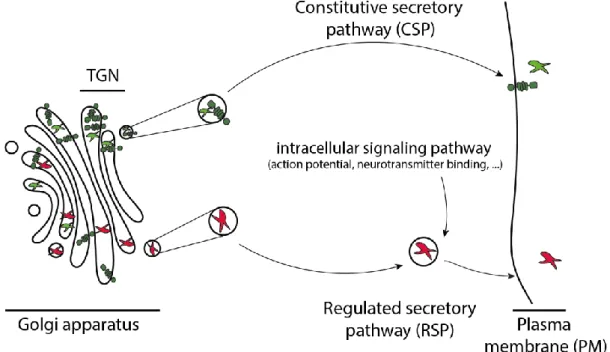
Figure 27: De novo production of vesicles is made in TGN.. ... 46
Figure 28: ER network is present in neurites. ... 47
Figure 29: different endocytic modes exist. . ... 48
Figure 30: TGN vesicles are secreted through two pathways, CSP or RSP. ... 49
Figure 31: DCVs are dense and bigger than SVs. ... 50
18
Figure 34: secretory vesicles carry specific cargoes. ... 53
Figure 35: excitatory and inhibitory synapses exhibit different structural characteristics. ... 55
Figure 36: endosomal vesicles carry specific cargoes.. ... 56
Figure 37: MTs constitute tracks for neuronal transport of vesicle.. ... 59
Figure 38: Tau homeostasis is crucial for MT stabilization which directly affects neuron survival. ... 59
Figure 39: Tau is mostly axonal whereas MAP-2 is dendritic. ... 60
Figure 40: MT acetylation regulates molecular motor binding to MTS. ... 62
Figure 41: kinesin-1 and -3 exhibit different behavior according to MT PTM.. ... 63
Figure 42: 3D representation of kinesin-1 and -3 structure. ... 65
Figure 43: kinesin progression on MTs is ATP dependent.. ... 66
Figure 44: kinesins within a family exhibit different structures. ... 67
Figure 45: kinesins display specific velocity distributions. ... 68
Figure 46: kinesin number on a vesicle influences its velocity and run length. ... 71
Figure 47: involvement of different kinesins on one vesicle display specific velocity distribution.. .... 71
Figure 48: dynein-1 structure ... 72
Figure 49: dynein regulators specify dynein mediated transport of cargoes. ... 73
Figure 50: dynactin structure serves dynein functions. ... 75
Figure 51: Hook and BicD acts as dynein regulators ... 75
Figure 52: kinesin and dynein are present on the same vesicle. ... 76
Figure 53: HTT interacts with both kinesin and dynein acting as a scaffold protein. ... 77
Figure 54: microfluidics device allows the study of axonal transport in connected neurons. ... 79
Figure 55: kymographs are the 2D representation of a vesicle transport ... 79
Figure 56: the selective recruitment model. ... 80
Figure 57: the tug-of-war model explains the bidirectional and non-processive transport. ... 81
Figure 58: the coordination model explains the fast and processive transport of vesicles. ... 82
Figure 59: the steric disinhibition model relies on a cooperation between the two motors. ... 82
Figure 60: Molecular model of an average SV. ... 87
Figure 61: SP interact with each other to regulate exocytosis. ... 88
Figure 62: APP might be present on SVs. ... 91
Figure 63: SV diameter is around 40 nm. ... 91
Figure 64: SV size is dependent on SP steric hindrance. ... 91
Figure 65: SV are organized in three pools: RRP, recycling pool and the reserve pool. ... 92
Figure 66: RRP vesicles are docked to the PM.. ... 93
Figure 67: CME forms clathrin coat around the endocytosed vesicle.. ... 95
19
Figure 69: synapsin regulates the reserve pool size by sticking the SV all together. ... 97
Figure 70: glutamate loading in SVs requires electric and chemical components... 100
Figure 71: SVPs formed from the Golgi are the result of a specific and SNARE-dependent SP segregation. ... 101
Figure 72: KIF1A dimer structure. ... 103
Figure 73: KIF1A also transports BDNF vesicles and BACE1 vesicles. ... 104
Figure 74: KIF1A activation follows KIF1A dimerization on the vesicle. ... 106
Figure 75: KIF1A affinity for MT is increased thanks to the K-loop. ... 106
Figure 76: DENN/MADD complex acts as an adaptor for KIF1A recruitment on SVP. ... 108
Figure 77: liprin-α acts as aKIF1A captor at dendritic spines. ... 108
Figure 78: KIF1A KO mice exhibit a decrease in the number of SV at the synapse. ... 109
Figure 79: KIF1A mutation in human causes cerebellar atrophy. ... 110
Figure 80:KIF1A mutation decreases KIF1A velocity in vitro. ... 110
Figure 81: KIF1A mutations in human diseases. ... 111
Figure 82: SV cycle life lasts for more than 92h, from SVP biosynthesis to degradation. ... 112
Figure 83: 3D representation of HTT structure. ... 114
Figure 84: HT interacts with many proteins involved in large cellular functions. ... 115
Figure 85: HTT regulates dynamin-1 activation. ... 116
Figure 86: HTT regulates transcription. ... 117
Figure 87: HTT adopt multiple conformations according to the environment. ... 118
Figure 88: weak interaction between C- and N-term. ... 118
Figure 89: HD provokes neurodegeneration leading to a reduction of brain weight. ... 119
Figure 90: HTT structures are affected by polyQ stretch. ... 121
Figure 91: APP cleavage by secretases produces many fragments ... 137
Figure 92: APP processing is dependent on APP transport and trafficking ... 139
Figure 93: AD provokes neurodegeneration of the cortex and the hippocampus ... 141
Figure 94: unphosphorylatable form of HTT rescues AD mouse model phenotypes. ... 173
Figure 95: Akt is overactivated in AD. ... 174
Figure 96: IGF-1 pathway regulates cell survival through Akt activation. ... 175
Figure 97: balance of APP level at the synapse is important for neuronal homeostasis ... 177
Figure 98: BDNF level is crucial for neuronal homeostasis and subsequent behavior. ... 195
Figure 99: exocytosis is regulated by SNAREs. ... 197
Figure 100: facilitation results in a higher release of NT at the second stimulation... 198
Figure 101: facilitation might be due to SVs forming a reserve for docked SVs. ... 199
20
Figure 103: DLS and DMS are both involved in the procedural memory formation. ... 201
Figure 104: HTT acts as a scaffold for SVP transport.. ... 242
Figure 105: SV number is crucial for neuronal homeostasis and subsequent behavior. ... 245
Figure 106: HTT acts as a scaffold for glycolytic enzymes on the vesicle ... 251
Figure 107: DENN/MADD cellular functions. Scheme from Miyoshi & Takai, 2004 ... 253
Figure 108: APP transport and post signaling are impaired in HdhCAG140+/- corticostriatal network. ... 254
Figure 109: Aβ peptides regulates SV cycle... 256
Figure 110: S421 phosphorylation role in the homeostatic regulation of neurotransmission. ... 261
21
List of abbreviations
Ach Acetylcholine AD Alzheimer’s Disease ADP Adenosin DiPhosphate AIS Axon Initial Segment
ALS Amyotrophic Lateral Sclerosis AP Action Potential
APP Amyloïd Precursor Protein ATP Adenosin TriPhosphate ASO AntiSens Oligonucleotde Aβ Amyloïd beta
BDNF Brain Derived Neurotrophic Factor
CA Cornu Ammonis
CME Clathrin-Mediated Endocytosis CMT Charcot-Marie-Tooth disease CNS Central Nervous System
CSP Constitutively Secretory Pathway DCV Dense Core Vesicle
DCX Doublecortin
DLS DorsoLateral Striatum DMS DorsoMedial Striatum EC Entorhinal Cortex
EPSC Excitatory PostSynaptic Current
ER Endoplasmic Reticulum
FAD Familial form of Alzheimer’s Disease FAT Fast Axonal Transport
GABA Gamma-AminoButyric Acid GDP Guanosine DiPhosphate GPe Globus Pallidus externus GPi Globus Pallidus internus GTP Guanosine TriPhosphate HD Huntington’s Disease
HSANII Hereditary Sensory and Autonomic Neuropathy type II
HSP Hereditary Spastic Paraplegia HTT Huntingtin
IGF-1 Insulin-like Growth Factor-1 IPSC Inhibitory PostSynaptic Current JIP1 JNK-interacting Protein-1 JNK c-Jun N-terminal Kinase LDP Long-Term Depression LTP Long-Term Potentiation
MAP Microtubule Associated Protein MSN Medium Spiny Neuron
22 MVB Multi Vesicular Body
NDs Neurological Disorders
NMDAr N-Methyl-D-Aspartate receptor NT NeuroTransmitter
PD Parkinson’s Disease
PEHO Progressive encephalopathy with Edema, Hypsarrhythmia and Optic atrophy
PM Plasma Membrane
PNS Peripheric Nervous System PSC PostSynaptic Current PSD PostSynaptic Density
PTM Post-Translational Modification
RRP Readily Releasable Pool RSP Regulatory Secretory Pathway
SNARE Soluble NSF (N-ethylmaleimide Sensitive Fusion) Attachment Protein REceptor
SP Synaptic Protein
STED STimulated Emission Depletion
STORM Stochastic Optical Reconstruction Microscopy
SV Synaptic Vesicle
SVP Synaptic Vesicle Precursor TGN Trans-Golgi Network
23
Introduction
This work focuses on the consequences of Huntingtin (HTT) phosphorylation at S421 on cellular processes and mouse behavior in both physiological and pathological contexts. In order to appreciate and highlight the relevance of this study, we propose a top-bottom presentation of this work, from the macroscopic behavior to the nanometer structures responsible for its establishment.
The mouse brain will be first described as an assembly of structures communicating between themselves through neurons to regulate behavior.
Then, the neuronal communication and survival are described as relying on transport of different vesicles through the interaction with molecular motors. Later, the attention is brought to the role of adaptors on vesicular complexes modulating neuronal transport. The third chapter describes axonal transport of a specific type of vesicles, the synaptic vesicle precursors.
Finally, the fourth chapter focuses on HTT, described as a specific adaptor by its structure and functions, allowing the scaffolding of many proteins responsible for transport modulation. Then, we will understand the consequences of HTT phosphorylation at S421 on axonal transport.
24
Chapter 1 - Behaviors are the result of the communication between central nervous
system structures through neuronal network establishment
Behavior can be defined as the result of an information processed by the brain through neuronal connections able to select and modulate the nervous message. This chapter focuses on understanding how interconnected structures regulate behaviors based on the structural and functional properties of neural cells. Finally, attention will be brought to learning and memory mechanisms based on neuronal connection changes.
1. The brain as a combination of structures
a. Definition
Nervous system in human and in mammals in general is composed by the central nervous system (CNS) and the peripheral nervous system (PNS). The CNS is composed by the brain and the spinal cord.
Human brain differs from mouse brain from its organization in 4 lobes and its convoluted shape defined by grooves and gyri (Snyder et al., 2018) (figure 1). This structural property is thought to be an evolutionary adaptation to pack an increasing number of neurons into a limited space represented by the skull.
Brain is composed of six regions: the medulla, the pons, the midbrain, the cerebellum, the diencephalon, and the telencephalon. The medulla, pons and midbrain can be gathered in the brain stem controlling automatic functions. The cerebellum controls the body balance and the diencephalon regroups the thalamus and the hypothalamus. Hypothalamus regulates body temperature, hunger, sleep, emotion whereas thalamus is involved in movement and cognition through connection with other structures.
Figure 1: Human brain is more complex and developed than mouse brain. Comparison of the organization and the structure
25 The telencephalon is composed of two hemispheres
containing among others the cortex at the periphery, basal ganglia, and hippocampus (figure 2). In mice, basal ganglia are mostly formed by the striatum (figure 2). These structures are divided into areas exhibiting specific functions. The cortex regulates perceptual, motor and cognitive functions whereas basal ganglia play an important role in controlling movement. Hippocampus is associated with short-term and explicit memory. Neurodegeneration of specific regions in diseases such as PD, AD and HD is responsible for specific symptoms
affecting memory, psychiatry and motor skills in each of these diseases.
b. Examples of brain structures in mouse brain: cortex, striatum and hippocampus
This work focuses on the understanding of the basis of communication between two types of neurons within the cortex or between cortex and striatum or cortex and hippocampus. Thus, the following part is dedicated to the structural and functional presentation of these structures in a mouse brain.
i. Cortex
The cortex at the periphery of the brain (neocortex)(figure 3) is organized into 6 layers and several columns of neurons, organized according to their role (Bayer & Altman, 1991). Each layer has intrinsic properties like width, density, type and morphology of neurons and connections to other structures (figure 4). Indeed, each layer receives specific inputs and send out outputs to specific structures.
Figure 2: brain is the sum of structures. MRI
reconstitution of a mouse brain. Blue: cortex, pink: hippocampus, green: striatum (Bruyère et al., 2020)
Figure 3: 3D reconstitution of the cortex (left) opposed to a coronal slice in which the cortex is colored in blue (right).
Figure 4: cortex is organized in layers of neurons. Cortical organization into layers in S1 barrel cortex. From Harris &
26 The first layer from the pia mater, layer I, is called molecular layer and contains mostly dendrites and axons from deeper cortical layers. Layer II (external granular cell layer) and III (external pyramidal cell
layer) contain respectively small and large pyramidal cells projecting to the same or other cortical
areas, promoting intracortical communication. Layer IV, also called internal granular cell layer, contains small spherical neurons crucial for their roles in sensory function since this layer is the main recipient for sensory inputs from the thalamus. Layer V, the internal pyramidal cell layer, contains large pyramidal cells exhibiting a thick primary apical dendrite (Genc et al., 2019) (figure 5) and projecting axons communicating with the cortex and subcortical structures as the striatum. Finally, layer VI, the
polymorphic layer, sends its projections to other cortical areas.
The layer V is of interest in this study because it is the main layer projecting to the striatum (McGeorge & Faull, 1989).
According to the column division of the cortex, layer V neurons project to different part of the striatum: neurons residing in the associative and cingulate cortex project to the dorsomedial striatum (DMS) whereas sensorimotor neurons project to the dorsolateral striatum (DLS) (Costa et al., 2004; Mannella et al., 2013) (figure 12).
Although many cell types are encountered in the cortex, 80% or more of cortical neurons are excitatory (Georgiev & Glazebrook, 2018; K. D. Harris & Shepherd, 2015). Thus, the signal sent from the cortex is mainly exciting the targeted areas.
Cortical neurons also communicate with the hippocampus via a specific area, named as the entorhinal cortex (EC), that acts as an interface between the neocortex and the hippocampus. Interestingly in AD, the entorhinal cortex is one of the first structures to degenerate, underlying its relevance in the memory formation (Heiko Braak et al., 2006; Schmitz & Nathan Spreng, 2016).
A
Figure 5: layer V. Layer V localization within a brain slice (A), within the cortex (B) and cortical pyramidal neurons
27
ii. Striatum & basal ganglia
In human, striatum is composed of caudate nucleus and putamen. In mouse, the two structures are not anatomically distinct and referred as striatum. Striatum is the major input structure of the basal ganglia which englobes the striatum, the globus pallidus (internal and external nuclei), the substancia nigra (pars compacta and pars reticula) and the subthalamic nucleus (A. Y. Kim et al., 2020) (figure 7). The striatum receives projections from the cortex (layer V), the thalamus, the limbic system and the brain stem (figure 10).
Striatum is made of three compartments anatomically, biochemically and functionally different: an extrastriosomal matrix, reticular patches called striosomes and the recently added annular compartment (Perrin & Venance, 2019). The matrix contains the neurons mostly found in the striatum, the GABAergic medium spiny neurons (MSNs), named by the abundance of spines on their dendrites (Gritton et al., 2019). As an example, dorsal striatum is composed of 95% of MSNs (Jiang & North, 1991).
MSN activity is driven by cortical inputs (Wilson, 1995) forming synapses with MSN spines only. MSN activity can be modulated by dopamine projections from the substantia nigra pars compacta or by the action of
GABAergic interneurons (Parvalbumin, Neuropeptide Y, somatostatin) or cholinergic interneurons (CHI). Despite the interneuron low representation (<5% of each interneurons types), they play an essential role (Gritton et al., 2019). PV interneurons would best predict movement, whereas CHI interneurons have a selective role in recruiting and synchronizing MSN activity through the occurrence of a movement, irrespectively to a reward (Gritton et al., 2019).
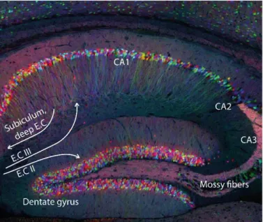
iii. Hippocampus
Hippocampus was named after its unusual and very organized sea-horse shape (figure 8). The hippocampus receives projections from the layer II or III of the entorhinal cortex (EC), and projects to subiculum or deep layers of the EC (figure 9). These bidirectional connections confer to the hippocampus a fundamental role in learning and memory.
Figure 7: 3D reconstitution of the
striatum (left) opposed to a coronal slice in which the striatum is colored in green (right).
Figure 6: 3D representation of
basal ganglia organization in a mouse brain. Green: striatum, yellow: globus pallidus external, red: globus pallidus internal, pink: subthalamic nucleus, blue: substancia negra pars compacta and purple: substancia negra pars reticula. From Kim et al., 2020
Figure 8: 3D reconstitution of
the hippocampus (left) opposed to a coronal slice in which the hippocampus is colored in pink (right).
28 Cell bodies are densely concentrated in Cornu Ammonis (CA) areas and are discriminated according to their localization and functions: CA1, CA2 or CA3 (figure 9). Although hippocampus cells are in majority excitatory pyramidal cells, similar to the pyramidal cell in the cortex, astrocytes are known to form tripartite synapses regulating neuronal processes (Bosson et al., 2017).
2. Structures communication results in a behavior
In the past, several experiments of ligation and lesion were realized in mammals proving that brain structures alone, disconnected of other structures, cannot play their role (Drewe, 1974; N. G. Müller & Knight, 2006; Nordborg & Johansson, 1995). Indeed, brain structures need to be interconnected and need to act cooperatively to produce a behavior adapted to the external stimuli. Each behavior is the product of a signal between and/or within brain structures. Neuronal networks resulting into a behavior can differ from an organism to the other, even between mammals. For example, 25% of human striatum possess different connectivity fingerprints compared to mice (Balsters et al., 2020). Despite these discrepancies, it is important to understand biological mechanisms in both physio- and pathological contexts in a simpler organism like the mouse at a first instance, in order to further apply the knowledge to human physiology and disease mechanisms (Collins et al., 2019).
This paragraph focuses on neuronal networks responsible for mouse behaviors studied during this work in physiological condition (procedural memory) and pathological condition in mouse model of human neurological disorders like AD (explicit memory) and Rett syndrome (motor coordination).
Figure 9: cortex and hippocampus form a network. Hippocampus
29
a. Corticostriatal network & motor coordination
Thanks to its connections with the cortex, basal ganglia, limbic system and the thalamus, the striatum is critical for the control of voluntary movements, motor learning but is also able to link emotions, rewards, executive functions, and mood. In this paragraph, we focus on the striatal control of the voluntary movements.
The crucial role of the corticostriatal network in controlling the movements is illustrated by symptoms observed in neurological disorders in which the striatum is specifically affected. In HD, the degeneration of the cortex and the striatum leads to the apparition of hyperkinesia often referred as chorea. Rett syndrome provoking motor infirmity is another example illustrating the central role of the corticostriatal network.
Within the corticostriatal network, two cellular pathways are regulating the control of voluntary movements: the permissive and monosynaptic direct pathway and the inhibitory and polysynaptic indirect pathway (figure 10). Both pathways implicate
MSNs (Brimblecombe & Cragg, 2015; Crittenden & Graybiel, 2011; Perrin & Venance, 2019) receiving excitatory afferences from neurons of cortical layer V within the motor or associative areas (respectively projecting to the dorsolateral or dorsomedial striatum). The direct pathway is then described as MSNs projecting to and inhibiting the GPi/SNr, nuclei known to inhibit the thalamus. Finally, the stimulated thalamus projects back to the cortex, completing the loop essential for smoothly executed movement. On the other hand, the indirect pathway results in an inhibition of the thalamus through a GPe-mediated inhibition of the SNr or the STN (in the latter case, we term this network the hyperdirect pathway).
Chorea in HD is thought to be due to a loss of MSNs projecting to the GPe leading to an excessive inhibition of the subthalamic nucleus and a subsequent reduction in the basal ganglia output from the thalamus. This network dysfunction could explain the involuntary movements. However, rigidity and akinesia observed in the later stages of HD would impact the MSNs projecting to the GPi/SNr. Removing inhibition from those neurons could convert hyperkinetic movements into hypokinetic problems (rigidity and akinesia).
Figure 10: movements are controlled by two loops involving cortex and basal ganglia. Direct and indirect
pathways of the corticostriatal loop. Green: striatum, yellow: globus pallidus external, red: globus pallidus internal, pink: subthalamic nucleus, blue: substancia negra pars compacta and purple: substancia negra pars reticula. Adapted from Kim et al., 2020
30 Cortical inputs from associative areas to MSNs can target DMS and control what we describe as goal directed behavior (Gangarossa et al., 2020; Hawes et al., 2015; Koralek et al., 2012; Ma et al., 2018; Shan et al., 2014), which is the decision of an action regarding the outcome in a specific situation and is directly dependent on the control of voluntary movements.
b. Learning & Memories
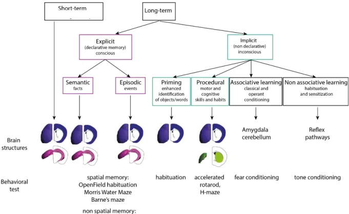
Learning and memory are behaviors often attributed to superior mammals like humans or primates but mice, flies or even worms also exhibit a certain form of learning and memory (L. B. Li et al., 2016; Phan et al., 2019). They are characterized by the sequence of three steps: the encoding of a new information into the neurons, the consolidation of this information for long-term memory and the retrieval.
Several forms of learning and memories exist and they rely on different brain structures (Camina & Güell, 2017; Quillfeldt, 2010) (figure 11) (to know more about memories: http://memorya.org). Long-term memory is opposed to the short-Long-term memory which can be verbal or visual, lasting up to 1 minute and recruited to be used immediately (remembering a phone number for instance). Mice take advantage of the short-term working memory when trying to escape from a maze for few minutes. Differently, long-term memory can last minutes, days, months, up to lifespan. The shift, after few seconds, of encoding between forgetfulness (characterizing the short-term memory) and the maintenance of the memory characterizes the long-term memory and is based on the consolidation of the memory at both synaptic and system levels. The process of consolidation, that can occur during sleep, allows a reinforcement of the memory traces recently encoded that are thus, labile and fragile (Kitamura et al., 2017; Tonegawa et al., 2018). At the cellular level, consolidation reinforces the connections of specific cells: the engram cells. By modulating the activation of the engram cell, it is possible to activate or inhibit the retrieval (Tonegawa et al., 2018). Changes in the physical or chemical properties of the neurons (synaptic consolidation) with time create a new organization in time and in space of the stimulated neuronal network (systems consolidation). For instance, long term explicit memory is known to appear in the hippocampus during the encoding phase and is then transferred to the cortex thanks to consolidation processes. Engrams cells are known and found within corticohippocampal network (Kitamura et al., 2017) but might also be present in corticostriatal networks as revealed by their specific increase in activity within the striatum upon cortical stimulation and after training (Badreddine et al., under revisions).
Long-term memory can refer to the explicit memory, highly flexible or to the implicit memory which stores knowledge acquired without conscious effort. Explicit memory is the memory intentionally recovered of facts, concepts (semantic) or personal experiences (episodic). This memory is stored in
31 the cortex and in the hippocampus. A mouse uses its explicit memory when remembering the localization of the platform in the Morris Water Maze. Implicit memory, tightly dependent on the conditions of learning, is involved in several processes like priming, procedural memory and
(non)associative memory. Priming improves the perception of a word or an object by prior exposure.
Associative learning associates an event with another; it is the case for fear conditioning experiments where an event (sound) is associated with electric foot shock. Non-associative learning is implicated through a change in the response of a stimuli due to repeated exposure.
Finally, procedural memory, is the memory of habits and is mobilized in human when we learn how to ride a bike or play an instrument. Habits can be defined by a stimulus-response association created only by the repetition of the stimuli rather than the desire or the fear of the outcome (associative learning). This memory is acquired through trials and errors, practice, and experience. To give a clear example, a mouse forms its procedural memory when falling off the accelerating rod repeatedly. At the end of the training, adapting its movements to the rod speed has become a habit. This type of
memory is known to be dependent on the corticostriatal network (Bosson et al., 2017; Costa et al., 2004; Perrin & Venance, 2019; Yin et al., 2009).
32
i. Corticohippocampal and corticocortical networks & explicit memory
Thanks to its connection with the cortex, the hippocampus receives sensory and spatial information and is able to store this information for days to a lifetime period. The ultimate and long-term storage takes place, after consolidation, in the cortex through hippocampi output towards the deep layers of the entorhinal cortex or the subiculum.
Corticohippocampal network is defined by two pathways called perforant pathways: a monosynaptic
and direct pathway and a trisynaptic and indirect pathway. In the direct pathway, projections from
layer III of the entorhinal cortex form synapses with CA1 neurons which project back to the cortex. The indirect pathway is defined by the layer II of the entorhinal cortex projecting to the granule cells of the dentate gyrus through the mossy fibers which then project to the CA1 neurons through the Schaffer collateral. The existence of these two pathways is thought to be necessary for learning and memory because they allow the comparison of the outputs received through both pathways.
Spatial memory evaluated by the Morris-Water Maze test in mouse is dependent on cortical and hippocampal plasticity. In fact, a spatial map of the testing environment is formed in the hippocampus of the tested mouse through firing pattern of specific neurons in CA1 and CA3 regions: the place cells. Although spatial memory is hippocampus-dependent, it seems that is not the case for object recognition which is thought to depend more on the cortex (Oliveira et al., 2010).
ii. Corticostriatal network & procedural memory
As detailed earlier, movement control is dictated through the loop involving mainly the cortex, the basal ganglia, and the thalamus. However, movement control is not only useful for goal directed behavior but also for learning and acquisition of new motor skills and habits, what we identify as the procedural memory (Costa et al., 2004; Graybiel & Grafton, 2015; Hawes et al., 2015; Hyungju Park et al., 2014; Yin et al., 2009). Interestingly, procedural memory, mainly dictated by the corticostriatal network, is known to be impaired in HD where this
network is the first to degenerate (Heindel et al., 1989; Van Asselen et al., 2012). Cortical inputs reside in either the associative cortex projecting to DMS, either the sensorimotor cortex projecting to the DLS (Mannella et al., 2013; Miyachi et al., 1997, 2002) (figure 12). Regarding striatal outputs during
procedural learning, both direct and indirect Figure 12: cortical projections target different striatal zones. associative cortex projects towards the DMS (left)
whereas sensorimotor cortex projects towards DLS, from Corbit et al., 2017.
33 pathways seem to be used since both DLS outputs are strengthened (O’Hare et al., 2016).
Two phases are involved in the learning of a new motor skill: a fast acquisition phase relying on the goal-directed behavior, and a slow mastery phase where the movement becomes unconscious (or implicit) and the habit is formed (Corbit et al., 2017; Costa et al., 2004; Yin et al., 2009). Neuronal mechanisms responsible for the establishment of procedural memory are still being investigated and two models co-exist in the literature to explain the roles of each network. The first model relies on a control shift from associative cortex-DMS network, responsible for the early learning of a task, to the sensorimotor-DLS network later during the training (Costa et al., 2004; Yin et al., 2009). The other model relies on a co-engagement of the two networks during learning and competing for control (Kupferschmidt et al., 2017; Perrin & Venance, 2019).
Thus, communication between two neurons is crucial for the emergence of a behavior such as learning and memory. Consequently, it is important to understand how two neurons communicate. In the following paragraph, we will focus on how neuronal morphology serves neuronal functions and communication.
3. The neuron: the structural and functional unit of the brain
Brain structures’ communication is based on the functional transmission of information between two neurons. Before detailing neuron specificities, it is relevant to recapitulate the main hallmarks of the physiology of cells.
a. Cellular physiology
Cellular physiology represents the elements regulating the functions of a cell, among which the cellular homeostasis, regulated by cellular signaling pathways, is crucial for the establishment of the cell state equilibrium.
i. Cellular homeostasis
First conceptualized by Claude Bernard in the 19th century who investigated the stability of the “milieu interieur”, the cellular homeostasis has then been pointed out by Cannon for its crucial role in survival (Cannon & Rosenberg, 1932). Cellular homeostasis can be defined as a dynamic equilibrium of a cellular steady state (variable, set point) established by the response of the cell (feedback) from the reception of intra and extra-cellular events (information from a sensor) (Styr & Slutsky,
Figure 13: cellular homeostasis can be defined as a closed feedback loop regulating cellular functions. From Macleod & Zinsmaier, 2006
34 2018) (figure 13). When it is impaired in neurons, it is thought to be the source of neurological disorders like AD, PD and HD (D. Fernandes & Carvalho, 2016; Soukup et al., 2018). Cellular homeostasis depends partly on protein homeostasis since both protein quantity and quality are crucial for the good functioning of the cell. Thus, physiological protein levels and clearance of the damaged protein are important for the cell to keep its homeostasis. For instance, Aβ peptides have been shown to be toxic for neuronal functions if they are either at high or low concentration (Abramov et al., 2009; Puzzo et al., 2008).
ii. Cellular signaling pathways
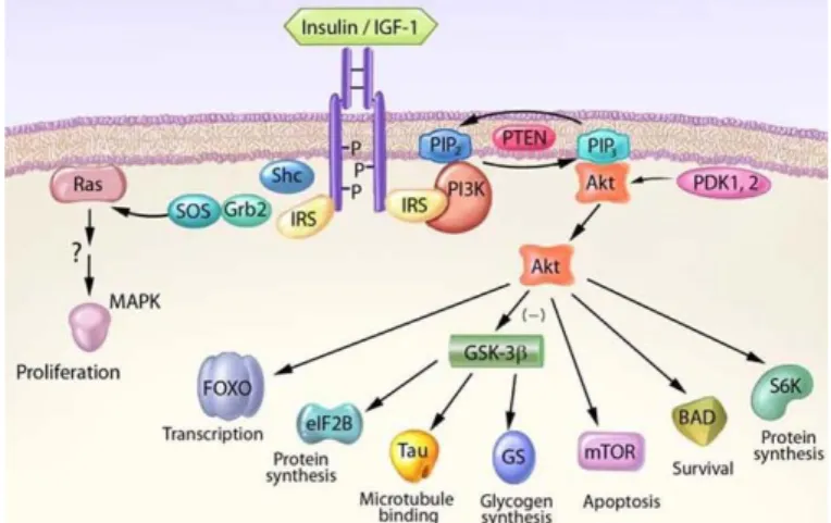
Cellular homeostasis is regulated by an entire set up of cellular pathways. A dozen of signaling pathways are described in the literature and one particularly interesting is the IGF-1/Akt pathway. IGF-1 (Insulin -like growth factor-1) is a
growth factor able to activate many signaling pathways, thus responsible for several functions in cells like survival and differentiation (Bondy et al., 2006). When bound to its tyrosine kinase receptor at the surface of the cell, it causes the activation of intrinsic tyrosine kinases. Targeted protein like IRS-1 and Shc will then be phosphorylated leading to the activation of
several pathways including the one relying of the PI3K/Akt and the MAP kinase (ERK). PI3K activation leads to an Akt recruitment to the plasma membrane where it is phosphorylated by PDK-1 and -2 on Thr308 and Ser473. Akt, a serine/threonine kinase, is known for its roles in cell development, growth and survival when activated. This diversity of functions is the results of the many Akt targets like GSK-3β, Bad, caspases, mTOR and FOXO, promoting cell survival and inhibiting apoptosis (W. H. Zheng & Quirion, 2006) (figure 14). For instance, by inhibiting GSK-3β, Akt is able to prevent Tau accumulation observed in AD (Y. S. Hu et al., 2013; Kitagishi et al., 2014).
Thus, as any other cell, a neuron needs this equilibrium state to survive and perform its roles. However, a neuron is a specific cell in which cellular homeostasis can differ according to its type, its compartments, and its roles.
Figure 14: IGF-1 pathway regulates cell survival through Akt activation. From Bondy et al., 2006
35
b. The neuron: a highly specialized cell
A neuron is a specific cell whose shape is crucial for its function and vice versa.
i. Different types of neurons
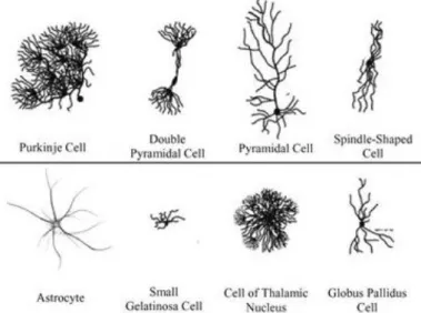
Throughout the brain, dozens of neuron types exist and are characterized by a morphology matching their functions (Weissman et al., 2007) (figure 15).
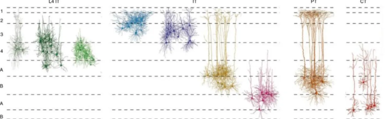
For instance, pyramidal neurons in the cortex have been described at the end of the 19th century by Ramón y Cajal, famous for its functional and morphological drawings of neurons based on Golgi staining’s (Llinás, 2003) (figures 15 and 16). Pyramidal cells present a primary and apical dendrite connected to the soma in a specific layer projecting its axons in specific output structures. Although this type of neurons is predominantly present within the cortex, one of Ramón y Cajal students, Rafael Lorente de Nó later identified more than 40 types of cortical neurons based on the distribution of the dendrites and axons.
Brain structures are often characterized by their types of neurons: Purkinje cells, presenting a huge dendritic arborization with many spines, are only found in the cerebellum whereas MSNs characterized by a high dendritic spine number are striatum-specific (Klapstein et al., 2001) (figure 15 and 17). These characteristics can be explained by the fact that the neuronal shape and neuronal function influence each other. For instance, MSNs are thought to compensate their smaller number within the striatum, compared to the number of cortical projecting neurons, by the high density of dendritic spines, emphasizing their central role in receiving and selecting the information from the different cortical areas (figure 17).
Figure 16: pyramidal cells display a specific organization. Santiago Ramón y Cajal, Golgi
stained pyramidal cells of the cerebral cortex (detail) ink and pencil on paper. From Cajal Institute, Spanish National Research Council
Figure 15: neuronal shapes define neuronal functions. From
36
ii. A polarized structure
Both cell shape and neuronal function imply a polarity within the cell. All neurons are compartmentalized with neurites receiving or sending the signal in order to communicate. Polar morphology and function directly depend on the organization of the cytoskeleton (Kelliher et al., 2019).
1. Neuronal compartments
Neuronal communication is supported by a specific spatial compartmentalization of the neuron separating the neurite receiving the signal, the dendrite (input) and the one sending the signal, the axon (output) (figure 18). Between those two neurites stand the soma and its nucleus.
Figure 17: morphologic properties differentiate cortical pyramidal cells from MSN. 3D
reconstitution of a pyramidal neuron (A) and a MSN (B), adapted from Klapstein et al., 2001.
Figure 18: neuronal networks are formed with a presynaptic neuron and a postsynaptic neuron communicating through a synapse. Schematic representation of neuronal organization adapted from Leterrier, 2018.