Université de Montréal
Short-term memory of temporal aspects of noxious and
innocuous thermal sensation: psychophysical and fMRI
studies
par Mina Khoshnejad
Département de Neurosciences Faculté de Médecine
Thèse présentée à la Faculté des études supérieures en vue de l’obtention du grade de Philosophiae Doctor (Ph.D.)
en sciences neurologiques
Sept, 2017
Université de Montréal
Faculté des études supérieures et postdoctorales
Cette thèse intitulée :
Short-term memory of temporal aspects of noxious and innocuous thermal sensation: psychophysical and fMRI studies
Présentée par : Mina Khoshnejad
a été évaluée par un jury composé des personnes suivantes :
Dr. Elaine Chapman, président-rapporteur Dr. Pierre Rainville, directeur de recherche
Dr. Pierre Jolicoeur, membre du jury Dr. Ron Kupers, examinateur externe
Résumé
La douleur peut être considérée comme un système de protection qui signale une menace et qui nous avertit des dégâts imminents aux tissus. En tant que mécanisme de défense, il nécessite l'apprentissage et la mémoire des expériences du passé pour la survie et les comportements liés à la douleur. Par conséquent, notre expérience de la douleur actuelle est fortement influencée par les expériences antérieures et l'apprentissage. Cependant, malgré son importance, notre compréhension actuelle de l'interaction entre le système de la douleur et le système de mémoire est très limitée. La mémoire de la douleur est un sujet de recherche très vaste. Il nécessite une compréhension des mécanismes impliqués à chaque étape du système de mémoire (mémoire immédiate, à court terme et à long terme) et l'interaction entre eux. Parmi les étapes multiples de la mémoire, la mémoire à court terme de la douleur est une zone qui est moins recherchée, alors qu'il existe une énorme quantité de recherche neuroscientifique dans la mémoire à court terme sur d'autres modalités, en particulier la vision. L'étude de la mémoire à court terme de la douleur est particulièrement importante car cette trace de la mémoire à court terme de la douleur est ensuite convertie en mémoire à long terme et affecte ensuite les expériences futures de la douleur. Cette thèse est largement axée sur la mémoire à court terme de la douleur.
La complexité et la multi dimensionnalité de la douleur ajoutent encore un autre élément à la recherche sur la mémoire de la douleur. Par exemple, la trace de la mémoire de la douleur peut contenir des traces de mémoire de diverses composantes de la douleur telles que la réponse sensorielle affective, cognitive et motrice et l'interaction entre elles. Par conséquent, une première étape dans l'exploration neuroscientifique de la mémoire de la douleur nécessite la réduction de l'expérience de la douleur tout en englobant tous ces différents composants à un seul composant. Dans la recherche présentée ici, nous avons généralement examiné cela par des instructions d'attention ‘ top-down’ pour assister à la dimension sensorielle de la douleur. La recherche précédente sur la mémoire à court terme de la douleur a également porté principalement sur la dimension sensorielle de la douleur. Cependant, parmi les dimensions
sensorielles de la douleur, la mémoire à court terme de l'intensité et de la dimension spatiale de la douleur a fait l'objet de recherches antérieures. Malgré son importance, la dimension temporelle de la douleur est restée complètement inexplorée dans la recherche sur la mémoire de la douleur.
La recherche menée dans cette thèse est consacrée à l'exploration de la mémoire à court terme de la durée de la douleur. La durée de la douleur peut être suivie de manière indépendante, mais peut également être suivie conjointement avec la dimension d'intensité telle que le suivi dynamique de l'intensité de la douleur dans le temps. Les études menées dans cette thèse traitent spécifiquement du traitement isolé de la durée de la douleur ainsi que du traitement conjoint de la dimension durée / intensité de la douleur.
La première étude psychophysique a exploré la nature de la représentation mentale du modèle de mémoire de la douleur thermique dynamique et a également été conçue pour aborder les différences de la dimension sensorielle et affective de la douleur thermique dans la mémoire à court terme. La deuxième étude psychophysique portait sur les propriétés de la mémoire à court terme de la sensation thermique non douloureux en comparant le suivi dynamique de la sensation et le suivi isolé de la durée d'un événement thermique non douloureux. La troisième étude poursuit l'exploration du traitement dynamique de la durée conjointement avec l'intensité par rapport au traitement isolé de la durée dans la mémoire à court terme en utilisant des stimuli thermiques douloureuse une résonance magnétique fonctionnelle (IRMF).
Dans l'ensemble, les résultats des études psychophysiques ont montré une transformation significative de la durée et de la dynamique de la sensation thermique douloureux et non-douloureux dans la mémoire à court terme; comme la perte d'informations somatosensorielles temporelles en mémoire. Nous avons en outre montré une amélioration du rappel de la durée dans le suivi dynamique de la durée, en comparaison avec le suivi de la durée isolée. Nous avons également montré des différences dans les corrélats neuronaux de la mémoire à court terme de la durée de douleur par rapport à la dynamique de douleur. L'étude de l'IRMF a montré des similitudes frappantes dans les corrélats neuronaux sous-jacents à la
mémoire à court terme de douleur et d'autres modalités telles que la contribution des coticés fronto-pariétales ainsi que les corticaux sensoriels impliqués dans le traitement perceptuel.
Abstract
Pain can be viewed as a protective system that signals threat and alerts us to impending tissue damage. As a defense mechanism, it necessitates the learning and memory of past painful experiences for survival and pain-related behavior. Therefore our current pain experience is heavily influenced by previous experiences and learning. However, despite its importance, our current understanding of the interaction between the pain system and the memory system is very limited. Pain memory is a very broad topic of research on its own. It requires an understanding of the mechanisms involved at each stage of the memory system (immediate, short-term, and long-term memory), and the interaction among them. Among the multiple stages of memory, the short-term memory of pain is an area that is less researched, while there are enormous amount of neuroscientific research in short-term memory of other modalities, particularly vision. Investigation of the short-term memory of pain is especially important as the short-term memory trace of pain is converted to long-term memory and subsequently affects future pain experiences. This thesis is broadly focused on the short-term memory of pain.
The complexity and multi-dimensionality of pain adds yet another element to the research on pain memory. For example, the memory trace of pain may contain memory traces of various components of pain such as sensory, affective, cognitive, and motoric responses, and the interactions among them. Therefore, an initial step in the neuroscientific exploration of pain memory requires narrowing down the pain experience, which encompasses all of these various components, to one single component. In the research presented here, we achieved this using top-down attentional instructions to attend to the sensory component of pain. The previous research on short-term memory of pain also focused mainly on the sensory component of pain. However, within the sensory component of pain the short-term memory of intensity and spatial dimension of pain has been the focus of previous research. Despite its importance, the temporal dimension of pain remained completely unexplored in pain memory research.
Thus, the research conducted in this thesis is devoted to the exploration of short-term memory of the duration of pain. Pain duration can be tracked independently, but it can also be tracked conjointly with intensity, such as in dynamic tracking of pain intensity over time. The studies addressed in this thesis examined the isolated processing of pain duration as well as conjoint processing of the duration and intensity of pain.
The first psychophysical study explored the nature of the mental representation of the memory template of dynamic thermal pain sensation and, additionally, addressed the differences between the sensory versus affective dimensions of thermal pain sensation in short-term memory. The second psychophysical study focused on properties of the short-term memory of innocuous thermal sensation by comparing dynamic tracking of sensation versus isolated tracking of duration of an innocuous thermal event. The third study explored the dynamic processing of duration conjointly with intensity, versus the isolated processing of duration in short-term memory, using noxious thermal stimuli and functional magnetic resonance imaging (fMRI).
Overall, the results of the psychophysical studies showed significant transformation of duration and dynamics information of noxious and innocuous thermal sensation in short-term memory, such as loss of temporal somatosensory information. Additionally, we showed improvement in duration recall during dynamic tracking versus isolated tracking of duration. The fMRI study revealed differences in neural correlates of short-term memory of pain duration versus pain dynamics. Importantly, it also showed striking similarities between neural correlates underlying the short-term memory of pain and those underlying other modalities, such as a contribution of fronto-parietal cortices as well as sensory cortices involved in perceptual processing.
Table of Contents
Résumé ... iii
Abstract ... vi
Table of Contents ... viii
List of Tables ... xi
List of Figures ... xii
List of Abbreviations ... xv
Acknowledgements ... xvii
Chapter 1: General Introduction ... 18
Chapter 2: Pain System ... 21
2.1. Introduction ... 22
2.2. Somatic sensation... 23
2.2.1. First-order neuron ... 23
2.2.2. Second-order neuron ... 25
2.2.3. Third-order neuron ... 25
2.3. Cortical and subcortical regions involved in pain processing ... 28
2.3.1. Primary and secondary somatosensory cortex ... 28
2.3.2. Insula ... 30
2.3.3. Anterior Cingulate Cortex (ACC) ... 32
2.3.4. Basal Ganglia (BG) ... 34
2.3.5. Prefrontal cortex (PFC) ... 35
2.3.6. Motor cortex... 36
2.4. Controversies regarding the pain matrix ... 38
Chapter 3: Short-term memory system ... 40
3.1. An overview ... 41
3.2. Theoretical frameworks and properties of different types of memory ... 41
3.3. Definition of working memory and general overview ... 44
3.5. Role of attention in working memory ... 46
3.6. Contribution of prefrontal cortex ... 47
3.6.1. Storage ... 48
3.6.2. Top-down control... 50
3.6.3. Alternative accounts... 51
3.7. Contribution of sensory cortices ... 52
3.7.1. Visual modality ... 52
3.7.2. Tactile modality ... 55
3.8. Contribution of the posterior parietal cortex ... 58
3.8.1. Storage ... 59
3.8.2. Top-down control... 60
Chapter 4: Review of short-term memory studies of pain ... 62
4.1. General overview ... 63
4.2. Sensory short-term memory of pain ... 64
4.2.1. Review of behavioral measures of recall performance ... 66
4.2.2. Review of imaging results ... 68
4.3. Discussion ... 78
Chapter 5: Review of time-perception literature ... 87
5.1. An overview ... 88
5.2. Basic definitions and multiple categories of research ... 88
5.3. Range of duration ... 90
5.4. Theoretical models ... 91
5.5. Modulation in perceived duration by stimulus characteristics ... 92
5.6. Psychophysical studies on the contribution of cognitive factors to duration judgment 93 5.7. Neural substrate of timing ... 95
Chapter 6: Articles ... 101
6.1. General objectives and hypotheses of the thesis ... 102
Article 1: Remembering the dynamic changes in pain intensity and pain unpleasantness: A psychophysical study ... 104
Article 2: The delayed reproduction of long time intervals defined by innocuous thermal
sensation ... 141
Article 3: Brain processing of the temporal dimension of acute pain in short-term memory ... 169
Chapter 7: General conclusion ... 207
7.1. An overview ... 208
7.2. General summary of the main results of the studies ... 209
7.3. Clinical importance of pain memory research ... 212
7.4. Limitations of experimental studies of pain memory ... 213
7.5. Future research avenues ... 215
7.6. Conclusion ... 216
List of Tables
Chapter 4:Table 1. Brain activations in studies using delayed discrimination paradigms. ... 70 Table 2. Brain activation in studies using delayed recall paradigms. ... 70
Chapter 6 – Article 1:
Table 1. Significant main effects and interactions defined by ANOVA on the components 1-3. ... 122 Table S1. 4-Way ANOVA results on the extracted parameters from VAS curves (significant results with p=<0.01 are highlighted). ... 138 Table S2. 4-Way ANOVA results on the components (Comp1-3) obtained from the PCA (significant results with p=<0.01 are highlighted). ... 138 Table S3. Linear correlation analysis results (Pearson-r) on the extracted parameters (significant results with r=>0.5 are highlighted). ... 140
Chapter 6 – Article 2:
Table 1. Summary of ANOVAs... 154
Chapter 6 – Article 3:
Table 1. Contrasts of interests tested across the two models (models 1-2). ... 182 Table 2. Global Effect of the Duration Task vs. the Intensity (control) Task (Model 1b). .... 188 Table 3. Delay Effect of the Dynamic Task vs. the Intensity Task (control) on brain activity (Model 2a). ... 191 Table 4. Delay Effect of the Duration Task vs. the Intensity Task (control) on brain activity (Model 2b). ... 191
List of Figures
Chapter 4 – Review of short-term memory studies of pain
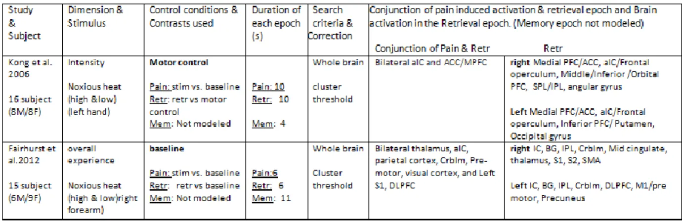
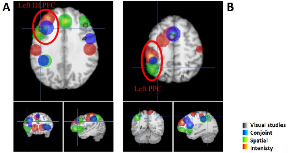
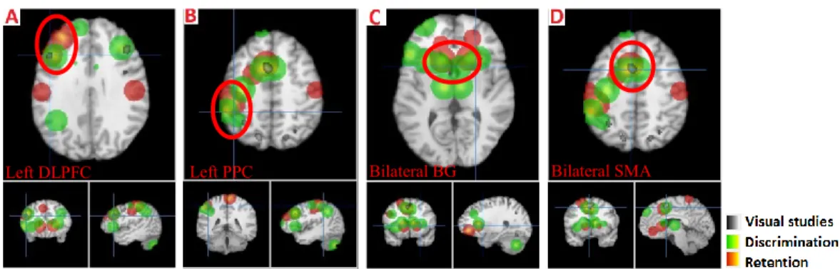
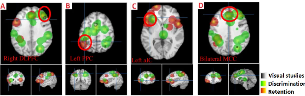
Figure 1. Fronto-parietal activation in the retention epoch... 72 Figure 2. Activation in the retention (red) and discrimination (green) phases of the intensity dimension. ... 74 Figure 3. Activation in the discrimination (green) and delayed recall (blue) phases of delayed discrimination and delayed recall paradigms, respectively. ... 75 Figure 4. Activation in the retention (red) and discrimination (green) phases of the spatial dimension. ... 76 Figure 5. Activation in the retention (red) and discrimination (green) phase of the conjoint intensity/spatial dimension... 78
Chapter 6 – Article 1:
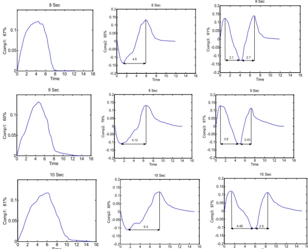
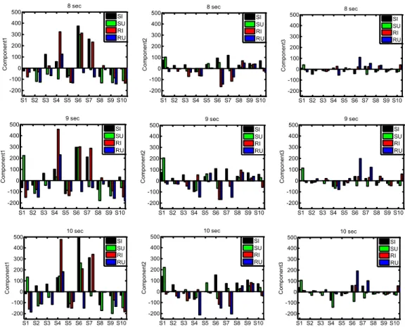
Figure 1. Temporal profile of the actual stimulus delivered consists of three phases: onset, plateau, and offset. ... 110 Figure 2. Mean VAS profiles for 8 sec, 9 sec and 10 sec trials in each subject (subject 1 to 10 from left to right) in the conditions simultaneous intensity (SI - black), simultaneous unpleasantness (SU - green), retrospective intensity (RI - red), and retrospective unpleasantness (RU - blue). ... 116 Figure 3. Coefficients associated with the first three components of the PCA (Comp1-3) across three durations (first row 8 sec data, second row 9 sec data, and third row 10 sec data). ... 118 Figure 4. PCA outputs (Scores) for each individual across the four conditions (mean of 4 trials per condition) and across the three tested durations; first row 8 sec data, second row 9 sec data, and third row 10 sec data... 119 Figure 5. Reconstructed VAS curves for each condition (SI, SU, RI and RU) with the use of only component1, component1 and 2, and finally component 1, 2 and 3 (left to right). ... 121
Figure 6. Classification of the two conditions (SI: simultaneous intensity and SU: simultaneous unpleasantness) in the space of component 1 and component 2 with a linear classifier (represented by the line separating the two shaded areas). ... 123 Figure S1. Typical raw time course of continuous e-VAS ratings (left panel) and its derivative (right panel) showing some of the parameters extracted. ... 135 Figure S2. Effects of memory and stimulus duration (8-9-10s) on the time to maximal pain (TPM, left panel) and total time (TT, right panel). ... 139
Chapter 6 – Article 2:
Fig 1. Stimulus temporal profiles in the continuous (a) and segmented (b) conditions. ... 150 Fig 2. Mean and standard error of duration in synchronized dynamics and delayed dynamics conditions across continuous and segmented trials. ... 155 Fig 3. Mean and standard error of segments’ duration in synchronized dynamics and delayed dynamics conditions for the segmented trials. ... 156 Fig 4. Mean and standard error of duration in delayed dynamics and delayed duration conditions across continuous and segmented trials. ... 157
Chapter 6 – Article 3:
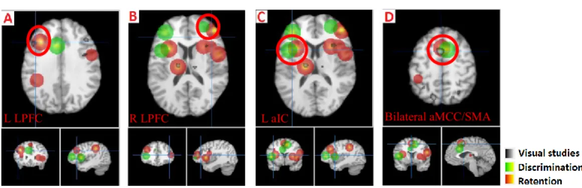
Figure 1. Experimental stimuli and tasks. ... 177 Figure 2. Behavioural results for (A) reproduced duration in dynamic and duration tasks, (B) reproduced maximum pain in the dynamic task, and (C) reproduced overall pain in the intensity task. ... 185 Figure 3. Cortical activation of the global task effects (Model 1) in the duration vs. intensity tasks (A. Dur; contrast 1b in Table 1; see coordinates of local maxima in Table 2) and dynamic vs. duration tasks (B. Dyn vs. Dur; contrast 1c in Table 1). ... 187 Figure 4. Cortical activation associated with the delay period of the dynamic (A. contrast 2a) and duration task (B. Contrast 2b) compared to the control intensity task and for the contrast between the dynamic and duration tasks (C. contrast 2c), as assessed with Model 2. ... 190
Figure 5. Results of the parametric modulation of the retention epoch of the dynamic task with reproduced duration. ... 192 Figure S1. Mean beta values extracted for the target task (dark grey) and the control task (light grey) for the clusters identified for the task contrasts in model 1 (Global task model). ... 205 Figure S2. Mean beta values extracted for the target tasks (dark grey) and the control task (light grey) for the clusters identified for the task contrasts in model 2 (Task-epochs model). ... 206
List of Abbreviations
ACC anterior cingulate cortex aIC anterior insular cortex ANOVA analysis of variance BG basal gangliaBOLD blood-oxygen-level-dependent CNV contingent negative variation DLPFC dorsolateral prefrontal cortex DMS delayed match-to-sample DPA delayed paired association ERP event-related potential
e-VAS electronic visual analogue scale FDR false discovery rate
fMRI functional magnetic resonance imaging FWER family-wise error rate
GLM general linear model IC insular cortex
IFG inferior frontal gyrus IPS intra-parietal sulcus IT inferotemporal cortex LTM long-term memory MCC mid cingulate cortex MEG magnetoencephalography MFG medial frontal gyrus mPFC medial PFC
MT middle temporal cortex MTG middle temporal gyrus MVPA multivariate pattern analysis NS nociceptive specific
OFC orbitofrontal cortex OFC orbitofrontal cortex PAG periaqueductal grey
PCA principal component analysis PD Parkinson’s disease
PM pain matrix PMC premotor cortex
PPC posterior parietal cortex
rTMS repetitive transcranial magnetic stimulation SFG superior frontal gyrus
SI primary somatosensory cortex SII secondary somatosensory cortex SMA supplementary motor area SMG supramarginal gyrus SPG superior parietal gyrus SPL superior parietal lobule STM short-term memory STT spinothalamic tract
TMS transcranial magnetic stimulation V1 primary visual cortex
VAS visual analogue scale
VLPFC ventrolateral prefrontal cortex VMPFC ventromedial prefrontal cortex VMpo ventromedial posterior nucleus VPI ventral posterior inferior VPL ventral posterior lateral VPM ventral posterior medial VWM visual working memory WDR wide dynamic range
Acknowledgements
First, I would like to thank my supervisor, Dr. Pierre Rainville, whose excellent guidance, support and encouragement, allowed me to pursue my interests, and develop passion for the research I have been doing. I totally indebted to him, for his great mentorship, and for his continual support in allowing me to explore and peruse my curiosity and interests.
I also thank Kristina Martinu, for her generous help in acquiring the psychophysical and fMRI data. She has been a great friend to me, and supported me in so many ways.
I especially thank my husband Nima, who was so supportive through the thick and the thin, during my Phd Journey. I dedicate this to him and our 3 year old son who is the joy of our life.
The perception of pain is a complex phenomenon involving sensory, affective, cognitive, and motor components that result from a distributed neural processing network of cortical and subcortical regions. Recent advances in pain research expanded our understanding of interactions among multiple components of pain and the mechanisms underlying the perception of pain. One of the important factors that contributes to our perception of pain is the memory of past pain experiences. However, little research has been done concerning the mechanisms and neural processes involved in the memory of pain. Pain memory is a very broad topic encompassing a classical interactive multi-stage information processing system that comprises immediate, short-term, and long-term memory. There are some studies on immediate memory and some clinical studies on long-term recall of pain. However, only a few studies have examined short-term memory of pain. Understanding the mechanisms and properties of short-term memory of pain is important as it is an intermediate stage that relates immediate memory and long-term memory. In particular, the transformation of pain-related information that occurs in short-term memory is relayed to long-short-term memory and is known to impact the perception of future pain. This thesis is broadly focused on the topic of explicit short-term memory of pain.
With regard to multiple components of pain, the sensory aspect of pain is a most prominent aspect and consists of multiple attributes such as intensity, spatial, and duration. However, neuroscientific examination of sensory characteristics of pain has largely focused on the intensity and spatial attributes. In particular, no previous research has explored the short-term memory of pain duration. The experimental studies described in this thesis are designed to provide some understanding of properties and neural correlates of the temporal processing of pain as well as the conjoint processing of duration and intensity of pain in short-term memory.
The organization of this thesis is described here. Chapter 2 provides an overview of the pain system and the functional roles of cortical and sub-cortical regions generally involved in processing pain-related information. Chapter 3 provides a summary of the memory system followed by a comprehensive overview of the functional roles of the
main cortical areas involved in short-term memory of visual and tactile modality. Chapter 4 is devoted to a review of the few studies that have examined short-term memory of pain. This chapter is in preparation for submission as a review paper. Chapter 5 is a general overview of the theoretical perspective, properties, and neural mechanisms involved in time perception. Chapter 6 comprises three published articles as follows: Article 1 is a psychophysical study concerning the nature of mental representations for the retention of pain dynamics and the ensuing transformation of pain-related information in short-term memory; Article 2 is a second psychophysical study investigating the properties of short-term memory of the duration of innocuous thermal stimuli; and Article 3 is an imaging study on short-term memory of pain dynamics and duration. General conclusions are proposed in Chapter 7.
2.1. Introduction
Pain is an unpleasant sensory experience associated with tissue damage. The perception of pain is evoked by high threshold mechanical, thermal, and noxious chemical stimuli, and is detected by specialized peripheral sensory neurons called nociceptors. Nociceptors in turn transfer information about noxious stimuli through multiple ascending pathways to the neocortex, where the perception of pain is elicited. Pain is highly individual and subjective, and is elicited by interactions among multiple processing components.
Noxious stimuli provoke multiple autonomic and physiological responses such as withdrawal reflexes, increases in heart rate, and changes in skin conductance, as well as behavioral responses such as the generation of unpleasant emotional states accompanied by a motivational force to escape or avoid the potential harm. First established in a formal integrative neuropsychological model by Melzack and Casey, pain is generally recognized as a multidimensional experience comprising of sensory, affective, motivational and cognitive components (Melzack and Casey 1968). The sensory-discriminative component comprises of multiple aspects such as recognition of the noxious nature of the stimuli, the quality of pain (burning, pricking, stabbing, etc.), as well as its location, intensity, and duration. The affective component of pain is itself separated into two sub-components: immediate unpleasantness of the experience as well as a more elaborate reflection of the long-term meaning and consequences of pain, which is termed secondary pain affect. The cognitive component involves attention, anticipation, and memory of past experiences, and through interactions with other components modulates our perception of pain. It has been discussed that there is a causal relationship between multiple components of pain such that the sensory-discriminative dimension leads to an immediate emotional reaction that integrates with past memories of having pain and in turn leads to secondary pain affect (Wade et al. 1996).
Traditionally, it has been taught that these components, especially the sensory vs. affective components, are dissociated and processed in parallel via lateral and medial pain
pathways, with the former mainly processed in post-central gyrus and the latter processed in limbic areas such as the anterior cingulate cortex. However, recently there has been a shift to the acknowledgment that the sensation of pain is a complex phenomenon and that its various components interact with each other. In this chapter, a brief review of the transmission of nociceptive information from the spinal cord to the cortex is provided (section 2.2), followed by a description of the role of cortical and sub-cortical structures involved in processing pain-related information (section 2.3). Finally, recent ideas involving the specificity of the identified cortical pain network are discussed in section 2.4.
2.2. Somatic sensation
Somatic sensation is divided into four distinct sub-modalities: touch, proprioception, pain, and temperature sense. Like other sensory modalities, somatic sensation is a result of signal processing at various stages (peripheral, spinal, brain stem, and cerebral cortex) evoked by somatic sensory event. The touch and proprioception modalities are transmitted by dorsal column medial lemniscal pathways to the thalamus and cerebral cortex, while information about pain and temperature sensation is transmitted by the anterolateral system. As the work in this thesis used noxious and innocuous thermal stimuli, a description of major ascending pathways of the anterolateral system is provided. Pain and temperature pathways generally consist of a three-neuron chain that transmits pain and temperature information from the periphery to the cerebral cortex. The main components of the pain and temperature pathways are described below.
2.2.1. First-order neuron
Transmission of pain begins with activation of nociceptors in response to noxious stimuli in the surrounding tissues (for a review of cellular and molecular mechanisms see
Basbaum et al. 2009). Nociceptors are the peripheral ending terminals of the first-order sensory neurons and are found in skin, muscle, joints, and viscera, and are activated by noxious cold, heat, intense mechanical stimuli, and a variety of chemical mediators. The first-order sensory neurons innervating limbs and the trunk have their cell bodies located in dorsal root ganglia of the spinal cord and their axons terminate predominantly in the dorsal horn of the spinal cord. The first-order sensory neurons innervating the head and face have their cell bodies in the trigeminal ganglion and their axons terminate in the trigeminal nucleus in the medulla.
There are several classes of nociceptor: I) unimodal, II) polymodal, and III) silent (Kandel et al. 2000). The unimodal nociceptors consist of thermoreceptors that selectively respond to noxious thermal stimuli (both cold <5 °C and hot > 45 °C temperatures) or mechanoreceptors that selectively respond to intense pressure. These nociceptors transmit information via small diameter thinly myelinated Aδ fibers with a fast conduction velocity of 5-30 m/s. Polymodal nociceptors respond to noxious stimuli of mechanical, thermal, and chemical nature. They transmit the nociceptive information via unmyelinated C fibers with a slow conduction velocity of about 1 m/s. Unimodal and polymodal nociceptors innervate skin and deep tissues. Silent nociceptors innervate viscera and are difficult to activate within normal range of noxious stimulus intensity. They become active after tissue damage in response to endogenous chemical stimuli (inflammatory mediators) associated with tissue injury, and respond to noxious thermal or mechanical stimuli after becoming activated. They also transmit information through C fibers. The basic function of nociceptors is to transmit information to second-order neurons about actual or potential tissue damage.
Transmission of temperature information begins with the activation of thermal
receptors, which are the peripheral ending of first-order neurons whose cell bodies are
located in dorsal root ganglions and whose axons terminate in the dorsal horn. Thermal receptors fire as a function of temperature difference between skin and objects contacting the body, or the air. Thermal receptors have a steady-state firing rate at normal skin temperature. Warming the skin evokes an increase in the firing rate of the warmth
receptors and decreases of the firing rate of cold receptors. Cooling the skin produces opposite effects: an increase in the firing rate of cool receptors and a decrease in the firing rate of warmth receptors. The relative activity by warmth and cool receptors evokes the perception of temperature. Thermal receptors transfer information by Aδ and C fibers to the dorsal horn of the spinal cord (Kandel et al. 2000).
2.2.2. Second-order neuron
By activation of nociceptors, noxious mechanical, thermal, or chemical energy is transformed to electric action potentials by the membrane receptors through transduction phenomenon and transmit nociceptive information to the second-order neurons predominantly in the dorsal horn of the spinal cord. These neurons in the dorsal horn are classified as nociceptive specific (NS), which respond exclusively to noxious stimuli, or wide-dynamic-range (WDR), which respond to both noxious and innocuous stimuli. Projection neurons are mainly from lamina I (consisting of both NS and WDR), lamina V (WDR neurons), and to a lesser degree from lamina VI, VII, and VIII. Nociceptors innervating the head and face are transmitted similarly to the second-order neurons located in the trigeminal nucleus in the medulla (Kandel et al. 2000).
2.2.3. Third-order neuron
The axons of second-order neuron decussate and ascend via the anterolateral system to innervate the third-order neurons in supra-spinal targets such as the thalamus, the periaqueductal gray (PAG), and brain stem nuclei. The anterolateral system consists of multiple pathways, including a direct pathway to the thalamus via the spinothalamic tract, as well as indirect pathways to the thalamus by way of synapses in the reticular formation of the medulla via the spinoreticular tract, brainstem nuclei via the spinomesencephalic tract, or the hypothalamus via spinohypothalamic tract.
The spinothalamic tract (STT), which is the main ascending pain pathway, originates mainly from second-order neurons in lamina I and V of spinal dorsal horn and transfers noxious and thermal information from the limbs and trunk directly to the contralateral thalamus. Pain and temperature sensation from the head, face, and neck is transferred by an analogous pathway called the trigeminothalamic tract to the thalamus. The main somatosensory relay nuclei of the STT is the ventral posterior nuclear group, which includes the ventral posterior lateral (VPL), ventral posterior medial (VPM), and ventral posterior inferior (VPI) nuclei. While the VPL receives information from the STT, the VPM receives information from the trigeminothalamic tract. There is controversy with regard to the projection sites to VPL nuclei from the dorsal horn: while some suggest that they receive projections from lamina V (Craig 2006), others assert that superficial layers (lamina I) mainly project to the VPL (Willis, Zhang et al. 2001).
It has been shown that VPL and VPM nuclei consist of thermo-sensitive and nociceptive neurons (Apkarian and Shi 1994; Bushnell et al. 1993). In addition, electrophysiological studies in awake monkeys showed that VPM neurons show graded responses to increasing intensity of somatosensory stimulation, which suggests that they play a role in the sensory-discriminative aspect of nociception and thermal sensation (Bushnell et al. 1993). It has been shown that VPL/VPM neurons project to primary somatosensory cortex (SI) (Gingold et al. 1991). VPI nuclei receive projections from both lamina I and V (Craig 2006), and project to secondary somatosensory cortex (SII) (Friedman and Murray 1986; Stevens et al. 1993). The three nuclei of the ventral posterior nuclear group are thought to contribute primarily to sensory-discriminative aspects of pain.
The STT also has dense projections to multiple nuclei in the posterior complex (PO) such as the suprageniculate nucleus (Sg) and limitans nucleus (L) (Boivie 1979; Davidson et al. 2008; Ralston and Ralston 1992). Another group of important posterior thalamic nuclei that was more recently identified is the posterior part of the ventromedial nuclei (VMpo), which is specifically activated by noxious and thermal stimulation and receives projection from lamina I (Craig et al. 1994; Craig 2006; Dostrovsky and Craig
1996). It has been shown that VMpo projects to the posterior insula (Craig 2014), an area that has is well-known to be involved in pain processing (Garcia-Larreaa and Magnina).
Other termination sites of the STT include the medial division of the thalamus such as the dorsal medial nuclei (MD) (Apkarian and Hodge 1989; Craig 2004). MD nuclei project to the lateral and medial prefrontal cortex (PFC), orbitofrontal cortex (OFC) (Ray and Price 1993), and anterior insula (Mufson and Mesulam 1984). In addition, the intralaminar nuclear group also receives projections from the STT (Boivie 1979) and projects to anterior cingulate cortex (ACC) (Vogt et al. 1979), which also has a well-known role in pain affect.
The spinoreticular tract originates from laminae V and VIII of the dorsal horn. Most of the axons of the spinoreticular tract ascend ipsilaterally and transmit nociceptive and thermal information to the brainstem reticular formation, prior to making synapses in the thalamus. The major thalamic recipient of this tract is the intralaminar nuclear group, which projects to limbic areas such as the ACC and likely contributes to affective components of pain (e.g. alerting).
The spinomesencephalic tract originates from laminae I and V of the dorsal horn and projects to midbrain structures such as the periaqueductal gray (PAG). The PAG gives rise to fibers that modulate nociceptive transmission through descending pain inhibition systems via projections back to the spinal dorsal horn. In addition, some spinomesencephalic fibers project to parabrachial nuclei at the junction of the midbrain and pons, which in turn project to the amygdala (Bernard et al. 1992) and contribute to the affective component of pain and possibly to pain-related associative learning (e.g. fear-conditioning) (Rogan et al. 1997).
The spinohypothalamic pathway arises from neurons in laminae I, V, and VIII; projects directly to the hypothalamus; and is well-positioned to regulate autonomic and metabolic responses to pain (Kandel et al. 2000).
2.3. Cortical and subcortical regions involved in pain
processing
Growing neuroscientific evidence indicates the involvement of a distributed network of pain processing areas often referred to as the ‘pain matrix’ (PM) including primary and secondary somatosensory cortices, anterior cingulate cortex (ACC), insular cortex (IC), the basal ganglia (BG), and frontal and prefrontal cortices (PFC) (Apkarian et al. 2005; Duerden and Albanese 2013; Treede et al. 1999). An important anatomical study using injection of herpes virus in monkeys to track cortical areas receiving spinothalamic input demonstrated the targeting of SI, SII, IC and ACC (Dum, Levinthal et al. 2009), and therefore provide strong evidence for the nociceptive activity of the PM. The joint activation of these regions is necessary for the conscious experience of pain. In the following sections, our current understanding of the functional role of these areas is described.
2.3.1. Primary and secondary somatosensory cortex
Electrophysiological investigations revealed the existence of nociceptive neurons in primary somatosensory cortex. Single-cell studies on anesthetized monkeys show that the discharge rate of SI neurons is modulated by changes in the intensity of a pain stimulus, and that the temporal course of their discharge rate corresponds to stimulus duration (Chudler, Anton et al. 1990, Kenshalo and Isensee 1983). In awake monkeys, it has also been shown that the discharge rate of nociceptive neurons in SI is augmented by increases in stimulus intensity, and is correlated with the monkey’s speed of detection of graded increases in stimulus intensity (Kenshalo Jr, Chudler et al. 1988). Intensity encoding properties have also been observed in imaging studies. For example, it has been shown that graded changes in the intensity of pain sensation correlate with BOLD (
blood-oxygen-level dependent) activation in distributed cortical areas including contralateral SI
inconsistencies regarding the role of SI in pain perception. In particular, it has been argued that attention to pain modulates the activity in SI (Bushnell, Duncan et al. 1999). Further evidence for the involvement of SI in intensity encoding of pain come from a study showing that hypnotic suggestion used to modulate pain intensity selectively caused changes in SI activity (Hofbauer et al. 2001). Imaging studies also suggest that SI is involved in dynamic encoding of pain intensity. Porro and colleagues showed that the temporal profile of changes in pain intensity over several minutes was positively correlated to the changes in BOLD signal in SI (Porro, Cettolo et al. 1998). This suggests that SI is involved in encoding both temporal and intensity aspects of pain. Electrophysiological studies have additionally shown that neurons in SI are somatotopically organized and have small receptive fields (Nelson, Sur et al. 1980, Pons, Garraghty et al. 1985), making them suitable for spatial localization. Somatotopic organization in SI has also been observed by imaging studies in humans using noxious stimuli (Bingel, Lorenz et al. 2004; Ogino, Nemoto et al. 2005). In fact, it has been shown that transcranial magnetic stimulation (TMS) over SI disrupts the spatial localization of painful stimuli (Porro, Martinig et al. 2007). Overall, electrophysiological and imaging studies suggest that SI is involved in the encoding of spatial location, intensity, and duration of noxious stimuli.
In contrast to SI, electrophysiological studies in nonhuman primates have shown that relatively small populations of SII neurons are responsive to noxious stimuli, have large receptive fields, and poorly encode noxious stimulus intensity (Dong et al. 1989; Dong et al. 1994; Robinson and Burton 1980). However, the majority of imaging studies report activation of SII during pain processing (Duerden and Albanese 2013). Electrophysiological studies in monkeys have shown that SII neurons encode stimulus duration by their firing rate (Dong et al. 1989). The involvement of SII in the sensory-discriminative aspect of pain is also suggested by imaging studies in humans showing that the magnitude of SII activation is correlated with the ratings of the sensory-discriminative dimension of pain (Maihofner et al. 2006; Maihofner and Kaltenhauser 2009). Moreover, TMS over the SII has been shown to disrupt the perception of the
intensity of a noxious stimulus (Lockwood, Iannetti et al. 2013). This is in line with a study showing a correlation of BOLD activation in SII with graded increases in stimulus intensity (Coghill et al. 1999). Electrophysiological studies have also shown that neurons in area 7b have multi-sensory characteristics (e.g. respond to fearful visual stimuli and noxious stimuli) (Dong et al. 1994). An imaging study also showed that SII is activated when participant watched pain-evoking images (images of pain applied to hand/foot area) (Vachon-Presseau et al. 2012). It has been suggested that neurons in area 7b might have a role in attention to and detection of potentially harmful stimuli (Dong et al. 1994; Robinson and Burton 1980). This finding is replicated in imaging studies in humans where it has been shown that graded increases in the intensity of noxious stimuli causes a sharp increase in SII activation at stimuli intensities above pain threshold (in comparison to SI, which shows graded increases in activation corresponding to graded increases in stimulus intensity) (Timmermann et al. 2001). Additionally, lesion studies have shown that lesions to SII and adjacent parietal operculum cause alterations in pain threshold (Greenspan and Winfield 1992; Greenspan et al. 1999). Overall, the results of electrophysiological, imaging, and lesion studies suggest a role of SII in the recognition of the noxious nature of stimuli, as well as in the sensory-discriminative aspect of pain perception.
2.3.2. Insula
Several lines of evidence point to the involvement of the insula in pain perception. Single-cell recordings in monkeys revealed the existence of nociceptive neurons in the insula (Robinson and Burton 1980; Zhang et al. 1999). Imaging studies also consistently report robust activation in bilateral insula in response to noxious stimulation (Duerden and Albanese 2013). Lesions studies have shown elevated heat pain thresholds in patients with lesions in the posterior insula (Greenspan and Winfield 1992; Greenspan et al. 1999). In an early study it was shown that when patients with insular lesions were tested with supra-threshold noxious stimuli, they exhibited pain asymbolia, a condition in which
individuals can recognize noxious stimuli as painful but exhibit inappropriate affective responses (Berthier et al. 1988). The converging lines of evidence from electrophysiological, imaging, and lesion studies highlight the key role of the insula in pain perception; however, its exact role in processing pain information is not yet clear.
In recent years, our understanding of the functional roles of anterior vs. posterior parts of the insula has improved. The posterior part of the insula is somatotopically organized and may be involved in sensory aspects of pain processing (Brooks, Zambreanu et al. 2005; Henderson et al. 2007). Some researchers argue that the posterior insula is specific to pain processing (Segerdahl et al. 2015) while other areas in the PM might be more involved in secondary cognitive processes such as attention, expectation, and memory. On the other hand, the anterior insular cortex is more implicated in affective and cognitive aspects of pain (Garcia-Larrea and Peyron 2013; Schweinhardt and Bushnell 2010), although Coghill and colleagues have suggested the insula is part of a large network whose activity is associated with intensity encoding of noxious stimuli (Coghill et al. 1999). For example, the insula has been involved in subjective feelings of pain in the absence of stimuli (Kong et al. 2006) or when the noxious stimuli are specifically attended to (Brooks et al. 2002). Moreover, in recent years pain has been conceived as a homeostatic emotion and associated with interoceptive feelings (i.e. feelings from the body) (Craig 2003). It has been argued that right anterior insular cortex is the neural basis for the integration of all interceptive signals from the body and interoceptive awareness (i.e. feeling of bodily changes) (Craig 2009). This is supported by studies showing that interoceptive feelings are associated with aIC activation (Critchley et al. 2004). Overall, insights from these various studies supports the distinct roles of posterior vs. anterior insula, and a posterior-to-anterior information flux in the insula has been suggested (Frot et al. 2014).
The reciprocal connections of the insula with the prefrontal cortex, ACC, amygdala, parahippocampal gyrus, and SII suggest a multi-faceted role of this area in pain processing (Mufson et al. 1981; Mesulam and Mufson 1982). For example, it has been suggested that such extensive connectivity subserves tactile and pain-related
learning and memory (Lenz et al. 1997). The insula has also been shown to play a key role in pain modulation in placebo studies (for an example see Craggs et al., 2007).
Together the results of electrophysiological, imaging, and lesion studies point to the involvement of the insula in a wide variety of functions related to pain processing including learning, memory, evaluative processes, expectation, and affective processing.
2.3.3. Anterior Cingulate Cortex (ACC)
The anterior cingulate cortex is an integral part of the pain network and several lines of evidence pinpoint its involvement in pain processing. For example, electrophysiological investigation in patients undergoing cingulotomy confirms that the ACC contains neurons that respond to noxious stimulation (Hutchison et al. 1999). In addition, robust activation of the ACC is consistently reported in neuroimaging studies of acute experimental pain (Duerden and Albanese 2013).
The ACC is part of the limbic system and historically it was considered to be related to the affective dimension of pain (for a review on the interaction between emotion and pain in the ACC see Vogt 2005). Animal studies also strongly favor the involvement of the ACC in affective processing of pain (for a review see Fuchs et al. 2014). Surgical removal of the ACC is also sometimes performed to alleviate the suffering of chronic pain patients (Agarwal et al. 2016). Patients with lesions in the ACC still experience pain and can localize it, but they seem to associate less emotional unpleasantness with it (Foltz and White 1962). One study providing direct evidence for the involvement of the ACC in pain affect is a PET study in which subjects were instructed to manipulate the unpleasantness of their pain through hypnosis (Rainville et al. 1997). This study showed that subjective ratings of pain unpleasantness were correlated with activation in the ACC. Overall there is strong evidence for the involvement of the ACC in pain affect.
However, there is also strong evidence for projections from the cingulate cortex to motor cortices (Dum et al. 2009; Morecraft and Van Hoesen 1992) and spinal cord (Dum and Strick 1991; Dum and Strick 1996) in monkeys, which suggests a role of the cingulate cortex in motor control. It has been shown that cingulate motor areas are somatotopically organized (He et al. 1995), and a meta-analysis showed that cingulate motor areas overlap with pain processing areas (Dum et al. 2009). Thus, activation in the cingulate cortex might be related to the generation of motor responses to painful stimuli.
On the other hand, there is also evidence for a somatotopic organization in ACC, which may pinpoint its possible involvement in sensory-discriminative aspect of pain (Arienzo et al. 2006). There is also fMRI evidence that shows some regions in ACC can differentiate between different levels of noxious stimulation and can therefore code for stimulus intensity (Buchel et al. 2002). Also, some lesion studies showed a reduction in subjective intensity of pain following cingulotomy (Davis et al. 1994; Talbot et al. 1995). Altogether, neuroscientific evidence strongly suggests a role for the ACC in processing the sensory-discriminative aspect of pain.
The ACC is typically discussed in relation to a variety of cognitive processes and therefore it has also been discussed as having a role in secondary processes important for pain perception. For example, the ACC is shown to be involved in attention toward pain (Peyron et al. 1999). However, another fMRI study showed a functional distinction in ACC for cognitive vs. pain-related processes (Davis et al. 1997). Therefore, there seems to be evidence for the involvement of the ACC in processing cognitive aspects of pain.
There is also evidence for the involvement of the ACC in pain modulation. Real-time fMRI experiments have shown that the degree to which an individual modulates their pain is reflected ACC activity (Chapin et al. 2012; deCharms et al. 2005). Placebo analgesia was also associated with increased activation in the ACC (Petrovic et al. 2002). This suggests a possible role of the ACC in pain modulation.
Overall, although the ACC has been mainly implicated in processing the affective aspect of pain, the response properties of the ACC are also generally consistent with a
role in processing sensory-discriminative and cognitive aspects of pain, as well as pain modulation.
2.3.4. Basal Ganglia (BG)
Several lines of evidence support the involvement of the basal ganglia in pain processing. Neurophysiological studies in monkeys (Romo and Schultz 1989) and rats (Bernard et al. 1992; Chudler et al. 1993; Chudler and Dong 1995; Chudler 1998) have revealed the existence of nociceptive neurons in the BG. Lesion studies also show that patients with lesions involving the putamen have reduced pain sensitivity and reductions in pain-related cortical activation (Starr et al. 2011). Moreover, Parkinson’s disease patients that have undergone pallidotomy have significant reductions in Parkinson’s-related pain (Honey et al. 1999). The BG has also been consistently reported to be activated in fMRI studies of acute experimental pain (Borsook et al. 2010; Duerden and Albanese 2013). Altogether, neuroscientific evidence confirms the involvement of the BG in pain processing, although their exact role is still under debate.
Neurophysiological studies have shown that some of the nociceptive neurons in BG encode stimulus intensity in their firing rates (Chudler 1998). FMRI studies have also shown that spatial information about noxious stimuli such as laterality is reflected in putamen activation (Bingel et al. 2002). Another fMRI study showed that the putamen is somatotopically organized in response to hand- and foot-related noxious stimulation (Bingel et al. 2004). Together, these studies suggest a role of the BG in processing the sensory-discriminative aspect of pain.
Some studies suggest the BG are also involved in processing the emotional component of pain. For example, an fMRI study showed that activation in the BG was associated with affective qualities of pain (Scott et al. 2006). The BG has also been shown to be involved in cognitive aspects of pain such as reward processing for pain termination. For example, one fMRI study showed positive vs. negative BOLD signal
changes in response to pain onset vs. offset in the nucleus accumbens, a structure that has been long implicated in reward-aversion processing (Becerra et al. 2001). Together, these results provide evidence for the involvement of the BG in processing emotional and cognitive aspects of pain.
Moreover, the role of the BG has been discussed in relation to sensorimotor integration (see review: Chudler and Dong 1995) and orientation of the organism that enables the generation of motoric responses to noxious stimuli, such as withdrawal reflexes. This is consistent with the known role of the BG in motor processing and their connectivity with areas such as the PFC and motor cortices.
The BG have also been implicated in pain modulation. For example, suppression of pain sensation while enduring pain was associated with reduction of BOLD responses in the caudate (Freund et al. 2007; Freund et al. 2009). Moreover, activation in the caudate has been associated with placebo responses to pain (Scott et al. 2008).
Altogether, the BG is likely to be involved in most aspects of pain processing including the sensory-discriminative, affective, and cognitive dimensions of pain as well as motor responses and pain modulation.
2.3.5. Prefrontal cortex (PFC)
Regions of the prefrontal cortex such as the medial PFC (mPFC), dorsolateral PFC (DLPFC), ventrolateral PFC (VLPFC), and orbitofrontal cortex (OFC) have been implicated in both pain processing and pain modulation. Prefrontal activity during pain is generally related to cognitive processing of pain sensation.
Imaging studies have shown that PFC activation reflects attention toward the stimuli (Peyron et al. 1999). There is evidence that prefrontal activity is associated with anticipation of pain (Ploghaus et al. 1999). An anticipatory role of the PFC is generally discussed in relation to cognitive modulation of pain. For example, placebo studies have shown that placebo analgesia was associated with increased PFC activation during
anticipation of pain (Wager, Rilling et al. 2004), which may trigger descending modulation.
Moreover, it has been shown that perceived controllability over experimentally induced pain is associated with greater activation in the VLPFC during the anticipation of pain, and lower pain ratings (Salomons, Johnstone et al. 2007). It has been discussed that this may be due to PFC involvement in regulating the emotional aspect of uncontrollable pain. In another study extensive bilateral DLPFC and OFC activation was observed in the differences between normal heat pain and equally intense heat allodynia which discussed to be related in mediating emotional responses to pain caused by inflammation (Lorenz et al. 2002). In a later study the same group showed that PFC cortex activation in heat allodynia provide a top-down regulation of pain by modulating the cortico-cortical, and cortico-subcortical network involved in pain processing (Lorenz et al. 2003). It has also been shown that performance of a cognitively demanding task results in lower pain ratings and enhanced activation of the OFC (Petrovic et al. 2000). This may suggest a role of OFC in pain modulation.
Overall, converging lines of evidence pinpoint to the involvement of PFC in cognitive aspect of pain processing as well as pain modulation.
2.3.6. Motor cortex
Pain inevitably causes an orienting response towards the site of injury and movement away from the painful stimulus. Therefore motor-related structures are generally expected to be involved in the response to painful stimulation. Motor areas including primary motor, pre-motor, and supplementary motor cortices are consistently reported to be activated in fMRI studies of acute experimental pain (Burns et al. 2016; Duerden and Albanese 2013). Additionally, it has also been shown that motor cortex stimulation can alleviate pain in chronic pain conditions (Garcia-Larrea et al. 1999; Nuti
et al. 2005). There is thus strong evidence for the involvement of the motor cortex in pain perception and modulation.
The relationship between pain and the motor cortex is very complex. Some insight for the nature of this relationship comes from studies on chronic pain. There is evidence that some chronic pain conditions such as complex regional pain syndrome and phantom limb pain are related to motor cortex reorganization (Mercier and Leonard 2011). There is also some evidence that motor training can provide pain relief. For example, in phantom limb patients it has been shown that virtual movement of the phantom limb by viewing a reflection of the intact limb in a mirror can alleviate pain (Ramachandran and Rogers-Ramachandran 1996). The effect of motor training on the reduction of pain in phantom limb patients is also reflected by changes in motor cortex activation that may be suggestive of the reversal of motor cortex reorganization in these patients. For example a fMRI study looking at the effects of mental imagery training of phantom arm/hand movement showed reduction in pain scores that were associated with changes in the activation of contralateral motor cortex in the hand/arm area (MacIver et al. 2008). These observations are in line with a view that these chronic pain conditions might be due to incongruity between motor intention, visual feedbacks, and proprioception (Harris 1999). Interestingly, another study showed that a series of limb movements with various degrees of sensory-motor conflict can induce the sensation of pain in healthy participants (McCabe et al. 2005). Altogether these results highlight the importance of sensory-motor integration for processing pain information in healthy volunteers and chronic pain patients.
Overall, converging lines of evidence from experimental studies of both acute and chronic pain highlight an important role of the motor cortex in the pain processing network. The role of the motor cortex is generally attributable to the orienting and withdrawal responses, as well as sensory-motor integration during the processing of pain-related information.
2.4. Controversies regarding the pain matrix
Pain is the conscious processing of nociceptive afferent input and is influenced by mnemonic, emotional, and cognitive factors. Accordingly, painful stimulation induces brain activation supporting functions such as attention, expectation, cognitive evaluation, and long-term/short-term memory processes. Thus, there are controversies regarding the functional interpretation of pain-evoked brain responses as a direct measure of the actual pain experience, as many of the so-called pain-related activations may reflect such secondary processes.
For example, Legrain and colleagues argue that a number of elements within the pain matrix (PM) can be triggered by any behaviorally-relevant stimulus, and thus are not specific to pain (Legrain et al. 2011). They describe the PM as a salience detection network responsible for detecting events that are potentially harmful to the body's integrity, regardless of stimulus modality (Legrain et al. 2011; Mouraux et al. 2011).
In addition, a recent fMRI study argues against the specificity of the PM as a measure of nociceptive processing (Liang et al. 2013). In this study, stimuli of four sensory modalities – tactile, painful, auditory and visual – were delivered in four separate fMRI scans, and three anatomical masks corresponding to the primary sensory cortices (PSC) for auditory, visual, and tactile/pain stimuli were defined for each participant. Multivariate pattern analysis (MVPA) demonstrated that each of the sensory stimuli types elicited patterns of neural activity that were not only distinct in their corresponding PSC, but also in non-corresponding PSC (for example, tactile and auditory stimuli could be differentiated using the fMRI responses sampled in auditory cortex). These results show that PSCs (including SI/SII) encode the modality of non-corresponding stimuli and are therefore not specific to pain. Moreover, Garcia-Larrea argues that the posterior insula is the only region in humans exhibiting features that are necessary and sufficient to generate the experience of pain (Garcia-Larrea 2012).
On the other hand, a recent study using a machine-learning-based regression technique showed that pain reports can be predicted from the pattern of fMRI activity
across brain regions associated with heat pain (Wager et al. 2013). These regions included the ventrolateral thalamus, posterior and anterior insula, SII, ACC, and PAG. Wager et al. showed that this neurologic signature for pain had more than 90% sensitivity and specificity for pain at the individual level. Moreover, this pain signature not only predicted pain report, but could also discriminate between pain versus warm, and pain versus anticipation.
Overall, there are conflicting views on the specificity of the PM for pain. However, with regard to the sensory aspect of pain, it seems to be a reasonable criterion that regions potentially involved in processing basic sensory aspects such as duration, intensity, and location of noxious stimulus should receive direct nociceptive input (Dum, Levinthal et al. 2009). Therefore the PM includes the candidate regions in processing basic sensory aspects of pain, most of which – as reviewed thoroughly in the previous section – are implicated in the sensory-discriminative aspect of pain.
3.1. An overview
Working memory (WM) has been one of the most studied topics in cognitive neuroscience. It is fundamental to the performance of many cognitive tasks such as problem solving or mental arithmetic, and a diverse set of daily activities such as remembering a phone number or playing chess. Cognitive neuroscience research on WM has largely used sensory or lexical stimuli to examine the properties and neural correlates of WM. As this thesis is focused on the sensory aspects of pain, a review of sensory WM is provided. Much of the information about sensory working memory comes from studies of the visual system; however, there is a growing body of literature addressing other sensory modalities. In particular, tactile WM is the closest to pain, and is reviewed along with the visual system in this chapter. In recent years advances in cognitive neuroscience of memory have enhanced our understanding of how our brain retains information. Here, a review of theoretical accounts, mechanisms, and neural systems involved in WM is provided.
3.2. Theoretical frameworks and properties of different types
of memory
Various theoretical views have been proposed to explain the processing of information in memory. In the early models, information processing of memory systems was thought to be serial across multiple stages (Sternberg 1966). The initial stage consists of what is generally called ‘sensory memory’ or ‘afterimage’ (Cowan 1988; Cowan 2015). which lasts only few hundred milliseconds, depending on the dynamic properties of sensory systems, but which contains large-capacity storage, and acts as a gateway for attended information to be incorporated into the memory structure.
At the second stage, the information enters a temporary storage referred to as ‘short-term memory’ responsible for short-term retention of information, which last
several seconds. An important characteristic of this stage that has been the focus of much research is its capacity limit (i.e. the maximum number of items that can be held in short-term memory). Miller (1956) suggested that the capacity of short-short-term memory is limited to about seven items, where each item is defined in terms of chunks of information. Later, Cowan (2001) demonstrated that this limit was closer to four items. Information held in short-term memory is susceptible to decay, but diverse rehearsal strategies can be employed to improve its short-term retention. The rehearsal strategies relate to internal representations of information that are formed such as sensory or analogue representations (i.e. the way things felt), or amodal representations (i.e. use of quantity or magnitude-estimate format), which are more resistant to decay.
Finally, the information transfers into the more stable ‘long-term memory’ with a large-capacity storage incorporating all previous knowledge learned. In a broad context, long-term memory is subdivided into conscious recollection about facts and events, which is termed explicit memory (or declarative memory), and unconscious, effortless recollection of knowledge, which is termed implicit memory (or non-declarative memory). Explicit memory in turn is further divided into semantic memory (i.e. knowledge about the world in general) and episodic memory (i.e. autobiographical memory of personal experiences). Implicit memory is subdivided into knowledge gained through previous exposure to related information (priming), associative responses (conditioning), or performance (i.e. procedural learning).
Since the work of Sternberg (1966), this multi-staged construct of memory has been improved. For example, Atkinson and Shiffrin incorporated interactions between multiple stages into the model (Atkinson and Shiffrin 1968), a property that has been further emphasized in later accounts (Baddeley and Hitch 1974; for reviews see Baddeley 2012 and Cowan 2008). In these later models, the structure of the short-term memory unit has been challenged. For example, Baddeley proposed that “different stores” are responsible for the maintenance of different kinds of information such as visuospatial and phonological information, and are under the control of a central executive unit that regulates the content of memory (Baddeley 1996). They later added another buffer (i.e.
episodic buffer) to the model, responsible for the integration of information across senses and time (Baddeley 2000; for a review, see Baddeley 2012). The model by Baddeley has been very influential but other prominent accounts have been proposed. Cowen challenged the idea of specialized storage buffers for maintenance of distinct types of information (Cowan 1999; Cowan 2015). This alternative view attributes an amodal functional role to attention for the short-term retention of information which interacts with information activated from long-term memory storage. Within this framework it is conceivable that the same network responsible for representing the perceptual information is re-activated for the maintenance of relevant information by bringing this information within the ‘focus of attention’.
Moreover, in recent accounts the operational role of short-term memory extended to the online manipulation of information and executive processes besides the general maintenance function. The term ‘working memory’ (WM) is used to reflect this additional involvement of dynamic operations on representations in short-term memory. Recent accounts of WM functioning describe the interplay between these two sub-processes. For example, the time-based resource-sharing model suggests that the processing and storage functions compete for attention (Barrouillet and Camos 2007). For recent reviews of theoretical models of WM and the implementation of their neural mechanism see D'Esposito (2007) and Martini et al (2015).
Working memory may involve the retention of basic stimulus-related features such as intensity, size, color, or location, for which we use the term ‘sensory working memory’ (i.e. coming from sensation) to separate it from non-sensory material such as lexical or semantic information. There has been much research performed on visual working memory and some on auditory and tactile working memory. In particular, the pioneering research and large body of literature on visual WM has advanced our understanding of different components and mechanism of WM. As the tactile modality is closest to pain, this chapter will provide a brief review of our current understanding of the neural mechanisms underlying tactile and visual WM.