Evaluation of matrix-assisted laser desorption/ionization time
of
flight mass spectrometry for the identification of
ceratopogonid and culicid larvae
I. C. STEINMANN1, V. PFLÜGER2, F. SCHAFFNER1, A. MATHIS1and C. KAUFMANN1*
1
Vector Entomology Unit, Institute of Parasitology, Vetsuisse Faculty, University of Zürich, Zürich, Switzerland 2
Mabritec SA, Riehen, Switzerland
(Received 3 July 2012; revised 31 August 2012; accepted 4 September 2012; first published online 19 October 2012)
S U M M A R Y
Matrix-assisted laser desorption/ionization time offlight mass spectrometry (MALDI-TOF MS) was evaluated for the rapid identification of ceratopogonid larvae. Optimal sample preparation as evaluated with laboratory-reared biting midges Culicoides nubeculosus was the homogenization of gut-less larvae in 10% formic acid, and analysis of 0·2 mg/ml crude protein homogenate mixed with SA matrix at a ratio of 1:1·5. Using 5 larvae each of 4 ceratopogonid species (C. nubeculosus, C. obsoletus, C. decor, and Dasyhelea sp.) and of 2 culicid species (Aedes aegypti, Ae. japonicus), biomarker mass sets between 27 and 33 masses were determined. In a validation study, 67 larvae belonging to the target species were correctly identified by automated database-based identification (91%) or manual full comparison (9%). Four specimens of non-target species did not yield identification. As anticipated for holometabolous insects, the biomarker mass sets of adults cannot be used for the identification of larvae, and vice versa, because they share only very few similar masses as shown for C. nubeculosus, C. obsoletus, and Ae. japonicus. Thus, protein profiling by MALDI-TOF as a quick, inexpensive and accurate alternative tool is applicable to identify insect larvae of vector species collected in thefield.
Key words: Culicoides, Ceratopogonidae, Culicidae, MALDI-TOF MS, identification, insect, larvae, vector.
I N T R O D U C T I O N
Culicoides biting midges (Diptera: Ceratopogonidae) are tiny haematophagous insects that can be a nuisance to humans and animals (Mellor et al. 2000). More importantly, they may cause chronic insect bite hypersensitivity in equines (Hellberg et al. 2009; Sloet van Oldruitenborgh-Oosterbaan et al.2009) and they are incriminated or suspected as vectors of a wide variety of pathogens, including nematodes and protozoa, but mainly of viruses such as bluetongue virus, African horse sickness virus, epizootic haemorrhagic disease virus (Mellor et al. 2000), the Toggenburg orbivirus (Planzer et al.2011) and the Orthobunya virus (‘Schmallenberg virus’) that very recently emerged in Europe (Hoffmann et al.2011; Rasmussen et al.2012). Options to control biting midges focus on insecticide treatments of host animals but the effectiveness of such interven-tions is controversial (Carpenter et al.2008; Bauer et al. 2009; Venail et al. 2011). Alternatively, the midges’ breeding sites can be targeted by applying chemical or biological agents to kill the larvae or by removing these habitats (Carpenter et al. 2008;
Ansari et al. 2011). Culicoides larvae are in general reported to dwell in aquatic and semi-aquatic habitats or are soil-living (Kettle and Lawson,1952). Specific breeding sites described in Europe are e.g. salt marshes, moorlands, livestock dung piles and silages (Kettle and Lawson, 1952; Blackwell and King, 1997; Uslu and Dik, 2007; Zimmer et al. 2008). Culicoides larvae are recognized by their lashing or eel-like swimming movements (Kettle and Lawson, 1952; Mellor et al. 2000) but their morphological identification, mainly based on features of the head and on the pigmentation (Kettle and Lawson,1952) is difficult. As an alternative, larvae can be reared to adults, whose morphological identification is better established, but this is a laborious and rather inefficient approach. Genetic identification by PCR has been described for many Culicoides species (summarized by Wenk et al.2012) but only a single or few (in multiplexed assays) species can be identified in a test and, thus, several tests may be required until a specimen, particularly of an un-expected or rare species, is identified.
Matrix-assisted laser desorption/ionization time offlight mass spectrometry (MALDI-TOF MS) is a well-established technique for high throughput, accurate and reproducible identification of clinically relevant micro-organisms (bacteria, yeasts, filamen-tous fungi) at low cost and with minimal sample preparation (Mellmann et al.2009; Santos et al.2010;
* Corresponding author: Vector Entomology Unit,
Institute of Parasitology, Vetsuisse Faculty, University of Zürich, Winterthurerstrasse 266a, CH-8057 Zürich, Switzerland. Tel: + 41 44 635 85 01. Fax: + 41 44 635 89 07. E-mail: [email protected]
Sauer and Kliem,2010; Stevenson et al.2010; Van Veen et al.2010). Organisms are rapidly identified by comparing their MALDI-TOF spectra with all the biomarker mass sets of reference specimens available in databases. This proteomic approach has also been applied in a few studies to identify metazoans, namely fish species (Mazzeo et al. 2008), plants (lentil varieties; Caprioli et al. 2010), ticks (Karger et al. 2012), and insects [Drosophila spp. (Campbell,2005; Feltens et al. 2010) and aphid species (Perera et al. 2005)]. Recently, we have demonstrated the
suit-ability of MALDI-TOF MS to characterize
Culicoides flies (Kaufmann et al. 2011, 2012). Whereas the different developmental life stages (nymphs, adults) of a hemimetabolous aphid yielded similar MALDI-TOF MS protein profiles (Perera et al.2005), no corresponding data are as yet available for the juvenile stages (larvae, pupae) of holometa-bolous insects.
The aim of this study was to develop the first reference database of MALDI-TOF biomarker mass sets to identify larval stages of holometabolous Ceratopogonidae, including the larvae of mosquitoes (Aedes aegypti, Ae. japonicus) as an outgroup.
M A T E R I A L S A N D M E T H O D S
Insect colonies
Culicoides nubeculosus biting midges (initially kindly provided by the IAH, Pirbright, UK) and mosqui-toes (Aedes aegypti [Rockefeller], kindly provided by the Swiss TPH, Basel, Switzerland); Ae. japonicus, field collected) were maintained at 24±0·5 °C, 85 ± 5% relative humidity under long-day conditions (14L, 10D). Larvae were fed with pulverized fish food (TetraMin®) and adults with 10% glucose solution. For egg production, bloodmeals were given through a Nescofilm®-membrane. Colonies of C. nubeculosus and Ae. aegypti were kept as described (Boorman,1974; Timmermann and Briegel, 1993). Aedes japonicus were reared fromfield-collected eggs from Switzerland.
Collection and isolation of ceratopogonid larvae
Larvae of Culicoides and Forcipomyia were obtained from the uppermost few centimetres of soil collected from putative breeding sites (e.g. humid, shaded, around decomposing plant material) in the Zürich region, either on a farm with dairy cows and pigs or around the Institute of Parasitology with cows and sheep in the near vicinity. Prior studies at these sites had shown that mainly C. obsoletus was present (as determined by genetic analyses; C. Kaufmann, unpublished observations). In addition, larvae of C. decor from Guadeloupe (Caribbean), collected at 1250 metres above sea level in water retained in leaf axils of bromeliads plants, were available. Larvae of
midges belonging to the genus Dasyhelea were obtained from vases from a cemetery in Zürich. For morphological identification (see below), some of the field-collected larvae of C. decor, Dasyhelea sp. and Forcipomyia sp. were reared to adults either in water in a mix of soil and ground TetraMin® as a food source (C. decor and Dasyhelea sp.) or in moist soil (Forcipomyia sp.).
Larvae were separated from soil samples by a combination of sequential sieving and flotation adapted from the literature (Kettle and Lawson, 1952; Khamala, 1975; Kline et al. 1975; Hribar, 1990). Briefly, the soil samples were washed with water through stacked analytical sieves (Retsch®, Haan, Germany) with mesh sizes of 2 mm, 600μm, 400μm, 200 μm and 150 μm. The material retained in the 2 mm mesh sieve was discarded, whereas the material from the other sieves was pooled in the 150μm sieve and subjected to flotation in a 30% (w/v) sugar solution (Kidder, 1997). Floating Culicoides biting midge larvae were identified by their typical eel-like swimming movements (Mellor et al. 2000), collected by pipette and washed with distilled water to remove remnants of the sugar solution. Larvae were kept overnight at 4 °C in distilled water.
Preparation of larvae
Larvae were rinsed 3 times with distilled water and carefully immobilized on a glass slide under a cover slip. Head length and width were recorded using a stereomicroscope (Olympus SZX10) and CellSens
software (Olympus Europa Holding GmbH,
Hamburg, Germany) in order classify the specimens into similar larval stages.
The larval gut with its content was removed using forceps (Regine Nr. 5, Morbio Inferiore, Switzerland) which were rinsed with distilled H2O
and 70% ethanol between each dissection. For genetic identification, the last 2 abdominal segments of all field-caught larvae were stored dry in a 1·5 ml Eppendorf tube at −20 °C. The remaining larval parts as well as unprocessed larvae to be used for MALDI-TOF MS analyses were stored separately in 70% ethanol at 4 °C.
Genetic identification
DNA was extracted from the last 2 abdominal segments of ceratopogonid larvae with the Qiamp
DNA mini kit (Qiagen, Hombrechtikon,
Switzerland) using the ‘tissue protocol’. DNA from abdomens of adults was isolated as described (Wenk et al. 2012). The DNA was used in species-specific real-time polymerase chain reactions (PCR) or conventional PCRs (Wenk et al. 2012) followed by sequencing of the purified amplicons (MinElute® PCR purification kit, Qiagen) by a private company 319 MALDI-TOF for identification of ceratopogonid larvae
(Synergene Biotech, Schlieren, Switzerland), and the sequences were blasted against GenBank (www.ncbi. nlm.nih.gov). In cases where novel sequences were obtained from field-collected larvae, specimens from the same sample were reared to adults that were morphologically and genetically characterized, allowing assignment of a species to the larval stages. Sequences are deposited in GenBank under Accession numbers JN657064–JN657080.
Sample preparation for MALDI-TOF MS
Residual ethanol from the larval samples was removed in a Barnstead Genevac miVac concentrator (Ipswich, England). Depending on dimensions determined under the stereomicroscope, single larvae were re-suspended in 15, 40, 60 or 240μl of homogenization solution (10% formic acid), then homogenized for 1 min using a manual homo-genizer (BioVortexer, Fisher Scientific, Wohlen, Switzerland) with disposable pellet pestles. After a short centrifugation (5000g for 30 s), the supernatant was transferred into a new 1·5 ml Eppendorf tube. Pools (n = 20) of C. nubeculosus were re-suspended in 100μl of homogenization solution and proceeded as above.
Crude protein concentration was determined using a modified Bio-Rad Protein Assay (Bradford, 1976) by adding NaOH to the diluted dye reagent to a final concentration of 0·13M. The absorbance
was measured with a Multiskan RC (Thermo Labsystems, Zürich, Switzerland) at λ=595 nm, and bovine serum albumin (Sigma Aldrich, Buchs, Switzerland), dissolved in 10% formic acid, was used as the standard. Adjusted with 10% formic acid to the desired concentration, the supernatant was mixed with SA matrix (saturated solution of sinapic acid in 60% acetonitrile and 0·3% trifluoaracetic acid) at a ratio 1:1·5 (homogenate to SA matrix) and incubated for 5 min at room temperature. Then 1μl aliquots of the mixture were spotted onto a steel target plate and air-dried prior to MALDI-TOF MS analysis.
Pupae of C. nubeculosus were processed as
described above for larvae with the exception that the gut was not removed (dissection not feasible because of the metamorphosis process). Adults of C. nubeculosus were processed according to the protocol of Kaufmann et al. (2011).
MALDI-TOF MS analysis
Protein mass fingerprints were obtained using
a MALDI-TOF Mass Spectrometry AXIMA™
Confidence machine (Shimadzu-Biotech Corp.,
Kyoto, Japan) with the specifications described earlier (Kaufmann et al. 2012). For the generation of biomarker mass sets, protein mass fingerprints
were determined in triplicate from 5 larvae of each of 3 haematophagous and 1 non-biting midge species as well as from 2 culicid species. From the laboratory-reared species, larval instars 3 and 4 were used, and similarly sized instars from the field-caught larvae were selected (instar not determined, but most probably instar 3 or 4). All spectra had at least 30 peaks. The peak lists were imported
into SARAMIS™ (AnagnosTec, Potsdam-Golm,
Germany), trimmed to a mass range of 2 to 20 kDa, and peaks with a relative intensity below 1% were removed. Peak lists were binned and average
biomarker masses were calculated using the
SARAMIS™ SuperSpectrum tool with an error of 800 ppm. The specificity of these potential biomarker masses was determined by comparison against the whole SARAMIS spectral archive and additional Mabritec-owned spectral data sets including more than 90 000 spectra covering >2700 different species of various taxa (mostly bacteria, but also fungi, eukaryotic cell lines, and few insects, see Kaufmann et al.2011,2012). In accordance with the SARAMIS user guidelines, the threshold for identification was set at 75% biomarker matches based on the reference data set. Peak matrix generation for unsupervised cluster analysis was done as described (Kaufmann et al.2011).
Validation study
For validation of the biomarker mass sets, 71 ceratopogonid (n = 59) and culicid (n = 12) larvae were analysed in duplicate. These included labora-tory-reared (n = 22) andfield-collected (n=49) larvae belonging to one of the species for which a biomarker mass set was generated. As non-target species, field-collected larvae of C. pulicaris (n = 1) and Forcipomyia sp. (n = 3) were used. Generated mass fingerprints were imported into SARAMIS software for auto-mated identification against >3400 biomarker mass sets, including insect species-specific ones, and, if required, for manual full spectra comparison against the insect reference library.
R E S U L T S
In thefirst step, MALDI-TOF profiles of a total of 228 C. nubeculosus larvae were analysed to evaluate the influence of parameters of sample preparation (e.g. parts of larvae used, protein concentration) to establish the proof-of-principle using protein pro fil-ing for holometabolous insect larvae.
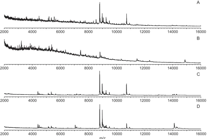
The presence of the larvae’s gut strongly impaired the protein profile, as shown in Fig. 1. The profile obtained from an entire C. nubeculosus larva is depicted in Fig. 1A, showing a high background noise and a suppression effect in the lower mass range as revealed by the baseline shift which is caused
2000 4000 6000 8000 10000 12000 14000 16000 m/z A B C D 2000 4000 6000 8000 10000 12000 14000 16000 2000 4000 6000 8000 10000 12000 14000 16000 2000 4000 6000 8000 10000 12000 14000 16000
Fig. 1. Matrix-assisted laser desorption/ionization time offlight (MALDI-TOF) mass spectra of Culicoides nubeculosus larvae in the range of 2 to 16 kDa. (A) entire larva; (B) dissected larval gut; (C) larva without gut and last 2 segments; (D) larva without gut, last 2 segments and head.
2000 4000 6000 8000 10000 12000 14000 m/z A B C 2000 4000 6000 8000 10000 12000 14000 2000 4000 6000 8000 10000 12000 14000
Fig. 2. Matrix-assisted laser desorption/ionization time offlight (MALDI-TOF) mass spectra profiles of Culicoides nubeculosus larvae (without gut and last 2 abdominal segments) at different raw protein concentrations in the range of 2 to 15 kDa. (A) 1·7 mg/ml, yielding 160 data counts; (B) 0·2 mg/ml, data count of 97 peaks– a qualitatively good spectrum; (C) 0·013 mg/ml, 13 data counts only.
321 MALDI-TOF for identification of ceratopogonid larvae
by gut components as revealed in Fig. 1B (profile of the dissected gut). Entire larvae without gut and larvae from which further parts were removed (last 2 segments,Fig. 1C; last 2 segments and head,Fig. 1D) yielded similar spectra of high quality.
The optimal protein concentration for MALDI-TOF MS analysis was assessed with dilutions of homogenates from 3 pools of 20 C. nubeculosus larvae (instar IV, 21 days post-hatching, gut and last 2
segments removed) and later confirmed with
Inten
si
ty
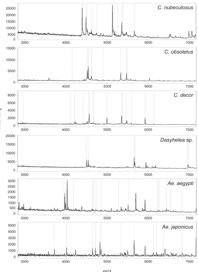
C. decor C. obsoletus m/z 0 1000 2000 3000 4000 5000 0 500 1000 1500 2000 2500 3000 0 5000 10000 15000 20000 0 2000 4000 6000 8000 0 5000 10000 15000 25000 0 5000 10000 15000 20000 Dasyheleasp. Ae. aegypti Ae. japonicus C. nubeculosus 3000 4000 5000 6000 7000 3000 4000 5000 6000 7000 3000 4000 5000 6000 7000 3000 4000 5000 6000 7000 3000 4000 5000 6000 7000 3000 4000 5000 6000 7000Fig. 3. Matrix-assisted laser desorption/ionization time offlight (MALDI-TOF) mass spectra of different larvae
(& 0·2 mg raw protein/ml). (A) Culicoides nubeculosus; (B) C. obsoletus; (C) C. decor; (D) Dasyhelea sp.; (E) Aedes
corresponding concentrations from single specimens of the same species (n = 9). Thus, in 7 re-duplicated dilutions, crude protein concentrations between
0·013 mg and 1·7 mg per ml were investigated
(Fig. 2) revealing spectra of high quality with regard to low noise, levelled baseline and number of mass data counts (97) for the 0·2 mg/ml concentration (Fig. 2B). Higher protein concentrations yielded generally higher data counts (160) but these ori-ginated from the noisiness of the spectrum and the baseline shift in the range between 2 and 6 kDa (Fig. 2A). With decreasing protein concentrations, the number of peaks declined and the spectra appeared noisy with baseline shifts (Fig. 2C).
Taken together, the recommended procedure for MALDI-TOF MS analyses of ceratopogonid larvae is to homogenize the gut-less larvae in 10% formic acid, mixing 0·2 mg/ml crude protein homogenate with SA matrix in a ratio of 1:1·5 and applying 1μl to mass spectrometry analyses.
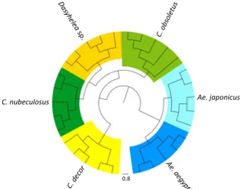
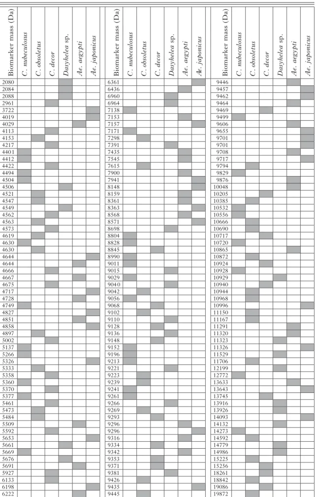
Total protein profiles were then generated from 5 larvae each of 3 biting midge species (C. nubeculosus, C. obsoletus, C. decor), 1 non-biting midge species from the genus Dasyhelea sp., and 2 mosquito species (Ae. aegypti and Ae. japonicus). Figure 3 shows 6 individual protein profiles in the range of 3 to 7 kDa of 1 larva from every species. Overall, the data counts included for protein profile generation per spectrum were between 30 and 124 in the mass range of 2 to 20 kDa. The whole protein profiles of 5 individuals per species (data count average of 67) were used to compile the total mass spectra for the 6 species in a dendrogram (Fig. 4), revealing that all larvae of the same species clustered on distinct branches. Based on this clear-cut clustering, species-specific biomarker mass sets could be generated using the SARAMIS™ SuperSpectrum tool (Fig. 3,Table 1). For automated
species identification of the different reference larvae, between 27 and 32 biomarker masses were deter-mined (Table 1).
Validation study
The accuracy of the reference database library was validated for all biomarker mass sets in a validation study. To this aim, 67 larvae belonging to the target species were used (laboratory-reared, n = 22, com-prising C. nubeculosus, n = 10; Ae. aegypti, n = 9; Ae. japonicus, n = 3; and field-collected, n=45, comprising C. obsoletus, n = 31; C. decor, n = 6; Dasyhelea sp., n = 8). In addition, larvae of field-collected C. pulicaris (n = 1) and of a non-biting midge Forcipomyia sp. (n = 3) were included as non-target species. From the investigated 67 specimens of the target species, 61 allowed correct identification by automated biomarker masses set analysis. From 54 of them, both profiles were suitable for automated identification whereas in 7 cases (C. obsoletus, n=2; C. decor, n = 1; Dasyhelea sp., n = 2; Ae. aegypti, n = 2) only a single profile allowed an automated identifi-cation as the second one had fewer than 75% biomarker mass matches, requiring a complete manual full spectra comparison against the whole reference data set library which, in all cases, revealed the correct identification. For 6 larvae (Dasyhelea sp., n = 1; Ae. aegypti, n = 3; Ae. japonicus, n = 2) of which neither profile was suitable for automated identifi-cation, complete manual full spectra comparison was done, resulting in the correct identification. In addition, none of the 4 specimens of the non-target species (C. pulicaris, n = 1; Forcipomyia sp., n = 3) yielded a positive identification, neither by
auto-mated biomarker identification nor by manual
comparison. Taken together, all larvae of the validation study were correctly identified using the protein profiling identification tool.
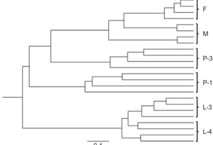
For 5 species for which validated biomarker masses of the larvae were determined (C. nubeculosus, C. obsoletus, C. pulicaris, Forcipomyia sp. and Ae. japonicus), the corresponding masses of the adult stages are also included in the reference database library (Kaufmann et al. 2011, 2012). None of the larvae were misidentified as adults. The reference biomarker mass sets of larvae (Table 1) and adults (Kaufmann et al. 2011, 2012) of C. nubeculosus, C. obsoletus and Ae. japonicus range between 27 and 33 masses but they are highly distinct as only 3 (C. nubeculosus) or 2 (C. obsoletus and Ae. japonicus) are shared within both sets in the range of ±800 ppm error. In addition, the protein profile changes during the complete metamorphosis as shown in the dendrogram (Fig. 5) displaying the individual spectra of larvae III and IV, early (1 day) and late (3 day) pupae, and adults of both sexes of C. nubeculosus (Fig. 5). All larvae III and IV cluster together,
C. nubeculosus Ae. japonicus
0.8
Fig. 4. Dendrogram of matrix-assisted laser desorption/ ionization time offlight (MALDI-TOF) mass spectra of larvae from 5 individuals of 6 insect species. Distance units correspond to the relative similarity calculated from the distance matrix.
323 MALDI-TOF for identification of ceratopogonid larvae
Table 1. MALDI-TOF MS reference biomarker masses of larvae Biomark er m ass (Da) C. nu becu losus C. obso let u s C. decor Dasyhelea sp. Ae. aegyp ti Ae. ja pon icus Biomark er m ass (Da) C. nu becu losus C. obso let u s C. decor Dasyhelea sp. Ae. aegyp ti A e. ja pon icus Biomark er m ass (Da) C. nu becu losus C. obso let u s C. decor Dasyhelea sp. Ae. aegyp ti Ae. ja pon icus 2080 6361 9446 2084 6436 9457 2088 6960 9462 2961 6964 9464 3722 7138 9469 4019 7153 9499 4029 7157 9606 4113 7171 9655 4153 7298 9701 4217 7391 9701 4401 7435 9708 4412 7545 9717 4422 7615 9794 4494 7900 9829 4504 7941 9876 4506 8148 10048 4521 8159 10205 4547 8361 10385 4549 8363 10532 4562 8568 10556 4563 8571 10666 4573 8698 10690 4619 8804 10717 4630 8828 10720 4630 8845 10865 4644 8990 10872 4644 9011 10924 4666 9015 10928 4667 9029 10929 4675 9040 10940 4717 9042 10944 4728 9056 10968 4749 9068 10996 4827 9102 11150 4851 9110 11167 4858 9128 11291 4897 9136 11320 5002 9148 11323 5137 9152 11326 5266 9196 11529 5326 9213 11706 5333 9221 12199 5358 9223 12772 5360 9239 13633 5370 9241 13643 5377 9261 13745 5461 9266 13916 5473 9269 13926 5484 9293 14093 5509 9296 14132 5592 9296 14273 5653 9316 14592 5661 9334 14779 5669 9342 14986 5676 9353 15225 5691 9371 15256 5927 9381 18261 6133 9426 18842 6198 9435 19086 6222 9445 19872 total 32 33 32 27 29 27
separated from the early pupae, whereas late pupae share the same main branch with the adults of the midges.
D I S C U S S I O N
Insect identification by protein profiling is still a rather novel entomological tool having been utilized in only a few studies (Campbell,2005; Feltens et al. 2010; Kaufmann et al. 2011; Perera et al. 2005), including the very recent application for the iden-tification of field-collected haematophagous biting midges (Kaufmann et al.2012). In the present study, the application of MALDI-TOF MS for the identification of larvae of holometabolous insects was evaluated for thefirst time. Biomarker mass sets could be established for 6 species, and all specimens investigated in the validation study were correctly identified.
Similar to the situation with adult midges
(Kaufmann et al. 2011), it was found that the gut content of the larvae strongly impaired the profile quality. Thus, the gut must be removed by dissection which is a more tedious task with larvae as compared to the situation with adults where simply cutting off the abdomen suffices. For plant vectors, like aphids, it was shown that the gut content, containing plant molecules of low molecular size (e.g. sugars and amino acids; Dinant et al.2010), does not impair the MALDI-TOF MS analysis (Perera et al.2005).
The gut-less larvae were manually homogenized in formic acid, and a raw protein concentration of approximately 0·2 mg/ml was determined as optimal for the mass spectrometry analyses. As a rule of thumb, this concentration can be obtained by homogenizing the ceratopogonid larvae in volumes
of 15–40 μl (depending on size) and the larger culicid larvae in 60μl (Ae. aegypti) or 240 μl (Ae. japonicus), respectively.
Crude protein purification (precipitation using trichloroacetic acid; metabolite segregation with acetone or methanol/chloroform) was applied in preliminary experiments but did not yield superior profiles (data not shown). As already discussed in a previous study by Kaufmann et al. (2011), a more elaborate purification of proteins/peptides including chromatography, as shown by Feltens and co-workers (2010) with Drosophila spp., provides high-quality profiles with higher data counts but, due to spectral heterogeneity within species, this approach does not necessarily result in a more ‘species-specific’ biomarker mass set.
Adult midges stored in 70% ethanol for up to 2 years (Kaufmann et al. 2011,2012) were reported to be suitable for mass spectrometry analyses albeit freshly collected specimens provided slightly better results (Kaufmann et al. 2012). For larvae, similar observations were made, and it is therefore re-commended to use fresh specimens for the generation of the biomarker mass sets. Nevertheless, as shown in the validation study, correct identification was achieved with larvae stored up to 4 months.
As expected, the generated biomarker mass sets for larvae strongly differed from those of the adults (and pupae), with only very few shared masses, and thus they cannot be used for the identification of the adults (and vice versa). In contrast, in hemimetabo-lous insects, Perera and co-workers (2005) when investigating the life cycle of the cowpea aphid Aphis craccivora found that most of the major
biomarker masses of juvenile stages (nymph
stages I–IV) were also present in adults and thus a single species-specific biomarker set can be
deter-mined. For holometabolous insects, clear-cut
biomarker sets were identified for larvae (this paper) and adults (Kaufmann et al.2012). The pupal stages, however, seem not be suitable for species identi
fi-cation by MALDI-TOF MS as their profiles
rapidly change during metamorphosis as shown in Fig. 5 for early (1 day) and late (3 days old) specimens. Thus, the identification of pupae pre-ferably is done by genetic or morphological analyses or by analysing adults which emerged in the lab (Rieb and Kremer, 1981; Uslu and Dik, 2007), e.g. by MALDI-TOF MS.
MALDI-TOF MS identification was straightfor-ward with the ceratopogonid larvae but less reliable with the larger culicid larvae using the same raw protein concentration as 42% of the investigated mosquito larvae could not be identified through automated identification but required manual com-parison. Thus, improvements in sample preparation are required for mosquitoes that should, for example, include analysis of the head and thorax only, which in culicid larvae easily can be separated from the
0.1 F M L-3 P-3 P-1 L-4
Fig. 5. Dendrogram of matrix-assisted laser desorption/ ionization time offlight (MALDI-TOF) mass spectra of different life stages of Culicoides nubeculosus. Each life stage (L-3, larva III; L-4, larva IV; P-1, pupa at 1 day, P-3, pupa at 3 days; F, female; M, male) is represented by 4 different individuals. Distance units correspond to the relative similarity calculated from the distance matrix.
325 MALDI-TOF for identification of ceratopogonid larvae
gut-containing abdomen because of their 3 tagmata system.
The applicability of MALDI-TOF MS for the identification of organisms was extended to include larvae of holometabolous insects. In the case of biting midges, this fast, cheap, and reliable diagnostic method might be of value to identify breeding habitats that can be targeted in control programmes.
A C K N O W L E D G E M E N T S
We thank Simon Carpenter and Eric Denison (Institute for Animal Health, Pirbright, UK) for the initial supply of Culicoides nubeculosus for the laboratory colonies and Pie Müller (Swiss Tropical and Public Health Institute, Basel) for eggs of laboratory-colonized Aedes aegypti. We also like to thank Stefanie Wagner (Institute of Parasitology, University of Zürich) for providingfield-collected eggs of Ae. japonicus.
F I N A N C I A L S U P P O R T
We greatly acknowledge the financial support by
the Federal Veterinary Office (grant 1.08.10) and the University of Zürich (‘Forschungskredit’, C.K., personal grant no. 55080502_4974).
R E F E R E N C E S
Ansari, M. A., Pope, E. C., Carpenter, S., Scholte, E. J. and Butt, T. M. (2011). Entomopathogenic fungus as a biological control for an important vector of livestock disease: the Culicoides biting midge. PLoS One6, e16108. Bauer, B., Jandowsky, A., Schein, E., Mehlitz, D. and Clausen, P.-H. (2009). An appraisal of current and new techniques intended to protect bulls against Culicoides and other haematophagous Nematocera: the case of Schmergow, Brandenburg, Germany. Parasitology Research105, 359–365.
Blackwell, A. and King, F. C. (1997). The vertical distribution of Culicoides impunctatus larvae. Medical and Veterinary Entomology11, 45–48. Boorman, J. (1974). The maintenance of laboratory colonies of Culicoides variipennis (Coq.), C. nubeculosus (Mg.) and C. riethi Kiff. (Diptera, Ceratopogonidae). Bulletin of Entomological Research64, 371–377. Bradford, M. M. (1976). A rapid and sensitive method for the quantitation of microgram quantities of protein utilizing the principle of protein-dye binding. Analytical Biochemistry72, 248–254.
Campbell, P. M. (2005). Species differentiation of insects and other multicellular organisms using matrix-assisted laser desorption/ionization time offlight mass spectrometry protein profiling. Systematic Entomology 30, 186–190.
Caprioli, G., Cristalli, G., Ragazzi, E., Molin, L., Ricciutelli, M., Sagratini, G., Seraglia, R., Zuo, Y. and Vittori, S. (2010). A preliminary matrix-assisted laser desorption/ionization time-of-flight approach for the characterization of Italian lentil varieties. Rapid Communications in Mass Spectrometry24, 2843–2848.
Carpenter, S., Mellor, P. S. and Torr, S. J. (2008). Control techniques for Culicoides biting midges and their application in the U.K. and northwestern Palaearctic. Medical and Veterinary Entomology22, 175–187.
Dinant, S., Bonnemain, J. L., Girousse, C. and Kehr, J. (2010). Phloem sap intricacy and interplay with aphid feeding. Comptes Rendus Biologies 333, 504–515.
Feltens, R., Gorner, R., Kalkhof, S., Groger-Arndt, H. and von Bergen, M. (2010). Discrimination of different species from the genus Drosophila by intact protein profiling using matrix-assisted laser desorption ionization mass spectrometry. BMC Evolutionary Biology10, 95. Hellberg, W., Mellor, P. S., Torsteinsdottir, S. and Marti, E. (2009). Insect bite hypersensitivity in the horse: comparison of IgE-binding proteins in salivary gland extracts from Simulium vittatum and Culicoides nubeculosus. Veterinary Immunology and Immunopathology132, 62–67. Hoffmann, B., Scheuch, M., Höper, D., Jungblut, R., Holsteg, M., Schirrmeier, H., Eschbaumer, M., Goller, K. V., Wernike, K., Fischer, M., Breithaupt, A., Mettenleiter, T. C. and Beer, M. (2011).
Novel orthobunyavirus in cattle, Europe, 2011. Emerging Infectious Disease 18, 469–472.
Hribar, L. J. (1990). A review of methods for recovering biting midge larvae (Diptera: Ceratopogonidae) from substrate samples. Journal of Agricultural Entomology7, 71–77.
Karger, A., Kampen, H., Bettin, B., Dautel, H., Ziller, M., Hoffmann, B., Süss, J. and Klaus, C. (2012). Species determination and characterization of developmental stages of ticks by whole-animal matrix-assisted laser desorption/ionization mass spectrometry. Ticks and Tick-borne Diseases3, 78–89.
Kaufmann, C., Schaffner, F., Ziegler, D., Pflüger, V. and Mathis, A. (2012). Identification of field-caught Culicoides biting midges using matrix-assisted laser desorption/ionization time of flight mass spectrometry. Parasitology139, 248–258.
Kaufmann, C., Ziegler, D., Schaffner, F., Carpenter, S., Pflüger, V. and Mathis, A. (2011). Evaluation of matrix-assisted laser desorption/ ionization time offlight mass spectrometry for characterization of Culicoides nubeculosus biting midges. Medical and Veterinary Entomology25, 32–38. Kettle, D. S. and Lawson, J. W. (1952). The early stages of British biting midges Culicoides Latreille (Diptera: Ceratopogonidae) and allied genera. Bulletin of Entomological Research43, 421–467.
Khamala, C. P. (1975). Breeding habitats and biting activities of Culicoides (Diptera: Ceratopogonidae) at Lake Nakuru National Park, Kenya, with special reference to C. trifasciellus Goetghebuer. East African Medical Journal52, 405–412.
Kidder, T. R. (1997). Sugar reflotation: An alternative method for sorting flotation-derived heavy fraction samples. Journal of Field Archaeology 24, 39–45.
Kline, D. L., Dukes, J. C. and Axtell, R. C. (1975). Salt marsh Culicoides (Diptera: Ceratopogonidae): comparison of larval sampling methods. Mosquito News35, 147–150.
Mazzeo, M. F., Giulio, B. D., Guerriero, G., Ciarcia, G., Malorni, A., Russo, G. L. and Siciliano, R. A. (2008). Fish authentication by MALDI-TOF mass spectrometry. Journal of Agricultural and Food Chemistry56, 11071–11076.
Mellmann, A., Bimet, F., Bizet, C., Borovskaya, A. D., Drake, R. R., Eigner, U., Fahr, A. M., He, Y., Ilina, E. N., Kostrzewa, M., Maier, T., Mancinelli, L., Moussaoui, W., Prevost, G., Putignani, L., Seachord, C. L., Tang, Y. W. and Harmsen, D. (2009). High inter-laboratory reproducibility of matrix-assisted laser desorption ionization-time offlight mass spectrometry-based species identification of nonferment-ing bacteria. Journal of Clinical Microbiology47, 3732–3734.
Mellor, P. S., Boorman, J. and Baylis, M. (2000). Culicoides biting midges: their role as arbovirus vectors. Annual Review of Entomology45, 307–340.
Perera, M. R., Vargas, R. D. F. and Jones, M. G. K. (2005). Identification of aphid species using protein profiling and matrix-assisted laser desorption/ ionization time-of-flight mass spectrometry. Entomologia Experimentalis et Applicata117, 243–247.
Planzer, J., Kaufmann, C., Worwa, G., Gavier-Widen, D., Hofmann, M. A., Chaignat, V. and Thur, B. (2011). In vivo and in vitro propagation and transmission of Toggenburg orbivirus. Research in Veterinary Science91, e163–168.
Rasmussen, L. D., Kristensen, B., Kirkeby, C., Rasmussen, T. B., Belsham, G. J., Bodker, R. and Botner, A. (2012). Culicoids as vectors of Schmallenberg virus. Emerging Infectious Disease18, 1204–1206. Rieb, J. P. and Kremer, M. (1981). Ecologie des Cératopogonidés de la plaine d’Alsace. III. Cycle évolutif des Culicoides (Diptères, Cératopogonidés) d’un gîte fluviatile. Annales de Parasitologie Humaine et Comparée56, 423–439.
Santos, C., Paterson, R. R. M., Venancio, A. and Lima, N. (2010). Filamentous fungal characterizations by matrix-assisted laser desorption/ ionization time-of-flight mass spectrometry. Journal of Applied Microbiology 108, 375–385.
Sauer, S. and Kliem, M. (2010). Mass spectrometry tools for the classification and identification of bacteria. Nature Reviews Microbiology 8, 74–82.
Sloet van Oldruitenborgh-Oosterbaan, M. M., van Poppel, M., de Raat, I. J., van den Boom, R. and Savelkoul, H. F. (2009). Intradermal testing of horses with and without insect bite hypersensitivity in The Netherlands using an extract of native Culicoides species. Veterinary Dermatology20, 607–614.
Stevenson, L. G., Drake, S. K., Shea, Y. R., Zelazny, A. M. and Murray, P. R. (2010). Evaluation of matrix-assisted laser desorption ionization-time offlight mass spectrometry for identification of clinically important yeast species. Journal of Clinical Microbiology48, 3482–3486. Timmermann, S. E. and Briegel, H. (1993). Water depth and larval density affect development and accumulation of reserves in laboratory
populations of mosquitoes. Bulletin of the Society of Vector Ecologists18, 174–187.
Uslu, U. and Dik, B. (2007). Description of breeding sites of Culicoides species (Diptera: Ceratopogonidae) in Turkey. Parasite 14, 173–177.
Van Veen, S. Q., Claas, E. C. J. and Kuijper, E. J. (2010). High-throughput identification of bacteria and yeast by matrix-assisted laser desorption ionization-time offlight mass spectrometry in conventional medical microbiology laboratories. Journal of Clinical Microbiology 48, 900–907.
Venail, R., Mathieu, B., Setier-Rio, M. L., Borba, C., Alexandre, M., Viudes, G., Garros, C., Allene, X., Carpenter, S., Baldet, T. and
Balenghien, T. (2011). Laboratory andfield-based tests of deltamethrin insecticides against adult Culicoides biting midges. Journal of Medical Entomology48, 351–357.
Wenk, C., Kaufmann, C., Schaffner, F. and Mathis, A. (2012). Molecular characterisation of Swiss Ceratopogonidae (Diptera) and evaluation of real-time PCR assays for the identification of Culicoides biting midges. Veterinary Parasitology184, 258–266.
Zimmer, J. Y., Haubruge, E., Francis, F., Bortels, J., Simonon, G., Losson, B., Mignon, B., Paternostre, J., De Deken, R., De Deken, G., Deblauwe, I., Fassotte, C., Cors, R. and Defrance, T. (2008). Breeding sites of bluetongue vectors in northern Europe. Veterinary Record162, 131.
327 MALDI-TOF for identification of ceratopogonid larvae