HAL Id: hal-02661791
https://hal.inrae.fr/hal-02661791
Submitted on 30 May 2020
HAL is a multi-disciplinary open access
archive for the deposit and dissemination of
sci-entific research documents, whether they are
pub-lished or not. The documents may come from
teaching and research institutions in France or
abroad, or from public or private research centers.
L’archive ouverte pluridisciplinaire HAL, est
destinée au dépôt et à la diffusion de documents
scientifiques de niveau recherche, publiés ou non,
émanant des établissements d’enseignement et de
recherche français ou étrangers, des laboratoires
publics ou privés.
Identification of a variety of staphylococcus species by
matrix-assisted laser desorption ionization-time of flight
mass spectrometry
Damien Dubois, David Leyssene, Jean-Paul Chacornac, Markus Kostrzewa,
Pierre Olivier Schmit, Régine Talon, Richard Bonnet, Julien Delmas
To cite this version:
Damien Dubois, David Leyssene, Jean-Paul Chacornac, Markus Kostrzewa, Pierre Olivier Schmit, et
al.. Identification of a variety of staphylococcus species by matrix-assisted laser desorption
ionization-time of flight mass spectrometry. Journal of Clinical Microbiology, American Society for Microbiology,
2010, 48 (3), pp.941-945. �10.1128/JCM.00413-09�. �hal-02661791�
Published Ahead of Print 23 December 2009.
10.1128/JCM.00413-09.
2010, 48(3):941. DOI:
J. Clin. Microbiol.
Richard Bonnet and Julien Delmas
Markus Kostrzewa, Pierre Olivier Schmit, Régine Talon,
Damien Dubois, David Leyssene, Jean Paul Chacornac,
Mass Spectrometry
Laser Desorption Ionization-Time of Flight
Species by Matrix-Assisted
Staphylococcus
Identification of a Variety of
http://jcm.asm.org/content/48/3/941
Updated information and services can be found at:
These include:
REFERENCES
http://jcm.asm.org/content/48/3/941#ref-list-1
at:
This article cites 34 articles, 14 of which can be accessed free
CONTENT ALERTS
more»
articles cite this article),
Receive: RSS Feeds, eTOCs, free email alerts (when new
http://journals.asm.org/site/misc/reprints.xhtml
Information about commercial reprint orders:
http://journals.asm.org/site/subscriptions/
To subscribe to to another ASM Journal go to:
on October 24, 2012 by INRA Centre de Versailles-Grignon
http://jcm.asm.org/
JOURNAL OF
CLINICAL
MICROBIOLOGY, Mar. 2010, p. 941–945
Vol. 48, No. 3
0095-1137/10/$12.00
doi:10.1128/JCM.00413-09
Copyright © 2010, American Society for Microbiology. All Rights Reserved.
Identification of a Variety of Staphylococcus Species by Matrix-Assisted
Laser Desorption Ionization–Time of Flight Mass Spectrometry
䌤
Damien Dubois,
1,2David Leyssene,
1,2Jean Paul Chacornac,
3Markus Kostrzewa,
4Pierre Olivier Schmit,
5Re
´gine Talon,
3Richard Bonnet,
1,2and Julien Delmas
1,2*
CHU Clermont-Ferrand, Centre de Biologie, Laboratoire de Bacte
´riologie Clinique, Clermont-Ferrand F-63003, France
1;
Universite
´ d’Auvergne Clermont-1, Faculte
´ de Me
´decine, Laboratoire de Bacte
´riologie, JE2526, USC-INRA 2018,
Clermont-Ferrand F-63001, France
2; INRA, Centre de Clermont-Ferrand-Theix, UR 454, Microbiologie,
63122 Saint-Genes Champanelle, France
3; Bruker Daltonik GmbH, Leipzig, Germany
4; and
Bruker Daltonique, Wissembourg, France
5Received 25 February 2009/Returned for modification 5 August 2009/Accepted 11 December 2009
Whole-cell fingerprinting by matrix-assisted laser desorption ionization–time-of-flight mass spectrometry
(MALDI-TOF MS) in combination with a dedicated bioinformatic software tool (MALDI Biotyper 2.0) was
used to identify 152 staphylococcal strains corresponding to 22 staphylococcal species. Spectra of the 152
isolates, previously identified at the species level using a sodA gene-based oligonucleotide array, were analyzed
against the main spectra of 3,030 microorganisms. A total of 151 strains out of 152 (99.3%) were correctly
identified at the species level; only one strain was identified at the genus level. The MALDI-TOF MS method
revealed different clonal lineages of Staphylococcus epidermidis that were of either human or environmental
origin, which suggests that the MALDI-TOF MS method could be useful in the profiling of staphylococcal
strains. The topology of the dendrogram generated by the MALDI Biotyper 2.0 software from the spectra of 120
Staphylococcus reference strains (representing 36 species) was in general agreement with that inferred from the
16S rRNA gene-based analysis. Our findings indicate that the MALDI-TOF MS technology, associated with a
broad-spectrum reference database, is an effective tool for the swift and reliable identification of Staphylococci.
Most staphylococci are harmless and reside normally on the
skin and mucous membranes of humans and other organisms
(16, 22, 34). Staphylococcal strains are isolated from various
food products in which they are involved in fermentation (18,
29). Staphylococcus species can cause a wide variety of diseases
in humans and other animals (2, 22, 30–32, 35). S. aureus is a
major pathogen in human infections (31). Several other
Staph-ylococcus species have also been implicated in human
infec-tions, notably S. saprophyticus, S. epidermidis, S. lugdunensis,
and S. schleiferi (4, 16, 31, 34). Coagulase-negative
staphylo-cocci (CoNS) have emerged as predominant pathogens in
hos-pital-acquired infections (4, 16, 31, 34). One of the major
challenges of daily diagnostic work is therefore to identify
Staphylococcus species.
Several manual and automated methods based on
pheno-typic characteristics have been developed for the identification
of Staphylococci (12, 24). Unfortunately, these systems have
their limitations, mostly due to phenotypic differences between
strains from the same species (6, 10, 19, 21). Over the last 10
years, many genotypic methods based on the analysis of
se-lected DNA targets have been designed for species-level
iden-tification of most common isolated CoNS (20, 26, 33). The
sequence polymorphism of the sodA gene has significant
dis-criminatory power (20) and allows the development of assays
based on DNA chip technologies (“Staph array”) (8).
Re-cently, matrix-assisted laser desorption ionization–time of
flight mass spectrometry (MALDI-TOF MS) using protein
“fingerprints” was used for the identification of
microorgan-isms (1, 3, 5, 9, 11, 14, 25, 36). In the present study, we assessed
the ability of the MALDI Biotyper system (Bruker Daltonique,
Wissembourg, France) to identify Staphylococcus species of
clinical and environmental origins previously identified by
sodA gene-based oligonucleotide array (8).
MATERIALS AND METHODS
Bacterial strains.A total of 152 blind-coded Staphylococcus strains from our collection, isolated from clinical (n⫽ 60) and food and plant samples (n ⫽ 92) and representing 22 species, were studied (Table 1). All strains were previously identified as belonging to a specific species by the oligonucleotide “Staph array” (8). This system associates PCR amplification of the sodA gene with an oligo-nucleotide-based array to efficiently discriminate between the Staphylococcus species. Identifications found with this array were identical to those obtained by sequencing the internal fragment sodA (sodAint). In addition, the Vitek 2 system has been used with the Gram-positive (GP) identification card (bioMe´rieux, Marcy l’Etoile, France) to identify Staphylococcus strains. If the GP card indi-cated the possibility of two or three species, supplementary tests (pigmentation, hemolysis, or novobiocin resistance) were then performed to determine the identification.
Sample preparation. Sample preparation for MALDI-TOF MS was per-formed as previously described (14). Briefly, a few colonies of a fresh overnight culture grown on Columbia blood agar at 37°C under aerobic conditions were suspended in 300l distilled water to which 900 l absolute ethanol was added. The mixture was centrifuged at 12,000⫻ g for 2 min, and the supernatant was discarded. Ten microliters of formic acid (70%) was added to the pellet and mixed thoroughly by pipetting before the addition of 10l of acetonitrile to the mixture. The mixture was centrifuged at 12,000⫻ g for 2 min. One microliter of the supernatant was placed onto a steel target plate and air dried at room temperature. Each sample was overlaid with 2l saturated solution of ␣-cyano-4-hydroxycinnamic acid in 50% acetonitrile–2.5% trifluoroacetic acid and air dried at room temperature. MALDI-TOF MS measurements were performed with a Bruker Ultraflex II MALDI-TOF/TOF (tandem TOF) instrument equipped with 200-Hz Smartbeam laser technology. Spectra were recorded in the
* Corresponding author. Mailing address: Laboratoire de Bacte
´ri-ologie, 28 place H. Dunant, 63001 Clermont-Ferrand, France. Phone:
33 4 73 75 49 20. Fax: 33 4 73 75 49 22. E-mail: julien.delmas@u
-clermont1.fr.
䌤
Published ahead of print on 23 December 2009.
941
on October 24, 2012 by INRA Centre de Versailles-Grignon
http://jcm.asm.org/
positive linear mode within a mass range of 2,000 to 20,000 Da. Five hundred laser shots were recorded for each spectrum.
Data analysis.Raw spectra of the Staphylococcus strains were analyzed by MALDI Biotyper 2.0 software (Bruker Daltonique) with default settings. The whole process from MALDI-TOF MS measurement to identification was per-formed automatically without any user intervention. The peak lists generated were used for matches against the reference library directly using the integrated pattern-matching algorithm of the software. The software provides a log score, and the cutoff log score of 2 was used to validate identification at the species level, as recommended by the manufacturer. For strain classification, the cre-ation of the dendrogram is based on cross-wise minimum spanning tree (MSP) matching. Similar MSPs result in a high matching score value. Each MSP is matched against all MSPs of the analyzed set. The list of score values is used to calculate normalized distance values between the analyzed species, resulting in a matrix of matching scores. The visualization of the respective relationship be-tween the MSPs is displayed in a dendrogram using the standard settings of the MALDI Biotyper 2.0 software. Species with distance levels under 500 have been described as reliably classified (25).
RESULTS AND DISCUSSION
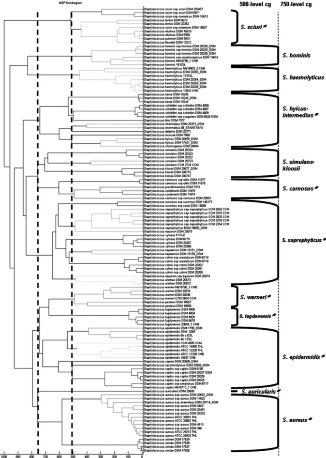
Classification of staphylococcal reference strains.
A
score-oriented dendrogram was generated on the basis of the 120
reference strains in the Bruker MALDI Biotyper 2.0 database
(Fig. 1). This dendrogram revealed, with the default critical
distance level of 500, 4 of the 11 cluster groups defined by
Poyart et al. from the phylogenetic analysis based on the sodA
gene (20): S. sciuri, S. warneri, S. lugdunensis, and S. auricularis
(Fig. 1). These cluster groups were also in accordance with
those established by the phylogenetic analysis of the 16S rRNA
gene (28). With a critical distance level of 750, the dendrogram
revealed overall the cluster groups of 16S rRNA genes, with
some variations (Fig. 1) (17, 28). The 16S rRNA gene-based
cluster group S. haemolyticus including S. haemolyticus and S.
hominis was divided into branches which linked at a distance
level of 850. Unlike in other phylogenetic analyses, in which S.
kloosii belonged to S. saprophyticus cluster group, this species
was clustered within the S. simulans group in the MALDI-TOF
MS-based dendrogram (13, 20, 28). Inside the S. saprophyticus
cluster group, the two strains S. equorum subsp. equorum DSM
20675 and DSM 20674 belonged to two distinct clusters. It is
noteworthy that the S. lutrae and S. schleiferi species each
constituted a well-defined clade inside the S. hyicus-S.
interme-dius cluster group, like S. succinus, S. xylosus, S. cohnii, and S.
arlettae in the S. saprophyticus cluster group and S. capitis in the
S. epidermidis cluster group.
Although some differences were observed, the topology of
the MALDI-TOF MS-based phylogenetic tree was in more
general agreement with that inferred from the analysis of the
16S rRNA gene sequences than that based on the sodA gene
(20, 28). The proteins analyzed by MALDI Biotyper
corre-spond to predominant proteins, such as ribosomal proteins.
The probable coevolution of the ribosomal proteins and
rRNAs may explain the similarities between 16S rRNA gene
and MALDI-TOF-based dendrograms (23).
MALDI-TOF MS could thus be a valuable tool in
phylopro-teomics. It might serve as a technique for protein profiling,
which is recommended as an additional test for the description
of new staphylococcal species (7).
Species identification of staphylococcal isolates.
Our
den-drogram showed that the 36 reference Staphylococcus species
were correctly distinguished. S. vitulinus and S. pulvereri, which
have recently been shown to be one single species (27), were
not differentiated by the MALDI-TOF MS technique. This
technique seemed to be suitable for the differentiation of
Staphylococcus isolates at the species level.
MALDI-TOF MS spectra were then obtained for all clinical
and environmental staphylococcal isolates. A total of 151
strains out of 152 (99.3%) were identified at the species level
by the MALDI Biotyper. One S. saprophyticus strain out of 16
was identified as S. xylosus, with a log score of 1.995. It was
therefore identified at the genus level. The system identified
both clinical (14 different species) and environmental (17
dif-ferent species) strains without any complementary tests.
Carbonnelle et al. used the MALDI-TOF technology to
identify a clinical collection of Staphylococcus strains
consti-tuted by the four major clinically relevant Staphylococcus
spe-cies (S. aureus, S. epidermidis, S. warneri, and S. haemolyticus)
(3). Their reference database contained 22 staphylococci
ref-erence strains, including 17 staphylococcal species. In our
study, we identified 22 Staphylococcus species from the
MALDI Biotyper database, which comprised the spectra of
3,030 microorganisms, corresponding to Gram-negative and
Gram-positive bacteria, whose 120 staphylococcal strains
rep-resented 36 staphylococcus species. A fortiori, the MALDI
Bio-typer did not require any initial assessment such as Gram
staining or a catalase test, unlike the technology developed by
Carbonnelle et al., which requires a presumptive identification
of Staphylococcus at the genus level.
The MALDI Biotyper database comprised 36 staphylococcal
species. The Staphylococcus genus comprises 39 validly
de-scribed species (J. P. Euze
´by, List of bacterial names with
standing in nomenclature, 5 November 2008, posting date;
http://www.bacterio.cict.fr/). The three species S. pettenkoferi,
S. pseudintermedius, and S. gallinarum were absent from the
MALDI Biotyper database. The MALDI Biotyper database
TABLE 1. Clinical and environmental staphylococcal strains used
in this study
Species (“Staph array” identification)
No. of isolates
Clinical Environmental Total
S. arlettae
0
1
1
S. aureus
6
1
7
S. capitis
5
2
7
S. caprae
1
0
1
S. carnosus
0
7
7
S. cohnii subsp. ureal
2
2
4
S. delphini
1
0
1
S. epidermidis
11
8
19
S. equorum
0
13
13
S. fleurettii
0
2
2
S. haemolyticus
5
1
6
S. hominis
5
1
6
S. lugdunensis
5
0
5
S. pasteuri
0
8
8
S. saprophyticus
6
10
16
S. schleiferi
2
0
2
S. sciuri
1
2
3
S. simulans
4
0
4
S. succinus
0
9
9
S. vitulinus
0
5
5
S. warneri
6
6
12
S. xylosus
0
14
14
Total
60
92
152
942
DUBOIS ET AL.
J. CLIN. MICROBIOL.
on October 24, 2012 by INRA Centre de Versailles-Grignon
http://jcm.asm.org/
FIG. 1. Classification of staphyloccal reference strains. Shown is a score-oriented dendrogram of staphylococcal reference strains included in
the database. The terms “500-level cg” and “750-level cg” define cluster groups (cg) based on the branching pattern using critical distance levels
of 500 and 750, respectively.
#, cluster groups as defined by the phylogenetic analysis of 16S rRNA genes.
943
on October 24, 2012 by INRA Centre de Versailles-Grignon
http://jcm.asm.org/
contains four species (S. pasteuri, S. delphini, S. fleurettii, and S.
succinus) absent from the Vitek 2 Gram-positive (GP) card
database (bioMe
´rieux, La-Balme les Grottes, France).
More-over, according to the previously published data from our
strain collection, the Vitek 2 system had misidentified or not
identified 1 out of 7 S. capitis, 2 out of 7 S. carnosus, 11 out of
13 S. equorum, 1 out of 4 S. simulans, and 1 out of 5 S. vitulinus
strains (6). This suggests that the MALDI-TOF MS technique
is better fitted to the identification of staphylococcal strains
that are rarely isolated in clinical bacteriology than automated
biochemical methods. In addition, sample preparation and
analysis with the MALDI-TOF MS technique are easier and
more time-saving (approximately 20 min for one identification)
than with genotypical methods, and result acquisition is faster
than with biochemical methods.
Biodiversity of S. epidermidis species.
The dendrogram of
Fig. 2 shows protein “fingerprint” heterogeneity of clinical and
environmental (salt meat originated) S. epidermidis strains.
Clinical isolates are gathered within two cluster groups and two
isolated strains, whereas meat origin isolates displayed one
distinct cluster group and one isolated strain. Diversity within
the S. epidermidis species has been observed for human origin
S. epidermidis and animal origin S. epidermidis (15). The
MALDI-TOF MS method might have enough discriminatory
power to group isolates below the species level.
Conclusion.
MALDI-TOF MS associated with MALDI
Bio-typer software appears a reliable and accurate tool for the
identification of Staphylococcus species. However, further
studies are required to test this technology with a large
collec-tion of staphylococci of diverse origins. The speed and the
simplicity of sample preparation and result acquisition
associ-ated with minimal consumable costs make this method well
suited for routine and high-throughput use. Hence, it is an
excellent alternative to traditional methods in food processing
and medical care laboratories. It may also be used for the
analysis of clonal and/or taxonomic relationships.
REFERENCES
1. Barbuddhe, S. B., T. Maier, G. Schwarz, M. Kostrzewa, H. Hof, E. Domann, T. Chakraborty, and T. Hain.2008. Rapid identification and typing of
Lis-teria species using matrix-assisted laser desorption ionization–time of flight
mass spectrometry. Appl. Environ. Microbiol. 74:5402–5407.
2. Bergonier, D., R. de Cremoux, R. Rupp, G. Lagriffoul, and X. Berthelot. 2003. Mastitis of dairy small ruminants. Vet. Res. 34:689–716.
3. Carbonnelle, E., J. L. Beretti, S. Cottyn, G. Quesne, P. Berche, X. Nassif, and A. Ferroni.2007. Rapid identification of staphylococci isolated in clinical microbiology laboratories by matrix-assisted laser desorption ionization– time of flight mass spectrometry. J. Clin. Microbiol. 45:2156–2161. 4. Choi, S. H., J. H. Woo, J. Y. Jeong, N. J. Kim, M. N. Kim, Y. S. Kim, and J.
Ryu.2006. Clinical significance of Staphylococcus saprophyticus identified on blood culture in a tertiary care hospital. Diagn. Microbiol. Infect. Dis. 56:337–339.
5. Claydon, M. A., S. N. Davey, V. Edwards-Jones, and D. B. Gordon. 1996. The rapid identification of intact microorganisms using mass spectrometry. Nat. Biotechnol. 14:1584–1586.
6. Delmas, J., J. P. Chacornac, F. Robin, P. Giammarinaro, R. Talon, and R. Bonnet.2008. Evaluation of the Vitek 2 system with a variety of Staphylo-coccus species. J. Clin. Microbiol. 46:311–313.
7. Freney, J., W. E. Kloos, V. Hajek, J. A. Webster, M. Bes, Y. Brun, and C. Vernozy-Rozand.1999. Recommended minimal standards for description of new staphylococcal species. Subcommittee on the taxonomy of staphylococci and streptococci of the International Committee on Systematic Bacteriology. Int. J. Syst. Bacteriol. 49:489–502.
8. Giammarinaro, P., S. Leroy, J. P. Chacornac, J. Delmas, and R. Talon. 2005. Development of a new oligonucleotide array to identify staphylococcal strains at species level. J. Clin. Microbiol. 43:3673–3680.
9. Grosse-Herrenthey, A., T. Maier, F. Gessler, R. Schaumann, H. Bohnel, M. Kostrzewa, and M. Kruger.2008. Challenging the problem of clostridial identification with matrix-assisted laser desorption/ionization-time-of-flight mass spectrometry (MALDI-TOF MS). Anaerobe 14:242–249.
10. Ieven, M., J. Verhoeven, S. R. Pattyn, and H. Goossens. 1995. Rapid and economical method for species identification of clinically significant coagu-lase-negative staphylococci. J. Clin. Microbiol. 33:1060–1063.
11. Keys, C. J., D. J. Dare, H. Sutton, G. Wells, M. Lunt, T. McKenna, M. McDowall, and H. N. Shah.2004. Compilation of a MALDI-TOF mass spectral database for the rapid screening and characterisation of bacteria implicated in human infectious diseases. Infect. Genet. Evol. 4:221–242. 12. Kim, M., S. R. Heo, S. H. Choi, H. Kwon, J. S. Park, M. W. Seong, D. H. Lee,
K. U. Park, J. Song, and E. C. Kim.2008. Comparison of the MicroScan, VITEK 2, and Crystal GP with 16S rRNA sequencing and MicroSeq 500 v2.0 analysis for coagulase-negative staphylococci. BMC Microbiol. 8:233. 13. Kwok, A. Y., S. C. Su, R. P. Reynolds, S. J. Bay, Y. Av-Gay, N. J. Dovichi, and
A. W. Chow. 1999. Species identification and phylogenetic relationships based on partial HSP60 gene sequences within the genus Staphylococcus. Int. J. Syst. Bacteriol. 49:1181–1192.
14. Mellmann, A., J. Cloud, T. Maier, U. Keckevoet, I. Ramminger, P. Iwen, J. Dunn, G. Hall, D. Wilson, P. Lasala, M. Kostrzewa, and D. Harmsen.2008. Evaluation of matrix-assisted laser desorption ionization-time-of-flight mass spectrometry in comparison to 16S rRNA gene sequencing for species iden-tification of nonfermenting bacteria. J. Clin. Microbiol. 46:1946–1954. 15. Nagase, N., A. Sasaki, K. Yamashita, A. Shimizu, Y. Wakita, S. Kitai, and J.
Kawano.2002. Isolation and species distribution of staphylococci from ani-mal and human skin. J. Vet. Med. Sci. 64:245–250.
16. Otto, M. 2009. Staphylococcus epidermidis—the ‘accidental’ pathogen. Nat. Rev. Microbiol. 7:555–567.
17. Pantucek, R., I. Sedlacek, P. Petras, D. Koukalova, P. Svec, V. Stetina, M. Vancanneyt, L. Chrastinova, J. Vokurkova, V. Ruzickova, J. Doskar, J. Swings, and V. Hajek.2005. Staphylococcus simiae sp. nov., isolated from South American squirrel monkeys. Int. J. Syst. Evol. Microbiol. 55:1953– 1958.
18. Papamanoli, E., P. Kotzekidou, N. Tzanetakis, and E. Litopoulou-Tzan-etakis. 2002. Characterization of Micrococcaceae isolated from dry fer-mented sausage. Food Microbiol. 19:441–449.
19. Perl, T. M., P. R. Rhomberg, M. J. Bale, P. C. Fuchs, R. N. Jones, F. P. Koontz, and M. A. Pfaller.1994. Comparison of identification systems for Staphylococcus epidermidis and other coagulase-negative Staphylococcus species. Diagn. Microbiol. Infect. Dis. 18:151–155.
20. Poyart, C., G. Quesne, C. Boumaila, and P. Trieu-Cuot. 2001. Rapid and accurate species-level identification of coagulase-negative staphylococci by using the sodA gene as a target. J. Clin. Microbiol. 39:4296–4301. 21. Rhoden, D. L., and J. M. Miller. 1995. Four-year prospective study of
STAPH-IDENT system and conventional method for reference identifica-tion of Staphylococcus, Stomatococcus, and Micrococcus spp. J. Clin. Mi-crobiol. 33:96–98.
22. Rich, M. 2005. Staphylococci in animals: prevalence, identification and an-timicrobial susceptibility, with an emphasis on methicillin-resistant Staphy-lococcus aureus. Br. J. Biomed. Sci. 62:98–105.
FIG. 2. Score-oriented dendrogram of clinical (C) and
environ-mental (E) S. epidermidis isolates.
944
DUBOIS ET AL.
J. CLIN. MICROBIOL.
on October 24, 2012 by INRA Centre de Versailles-Grignon
http://jcm.asm.org/
23. Ryzhov, V., and C. Fenselau. 2001. Characterization of the protein subset desorbed by MALDI from whole bacterial cells. Anal. Chem. 73:746–750. 24. Sampimon, O. C., R. N. Zadoks, S. De Vliegher, K. Supre, F. Haesebrouck,
H. W. Barkema, J. Sol, and T. J. Lam.2009. Performance of API Staph ID 32 and Staph-Zym for identification of coagulase-negative staphylococci isolated from bovine milk samples. Vet. Microbiol. 136:300–305. 25. Sauer, S., A. Freiwald, T. Maier, M. Kube, R. Reinhardt, M. Kostrzewa, and
K. Geider.2008. Classification and identification of bacteria by mass spec-trometry and computational analysis. PLoS One 3:e2843.
26. Sivadon, V., M. Rottman, S. Chaverot, J. C. Quincampoix, V. Avettand, P. de Mazancourt, L. Bernard, P. Trieu-Cuot, J. M. Feron, A. Lortat-Jacob, P. Piriou, T. Judet, and J. L. Gaillard.2005. Use of genotypic identification by sodA sequencing in a prospective study to examine the distribution of co-agulase-negative Staphylococcus species among strains recovered during septic orthopedic surgery and evaluate their significance. J. Clin. Microbiol. 43:2952–2954.
27. Svec, P., M. Vancanneyt, I. Sedlacek, K. Engelbeen, V. Stetina, J. Swings, and P. Petras.2004. Reclassification of Staphylococcus pulvereri Zakrze-wska-Czerwinska et al. 1995 as a later synonym of Staphylococcus vitulinus Webster et al. Int. J. Syst. Evol. Microbiol. 54:2213–2215.
28. Takahashi, T., I. Satoh, and N. Kikuchi. 1999. Phylogenetic relationships of 38 taxa of the genus Staphylococcus based on 16S rRNA gene sequence analysis. Int. J. Syst. Bacteriol. 49:725–728.
29. Talon, R., S. Leroy-Setrin, and S. Fadda. 2002. Bacterial starters involved in the quality of fermented meat products, p. 175–191. In F. Toldra (ed.),
Research advances in quality of meat and meat products. Research Signpost, Trivandrum, Kerala, India.
30. Taponen, S., and S. Pyorala. 2009. Coagulase-negative staphylococci as cause of bovine mastitis—not so different from Staphylococcus aureus? Vet. Microbiol. 134:29–36.
31. Tenover, F. C., and R. P. Gayne. 2000. The epidemiology of Staphylococcus infections, p. 414–421. In J. I. Rood (ed.), Gram-positive pathogens. Amer-ican Society for Microbiology, Washington, DC.
32. van Duijkeren, E., D. J. Houwers, A. Schoormans, M. J. Broekhuizen-Stins, R. Ikawaty, A. C. Fluit, and J. A. Wagenaar.2008. Transmission of methi-cillin-resistant Staphylococcus intermedius between humans and animals. Vet. Microbiol. 128:213–215.
33. Vliegen, I., J. A. Jacobs, E. Beuken, C. A. Bruggeman, and C. Vink. 2006. Rapid identification of bacteria by real-time amplification and sequencing of the 16S rRNA gene. J. Microbiol. Methods 66:156–164.
34. von Eiff, C., G. Peters, and C. Heilmann. 2002. Pathogenesis of infections due to coagulase-negative staphylococci. Lancet Infect. Dis. 2:677–685. 35. Weese, J. S., J. Rousseau, J. L. Traub-Dargatz, B. M. Willey, A. J. McGeer,
and D. E. Low.2005. Community-associated methicillin-resistant Staphylo-coccus aureus in horses and humans who work with horses. J. Am. Vet. Med. Assoc. 226:580–583.
36. Winkler, M. A., J. Uher, and S. Cepa. 1999. Direct analysis and identification of Helicobacter and Campylobacter species by MALDI-TOF mass spectrom-etry. Anal. Chem. 71:3416–3419.