Any correspondence concerning this service should be sent
to the repository administrator: [email protected]
This is an author’s version published in:
http://oatao.univ-toulouse.fr/24585
To cite this version:
Cornut, Julien and De Respinis, Sophie and Tonolla, Mauro
and Petrini, Orlando and Bärlocher, Felix and Chauvet, Eric
and Bruder, Andreas Rapid characterization of aquatic
hyphomycetes by matrix-assisted laser desorption/ionization
time-of-flight mass spectrometry. (2019) Mycologia, 111 (1).
177-189. ISSN 0027-5514
Official URL:
https://doi.org/10.1080/00275514.2018.1528129
Open Archive Toulouse Archive Ouverte
OATAO is an open access repository that collects the work of Toulouse
researchers and makes it freely available over the web where possible
Rapid characterization of aquatic hyphomycetes by matrix-assisted laser
desorption/ionization time-of-flight mass spectrometry
Julien Cornuta,b*, Sophie De Respinis a*, Mauro Tonollaa,c, Orlando Petrini d, Felix Bärlocher e, Eric Chauvet f, and Andreas Bruder a,b
aLaboratory of Applied Microbiology, University of Applied Sciences and Arts of Southern Switzerland, Via Mirasole 22A, 6501 Bellinzona,
Switzerland;bInstitute of Earth Sciences, University of Applied Sciences and Arts of Southern Switzerland, Trevano Campus, 6952 Canobbio,
Switzerland;cMicrobial Ecology Laboratory, Microbiology Unit, Department of Botany and Plant Biology, University of Geneva, Switzerland; dPOLE Pharma Consulting, Breganzona, Switzerland;eDepartment of Biology, Mount Allison University, Sackville, News Brunswick E4L1G7,
Canada;fEcoLab, Université de Toulouse, CNRS, UPS, INPT, 31062 Toulouse, France
ABSTRACT
Protein fingerprinting using matrix-assisted laser desorption/ionization time-of-flight mass spec-trometry (MALDI-TOF MS) is a rapid, reliable, and economical method to characterize isolates of terrestrial fungi and other microorganisms. The objective of our study was to evaluate the suitability of MALDI-TOF MS for the identification of aquatic hyphomycetes, a polyphyletic group of fungi that play crucial roles in stream ecosystems. To this end, we used 34 isolates of 21 aquatic hyphomycete species whose identity was confirmed by spore morphology and internal transcribed spacer (ITS1-5.8S-ITS2 = ITS) nuc rDNA sequencing. We tested the efficiency of three protein extraction methods, including chemical and mechanical treatments using 13 different protocols, with the objective of producing high-quality MALDI-TOF mass spectra. In addition to extraction protocols, mycelium age was identified as a key parameter affecting protein extraction efficiency. The dendrogram based on mass-spectrum similarity indicated good and relevant taxonomic discrimination; the tree structure was comparable to that of the phylogram based on ITS sequences. Consequently, MALDI-TOF MS could reliably identify the isolates studied and provided greater taxonomic accuracy than classical morphological methods. MALDI-TOF MS seems suited for rapid characterization and identification of aquatic hyphomycete species.
KEYWORDS
Aquatic fungi; ITS sequencing; mass spectrometry; morphology; phylogeny; proteomics; taxonomy
INTRODUCTION
Aquatic hyphomycetes (also known as freshwater hyphomycetes, amphibious fungi, or Ingoldian fungi) are a polyphyletic group of true fungi (Bärlocher1992; Belliveau and Bärlocher2005). Most species belong to Ascomycota and only about 10% to Basidiomycota (Belliveau and Bärlocher 2005; Baschien et al. 2006; Shearer et al.2007; Bärlocher2010). Aquatic hyphomy-cetes are widely distributed across most biomes, although some species appear to have restricted distri-butions (Bärlocher2010; Jabiol et al.2013; Duarte et al.
2016), occur on a wide range of plant material decom-posing in freshwaters, including leaves and wood, and play a major role in the recycling of organic matter (Cornut et al.2010; Krauss et al.2011).
Taxonomy of aquatic hyphomycetes has traditionally been based on morphology and ontogeny of their large and distinctly shaped spores with tetraradiate, sigmoid, or variously branched forms (Ingold1975; Webster and
Descals 1981). With more than 300 species of aquatic hyphomycetes described to date (Nikolcheva and Bärlocher2004; Shearer et al.2007), spore morphology may sometimes be insufficient for unambiguous iden-tification (Marvanová and Bärlocher 2001). Molecular techniques circumvent some of the shortcomings of morphological identification of spores, including diffi-culties in identifying mycelium and nonsporulating strains (Bärlocher 2010). This has led to a reassessment of the phylogeny of freshwater fungi (Nikolcheva and Bärlocher 2002; Baschien et al. 2006; Letourneau et al.2010; Seena et al. 2010, 2012) and to the identification of cryptic species within accepted morphological complexes (Roldán et al. 1989; Letourneau et al. 2010). Detailed taxonomic informa-tion may be provided by cloning-sequencing approaches (Bärlocher 2010, 2016). Next-generation sequencing (NGS) such as 454-pyrosequencing stands out among these new technologies as particularly
CONTACTAndreas Bruder [email protected]
promising for large-scale and high-throughput species identification in environmental samples and was suc-cessfully applied in studies of fungal ecology (Duarte et al. 2015; Wurzbacher et al. 2015; Bärlocher 2016). However, some of these established molecular techni-ques are still cost- and labor-intensive, especially if reliable identification requires sequencing of multiple genes (Baschien et al. 2013). Furthermore, unambigu-ous species identification is often hindered by incom-plete coverage of aquatic hyphomycete sequences in genomic databases (see, e.g., Clivot et al.2014; Duarte et al. 2015) and limitations of the commonly used internal transcribed spacer (ITS) barcode due to its short length (e.g., Duarte et al. 2014). Consequently, the choice among these established methods may strongly influence estimates of community diversity and composition (Fernandes et al.2015).
Matrix-assisted laser desorption/ionization time-of-flight mass spectrometry (MALDI-TOF MS) may pro-vide a valuable alternative to complement the estab-lished methods described above. MALDI-TOF MS is being routinely used for the identification of human bacterial and fungal pathogens (Li et al.2000; Fenselau and Demirev 2001; Bright et al. 2002; Erhard et al.
2008; Santos et al. 2010; Croxatto et al. 2012). The technique is based on the measurement of a large range of dominant constitutive proteins, such as ribo-somal and housekeeping proteins and cell-surface structures (Stackebrandt et al. 2005). Previous studies showed the potential of MALDI-TOF MS to character-ize and identify terrestrial filamentous fungi (De Respinis et al. 2010; Samuels et al. 2010), yeasts (Stevenson et al. 2010; Bader et al. 2011), dermato-phytes, and Aspergillus species (Santos et al. 2010; De Respinis et al.2013,2014,2015).
Compared with DNA sequencing, MALDI-TOF MS offers several advantages. It is very rapid, cost-effective, and reliable and can be applied to almost any type of organism if specific protocols and reference databases are developed (von Bergen et al.2009; Kaufmann et al.
2011; Calderaro et al. 2014; Ziegler et al. 2015). Furthermore, since a large range of ribosomal proteins is covered, MALDI-TOF mass spectra include much more genomic information than common DNA bar-codes. Applying the method to tissue grown under different environmental conditions may also hold the potential for proteomic studies of fungal phenotypic responses, which represents a crucial step in aquatic hyphomycete biology and ecology (Bärlocher 2010). However, current applications of MALDI-TOF MS to aquatic hyphomycetes are strongly limited by the lack of a reference database (Erhard et al. 2008), which requires identification of isolates by spore morphology
and/or DNA sequencing, with gene databases being themselves still incomplete for aquatic hyphomycetes (Clivot et al.2014; Duarte et al.2015).
Currently, the development of reference databases for reliable identification by MALDI-TOF MS is hin-dered by difficulties of obtaining high-quality spectra from filamentous fungi (De Respinis et al.2014). Lysis of fungal cell walls (mainly composed of various poly-saccharides; Kemptner et al. 2009) before protein extraction from mycelium seems necessary to obtain spectra of sufficient quality. However, this was chal-lenged by De Carolis et al. (2012), who reported good fungal species differentiation without cell-wall lysis using MALDI-TOF intact-cell mass spectrometry. Furthermore, different cell-lysis methods applied in earlier studies to various groups of fungi probably influence protein extraction efficiency (Hettick et al.
2008; Marinach-Patrice et al. 2009; Chalupová et al.
2012; De Respinis et al. 2014). To our knowledge, these methods have never been compared systematically.
The objective of our study was to address these crucial knowledge gaps, by (i) developing the applica-tion of MALDI-TOF MS to aquatic hyphomycetes; (ii) comparing protein extraction efficiency among differ-ent methods and protocols; and (iii) initiating the development of a spectral database that would allow rapid identification of aquatic hyphomycetes. We tested different protein extraction protocols based on previous applications of MALDI-TOF MS to fungi to produce mass spectra of 34 viable single-spore isolates belonging to 21 species within 14 genera. All isolates were simul-taneously characterized by spore morphology and internal transcribed spacer (ITS1-5.8S-ITS2 = ITS) nuc rDNA sequencing to compare the performance of these methods with that of MALDI-TOF MS.
MATERIALS AND METHODS
Isolates.—Thirty-four isolates belonging to 21 common species of aquatic hyphomycetes (TABLE 1) were derived from single spores trapped in naturally occurring foam or released from colonized leaf litter in eight streams in southern France following the procedure described by Cornut et al. (2015) and Descals (2005), and identified using standard taxonomic keys (Chauvet 1990; Gulis et al. 2005). Colonies growing in Petri dishes on 2% malt extract agar (2% MEA) were transplanted to 2% MEA covered with autoclaved cellophane film (Deti, Meckesheim, Germany) and incubated at 15 C until reaching 30–40 mm diam. Cellophane film is based on cellulose and facilitates detachment of mycelium for
DNA and MALDI-TOF MS analyses (Ang et al.
2013) without interfering with culture growth or identification using either method (data not shown). Subcultures of these strains were deposited at the Leibniz Institute DSMZ, German Collection of Microorganisms and Cell Cultures (TABLE 1).
ITS sequencing.—We used ITS sequencing to validate MALDI-TOF MS identifications, without intending to perform in-depth phylogenetic analyses. DNA was extracted from individual isolates by phenol-chloroform, and the ITS was amplified using the primers ITS1 and ITS4 (White et al. 1990). Sequences were deposited at GenBank and corresponding ID numbers are listed inTABLE 1.
Effects of different extraction protocols for MALDI-TOF MS.—We tested the effects of three (i.e., chemical, enzymatic, and mechanical) protein extraction methods on the number of accurately separated peaks in the spectra. We applied several
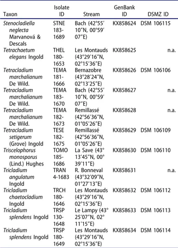
Table 1.Description of isolates studied and their identifiers in GenBank and in the collection of DSMZ (Deutsche Sammlung für Mikroorganismen und Zellkulturen).
Taxon IsolateID Stream GenBankID DSMZ ID Alatospora acuminata Ingold ALAC 180-1658 Les Montauds (43°29′16″N, 02°15′36″E) KX858600 DSM 106082 Alatospora acuminata Ingold ALAC 183-1668 Bach (42°55′ 10″N, 00°59′ 07″E) KX858602 DSM 106083 Alatospora pulchella Marvanová ALPU 130-1663 Le Lampy (43° 25′07″N, 02° 11′15″E) KX858601 DSM 106084 Anguillospora
crassa Ingold ANCR 130-1659 Le Lampy (43° 25′07″N, 02° 11′15″E) KX858603 DSM 106085 Anguillospora filiformis Greathead ANFI 130-1650 Le Lampy (43° 25′07″N, 02° 11′15″E) KX858604 n.a. Arbusculina moniliformis (Descals) Descals & Marvanová ABMO 182-1692 Remillassé (42°56′36″N, 01°05′26″E) KX858605 DSM 106087 Articulospora tetracladia Ingold ARTE 180-1647 Les Montauds (43°29′16″N, 02°15′36″E) KX858606 DSM 106088 Articulospora tetracladia Ingold ARTE 130-1660 Le Lampy (43° 25′07″N, 02° 11′15″E) KX858607 n.a. Clavariopsis aquatica De Wild. CLAQ 180-1651 Les Montauds (43°29′16″N, 02°15′36″E) KX858608 DSM 106090 Clavariopsis aquatica De Wild. CLAQ 180-1652 Les Montauds (43°29′16″N, 02°15′36″E) KX858609 DSM 106092 Clavariopsis aquatica De Wild. CLAQ 130-1676 Le Lampy (43° 25′07″N, 02° 11′15″E) KX858610 DSM 106093 Clavariopsis aquatica De Wild. CLAQ 182-1680 Remillassé (42°56′36″N, 01°05′26″E) KX858611 DSM 106091 Culicidospora aquatica Petersen CUAQ 130-1661 Le Lampy (43° 25′07″N, 02° 11′15″E) KX858612 DSM 106094 Flagellospora
curvula Ingold FLCU 130-1655 Le Lampy (43° 25′07″N, 02° 11′15″E) KX858613 n.a. Flagellospora
curvula Ingold FLCU 180-1662 Les Montauds (43°29′16″N, 02°15′36″E) KX858614 n.a. Lemonniera aquatica De Wild. LEAQ 185-1684 La Save (43° 13′45″N, 00° 39′11″E) KX858621 DSM 106098 Lemonniera
cornuta Ranzoni LECO 185-1685 La Save (43° 13′45″N, 00° 39′11″E) KX858620 DSM 106099 Lemonniera
terrestris Tubaki LETE 182-1672 Remillassé (42°56′36″N, 01°05′26″E) KX858623 DSM 106101 Lemonniera
terrestris Tubaki LETE 183-1669 Bach (42°55′ 10″N, 00°59′ 07″E) KX858622 DSM 106100 Lemonniera
terrestris Tubaki LETE 184-1681 Mouscailloux (43°28′37″N, 02°14′07″E) KX858615 DSM 106097 Neonectria lugdunensis Sacc. & Thérry
NELU 180-1645 Les Montauds (43°29′16″N, 02°15′36″E) KX858616 DSM 106102 Neonectria lugdunensis Sacc. & Thérry
NELU 130-1656 Le Lampy (43° 25′07″N, 02° 11′15″E) KX858617 DSM 106103 Neonectria lugdunensis Sacc. & Thérry
NELU 181-1664 Bernazobre (43°28′24″N, 02°13′25″E) KX858618 DSM 106104 Table 1.(Continued).
Taxon IsolateID Stream GenBankID DSMZ ID Stenocladiella neglecta Marvanová & Descals STNE 183-1689 Bach (42°55′ 10″N, 00°59′ 07″E) KX858624 DSM 106115 Tetrachaetum
elegans Ingold THEL 180-1653 Les Montauds (43°29′16″N, 02°15′36″E) KX858625 n.a. Tetracladium marchalianum De Wild. TEMA 181-1666 Bernazobre (43°28′24″N, 02°13′25″E) KX858626 DSM 106106 Tetracladium marchalianum De Wild. TEMA 183-1670 Bach (42°55′ 10″N, 00°59′ 07″E) KX858627 n.a. Tetracladium marchalianum De Wild. TEMA 182-1673 Remillassé (42°56′36″N, 01°05′26″E) KX858628 n.a. Tetracladium setigerum (Grove) Ingold TESE 182-1675 Remillassé (42°56′36″N, 01°05′26″E) KX858629 DSM 106109 Triscelophorus monosporus (Lind.) Hughes TOMO 185-1686 La Save (43° 13′45″N, 00° 39′11″E) KX858630 DSM 106110 Tricladium angulatum Ingold TRAN 4-1683 R. Bonneval(43°32′09″N, 01°27′13″E) KX858631 n.a. Tricladium chaetocladium Ingold TRCH 180-1646 Les Montauds (43°29′16″N, 02°15′36″E) KX858632 DSM 106112 Tricladium splendens Ingold TRSP 130-1648 Le Lampy (43° 25′07″N, 02° 11′15″E) KX858633 DSM 106113 Tricladium splendens Ingold TRSP 180-1649 Les Montauds (43°29′16″N, 02°15′36″E) KX858634 DSM 106114 Note. n.a. = not available.
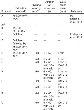
published protocols (Hettick et al. 2008; Marinach-Patrice et al.2009; Chalupová et al. 2012; De Respinis et al.2014) and our own modifications (TABLE 2). For this test, we used mycelium from three isolates, Lemonniera terrestris LETE 184-1681, Neonectria lugdunensis NELU 130-1656, and Tricladium splendens TRSP 180-1649, for which our standard extraction method (protocol A; De Respinis et al.
2014) did not yield satisfactory peak numbers (60–120 peaks; Erhard et al.2008). For all protocols, mycelium was scraped from a surface area of ca. 1 cm2
(containing both central and marginal parts of the colonies) of solid medium using a wet swab.
Chemical protocols.—In protocol A, both young and old mycelia were suspended in 300 µL of sterile deionized water to which 900 μL of >99.8% (v/v) ethanol was added. After vortexing and centrifugation (at 10 600 ×g, 2 min), the supernatant was discarded and the pellet air-dried for 5 min. The pellet was then dissolved in 40 μL of 70% formic acid by vortexing, and 40 μL of acetonitrile was added. After another vortexing
and centrifugation step (10 600 ×g, 2 min), 1 μL of the supernatant was spotted in quadruplicate directly on a MALDI-TOF MS target plate (Vitek MS-DS; bioMérieux, Marcy l’Etoile, France) and air-dried. One microliter of α-cyano-4-hydroxycinnamic acid matrix (Vitek MS CHCA matrix; bioMérieux) was added to each spot and air-dried before MALDI-TOF MS analysis (described below).
Protocol B was a modification of the technique described by Marinach-Patrice et al. (2009): 900 μL of >99.8% (v/v) ethanol was added to mycelium sus-pended in 300 µL sterile deionized water. After vortex-ing and centrifugation (10 600 × g, 2 min), the supernatant was discarded and the pellet air-dried. Fifty microliters of 80% trifluoroacetic acid was added to the pellet and vortexed. After incubation at room temperature for 30 min, 150 μL of sterile deionized water and 200 μL of acetonitrile were added. Samples were vortexed and centrifuged (10 600 ×g, 2 min), the supernatant was collected, and MALDI-TOF MS ana-lysis prepared as described above.
Enzymatic treatment.—Protocol C included a treatment based on the protocol described by Chalupová et al. (2012). The mycelium was suspended in a solution of 300 µL sterile deionized water, and 600 µL cellulase solution from Trichoderma sp. (5 Units; C1794; Sigma-Aldrich, St. Louis, Missouri, USA) was added. After incubation for 15 min at 37 C, the mycelium was washed three times in 1 mL of sterile deionized water and then suspended in 300 µL of sterile deionized water. After vortexing and centrifugation (10 600 × g, 2 min), the supernatant was collected and MALDI-TOF MS analysis was carried out as described above. Protocol D combined protocols C and A (TABLE 2).
Mechanical treatment (protocols E to L).—Following addition of acetonitrile, the suspension was directly mixed with glass beads and bead beating (Hettick et al. 2008) was carried out using a FastPrep-24 homogenizer (MP Biomedicals, Santa Ana, California, USA). After centrifugation (10 600 × g, 2 min), the supernatant was collected and used for MALDI-TOF MS analysis as described above.
Effects of mycelium age on extraction efficiency (protocol A1).—The effect of mycelium age was tested using mycelium taken from the edge of young colonies (<20 mm diam) of the three isolates described above. Proteins were extracted using protocol A.
Table 2.Protein extraction methods and protocols tested.
Protocol Extractionmethoda
Shaking velocity (m s−1) Duration of extraction (s) Glass beads diam (mm) Reference A 75EtOH-70FA-ACN — — — DeRespinis et al.2014 A1b — — — B 75EtOH-80TFA-ACN — — — C Cellulase — — — Chalupová et al.2012 D Cellulase followed by 75EtOH-70FA-ACN — — — E 75EtOH-70FA-ACN 4.0 1 × 60 1 mm F 6.0 1 × 40 1 mm G 6.0 3 × 60 with 30-s intervals 1 mm + 106–212 µm H 6.0 6 × 60, with 30-s intervals 1 mm + 106–212 µm I 4.0 1 × 60 150–212 µm J 6.0 1 × 40 150–212 µm K 6.0 3 × 60 with 30-s intervals 106–212 µm L 6.0 6 × 60 with 30-s intervals 106–212 µm
a75EtOH = 75% ethanol Aldrich); 70FA = 70% formic acid
(Sigma-Aldrich); ACN = acetonitrile (Sigma-(Sigma-Aldrich); 80TFA = 80% trifluoroacetic acid (Sigma-Aldrich).
bProtocol A1 was identical to protocol A except that it used young
Acquisition of MALDI-TOF mass spectra.—Based on an evaluation of extraction efficiency, protocol A1 was applied to all isolates, except forCulicidospora aquatica CUAQ 130-1661, Neonectria lugdunensis NELU 180-1645, Arbusculina moniliformis ABMO 182-1692, and Alatospora pulchella ALPU 130-1663, for which protocol A (young and old mycelia together) yielded higher protein extraction efficiency. MALDI-TOF MS was performed in quadruplicates per strain with a VITEK MS RUO mass spectrometer (AXIMA Confidence; bioMérieux) equipped with a 50 Hz nitrogen laser (pulse of 3 ns). Mass spectra were collected in positive linear mode in the range of 3000–20 000 mass-to-charge ratio (m/z) with delayed, positive ion extraction (delay time of 104 ns with a scale factor of 800) and an acceleration voltage of 20 kV. For each analysis, 100 averaged profile spectra were collected and those fulfilling the quality criteria (i.e., with peak intensity between 20 and 100 mV, cumulative intensity of all 100 spectra >3000 mV, main peaks resolution strictly higher than 600) were processed using the MALDI MS Launchpad 2.8 software (bioMérieux) with baseline correction, peak filtering, and smoothing procedures.
Phylogenetic analysis.—Sequences were generated with the primer ITS4 and processed using MEGA 6.0 (Tamura et al.2013) to construct a phylogenetic tree by neighbor-joining method with the Kimura two-parameter distances bootstrapped with 1000 replicates. Aquatic hyphomycete sequences (Letourneau et al.
2010; Seena et al. 2010; Baschien et al. 2013; Duarte et al. 2015) available from the National Center for Biotechnology Information (NCBI; http://www.ncbi. nlm.nih.gov) were also included and used as references. For isolates of species lacking a corresponding NCBI reference sequence, i.e., Lemonniera cornuta LECO 185-1685, Arbusculina moniliformis ABMO 182-1692, the three LETE Lemonniera terrestris isolates, and Culicidospora aquatica CUAQ 130-1661, identification was confirmed by morphological examination of conidia. In anticipation of the polyphyly to be expected in such a diverse group, ITS sequences of the Basidiomycota Boletus edulis and Russula cyanoxantha (from NCBI) were used as outgroups.
Statistical analysis.—Extraction efficiency was estimated as the number of peaks detected with each protocol using standardized postprocessing procedures (described above). We used a Friedman test with a post hoc Dunn’s test to verify whether peak numbers of the
three reference isolates depended on the extraction methods. All statistical analyses were performed with STATA 14 (StataCorp, College Station, Texas).
MALDI-TOF MS data.—The lists of MALDI-TOF MS peaks of individual analyses performed with standardized postprocessing procedures (described above; 0.08% error) were imported into the SARAMIS software package (bioMérieux), and data were analyzed using a proprietary single linkage agglomerative cluster analysis.
RESULTS
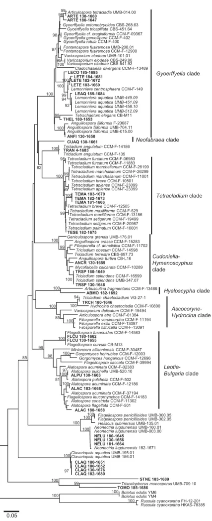
ITS sequencing.—Based on ITS data, the isolates clustered in different clades (FIG. 1), which correspond to the taxonomic groups recognized in earlier studies (e.g., Baschien et al. 2013). The phylogenetic tree contained characteristics that partly confirm results from earlier studies: (i) ITS sequences ofStenocladiella neglecta STNE 183-1689 (for which no reference ITS sequence is published) andTriscelophorus monosporus TOMO 185-1686 were clearly separated from all other isolates; (ii) Tricladium chaetocladium clustered with isolates of Neonectria lugdunensis and Hydrocina chaetocladia, respectively (FIG. 1); (iii) isolate Culicidospora aquatica CUAQ 130-1661 was the only aquatic hyphomycete in the Neofabraea clade; all other described taxa in Neofabraea are considered nonaquatic (Baschien et al. 2013); (iv) three species of Tricladium and two species of Anguillospora were interspersed among different clades, confirming results from previous studies (Belliveau and Bärlocher 2005; Bärlocher 2007; Seena et al.2010; Baschien et al.2013; Duarte et al.2015); and (v) isolates of T. marchalianum and T. setigerum clustered in the same clade together with other Tetracladium species for which reference sequences were available, corroborating earlier studies that suggested that Tetracladium is a relatively homogeneous genus (Nikolcheva and Bärlocher 2002; Letourneau et al.2010).
Evaluation of protein extraction protocols.— MALDI-TOF mass spectra obtained with the different protein extraction protocols (except those based on protocol C; see FIG. 2) were of good quality with clearly separated peaks. The overall median peak number of the 136 MALDI-TOF mass spectra of the 34 isolates was 65, ranging from 30 to 110, which is relatively low but within published quality criteria (Erhard et al. 2008). In general, the peak numbers
98 97 CLAQ 182-1680 CLAQ 180-1652 100 Gyoerffyella clade Ascocoryne-Hydrocina clade Articulospora tetracladia UMB-014.00
ARTE 130-1660 Gyoerffyella entomobryoides CBS-268.63 ARTE 180-1647 Gyoerffyella tricapillata CBS-451.64 Gyoerffyella rotula CCM-F-400 Gyoerffyella cf. craginiformis CCM-F-09367 Gyoerffyella gemellipara CCM-F-402 Fontanospora fusiramosa UMB-208.01 Fontanospora fusiramosa CCM-F-12900
Varicosporium elodeae UMB-101.01 Varicosporium elodeae CBS-249.90 Varicosporium elodeae CBS-541.92 Cladochasiella divergens CCM-F-13489 LECO 185-1685 LEAQ 185-1684 LETE 184-1681 LETE 183-1669 LETE 182-1672 Lemonniera centrosphaera CCM-F-149 Tetrachaetum elegans CB-M11 THEL 180-1653 Anguillospora filiformis F-20687 Anguillospora filiformis UMB-015.00 Anguillospora filiformis UMB-704.11 ANFI 130-1650
Tricladium angulatum CCM-F-139 TRAN 4-1683
Tricladium angulatum CCM-F-14186
Geniculospora grandis UMB-176.01
Anguillospora furtiva CB-L16 Tricladium obesum CCM-F-14598 ANCR 130-1659 Anguillospora crassa CCM-F-15283 Tricladium terrestre CBS-697.73 Filosporella cf. annelidica CCM-F-11702 Mycofalcella calcarata CCM-F-10289 Tricladium splendens CCM-F-16599 TRSP 180-1649 TRSP 130-1648
Tricladium splendens UMB-347.07
Filosporella exilis CCM-F-13097 Filosporella versimorpha CCM-F-11194 Filosporella fistucella CCM-F-13091 Articulospora atra CCM-F-01384 Varicosporium delicatum CCM-F-19494Hydrocina chaetocladia CCM-F-10890
Tricladium chaetocladium VG-27-1 TRCH 180-1646 ABMO 182-1692 Arbusculina fragmentans CCM-F-13486 Tetracladium furcatum CCM-F-06983 Tetracladium furcatum CCM-F-11883 Tetracladium marchalianum CCM-F-26199 Tetracladium marchalianum CCM-F-11001 Tetracladium breve CCM-F-10501 Tetracladium apiense CCM-F-23099 Tetracladium apiense CCM-F-23399 TEMA 183-1670 TEMA 182-1673 CLAQ 180-1651 TEMA 181-1666 Tetracladium breve CCM-F-12505 Tetracladium maxiliforme CCM-F-529 Tetracladium marchalianum CCM-F-26299 Tetracladium setigerum CCM-F-19499 Tetracladium setigerum CCM-F-20987 Tetracladium palmatum CCM-F-10001 TESE 182-1675 CUAQ 130-1661 Flagellospora fusarioides CCM-F-14583 FLCU 180-1662 Flagellospora curvula CB-M13 FLCU 130-1655 Miniancora allisoniensis CCM-F-30487 Gorgomyces honrubiae CCM-F-12003 Gorgomyces hungaricus CCM-F-12696 Flagellospora saccata CCM-F-39994 Alatospora pulchella UMB-520.10 Alatospora pulchella CCM-F-502 ALPU 130-1663 Alatospora acuminata CCM-F-02383 Alatospora acuminata CCM-F-12186 ALAC 183-1668 Alatospora acuminata CCM-F-37194 Flagellospora leucorhynchos CCM-F-14183 Alatospora flagellata CCM-F-501 Alatospora constricta CCM-F-11302 ALAC 180-1658
Flagellospora penicillioides UMB-300.05 Heliscus submersus UMB-135.01
STNE 183-1689 Neonectria lugdunensis UMB-160.01 Neonectria lugdunensis UMB-003.00 NELU 180-1645
Clavariopsis aquatica UMB-195.01 Clavariopsis aquatica UMB-156.01
CLAQ 130-1676 Cudoniella-Hymenoscyphus clade 99 98 99 94 100 95 97 100 100 100 98 98 100 100 100 99 82 99 99 100 100 99 100 100 94 85 100 100 100 96 100 98 81 100 100 100 85 100 97 100 100 100 99 100 100 100 Tetracladium maxiliforme CCM-F-13186
Flagellospora penicillioides UMB-302.05
Triscelophorus monosporus UMB-709.10
Russula cyanoxantha FH-12-201 Boletus edulis YM6
Hyaloscypha clade Tetracladium clade Neofabraea clade Leotia-Bulgaria clade 92 100
Lemonniera aquatica UMB-449.09 Lemonniera aquatica UMB-451.09 Lemonniera aquatica UMB-512.09 Lemonniera aquatica UMB-458.10
Boletus edulis YM4
Russula cyanoxantha HKAS-78385 Neonectria lugdunensis 182-1671
TOMO 185-1686
0.05
NELU 181-1664 NELU 130-1656
Figure 1.Phylogenetic tree based on ITS sequences of aquatic hyphomycetes. Neighbor-joining method using the Kimura two-parameter distances; bootstrap support of 80% or greater from 1000 replicates is shown in the internal nodes of the branches. Isolates used for this study are highlighted in bold.
obtained were similar for conspecific isolates (data not shown), with Tetracladium marchalianum being an exception (median peak numbers for TEMA 181-1666: 30; TEMA 182-1673: 88, and TEMA 183-1670: 56).
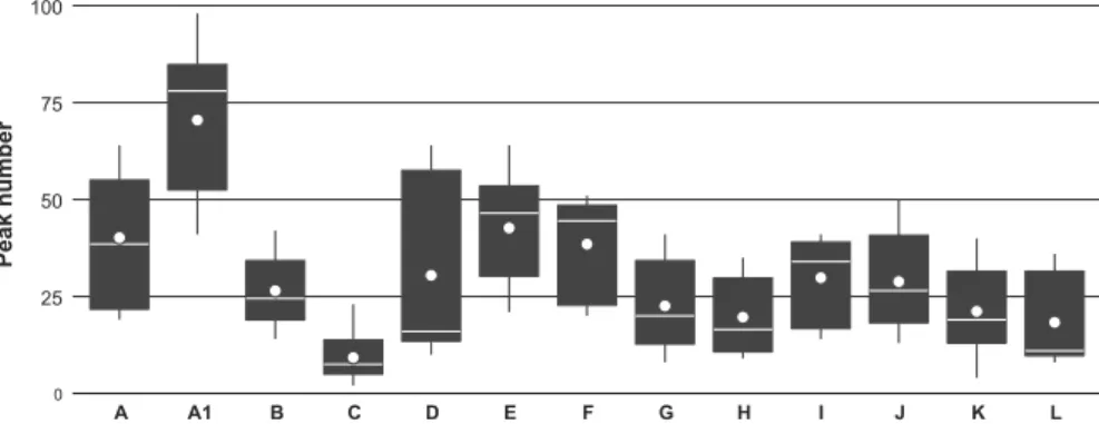
The statistical analysis revealed significant differences in peak numbers obtained from the isolates when using different protein extraction protocols (Friedman = 16.2; Kendall = 0.90; P = 0.0003), as shown by Dunn’s test. However, this statistical significance must be interpreted with caution because of the small sample size. Overall, protocol A1 (protocol A applied to young mycelium only) yielded highest peak numbers (FIG. 3), although signifi-cantly fewer peaks were detected from isolate TRSP 180-1649 than from the other two isolates tested (SUPPLEMENTARY FIG. 1).
MALDI-TOF MS analyses.—MALDI-TOF mass spectra showed substantial heterogeneity among different taxa, with similarities ranging from 20% to 36% at the genus or higher taxonomic rank (FIG. 4). Analytical variability of replicate measurements of the same isolates represented by two branches per strain was consistently lower than the variability among strains of the same species and especially than that among higher taxonomic units. In general, the homogeneity of some taxa revealed by sequencing (Clavariopsis aquatica, the genera Lemonniera and Tetracladium) and the heterogeneity within Tricladium and Anguillospora was confirmed by MALDI-TOF MS (FIGS. 1 and 4). In all cases, all isolates belonging to the same species clustered together with intraspecific
4000 6000 8000 10000 12000 14000 m/z F D C B A A1
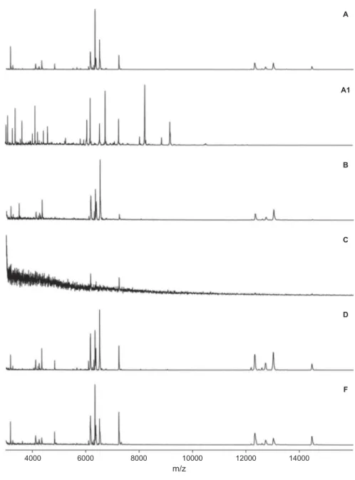
Figure 2.MALDI-TOF mass spectra of N. lugdunensis (NELU 130-1656) obtained using six different protocols (A, A1, B, C, D, F; for details, see Table 2). Relative intensities shown; averaged smoothing (width: 50 channels) and baseline subtracted (width: 500 channels), tolerance: 0.08%.
similarities of 58–84%. The conspecific isolates of Neonectria lugdunensis were an exception because they were highly heterogeneous, with only 36% similarity between NELU 180-1645 and the NELU 130-1656/NELU 181-1664 cluster. Nevertheless, these isolates also clustered together in the MALDI-TOF MS dendrogram (FIG. 4).
DISCUSSION
Potential of MALDI-TOF MS for aquatic hyphomycete taxonomy.—MALDI-TOF mass spectrometry yielded diagnostic spectra for all aquatic hyphomycete species of our collection. This corroborates earlier studies using MALDI-TOF MS that reported successful discrimination and/or identification of various fungal groups (Li et al. 2000; Erhard et al.2008; Hettick et al.2008; De Respinis et al.
2010,2013,2014,2015; Santos et al.2010; Theel et al.
2011). Although the performance strongly depended on the protein extraction protocol used (discussed below), MALDI-TOF MS was very powerful in discriminating even among closely related species that are difficult to distinguish based on spore morphology. For example, MALDI-TOF MS clearly resolved the Alatospora acuminata, A. constricta, and A. pulchella complex, the Lemonniera aquatica, L. cornuta, and L. terrestris complex, the Tetracladium marchalianum and T. setigerum group, and a group of species with sigmoid spores comprising Anguillospora crassa, A. filiformis, and Flagellospora curvula (FIG. 4).
Although cluster analysis of MALDI-TOF MS data leads to phenetic grouping of taxa that cannot be directly compared with phylogeny, the groupings obtained from our collection correlated well with that based on ITS sequencing. This suggests that MALDI-TOF MS provides reliable and reproducible
identification of aquatic hyphomycetes, with a taxonomic accuracy comparable to that of ITS sequencing. For example and in agreement with pre-vious findings, some anamorphic genera (e.g., Tetracladium) described previously as monophyletic (Nikolcheva and Bärlocher 2002) clustered together in our phylogenetic tree based on ITS, whereas other genera (e.g., Tricladium) described as polyphyletic (Baschien et al. 2013) were heterogeneous.
Evaluation of different protein extraction protocols. —Fungal cell walls consist mainly of polysaccharides, but their composition may vary both in quantity and quality among different species and even among strains (Kemptner et al. 2009). For this reason, exhaustive protein extraction and production of high-quality MALDI-TOF MS spectra of filamentous fungi may be challenging (De Respinis et al. 2014) and strongly dependent on extraction protocols. Most published studies show that cell-wall lysis before protein extraction is needed to obtain reliable and reproducible high-quality spectra from filamentous fungi (Hettick et al. 2008, 2011; Marinach-Patrice et al.2009; Cassagne et al.2011; De Respinis et al.
2014). In contrast, De Carolis et al. (2012) reported good differentiation of filamentous fungal species belonging to Aspergillus, Fusarium, and Mucorales, without pretreatment using MALDI-TOF intact-cell mass spectrometry where mycelium and spores were spotted directly on the MALDI-TOF MS target plate, and only absolute ethanol was added before adjunction of CHCA matrix. To our knowledge, De Carolis et al. (2012) were the only workers able to obtain good MALDI-TOF mass spectra without prior fungal cell-wall lysis.
Our comparison of 13 protein extraction protocols indicates that mechanical treatment by bead beating did not improve protein extraction efficiency. Similarly,
0 25 50 75 100 A A1 B C D E F G H I J K L Peak number
Figure 3.Box plots of the number of peaks detected with different extraction protocols used (A–L; see Table 2). The data are aggregates of three isolates: Lemonniera terrestris LETE 184-1681, Neonectria lugdunensis NELU 130-1656, and Tricladium splendens TRSP 180-1649. White line: median; white dot: mean; lower limit of the black box: 25th percentile; upper limit: 75th percentile; error bars: values within 1.5 interquartile range. Data of the individual isolates are presented in SUPPLEMENTARY FIG. 1.
CLAQ 180-1652 CLAQ 180-1651 CLAQ 182-1680 CLAQ 130-1676 FLCU 180-1662 FLCU 130-1655 TRSP 180-1649 ANCR 130-1659 TRSP 130-1648 LETE 184-1681 LETE 182-1672 LETE 183-1669 LECO 185-1685 LEAQ 185-1684 ARTE 130-1660 ARTE 180-1647 THEL 180-1653 ANFI 130-1650 TEMA 181-1666 TEMA 182-1673 TEMA 183-1670 TESE 182-1675 TRAN 4-1683 ALAC 180-1658 ALAC 183-1668 CUAQ 130-1661 NELU 130-1656 NELU 181-1664 NELU 180-1645 STNE 183-1689 TRCH 180-1646 ABMO 182-1692 ALPU 130-1663 TOMO 185-1686 Gyoerffyella clade Cudoniella-Hymenoscyphus clade Ascocoryne-Hydrocina clade Hyaloscypha clade Tetracladium clade Neofabraea clade Leotia-Bulgaria clade Leotia-Bulgaria clade Leotia-Bulgaria clade 0 10 20 30 40 50 60 70 80 90 100 %
Figure 4.Dendrogram (single linkage agglomerative clustering) based on the MALDI-TOF MS analysis of the aquatic hyphomycete isolates (using protocols A and A1, see details in text and inTable 2). Two MALDI-TOF mass spectra per isolate are shown to represent analytical variability; mass range: 3000–20 000 m/z, averaged smoothing (width: 50 channels) and baseline subtracted (width: 500 channels), tolerance: 0.08%.
treatment of mycelium with a nonspecific cellulase (Chalupová et al. 2012) yielded the poorest MALDI-TOF MS results in terms of quality and number of protein mass peaks. This was expected, because fungal cell walls do not contain cellulose. Future evaluations of protein extraction protocols should thus include other enzymes such as β-glucanase or chitinase to enhance fungal cell-wall lysis. Nevertheless, comparing spectra obtained with protocol C (only enzymatic treatment) with those obtained with protocol D (enzymatic plus chemical treatment), and assuming that the enzyme used was ineffective, lends further support to the importance of cell-wall lysis prior to protein extraction to achieve spectra of sufficient quality (FIG. 2). In conclusion, protocols using only chemical treatments were the most effective in our study, confirming earlier studies with other fungal groups (Bader et al. 2011; Cassagne et al.2011; De Respinis et al. 2014).
In addition, the use of young mycelium improved the quality of spectra obtained from fungal isolates, in particular the number of discernible peaks used for identification and construction of dendrograms. A comparison of discerned peaks from spectra of the same strain (NELU 130-1656) extracted with protocols A and A1, suggests that the spectra shared only a minority of peaks (FIG. 2). In our study, the protocol based on chemical protein extraction combined with the use of young mycelium (A1) was the most effective in producing high-quality mass spectra of aquatic hyphomycetes for species- and isolate-level identifica-tion. Qualitatively, these findings are consistent with those from other studies testing the effect of mycelium age on mass spectra quality (Santos et al. 2010; De Carolis et al. 2012; De Respinis et al. 2014). Further studies should aim at exploring the differences between spectra from mycelium of various ages to further improve the protocols. Given the importance of meth-odological considerations identified in our study (e.g., mycelium age, extraction protocol), we recommend a precautionary approach, i.e., to analyze fungal strains at a physiological state similar to the one used to create the reference database and using the same extraction protocols (see also Bader2013).
To our knowledge, this is the first study that used MALDI-TOF MS to characterize aquatic hyphomycetes. However, when compared with other applications, we observed lower peak numbers per spectrum obtained from some of our strains, especially compared with those from bacterial samples in other applications (S. De Respinis, unpubl. data). The cause for lower peak num-bers is currently unknown but may be related to the different cell-wall composition between bacteria and
fungi and even among fungal species and/or fungal strains, which seems an important factor for protein extraction efficiency (Kemptner et al.2009).
Conclusions.—Currently, the greatest challenges of MALDI-TOF MS applications to aquatic hyphomycetes include the lack of reference spectra in publicly accessible databases for species identification and the paucity of applications to metabolic proteins and of quantitative protocols. We have produced a first database based on an evaluated protocol as a reference for future taxonomic studies. To gain further insight into fungi of great ecological interest such as aquatic hyphomycetes, the taxonomic coverage of the database should be increased and the spectra of fungal species and/or isolates originating from different ecological conditions should be included.
In addition to taxonomic studies, analysis of protein mass fingerprints using MALDI-TOF MS may be a powerful tool to investigate and quantify genetic and phenotypic correlations, because the method can detect most dominant proteins simultaneously. Phenotypic variability in response to environmental conditions is probably reflected by protein expression, which can potentially be quantified by MALDI-TOF MS, assuming the development of additional quantita-tive protocols (Pan et al. 2009) and annotated genome sequences of study organisms. Providing an example for such applications, Howard and Boyer (2007) devel-oped protocols to quantify cyanobacteria microcystins from environmental samples using MALDI-TOF MS with internal standards. In aquatic hyphomycete ecology, elucidating the role of intraspecific diversity, phenotypic variability, and the organisms’ responses to environmental conditions may advance the mechanistic understanding not only of functional differences but also of interactions among strains and species with consequences for ecosystem functions (Ferreira et al.
2010; Fernandes et al. 2011). Our findings suggest that proteomic approaches using MALDI-TOF MS have the potential to refine our understanding of aquatic hypho-mycete biology. Because MALDI-TOF MS detects dominant proteins from mycelium, the technique also has great potential for assessing fungal responses to environmental conditions in ecological and ecotoxico-logical studies.
ACKNOWLEDGMENTS
We appreciate the expert technical assistance of Anna Paola Caminada, Alison Crenna, and Valentina Alesi (Laboratory of Applied Microbiology, Bellinzona, Switzerland) in the laboratory.
FUNDING
The project received financial support from the Department of Environment, Construction and Design of the Applied University of Southern Switzerland (SUPSI), and the Portuguese Foundation for Science and Technology to J.C. (FCT-SFRH/BPD/108779/2015).
ORCID
Sophie De Respinis http://orcid.org/0000-0001-7260-7264
Orlando Petrini http://orcid.org/0000-0002-1234-7832
Felix Bärlocher http://orcid.org/0000-0003-2404-625X
Eric Chauvet http://orcid.org/0000-0001-8676-392X
Andreas Bruder http://orcid.org/0000-0002-4591-491X LITERATURE CITED
Ang TN, Ngoh GC, Chua ASM. 2013. Development of a novel inoculum preparation method for solid-state fer-mentation—cellophane film culture (CFC) technique. Industrial Crops and Products 43:774–777, doi:10.1016/j. indcrop.2012.08.022
Bader O.2013. MALDI-TOF-MS-based species identification and typing approaches in medical mycology. Proteomics 13:788–799, doi:10.1002/pmic.201200468
Bader O, Weig M, Taverne-Ghadwal L, Lugert R, Groß U, Kuhns M.2011. Improved clinical laboratory identification of human pathogenic yeasts by matrix-assisted laser deso-rption ionization time-of-flight mass spectrometry. Clinical Microbiology and Infection 17:1359–1365, doi:10.1111/j.1469-0691.2010.03398.x
Bärlocher F. 1992. Research on aquatic hyphomycetes: his-torical background and overview. In: Bärlocher F, ed. The ecology of aquatic hyphomycetes. Berlin, Germany: Springer Verlag. p. 1–15, doi:10.1007/978-3-642-76855-2_1
Bärlocher F. 2007. Molecular approaches applied to aquatic hyphomycetes. Fungal Biology Reviews 21:19–24, doi:10.1016/j.fbr.2007.02.003
Bärlocher F. 2010. Molecular approaches promise a deeper and broader understanding of the evolutionary ecology of aquatic hyphomycetes. Journal of the North American Benthological Society 29:1027–1041, doi:10.1899/09-081.1
Bärlocher F. 2016. Aquatic hyphomycetes in a changing environment. Fungal Ecology 19:14–27, doi:10.1016/j. funeco.2015.05.005
Baschien C, Marvanová L, Szewzyk U. 2006. Phylogeny of selected aquatic hyphomycetes based on morphological and molecular data. Nova Hedwigia 83:311–352, doi:10.1127/0029-5035/2006/0083-0311
Baschien C, Tsui CK-M, Gulis V, Szewzyk U, Marvanová L.
2013. The molecular phylogeny of aquatic hyphomycetes with affinity to the Leotiomycetes. Fungal Biology 117:660–672, doi:10.1016/j.funbio.2013.07.004
Belliveau MJ-R, Bärlocher F.2005. Molecular evidence con-firms multiple origins of aquatic hyphomycetes. Mycological Research 109:1407–1417, doi:10.1017/ S0953756205004119
Bright JJ, Claydon MA, Soufian M, Gordon DB.2002. Rapid typing of bacteria using matrix-assisted laser desorption
ionisation time-of-flight mass spectrometry and pattern recognition software. Journal of Microbiological Methods 48:127–138, doi:10.1016/S0167-7012(01)00317-7
Calderaro A, Arcangeletti M-C, Rodighiero I, Buttrini M, Gorrini C, Motta F, Germini D, Medici M-C, Chezzi C, Conto F De.2014. Matrix-assisted laser desorption/ioniza-tion time-of-flight (MALDI-TOF) mass spectrometry applied to virus identification. Science Reports 4:6803, doi:10.1038/srep06803
Cassagne C, Ranque S, Normand A-C, Fourquet P, Thiebault S, Planard C, Hendrickx M, Piarroux R. 2011. Mould routine identification in the clinical laboratory by matrix-assisted laser desorption ionization time-of-flight mass spectrometry. PLoS ONE 6:e28425, doi: 10.1371/jour-nal.pone.0028425
Chalupová J, Sedlářová M, Helmel M, Řehulka P, Marchetti-Deschmann M, Allmaier G,Šebela M.2012. MALDI-based intact spore mass spectrometry of downy and powdery mildews. Journal of Mass Spectrometry 47:978–986, doi:10.1002/jms.3046
Chauvet E.1990. Hyphomycètes aquatiques du sud-ouest de la France. Gaussenia 6:3–31.
Clivot H, Cornut J, Chauvet E, Elger A, Poupin P, Guérold F, Pagnout C.2014. Leaf-associated fungal diversity in acid-ified streams: insights from combining traditional and molecular approaches. Environmental Microbiology 16:2145–2156, doi:10.1111/1462-2920.12245
Cornut J, Elger A, Lambrigot D, Marmonier P, Chauvet E.2010. Early stages of leaf decomposition are mediated by aquatic fungi in the hyporheic zone of woodland streams. Freshwater Biology 55:2541–2556, doi:10.1111/j.1365-2427.2010.02483.x
Cornut J, Ferreira V, Gonçalves AL, Chauvet E, Canhoto C.
2015. Fungal alteration of the elemental composition of leaf litter affects shredder feeding activity. Freshwater Biology 60:1755–1771, doi:10.1111/fwb.12606
Croxatto A, Prod’hom G, Greub G. 2012. Applications of MALDI-TOF mass spectrometry in clinical diagnostic microbiology. FEMS Microbiology Reviews 36:380–407, doi:10.1111/j.1574-6976.2011.00298.x
De Carolis E, Posteraro B, Lass-Flörl C, Vella A, Florio AR, Torelli R, Girmenia C, Colozza C, Tortorano AM, Sanguinetti M, Fadda G. 2012. Species identification of Aspergillus, Fusarium and Mucorales with direct surface ana-lysis by matrix-assisted laser desorption ionization time-of-flight mass spectrometry. Clinical Microbiology and Infections 18:475–484, doi:10.1111/j.1469-0691.2011.03599.x
De Respinis S, Monnin V, Girard V, Welker M, Arsac M, Cellière B, Durand G, Bosshard PP, Farina C, Passera M, Belkum A Van, Petrini O, Tonolla M. 2014. Matrix-assisted laser desorption ionization–time of flight (MALDI-TOF) mass spectrometry using the Vitek MS system for rapid and accurate identification of dermato-phytes on solid cultures. Journal of Clinical Microbiology 52:4286–4292, doi:10.1128/JCM.02199-14
De Respinis S, Tonolla M, Pranghofer S, Petrini L, Petrini O, Bosshard PP. 2013. Identification of dermatophytes by matrix-assisted laser desorption/ionization time-of-flight mass spectrometry. Medical Mycology 51:514–521, doi:10.3109/13693786.2012.746476
De Respinis S, Vogel G, Benagli C, Tonolla M, Petrini O, Samuels GJ. 2010. MALDI-TOF MS of Trichoderma:
a model system for the identification of microfungi. Mycological Progress 9:79–100, doi: 10.1007/s11557-009-0621-5
De Respinis S, Weissenhorn S, Bosshard PP, Tonolla M, Petrini L, Petrini O. 2015. Identification of some Aspergillus species in the Flavi and Fumigati sections by matrix-assisted laser desorption/ionisation time-of-flight (MALDI-TOF) mass spectrometry. Journal of Fungal Research 13:269–283, doi:10.13341/j.jfr.2014.2058
Descals E.2005. Techniques for handling Ingoldian fungi. In: Graça MAS, Bärlocher F, Gessner MO, eds. Methods to study litter decomposition: a practical guide. Dordrecht, The Netherlands: Springer. p. 129–141, doi:10.1007/ 1-4020-3466-0_19
Duarte S, Bärlocher F, Cassio F, Pascoal C. 2014. Current status of DNA barcoding of aquatic hyphomycetes. Sydowia 66:191–202, doi:10.12905/0380.sydowia66(2) 2014-0191
Duarte S, Bärlocher F, Pascoal C, Cássio F. 2016. Biogeography of aquatic hyphomycetes: current knowledge and future perspectives. Fungal Ecology 19:169–181, doi:10.1016/j.funeco.2015.06.002
Duarte S, Batista D, Bärlocher F, Cássio F, Pascoal C.2015. Some new DNA barcodes of aquatic hyphomycete species. Mycoscience 56:102–108, doi:10.1016/j.myc.2014.04.002
Erhard M, Hipler U-C, Burmester A, Brakhage AA, Wöstemeyer J.2008. Identification of dermatophyte species causing onychomycosis and tinea pedis by MALDI-TOF mass spectrometry. Experimental Dermatology 17:356–361, doi:10.1111/j.1600-0625.2007.00649.x
Fenselau C, Demirev PA. 2001. Characterization of intact microorganisms by MALDI mass spectrometry. Mass Spectrometry Reviews 20:157–171, doi:10.1002/mas.10004
Fernandes I, Pascoal C, Cássio F. 2011. Intraspecific traits change biodiversity effects on ecosystem functioning under metal stress. Oecologia 166:1019–1028, doi:10.1007/ s00442-011-1930-3
Fernandes I, Pereira A, Trabulo J, Pascoal C, Cássio F, Duarte S. 2015. Microscopy- or DNA-based analyses: which methodology gives a truer picture of stream-dwelling decomposer fungal diversity? Fungal Ecology 18:130–134, doi:10.1016/j.funeco.2015.08.005
Ferreira V, Gonçalves AL, Pratas J, Canhoto C. 2010. Contamination by uranium mine drainages affects fungal growth and interactions between fungal species and strains. Mycologia 102:1004–1011, doi:10.3852/09-248
Gulis V, Marvanová L, Descals E.2005. An illustrated guide to the common temperate species of aquatic hyphomy-cetes. In: Graça MAS, Bärlocher F, Gessner MO, eds. Methods to study litter decomposition: a practical guide. Dordrecht, The Netherlands: Springer. p. 135–149, doi:10.1007/1-4020-3466-0_21
Hettick JM, Green BJ, Buskirk AD, Kashon ML, Slaven JE, Janotka E, Blachere FM, Schmechel D, Beezhold DH.2008. Discrimination of Aspergillus isolates at the species and strain level by matrix-assisted laser desorption/ionization time-of-flight mass spectrometry fingerprinting. Analytical Biochemistry 380:276–281, doi:10.1016/j.ab.2008.05.051
Hettick JM, Green BJ, Buskirk AD, Slaven JE, Kashon ML, Beezhold DH. 2011. Discrimination of fungi by MALDI-TOF mass spectrometry. In: Fenselau C, Demirev P, eds. Rapid characterization of microorganisms by mass
spectrometry. Washington, DC: American Chemical Society. p. 3–35, doi:10.1021/bk-2011-1065.ch003
Howard KL, Boyer GL.2007. Quantitative analysis of cyano-bacterial toxins by matrix-assisted laser desorption ioniza-tion mass spectrometry. Analytical Chemistry 79:5980–5986, doi:10.1021/ac0705723
Ingold CT.1975. Illustrated guide to aquatic and water-borne Hyphomycetes (Fungi imperfecti) with notes on their biol-ogy. Freshwater Biological Association Scientific Publication no. 30. Ambleside, UK: Freshwater Biological Association. p. 86.
Jabiol J, Bruder A, Gessner MO, Makkonen M, McKie BG, Peeters ETHM, Vos VCA, Chauvet E. 2013. Diversity patterns of leaf-associated aquatic hyphomycetes along a broad latitudinal gradient. Fungal Ecology 6:439–448, doi:10.1016/j.funeco.2013.04.002
Kaufmann C, Ziegler D, Schaffner F, Carpenter S, Pflüger V, Mathis A.2011. Evaluation of matrix-assisted laser deso-rption/ionization time of flight mass spectrometry for characterization of Culicoides nubeculosus biting midges. Medical and Veterinary Entomology 25:32–38, doi:10.1111/j.1365-2915.2010.00927.x
Kemptner J, Marchetti-Deschmann M, Mach R, Druzhinina IS, Kubicek CP, Allmaier G.2009. Evaluation of matrix-assisted laser desorption/ionization (MALDI) preparation techniques for surface characterization of intact Fusarium spores by MALDI linear time-of-flight mass spectrometry. Rapid Communications in Mass Spectrometry 23:877–884, doi:10.1002/rcm.3949
Krauss G-J, Solé M, Krauss G, Schlosser D, Wesenberg D, Bärlocher F.2011. Fungi in freshwaters: ecology, physiol-ogy and biochemical potential. FEMS Microbiological Reviews 35:620–651, doi:10.1111/j.1574-6976.2011.00266.x
Letourneau A, Seena S, Marvanová L, Bärlocher F. 2010. Potential use of barcoding to identify aquatic hyphomycetes. Fungal Diversity 40:51–64, doi:10.1007/ s13225-009-0006-8
Li L, Garden RW, Sweedler J V.2000. Single-cell MALDI: a new tool for direct peptide profiling. Trends in Biotechnology 18:151–160, doi:10.1016/S0167-7799(00)01427-X
Marinach-Patrice C, Lethuillier A, Marly A, Brossas J-Y, Gené J, Symoens F, Datry A, Guarro J, Mazier D, Hennequin C.2009. Use of mass spectrometry to identify clinical Fusarium isolates. Clinical Microbiology and Infections 15:634–642, doi:10.1111/j.1469-0691.2009.02758.x
Marvanová L, Bärlocher F. 2001. Hyphomycetes from Canadian streams. VI. Rare species in pure cultures. Czech Mycology 53:1–28.
Nikolcheva L, Bärlocher F.2004. Taxon-specific fungal pri-mers reveal unexpectedly high diversity during leaf decom-position in a stream. Mycological Progress 3:41–49, doi:10.1007/s11557-006-0075-y
Nikolcheva LG, Bärlocher F.2002. Phylogeny ofTetracladium based on 18S rDNA. Czech Mycology 53:285–295.
Pan S, Aebersold R, Chen R, Rush J, Goodlett DR, McIntosh MW, Zhang J, Brentnall TA. 2009. Mass spec-trometry based targeted protein quantification: methods and applications. Journal of Proteome Research 8:787–797, doi:10.1021/pr800538n
Roldán A, Descals E, Honrubia M.1989. Pure culture studies on Tetracladium. Mycological Research 93:452–465, doi:10.1016/S0953-7562(89)80039-5
Samuels GJ, Ismaiel A, Bon M-C, De Respinis S, Petrini O.2010. Trichoderma asperellum sensu lato consists of two cryptic species. Mycologia 102:944–966, doi:10.3852/09-243
Santos C, Paterson RRM, Venâncio A, Lima N. 2010. Filamentous fungal characterizations by matrix-assisted laser desorption/ionization time-of-flight mass spectrometry. J ournal of Applied Microbiology 108:375–385, doi:10.1111/j.1365-2672.2009.04448.x
Seena S, Duarte S, Pascoal C, Cássio F. 2012. Intraspecific variation of the aquatic fungus Articulospora tetracladia: an ubiquitous perspective. PLoS ONE 7:e35884, doi:10.1371/journal.pone.0035884
Seena S, Pascoal C, Marvanová L, Cássio F. 2010. DNA barcoding of fungi: a case study using ITS sequences for identifying aquatic hyphomycete species. Fungal Diversity 44:77–87, doi:10.1007/s13225-010-0056-y
Shearer C, Descals E, Kohlmeyer B, Kohlmeyer J, Marvanová L, Padgett D, Porter D, Raja H, Schmit J, Thorton H, Voglymayr H. 2007. Fungal biodiversity in aquatic habitats. Biodiversity and Conservation 16:49–67, doi:10.1007/s10531-006-9120-z
Stackebrandt E, Päuker O, Erhard M. 2005. Grouping Myxococci (Corallococcus) strains by matrix-assisted laser desorption ionization time-of-flight (MALDI TOF) mass spectrometry: comparison with gene sequence phylogenies. Current Microbiology 50:71–77, doi: 10.1007/s00284-004-4395-3
Stevenson LG, Drake SK, Shea YR, Zelazny AM, Murray PR.
2010. Evaluation of matrix-assisted laser desorption ionization-time of flight mass spectrometry for identifica-tion of clinically important yeast species. Journal of Clinical Microbiology 48:3482–3486, doi:10.1128/ JCM.00687-09
Tamura K, Stecher G, Peterson D, Filipski A, Kumar S.2013. MEGA6: Molecular Evolutionary Genetics Analysis ver-sion 6.0. Molecular Biology and Evolution 30:2725–2729, doi:10.1093/molbev/mst197
Theel ES, Hall L, Mandrekar J, Wengenack NL. 2011. Dermatophyte identification using matrix-assisted laser desorption ionization–time of flight mass spectrometry. Journal of Clinical Microbiology49:4067–4071, doi:10.1128/JCM.01280-11
von Bergen M, Eidner A, Schmidt F, Murugaiyan J, Wirth H, Binder H, Maier T, Roesler U. 2009. Identification of harmless and pathogenic algae of the genusPrototheca by MALDI-MS. Proteomics Clinical Applications 3:774–784, doi:10.1002/prca.200780138
Webster J, Descals E. 1981. Morphology, distribution, and ecology of conidial fungi in freshwater habitats. In: Cole GC, Kendrick B, eds. Biology of the conidial fungi. London: Academic Press. p. 295–355.
White TJ, Bruns T, Lee S, Taylor JW.1990. Amplification and direct sequencing of fungal ribosomal RNA genes for phylo-genetics. In: Innis MA, Gelfand DH, Sninsky JJ, White TJ, eds. PCR protocols: a guide to methods and applications. San Diego, California: Academic Press. p. 315–322.
Wurzbacher C, Grimmett IJ, Bärlocher F.2015. Metabarcoding-based fungal diversity on coarse and fine particulate organic matter in a first-order stream in Nova Scotia, Canada. F1000Research 4:1378, doi:10.12688/f1000research.7359.1
Ziegler D, Pothier JF, Ardley J, Fossou RK, Pflüger V, Meyer S de, Vogel G, Tonolla M, Howieson J, Reeve W, Perret X.2015. Ribosomal protein biomarkers provide root nodule bacterial identification by MALDI-TOF MS. Applied Microbiology and Biotechnology 99: 5547–5562, doi:10.1007/s00253-015-6515-3