An in-house assay is superior to Sepsityper® for the direct MALDI-TOF identification of 1
yeast species in blood culture 2
3
Bidart-Coutton Marie,a Isabelle Bonnet, b Aurélie Hennebique,b Zine Eddine Kherraf, b 4
Hervé Pelloux,b Berger Françoisa, Muriel Cornet,b,c Sébastien Bailly,b,d,e Danièle 5
Maubonb,c, # 6
7
Clinatec, Pôle recherche, CHU de Grenoble, Grenoble, France a; Laboratoire de 8
Parasitologie-Mycologie, Institut de Biologie et de Pathologie, CHU de Grenoble, 9
Grenoble, France b; Laboratoire TIMC-IMAG-TheREx, UMR 5525 CNRS-UJF, 10
Université Grenoble Alpes, France c, U823, Grenoble Alpes University, 11
Grenoble, Franced ;UMR 1137 - IAME Team 5 – DeSCID, Inserm/ Paris Diderot, 12
Sorbonne Paris Cité University, Paris, Francee 13
14
Running Head: Direct identification of yeast in blood cultures 15
16
#Address correspondence to Danièle Maubon, dmaubon@chu-grenoble.fr 17
I.B., A.H. and Z.E.K. contributed equally to this work. 18
19 20
Abstract – max 50 words 21
We developed an in-house assay for the direct identification, by MALDI-TOF, of yeasts 22
in blood culture. Sixty-one representative strains from 12 species were analyzed in 23
artificial blood cultures. Our assay accurately identified 95 of 107 (88.8%) positive blood 24
cultures and outperformed the commercial Sepsityper® kit. 25
27
Prompt, appropriate, first-line antifungal treatment has been shown to improve 28
outcome in patients with fungemia, but identification is currently delayed by the need for 29
subculture from positive blood cultures (1, 2). Mass spectrometry techniques based on 30
MALDI-TOF technology have made it possible to identify species directly from positive 31
blood cultures (3). The sample contains human cells, and identification protocols must 32
lyse these cells efficiently (without disrupting the microorganism) and eliminate the 33
unwanted human proteins. In-house protocols based on the lytic agent saponin have been 34
described for bacterial identification (4, 5), but only few assays have been developed for 35
yeast-positive blood cultures (6–9). Bruker produces an easy-to-use kit, Sepsityper®, 36
which has yielded good results for the characterization of both bacteria and yeasts (10, 37
11). However, this commercial test has never been compared, for yeast identification, 38
with any in-house protocol. We developed an in-house protocol for the direct 39
identification, by MALDI-TOF, of the yeast species commonly isolated from blood 40
cultures. We compared the results obtained with our assay to those obtained with 41
Sepsityper®. 42
We used an artificial blood culture protocol (blood from healthy volunteers), to 43
facilitate the testing of large numbers of strains and species (12). We used 61 strains 44
previously identified by MALDI-TOF and representative of 12 species commonly 45
implicated in fungemia: Candida albicans (15), C. glabrata (9), C. parapsilosis (7), C. 46
kefyr (6), C. krusei (6), C. tropicalis (7), Cryptococcus neoformans (4), C. dubliniensis
47
(3), C. utilis (1), C. guillermondii (1), C. inconspicua (1), and Saccharomyces cerevisiae 48
(1). Six were isolated directly from blood cultures from patients. The strains were used to 49
inoculate (10 yeast cells/flask) Mycosis IC/F and BACTEC Plus Aerobic/F or BACTEC 50
plus Anaerobic/ F (C. glabrata only) flasks, and were incubated in BACTEC FX (Becton 51
Dickinson) until the flask tested positive for their presence. Each positive flask was 52
subjected to MALDI-TOF MS identification with a Microflex TM MALDI-TOF mass 53
spectrometer (Bruker Daltonics, Germany). We first compared the following agents: 54
0.8% saponin (as described in ref (4)), 1% Triton, 1.8% SDS (two other commonly used 55
lytic agents) and the Sepsityper® kit, on 17 strains. Two washing steps were included 56
after lysis, because the inclusion of an additional washing step had been shown to 57
improve identification scores ((11) and data not shown). Briefly, the lytic agent was 58
added to 1 ml of medium from each positive flask, which was then vortexed and pelleted 59
(16,000 RCF, two minutes). The supernatant was discarded and the pellet was washed 60
twice in sterile distilled water (saponin, Triton and SDS1.8 protocol) or Sepsityper® 61
washing buffer. It was then homogenized with an appropriate quantity of formic acid 62
(from 2 µl to 15 µl, depending on pellet volume; mean of 5 µl) and an equal volume of 63
acetonitrile was added. Following a final centrifugation (16,000 RCF, 2 min) 1 µl of 64
supernatant was dispensed, in duplicate, on a polished steel target and covered with 1 µl 65
α-cyano-4-hydroxycinnamic acid (HCCA) for MALDI-TOF analysis. The spectra 66
obtained were analyzed with the BioTyper 2.0 database (Bruker Daltonics). The 67
thresholds for interpretation were adapted as previously suggested (8, 13, 14). 68
Identification scores were classified into four categories: A [>2]; B [1.7-2]; C [1.4-1.7], 69
with the expected species proposed first and four times in a row; D [< 1.4] or not 70
identified. The first three categories yielded acceptable results for identification to species 71
level. In total, 305 tests were carried out (Table 1). According to our adapted scores, 72
95/107 (88.8%), 94/115 (81.7%), 16/42 (38%), 12/41 (29.2%) tests led to correct 73
identification for the SDS1.8, Sepsityper®, saponin and Triton protocols, respectively. 74
Protocols based on saponin and Triton were the least efficient for yeast identification in 75
blood cultures, possibly because they lysed human cells less efficiently, leading to a 76
mixed (yeast and human) and, thus, less specific spectrum profile. The spectrum obtained 77
with the SDS1.8 protocol was very similar to that obtained with the Sepsityper® kit 78
(Figure 1). Based on these results, we limited subsequent comparisons to the Sepsityper® 79
and SDS1.8 protocols. 80
We analyzed 44 strains grown in both Mycosis IC/F and BACTEC Plus/F flasks 81
with the SDS1.8 and Sepsityper® protocols (176 tests). Scores are reported as median 82
values (interquartile range). Score was transformed into a qualitative value, with four 83
classes corresponding to the identification categories (see above). The Wilcoxon signed 84
rank sum test and Spearman’s rank correlation analysis were used for quantitative scores 85
and Fisher’s exact test was used for qualitative scores. The results obtained with the two 86
protocols were well correlated (Spearman’s correlation coefficient r=0.70 p<0.001). 87
Median score was significantly higher with the SDS protocol (1.9 [1.7; 2.1]) than with 88
Sepsityper® (1.8 [1.55; 2.0]) (Wilcoxon test p=0.003). This difference remained 89
significant if separate analyses were carried out by vial type (Mycosis IC/F p<0.001 and 90
BACTEC Plus p=0.04) (figure 2) and for the non-Candida albicans yeasts (C. albicans: 91
p=0.16; non-C. albicans yeasts: p<0.001) (data not shown). A subgroup analysis by vial
92
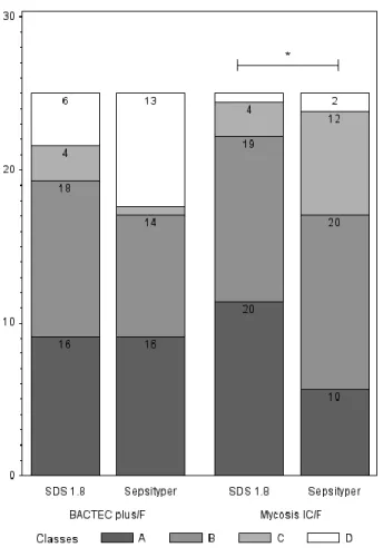
type showed that score class results were also better for the SDS assay than for the 93
Sepsityper® assay for Mycosis IC/F (p=0.04) (Figure 3). 94
This is the first comparison of an in-house protocol with the Sepsityper® kit for 95
the MALDI-TOF identification of yeasts in positive blood cultures. We found that lysis 96
with 1.8% SDS was globally superior to the Sepsityper® kit, particularly for yeasts 97
isolated from Mycosis IC/F medium and for non-albicans species of Candida. The two 98
protocols had similar durations (30 minutes), but the SDS1.8 protocol was cheaper than 99
the Sepsityper® protocol (by a factor of 220: €0.025 vs. €5.5 euros per test). The SDS 100
protocol yielded a correct identification rate of 100% (mean score 2 ± 0.19) in clinical 101
practice, in tests on nine flasks from six patients, but a prospective study is required to 102
confirm this performance in other vials containing patient’s blood. As the 0.8% saponin 103
procedure gave poorer results than previously reported for bacteria, rapid identification 104
procedures for positive blood cultures will probably need to be adapted according to the 105
results of direct examination. The SDS1.8 protocol appears to be an effective, cheap 106
alternative for yeast identification in positive blood cultures. 107
108
Acknowledgments 109
Authors have no conflict of interest to declare 110
References 111
1. Puig-Asensio M, Pemán J, Zaragoza R, Garnacho-Montero J, Martín-112
Mazuelos E, Cuenca-Estrella M, Almirante B, Prospective Population Study on 113
Candidemia in Spain (CANDIPOP) Project, Hospital Infection Study Group 114
(GEIH), Medical Mycology Study Group (GEMICOMED) of the Spanish Society of 115
Infectious Diseases and Clinical Microbiology (SEIMC), Spanish Network for 116
Research in Infectious Diseases. 2014. Impact of therapeutic strategies on the prognosis 117
of candidemia in the ICU. Crit. Care Med. 42:1423–1432. 118
2. Puig-Asensio M, Padilla B, Garnacho-Montero J, Zaragoza O, Aguado JM, 119
Zaragoza R, Montejo M, Muñoz P, Ruiz-Camps I, Cuenca-Estrella M, Almirante B, 120
CANDIPOP Project, GEIH-GEMICOMED (SEIMC), REIPI. 2014. Epidemiology 121
and predictive factors for early and late mortality in Candida bloodstream infections: a 122
population-based surveillance in Spain. Clin. Microbiol. Infect. Off. Publ. Eur. Soc. Clin. 123
Microbiol. Infect. Dis. 20:O245–254. 124
3. Posteraro B, De Carolis E, Vella A, Sanguinetti M. 2013. MALDI-TOF mass 125
spectrometry in the clinical mycology laboratory: identification of fungi and beyond. 126
Expert Rev. Proteomics 10:151–164. 127
4. Martiny D, Dediste A, Vandenberg O. 2012. Comparison of an in-house 128
method and the commercial SepsityperTM kit for bacterial identification directly from 129
positive blood culture broths by matrix-assisted laser desorption-ionisation time-of-flight 130
mass spectrometry. Eur. J. Clin. Microbiol. Infect. Dis. 31:2269–2281. 131
5. Meex C, Neuville F, Descy J, Huynen P, Hayette M-P, De Mol P, Melin P. 132
2012. Direct identification of bacteria from BacT/ALERT anaerobic positive blood 133
cultures by MALDI-TOF MS: MALDI Sepsityper kit versus an in-house saponin method 134
for bacterial extraction. J. Med. Microbiol. 61:1511–1516. 135
6. Marinach-Patrice C, Fekkar A, Atanasova R, Gomes J, Djamdjian L, 136
Brossas J-Y, Meyer I, Buffet P, Snounou G, Datry A, Hennequin C, Golmard J-L, 137
Mazier D. 2010. Rapid species diagnosis for invasive candidiasis using mass 138
spectrometry. PLoS ONE 5:e8862. 139
7. Ferroni A, Suarez S, Beretti J-L, Dauphin B, Bille E, Meyer J, Bougnoux M-140
E, Alanio A, Berche P, Nassif X. 2010. Real-Time identification of bacteria and 141
Candida species in positive blood culture broths by Matrix-Assisted Laser Desorption
142
Ionization-Time of Flight Mass spectrometry. J. Clin. Microbiol. 48:1542–1548. 143
8. Ferreira L, Sánchez-Juanes F, Porras-Guerra I, García MI, García-144
Sánchez JE, González-Buitrago JM, Muñoz-Bellido JL. 2011. Microorganisms direct 145
identification from blood culture by matrix-assisted laser desorption/ionization time-of-146
flight mass spectrometry: Blood culture pathogens direct identification by MALDI-TOF. 147
Clin. Microbiol. Infect. 17:546–551. 148
9. Spanu T, Posteraro B, Fiori B, D’Inzeo T, Campoli S, Ruggeri A, 149
Tumbarello M, Canu G, Trecarichi EM, Parisi G, Tronci M, Sanguinetti M, Fadda 150
G. 2012. Direct MALDI-TOF mass spectrometry assay of blood culture broths for rapid 151
identification of Candida species causing bloodstream infections: an observational study 152
in two large microbiology laboratories. J. Clin. Microbiol. 50:176–179. 153
10. Buchan BW, Riebe KM, Ledeboer NA. 2012. Comparison of the MALDI 154
Biotyper system using sepsityper specimen processing to routine microbiological 155
methods for identification of bacteria from positive blood culture bottles. J. Clin. 156
Microbiol. 50:346–352. 157
11. Yan Y, He Y, Maier T, Quinn C, Shi G, Li H, Stratton CW, Kostrzewa M, 158
Tang Y-W. 2011. Improved identification of yeast species directly from positive blood 159
culture media by combining Sepsityper specimen processing and Microflex analysis with 160
the matrix-assisted laser desorption ionization Biotyper system. J. Clin. Microbiol. 161
49:2528–2532. 162
12. Fricker-Hidalgo H, Lebeau B, Pelloux H, Grillot R. 2004. Use of the BACTEC 163
9240 system with Mycosis-IC/F blood culture bottles for detection of fungemia. J. Clin. 164
Microbiol. 42:1855–1856. 165
13. La Scola B, Raoult D. 2009. Direct Identification of bacteria in positive blood 166
culture bottles by Matrix-Assisted Laser Desorption Ionisation Time-of-Flight mass 167
spectrometry. PLoS ONE 4:e8041. 168
14. Idelevich EA, Grunewald CM, Wüllenweber J, Becker K. 2014. Rapid 169
Identification and susceptibility testing of Candida spp. from positive blood cultures by 170
combination of direct MALDI-TOF mass spectrometry and direct inoculation of Vitek 2. 171
PloS One 9:e114834. 172
173 174
Figure legends 175
Figure 1 Spectral profile obtained for Candida krusei isolated from Mycosis® blood 176
culture and subjected to various lysis protocols. SDS1.8 = 1.8% SDS; NC= negative 177
control (Mycosis® blood culture negative on day 6). Arrows indicate the three principal 178
nonspecific spectral signals present in negative controls but also identified in the saponin 179
and Triton protocols. 180
181
Figure 2: A. Boxplots of scores by protocol: **median score is significantly higher with 182
the SDS protocol than with Sepsityper® (p = 0.003). B. Boxplots of scores by protocols 183
and type of blood culture flasks: * significant difference between the two protocols for 184
BACTEC Plus (p=0.04) and **significant difference between the two protocols for 185
Mycosis IC/F (p<0.001). 186
187
Figure 3: Diagram showing the distribution of identification score classes, by protocol 188
and by type of culture vial. The classes correspond to the following scores: A: > 2; B: 189
1.7-2; C >1.4; D<1.4. * Subgroup analysis: class to class comparison was significantly 190
different between SDS1.8 and sepsityper® in the mycosis media (p = 0.04) 191
192 193
194
Table 1: Detailed results for each species and each protocol. SEP: Sepsityper®, SDS: 1.8% SDS, SAP: 0.8% saponin, TRI: 1% 195
Triton. M= Mycosis® media, B: bacterial media, NI= not identified. 196
197
SPECIES C. albicans C. glabrata C. parapsilosis C. kefyr C. krusei
PROTOCOL SEP SDS SAP TRI SEP SDS SAP TRI SEP SDS SAP TRI SEP SDS SAP TRI SEP SDS SAP TRI
FLASKS M B M B M B M B M B M B M B M B M B M B M B M B M B M B M B M B M B M B M B M B CLASS NI 1 2 1 2 1 1 1 2 1 2 1 1 1 1 2 1 1 2 4 1 1 3 1 3 1 1 1 1 <1.4 1 1 2 1 1 1 1 1 1 1 1 1 2 1 2 1 2 1 1 1.4-1.7 2 2 2 2 1 1 1 4 2 1 1 1 1 1 1 2 1 1.7-2 6 4 4 3 2 1 1 1 3 5 4 1 1 4 2 2 1 1 1 6 1 1 1 1 4 2 2 2 >2 4 4 6 3 2 4 3 4 1 1 2 3 4 4 2 2 1 2 4 3 3 n total =305 13 13 11 11 5 5 4 4 9 9 9 9 3 3 3 3 7 5 7 5 3 2 3 2 6 6 5 5 5 4 5 5 6 6 5 5 2 2 2 2 198 199
SPECIES C. tropicalis Cryptococcus neoformans C. dubliniensis C. utilis C. inconspicua C. guillermondii S. cerevisiae
PROTOCOL SEP SDS SAP TRI SEP SDS SAP TRI SEP SDS SAP TRI SEP SDS SEP SDS SEP SDS SEP SDS
FLASKS M B M B M B M B M B M B M B M B M B M B M B M B M B M B M B M B M B M B M B M B CLASS NI 1 1 1 1 1 1 2 2 2 2 2 1 1 1 <1.4 1 2 1 1 1.4-1.7 1 1 2 2 2 1 2 1 1 1 1 1.7-2 5 5 4 5 1 2 1 2 1 1 3 1 1 1 1 1 1 1 1 1 1 1 >2 1 1 1 1 1 1 n total =305 7 7 7 7 1 1 1 1 4 4 4 4 2 2 2 2 3 2 3 2 1 1 1 1 1 1 1 1 1 1 1 1 1 1 1 1 1 1 1 1 200 201
202 203
Figure 4 Spectral profile obtained for Candida krusei isolated from Mycosis® blood 204
culture and subjected to various lysis protocols. SDS1.8 = 1.8% SDS; NC= negative 205
control (Mycosis® blood culture negative on day 6). Arrows indicate the three principal 206
nonspecific spectral signals present in negative controls but also identified in the saponin 207
and Triton protocols. 208
209 210
211 212
Figure 5: A. Boxplots of scores by protocol: **median score is significantly higher with 213
the SDS protocol than with Sepsityper® (p = 0.003). B. Boxplots of scores by protocols 214
and type of blood culture flasks: * significant difference between the two protocols for 215
BACTEC Plus (p=0.04) and **significant difference between the two protocols for 216
Mycosis IC/F (p<0.001). 217
218 219
220 221
Figure 6: Diagram showing the distribution of identification score classes, by protocol 222
and by type of culture vial. The classes correspond to the following scores: A: > 2; B: 223
1.7-2; C >1.4; D<1.4. * Subgroup analysis: class to class comparison was significantly 224
different between SDS1.8 and Sepsityper® in the mycosis media (p = 0.04) 225


