Identi
fication of field-caught Culicoides biting midges using
matrix-assisted laser desorption/ionization time of
flight
mass spectrometry
CHRISTIAN KAUFMANN1, FRANCIS SCHAFFNER1, DOMINIK ZIEGLER2,
VALENTIN PFLÜGER2and ALEXANDER MATHIS1*
1
Vector Entomology Unit, Institute of Parasitology, Vetsuisse Faculty, University of Zürich, Zürich, Switzerland
2
Mabritec SA, Riehen, Switzerland
(Received 27 June 2011; revised 30 August 2011; accepted 5 September 2011; first published online 19 October 2011)
S U M M A R Y
Culicoides biting midges are of great importance as vectors of pathogens and elicitors of allergy. As an alternative for the identification of these tiny insects, matrix-assisted laser desorption/ionization time of flight mass spectrometry (MALDI-TOF MS) was evaluated. Protein massfingerprints were determined for 4–5 field-caught reference (genetically confirmed) individuals of 12 Culicoides species from Switzerland, C. imicola from France, laboratory-reared C. nubeculosus and a non-biting midge. Reproducibility and accuracy of the database was tested in a validation study by analysing 108 mostlyfield-caught target Culicoides midges and 3 specimens from a non-target species. A reference database of biomarker mass sets containing between 24 and 38 masses for the different species could be established. Automated database-based identification was achieved for 101 of the 108 specimens. The remaining 7 midges required manual full comparison with the reference spectra yielding correct identification for 6 specimens and an ambiguous result for the seventh individual. Specimens of the non-target species did not yield identification. Protein profiling by MALDI-TOF, which is compatible with morphological and genetic identification of specimens, can be used as an alternative, quick and inexpensive tool to accurately identify Culicoides biting midges collected in thefield.
Key words: Culicoides, biting midge, MALDI-TOF MS, identification, insect, vector.
I N T R O D U C T I O N
Culicoides biting midges are the proven or sus-pected biological vectors for a number of viruses, including bluetongue virus, African horse sick-ness virus, epizootic haemorrhagic disease virus
and Toggenburg orbivirus (Mellor et al. 2000;
Mellor and Hamblin, 2004; Paweska et al. 2005;
Meiswinkel et al. 2007; Carpenter et al. 2009;
Chaignat et al.2009). In addition, they are a nuisance pest and can cause insect bite hypersensitivity, particularly in Equids (Hellberg et al. 2009; Sloet
van Oldruitenborgh-Oosterbaan et al. 2009). In
order to study the significance of the various
Culicoides species with regard to their role as vectors or elicitors of allergy, tools for their rapid and easy
identification are required. The morphological
identification of these tiny (1–3 mm) insects to species
level is very difficult in many cases (Goffredo and
Meiswinkel, 2004; Meiswinkel et al. 2008; www.
culicoides.net) requiring the time-consuming ana-lyses of slide-mounted microscopical insect
prep-arations (Delécolle, 1985). Hence, in large
entomological surveys, trapped midges are grossly separated based on morphological features of wing patterns into Obsoletus group, Pulicaris group and
other Culicoides spp. (Goffredo and Meiswinkel,
2004). Several PCR-based tests have been developed for the specific identification of a number of species, in single- or multiplexed assays both in conventional
and in real-time PCR formats (compiled by
Kaufmann et al.2011and Wenk et al.2011). Matrix-assisted laser desorption/ionization time of flight mass spectrometry (MALDI-TOF MS) has emerged as an alternative technique for species
identification. This proteomic approach has come
of age for the high throughput, accurate and reproducible identification of clinically relevant microorganisms (bacteria, yeasts,filamentous fungi) at low cost and minimal preparation time (Mellmann et al.2009; Santos et al.2010; Sauer and Kliem,2010; Stevenson et al. 2010; van Veen et al. 2010). This technique has been described for the identification of metazoans, namelyfish species (Mazzeo et al.2008), plants (lentil varieties, Caprioli et al. 2010) and
insects (Drosophila fruit flies, Campbell, 2005;
Feltens et al. 2010; aphid species, Perera et al.
2005). In a recent study (Kaufmann et al.2011), we have demonstrated the suitability of MALDI-TOF MS analysis to reproducibly identify biomarker
masses from laboratory-reared C. nubeculosus,
* Corresponding author: Vector Entomology Unit,
Institute of Parasitology, Vetsuisse Faculty, University of
Zürich, Winterthurerstrasse 266a, CH-8057 Zürich,
Switzerland. Tel: + 41 44 635 85 01. Fax: + 41 44 635 89 07. E-mail: alexander.mathis@uzh.ch
independent of the insects’ sex, age and the duration of their storage in 70% ethanol up to 102 days. The only prerequisite was to remove the abdomen because host blood in female midges blurred the spectra in the first few days after the bloodmeal. Further, no differences were found in the spectra from thoraxes only or from thoraxes with head, legs and wings, leaving these parts for other purposes, e.g. morpho-logical and genetic identification.
With this study, we aimed at establishing a reference database of biomarker masses offield-caught biting midges and to evaluate this MALDI-TOF-based identification system in a blind study.
M A T E R I A L S A N D M E T H O D S
Insect origins and morphological and genetic identification
Ceratopogonid midges were caught between 2008 and 2010 on 10 cattle farms in 3 regions of Switzerland (Alps, Midland north of the Alps with Atlantic climate, South of the Alps with
Mediterranean climate; Kaufmann et al. 2009)
using Onderstepoort UV-light suction traps as
described (Venter and Meiswinkel, 1994). Whole
insects were stored between 2 and up to 24 months in 70% ethanol at room temperature. Culicoides spp. were identified under a stereo-microscope, based on wing pattern and morphological features (Delécolle,
1985). Culicoides imicola specimens originated from Corsica (France), and C. nubeculosus (initially pro-vided by the Institute for Animal Health, Pirbright, UK) were obtained from our in-house colony maintained according to Boorman (1974), with the exceptions that larvae were fed with pulverized Tetramin®only and that blood feeding was achieved
through a Nescofilm®-membrane with fresh
hepar-inized sheep blood. The non-biting midges of another ceratopogonid genus, Forcipomyia, were selected from one of the trapping sites from the Midland (Dittingen/BL; Kaufmann et al.2009), and individuals of the predominant morphotype of this genus were used for the study.
Individuals were dissected using a
stereo-microscope as described (Kaufmann et al. 2011).
From the abdomens, DNA was isolated with a kit (Qiamp DNA mini kit, Qiagen, Hildesheim, Germany) according to the manufacturer’s instruc-tion after mechanical homogenizainstruc-tion as described (Wenk et al.2011).
Genetic species confirmation was done by PCR/
sequencing of 585 bp of the mitochondrial cyto-chrome oxidase subunit I gene (mt COI). The
primers C1-J-1718-mod (5′-GGW GGR TTT
GGW AAY TGA YTA G-3′) and C1-N-2191-mod
(5′-AGH WCC AAA AGT TTC YTT TTT CC-3′)
were modified from previously described primers
(Dallas et al.2003) and were devised to be specific for
insects, by considering the corresponding sequences of Aedes aegypti, D. melanogaster, C. arakwae, C. dewulfi and Homo sapiens (Wenk et al. 2011). In addition, species conformation at a different locus (rDNA internal transcribed spacer 1, ITS1) accord-ing to Cêtre-Sossah and colleagues (2004) was occasionally performed. Direct sequencing of the amplicons was performed by a private company (Synergene, Schlieren, Switzerland).
Sample preparation and MALDI-TOF MS parameters
Thoraxes with head, wings and legs were manually homogenized in 20μl of formic acid (10%) and 5 μl
thereof were mixed with 7·5μl of SA (saturated
solution of sinapic acid in 60% acetonitrile, 40% H2O,
0·3% trifluoroacetic acid; Sigma-Aldrich, Buchs,
Switzerland). Oneμl was spotted within a maximum
of 2 h onto a steel target plate in quadruplet for reference spectra or in duplicate in the validation study, and air-dried at room temperature. Protein
mass fingerprints were obtained using a
MALDI-TOF Mass Spectrometry Axima™ Confidence
machine (Shimadzu-Biotech Corp., Kyoto, Japan) as described earlier (Stephan et al. 2010) with the threshold apex peak detection being performed in the dynamic threshold type setting using an offset to 0·020 mV and a threshold response factor of 1·2. All target plates were externally calibrated by Escherichia coli DH5α as the reference strain.
Peak matrix generation for unsupervised cluster analysis
Generated protein mass fingerprints were analysed
with SARAMIS software (Spectral Archive and
Microbial Identification System, AnagnosTec,
Potsdam-Golm, Germany). Binary matrix was gen-erated using the SARAMIS SuperSpectrum tool and exported to a textfile. Intensity and error columns were removed and average spectra were generated, eliminating masses with a presence lower than 50% within the 4 replicates. The adapted binary matrix was imported into PAST freeware, and multivariate cluster analysis was performed using the paired group dice algorithm. The generated dendrogram was
exported in nexus file format, and the dendrogram
illustration was performed by the FigTree freeware.
Generation of MALDI-TOF MS biomarker mass sets
Protein massfingerprints were determined in
quad-ruplets from the 69 reference Culicoides biting midges covering 14 species and one non-biting midge of
the genus Forcipomyia. Five genetically confirmed
specimens (only 4 for a cryptic species of C. grisescens denominated C. grisescens II; Wenk et al.2011) of the following species were included (origin: A: Alps,
M: Midland north of the Alps with Atlantic climate, S: South of the Alps with Mediterranean climate): (a) Obsoletus group: C. chiopterus (M and S); C. obsoletus (M and S); C. scoticus (M); (b) Pulicaris group: C. deltus (A); C. grisescens I (A); C. grisescens II (A); C. lupicaris (M); C. pulicaris (M); C. punctatus (M); (c) other Culicoides spp.: C. circumscriptus (S); C. dewulfi (M); C. festivipennis (M and S); C. imicola (Corsica, France); C. nubeculosus (from in-house colony, only 2 specimens genetically analysed).
Only reads with at least 50 peaks were further
included. The peak lists were imported into
SARAMIS, trimmed to a mass range of 2–20 kDa,
and peaks with a relative intensity below 1% were removed. Peak lists were binned and average bio-marker masses were calculated using the SARAMIS SuperSpectrum tool with an error of 800 ppm. The
specificity of these potential biomarker masses
was determined by comparison against the whole SARAMIS spectral archive and additional Mabritec-owned spectral data sets including more than 90 000 spectra covering >2700 different species of various taxa (mostly bacteria, but also fungi, eukaryotic cell lines, and a few insects; Kaufmann et al. 2011). In accordance with the SARAMIS user guidelines, the threshold for identification was set at 75% biomarker matches based on the reference data set.
Validation study
For the validation of the biomarker mass sets, 111 Culicoides biting midges were analysed in duplicate (blind study). These included midges caught in the field in the 3 regions of Switzerland and morpho-logically identified as belonging to one of the species for which a biomarker mass set was generated (C. chiopterus, n = 3; C. circumscriptus, n = 3; C. deltus,
n = 5; C. dewulfi, n=2; C. festivipennis, n=3;
C. grisescens, n = 2; C. lupicaris, n = 2; C. obsoletus (males), n = 5; C. obsoletus/scoticus (females), n = 37; C. pulicaris, n = 5; C. punctatus, n = 22; C. scoticus (males), n = 8). In addition, C. imicola (n = 1); C. nubeculosus (n = 10) from the colony and, as a nontarget species,field-caught C. pallidicornis (n=3) were included. All specimens from the validation study were genetically analysed.
Generated mass fingerprints were imported into
SARAMS software for automated identification
against >3400 biomarker mass sets, including 16 insect species-specific sets and, if required, for manual full spectra comparison against the 69 reference Culicoides biting midges.
R E S U L T S
Creating a reference database of biomarker masses MALDI-TOF MS reference spectra were
deter-mined for 12 field-caught Culicoides biting midge
species from Switzerland, including a cryptic species of C. grisescens (denominated C. grisescens II), C. imicola from France, from laboratory-reared C. nubeculosus as well as from 1 non-biting midge of the ceratopogonid genus Forcipomyia. In general, 5 individuals per species (total 74 specimens),
morphologically determined and confirmedbygenetic
analyses, were used to develop an MS reference database. The protein profiles of these Culicoides biting midges revealed between 50 and 169 peaks in the mass range of 2 to 20 kDa with an average of 99 peaks per spectrum. InFig. 1, profiles of individuals belonging to 6 biting midge species (i.e. C. obsoletus, C. scoticus, C. dewulfi, C. imicola, C. pulicaris, and C. punctatus) are shown for the range between 5 and 10 kDa.
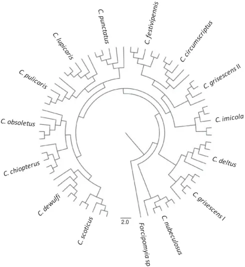
The total mass spectra of all 69 reference biting midges and of 1 Forcipomyia specimen as outgroup were used to generate a dendrogram (Fig. 2). Thus, all individuals of a species clustered on discrete branches. In particular, species that are indistin-guishable by morphology (i.e. females of C. obsoletus/ scoticus, the cryptic species C. grisescens I/II) are clearly distinguished by MALDI-TOF MS.
Based on this clear-cut clustering of the species,
specific biomarker mass sets, containing between
24 and 38 masses for the different species (Fig. 1,
Table 1), could be generated using SARAMIS and be used for automated Culicoides species identification.
The above mentioned morphologically similar
species had only few common biomarker masses, namely 4 (from 26 and 38, respectively, of C. obsoletus and C. scoticus) or 7 (from 25 and 30 of C. grisescens I/II) (Table 1).
Validation study
The reproducibility and accuracy of the reference database were tested in a second step in a validation study. Analyses against the biomarker mass sets of all reference species were performed for 2 profiles per individual. From 90 of the 108 specimens
investi-gated, both profiles allowed for an automated
identification. Eleven individuals yielded only 1
profile suitable for identification by automated
analysis, whereas the replicate had fewer than 75% biomarker matches, requiring manual full spectra
comparison which in all these cases confirmed the
result of the automated classification. From 7 midges, both profiles were of lower quality (displaying fewer masses and lower intensities), requiring manual full spectra comparison which in 5 of these cases consistently identified the species. In another case, only 1 of the duplicates yielded an analysable
profile. The seventh of these specimens,
morpho-logically and genetically identified as C. deltus, resulted in spectra which most closely resembled those derived of the reference C. deltus specimens
in one and of C. grisescens I in the other read. Finally, all 3 specimens of C. pallidicornis (non-target
species) did not yield identification neither by
automated biomarker identification nor by manual
comparison against the spectra of all reference specimens.
Except for 1 specimen with an ambiguous result (see above), all 107 biting midges from the evaluation
phase were correctly identified by MALDI-TOF
MS, either by automated biomarker identification
or by manual full spectra comparison. Initially,
discrepancies of the identification by morphology
and MALDI-TOF MS were obtained with 8 individuals. Genetic analyses of these insects were always in accordance with the MALDI-TOF MS results indicating that the morphological identi fi-cation was inaccurate. Thus, 2 of the 5 specimens morphologically identified as C. deltus in fact were C. pulicaris, 3 of 5 C. pulicaris were C. punctatus (2) and C. lupicaris (1), respectively, 2 C. grisescens turned out to be C. deltus and a specimen of C. lupicaris was misidentified as C. punctatus.
Intensity 150000 50000 40000 20000 0 15000 5000 0 60000 20000 0 60000 150000 50000 0 20000 0 0 m/z C. obsoletus C. scoticus C. dewulfi C. imicola C. pulicaris C. punctatus 5000 6000 7000 8000 9000 10000
Fig. 1. Matrix-assisted laser desorption/ionization time offlight (MALDI-TOF) mass spectra in the range of 5–10 kDa offield-caught Culicoides insects. Biomarker masses as eventually determined by SARAMIS are highlighted by dashed lines.
D I S C U S S I O N
MALDI-TOF MS using homogenates of whole insects has been shown in proof-of-principle studies by Campbell (2005) and Perera and colleagues (2005) to be suitable for differentiation of the few species they investigated (fruit flies, aphids). Feltens and colleagues (2010) applied this proteomic approach to discriminate 13 species of Drosophila, and the suitability of MALDI-TOF MS to characterize a haematophagous insect (C. nubeculosus) has recently been demonstrated (Kaufmann et al.2011). All these investigations relied on laboratory-reared insects. The study presented here is the first evaluation of
MALDI-TOF MS for species identification of
field-caught haematophagous insects. These included the Culicoides species from the Obsoletus and Pulicaris group that are the incriminated biological vectors of the bluetongue virus in Europe, but also C. imicola, the main vector in Africa, as well as other European species.
Using a very crude sample preparation method (mechanical grinding in 10% formic acid) it was possible to obtain specific and discrete biomarker
mass sets for the 14 Culicoides spp. investigated, and only few biomarker masses were shared by different species, even among closely related ones such as C. grisescens and a cryptic species (denominated C. grisescens II). Thus, it seems feasible that this approach can be expanded to include other Culicoides spp. Indeed, C. pallidicornis specimens, included in the validation phase as non-target organism, were not recognized by comparing with the spectra of these 14 species, and a unique biomarker mass set could also be calculated for C. pallidicornis (not shown).
Also using a minimal sample preparation approach (mechanical insect homogenization in water), the discrimination of sibling species of Drosophila by MALDI-TOF MS analyses has been reported (Campbell,2005). In contrast, when focusing on the analyses of proteins/peptides purified by chromato-graphy after mechanical homogenization under protein-denaturing conditions (6Murea), the di
ffer-entiation of closely related and laboratory-reared Drosophila spp. (i.e. D. miranda, D. pseudoobscura, belonging to the Obscura species group) was not possible (Feltens et al. 2010). Between 168 and 390 protein and peptide peaks in the range of 1·8 to
2.0
Fig. 2. Dendrogram of matrix-assisted laser desorption/ionization time offlight (MALDI-TOF) mass spectra of thoraxes from (4-) 5 individuals of 13 species offield-caught Culicoides biting midges and laboratory-reared C. nubeculosus. One individual of a Forcipomyia sp. is included as outgroup.
Table 1. MALDI-TOF MS reference biomarker masses for 14 Culicoides spp. and Forcipomyia sp. Biomark er ma ss (Da) C. chio pte rus C. circu ms criptus C. deltus C. dewu lfi C. fe stivipe n nis C. gri s esce n s I C. gri s esce n s II C. imic ol a C. lupi caris C. nu bec ulosus C. obso le tus C. puli c a ris C. pu nct atus C. sc oticus Forc ip o m y ia sp. 2527 2541 2581 2616 2624 2646 2801 2831 2844 2904 3034 3191 3200 3227 3234 3243 3252 3258 3271 3312 3433 3547 3587 3686 3716 3755 3761 3818 3830 3962 4018 4031 4039 4070 4086 4133 4174 4187 4247 4261 4287 4304 4392 4442 4453 4467 4483 4495 4514 4530 4548 4562 4579 4592 4650 4664 4678 4684 4692 4706 4716 4730 4742 4793 4814 4819 4881 4909 4930 4949 4965 5049 5063 5087 5127 5142 5153 5167 5182 5192 5205 5218 5231
Table 1 (Cont.) Biomarke r ma ss (Da ) C. chiopterus C. circumsc ri pt us C. deltus C. d e w u lf i C. fes tivipenn is C. gris esc ens I C. gris esc ens I I C. imicola C. lupica ris C. nubeculos us C. obsol etus C. pu lica ris C. pu nctatus C. scot ic us Forcipo myia s p . 5241 5254 5271 5288 5302 5320 5336 5348 5362 5383 5401 5413 5442 5451 5466 5494 5535 5591 5607 5637 5670 5687 5720 5738 5777 5833 5878 5903 6003 6033 6044 6133 6247 6360 6376 6389 6406 6459 6476 6492 6504 6519 6533 6551 6569 6579 6594 6602 6615 6630 6640 6653 6669 6685 6699 6716 6728 6743 6762 6797 6818 6837 6858 6986 7360 7373 7415 7434 7452 7475 7490 7512 7628 7706 7771 7854 7869 7969 8119 8169 8207 8305 8331
Table 1 (Cont.) Biomarke r ma ss (Da ) C. chiopterus C. circumsc ri pt us C. deltus C. d e w u lf i C. fes tivipennis C. gris esc ens I C. gris esc ens I I C. imicola C. lupica ris C. nubeculos us C. obsol etus C. pu lica ris C. pu nctatus C. scot ic us Forcipo myia s p . 8396 8413 8444 8485 8509 8525 8542 8554 8567 8591 8598 8614 8628 8668 8706 8720 8789 8805 8851 8876 8889 8913 8937 8947 8963 8977 9012 9023 9042 9055 9076 9099 9123 9166 9184 9193 9208 9223 9240 9256 9295 9339 9360 9371 9387 9408 9459 9471 9495 9576 9595 9615 9227 9641 9693 9708 9932 9992 10299 10630 10763 10790 10801 10834 10874 11329 11368 12227 12263 12273 12353 12494 12520 12723 12825 12901 12954 13345 13469 16704 19379 19549 Total 25 27 28 31 24 30 25 34 34 33 26 36 26 38 28
15 kDa, (average 236 peaks per spectrum) were identified in that study (present study: 50 to 169 peaks in the mass range of 2 to 20 kDa with an average peak number of 99), but spectral heterogeneity within species and a limited number of
species-specific peaks were noted. Feltens and colleagues
(2010) identified 18 potential protein markers by
electrospray ionization MS/MS, revealing that most of them stem from muscle tissue like myosin heavy
chain, troponin, tropomyosin, flightin or from
mitochondria-like ATP synthase subunits alpha and beta. In our study, mainly the muscle-containing thorax (and the head) were used for protein extraction because potential bloodmeal traces in the abdomen were shown to impair the MALDI spectra quality (Kaufmann et al. 2011). Thus, the spectra that we generated have a lower average datacount as com-pared to the study of Feltens and colleagues (2010) but they cover most of the potential discriminative and reproducible muscle tissue and mitochondrial proteins.
Although no emphasis was laid on phylogenetic analyses, the dendrogram shown inFig. 2reasonably
reflects the current comprehension of Culicoides
kinship. Thus, the species of the Obsoletus group (C. chiopterus, C. obsoletus, C. scoticus) cluster to-gether, also including C. dewulfi, which recently was suggested not to be a member of this group, accord-ing to genetic analyses at 3 loci (Schwenkenbecher et al.2009). The recognized species of the Pulicaris group are grouped in 2 clusters (C. lupicaris, C. pulicaris, C. punctatus; C. deltus, C. grisescens I).
Interestingly, the cryptic species denominated
C. grisescens II, which is morphologically indistin-guishable from the type species C. grisescens I but differs by approximately 10% at the barcode locus mt COX I gene (Wenk et al.2011), is not part of one of the Pulicaris group clusters. Thus, more in-depth investigations using MALDI-TOF MS, as described (Feltens et al. 2010), might be of value to address taxonomic and phylogenetic questions in the genus Culicoides.
In a previous study with C. nubeculosus (Kaufmann
et al. 2011), male and female specimens had
very similar spectra allowing derivation of a set of biomarker masses valid for both sexes. Similar spectra of males and females of D. melanogaster using a crude sample preparation were also reported (Campbell, 2005) whereas a differentiation of the two sexes of this fruitfly species was achieved based
on mass analyses of purified proteins/peptides
(Feltens et al.2010). In the present study, individuals of both sexes of all but 2 species were included either in the reference specimens or in those used in the validation study, and no discrimination between males and females was observed (data not shown). Only females were available from C. deltus and C. imicola. However, this is of minor importance because only females are haematophagous and thus
act as vectors and because the commonly used UV-traps primarily capture this gender. Further, only males of the rare species C. pallidicornis, which was used as the non-target species in the validation phase, were available. Considering thefindings with the other species. i.e. gender does not matter with
regard to identification by MALDI-TOF patterns,
the eventually calculated biomarker mass sets of these male specimens most probably are also valid to accurately identify females of this species.
The insects used in this work were stored in 70% EtOH which is considered suitable for subsequent
analyses by MALDI-TOF MS (Campbell, 2005;
Feltens et al.2010), and it was experimentally proven that such a storage up to around 3 months (102 days) yielded stable mass spectrum results (Kaufmann et al.2011). In the present study, it was observed that freshly caught species generated more data counts (peaks) than species which were kept for a longer period (> 3 months) in 70% ethanol after trapping (data not shown). Nevertheless, the biomarker mass sets compiled from the data of the reference
specimens allowed reliable identification in the
blind study midges kept over 2 years in 70% ethanol (at 4° C) (not shown).
Reproducibility and accuracy of the references were tested in a blind study of 111 morphologically identified biting midges mainly caught in the field all over Switzerland. Thus, a correct and unambiguous result was obtained in 110 of the 111 insects tested. Initial discrepancies between morphology on one side and mass analysis/genotyping on the other side were encountered in 8 individuals. All these morphologi-cally misidentified specimens belong to species from the Pulicaris group which are notoriously hard to distinguish with certainty as they are known to display natural variation in morphology as well as overlapping characters. Thus, the accuracy of the morphological characters employed in identification keys might be re-evaluated according to
MALDI-TOF MS or genetic identifications. Further, long
storage in ethanol might have blurred morphological features such as the wing patterns. Morphological misidentifications as identified by genetic data also were observed when selecting specimens for creating the reference database, but these individuals were excluded.
The application of MALDI-TOF MS for the identification of insects has just begun, and further investigations will generate more biomarker mass sets which preferably should be united in a single database. For Culicoides, a relatively low number
of very discrete biomarker masses were identified
(24–38) as compared to bacteria which usually are characterized by 30–50 such masses. In addition, it is not uncommon that more than 1 biomarker mass set is available in databases for a single species, e.g. there are >40 sets currently available for Staphylococcus aureus in the SARAMIS library of Anagnostec.
Larger datasets with Culicoides spp. might reveal
different sets for populations of a species from
different origins, although our limited data revealed
that individuals of 3 species (C. chiopterus,
C. festivipennis, C. obsoletus) that were collected at either site of the Alpine crest could be integrated in single sets each.
In conclusion, the presented results demonstrate
that protein profiling by MALDI-TOF can be used
as an alternative, quick and inexpensive tool to accurately identify Culicoides biting midges collected
in thefield, including cryptic species. MALDI-TOF
can be used complimentarily to other identification techniques (morphology, genetics) as only part of the insect need to be investigated. This approach has the potential to become the method of choice for a centralized, robust and high throughput screening of midge populations in connection with the surveil-lance of eventually emerging midge-transmitted agents.
A C K N O W L E D G M E N T S
We thank Simon Carpenter and Eric Denison (Institute for Animal Health, Pirbright, UK) for the initial supply of C. nubeculosus for the laboratory colonies, Jean-Claude Delécolle (Université de Strasbourg, France) for the C. imicola specimens and Jeannine Hauri for excellent technical assistance.
F I N A N C I A L S U P P O R T
We greatly acknowledge thefinancial support provided by the Swiss Federal Veterinary Office (grant 1.08.10) and the University of Zürich (‘Forschungskredit’, C.K., personal grant no. 4974).
R E F E R E N C E S
Boorman, J. (1974). The maintenance of laboratory colonies of Culicoides
variipennis (Coq.), C. nubeculosus (Mg.) and C. riethi Kiff. (Diptera,
Ceratopogonidae). Bulletin of Entomological Research64, 371–377.
Campbell, P. M. (2005). Species differentiation of insects and other
multicellular organisms using matrix-assisted laser desorption/ionization
time offlight mass spectrometry protein profiling. Systematic Entomology
30, 186–190.
Caprioli, G., Cristalli, G., Ragazzi, E., Molin, L., Ricciutelli, M., Sagratini, G., Seraglia, R., Zuo, Y. and Vittori, S. (2010). A preliminary
matrix-assisted laser desorption/ionization time-of-flight approach for the
characterization of Italian lentil varieties. Rapid Communications in Mass
Spectrometry24, 2843–2848.
Carpenter, S., Wilson, A. and Mellor, P. S. (2009). Culicoides and the emergence of bluetongue virus in northern Europe. Trends in Microbiology
17, 172–178.
Cêtre-Sossah, C., Baldet, T., Delécolle, J. C., Mathieu, B., Perrin, A., Grillet, C. and Albina, E. (2004). Molecular detection of Culicoides spp. and Culicoides imicola, the principal vector of bluetongue (BT) and African
horse sickness (AHS) in Africa and Europe. Veterinary Research 35,
325–337.
Chaignat, V., Worwa, G., Scherrer, N., Hilbe, M., Ehrensperger, F., Batten, C., Cortyen, M., Hofmann, M. and Thuer, B. (2009).
Toggenburg Orbivirus, a new bluetongue virus: initial detection, first
observations in field and experimental infection of goats and sheep.
Veterinary Microbiology138, 11–19.
Dallas, J. F., Cruickshank, R. H., Linton, Y. M., Nolan, D. V., Patakakis, M., Braverman, Y., Capela, R., Capela, M., Pena, I., Meiswinkel, R., Ortega, M. D., Baylis, M., Mellor, P. S. and
Mordue (Luntz), A. J. (2003). Phylogenetic status and matrilineal structure of the biting midge, Culicoides imicola, in Portugal, Rhodes and Israel.
Medical and Veterinary Entomology17, 379–387.
Delécolle, J.-C. (1985). Nouvelle contribution à l’étude systématique
et iconographique des espèces du genre Culicoides (Diptera:
Ceratopogonidae) du Nord-Est de la France. Université Louis Pasteur, Strasbourg, France.
Feltens, R., Gorner, R., Kalkhof, S., Groger-Arndt, H. and
von Bergen, M. (2010). Discrimination of different species from the
genus Drosophila by intact protein profiling using matrix-assisted
laser desorption ionization mass spectrometry. BMC Evolutionary Biology 10, 95.
Goffredo, M. and Meiswinkel, R. (2004). Entomological surveillance of
bluetongue in Italy: methods of capture, catch analysis and identification of
Culicoides biting midges. Veterinaria Italiana40, 260–265.
Hellberg, W., Mellor, P. S., Torsteinsdottir, S. and Marti, E. (2009). Insect bite hypersensitivity in the horse: comparison of IgE-binding proteins in salivary gland extracts from Simulium vittatum and Culicoides
nubeculosus. Veterinary Immunology and Immunopathology132, 62–67.
Kaufmann, C., Schaffner, F. and Mathis, A. (2009). Monitoring of
biting midges (Culicoides spp.), the potential vectors of the bluetongue virus the 12 climatic regions of Switzerland. Schweizer Archiv für Tierheilkunde
5, 205–513.
Kaufmann, C., Ziegler, D., Schaffner, F., Carpenter, S., Pflüger, V.
and Mathis, A. (2011). Evaluation of matrix-assisted laser desorption/
ionization time offlight mass spectrometry for characterization of Culicoides
nubeculosus biting midges. Medical & Veterinary Entomology25, 32–38.
Mazzeo, M. F., Giulio, B. D., Guerriero, G., Ciarcia, G., Malorni, A., Russo, G. L. and Siciliano, R. A. (2008). Fish authentication by
MALDI-TOF mass spectrometry. Journal of Agricultural and Food Chemistry56,
11071–11076.
Meiswinkel, R., Baldet, T., de Deken, R., Takken, W., Delécolle, J.-C. and Mellor, P. S. (2008). The 2006 outbreak of bluetongue in northern
Europe– the entomological perspective. Preventive Veterinary Medicine 87,
55–63.
Meiswinkel, R., van Rijn, P., Leijs, P. and Goffredo, M. (2007).
Potential new Culicoides vector of bluetongue virus in northern Europe.
Veterinary Record161, 564–565.
Mellmann, A., Bimet, F., Bizet, C., Borovskaya, A. D., Drake, R. R., Eigner, U., Fahr, A. M., He, Y., Ilina, E. N., Kostrzewa, M., Maier, T.,
Mancinelli, L., Moussaoui, W., Prevost, G., Putignani, L.,
Seachord, C. L., Tang, Y. W. and Harmsen, D. (2009). High inter-laboratory reproducibility of matrix-assisted laser desorption
ionization-time offlight mass spectrometry-based species identification of
nonferment-ing bacteria. Journal of Clinical Microbiology47, 3732–3734.
Mellor, P. S., Boorman, J. and Baylis, M. (2000). Culicoides biting
midges: their role as arbovirus vectors. Annual Review of Entomology45,
307–340.
Mellor, P. S. and Hamblin, C. (2004). African horse sickness. Veterinary
Research35, 445–466.
Paweska, J. T., Venter, G. J. and Hamblin, C. (2005). A comparison of the susceptibility of Culicoides imicola and C. bolitinos to oral infection with eight serotypes of epizootic haemorrhagic disease virus. Medical and
Veterinary Entomology19, 200–207.
Perera, M. R., Vargas, R. D. F. and Jones, M. G. K. (2005). Identification
of aphid species using protein profiling and matrix-assisted laser desorption/
ionization time-of-flight mass spectrometry. Entomologia Experimentalis
et Applicata117, 243–247.
Santos, C., Paterson, R. R. M., Venancio, A. and Lima, N. (2010). Filamentous fungal characterizations by matrix-assisted laser desorption/
ionization time-of-flight mass spectrometry. Journal of Applied Microbiology
108, 375–385.
Sauer, S. and Kliem, M. (2010). Mass spectrometry tools for the
classification and identification of bacteria. Nature Reviews Microbiology
8, 74–82.
Schwenkenbecher, J. M., Mordue Luntz, A. J. and Piertney, S. B.
(2009). Phylogenetic analysis indicates that Culicoides dewulfi should not be
considered part of the Culicoides obsoletus complex. Bulletin of Entomological
Research99, 371–375.
Sloet van Oldruitenborgh-Oosterbaan, M. M., van Poppel, M., de Raat, I. J., van den Boom, R. and Savelkoul, H. F. (2009). Intradermal testing of horses with and without insect bite hypersensitivity in The Netherlands using an extract of native Culicoides species. Veterinary
Dermatology20, 607–614.
Stephan, R., Ziegler, D., Pflüger, V., Vogel, G. and Lehner, A. (2010).
Rapid genus- and species-specific identification of Cronobacter spp. by
matrix-assisted laser desorption ionization-time offlight mass spectrometry.
Stevenson, L. G., Drake, S. K., Shea, Y. R., Zelazny, A. M. and Murray, P. R. (2010). Evaluation of matrix-assisted laser desorption
ionization-time offlight mass spectrometry for identification of clinically
important yeast species. Journal of Clinical Microbiology48, 3482–3486.
van Veen, S. Q., Claas, E. C. J. and Kuijper, E. J. (2010).
High-throughput identification of bacteria and yeast by matrix-assisted laser
desorption ionization-time offlight mass spectrometry in conventional
medical microbiology laboratories. Journal of Clinical Microbiology48,
900–907.
Venter, G. J. and Meiswinkel, R. (1994). The virtual absence of Culicoides imicola (Diptera: Ceratopogonidae) in a light-trap survey of the colder, high-lying area of the eastern Orange Free State, South Africa, and implications for the transmission of arboviruses. Onderstepoort Journal of Veterinary
Research61, 327–340.
Wenk, C. E., Kaufmann, C., Schaffner, F. and Mathis, A. (2011).
Molecular characterisation of Swiss Ceratopogonidae (Diptera) and
evaluation of real-time PCR assays for the identification of Culicoides biting