EXIT-SITE INFECTIONS IN PERITONEAL DIALYSIS: PREDICTIVE FACTORS FOR ADVERSE OUTCOMES
Filipa Silva, Joana Tavares, Sofia O Correia, Cristina Freitas, Olívia Santos, Maria João Carvalho, Jorge Malheiro, António Cabrita, Anabela Rodrigues
Peritoneal Dialysis Unit, Centro Hospitalar e Universitário do Porto, Portugal
Résumé
Les complications liées à l’infection chez les patients en dialyse péritonéale (DP) sont importantes. Notre objectif était d’évaluer le type d’infections d’orifice de sortie (ESI) et l’évolution naturelle chez une cohorte de patients admis en DP ces dix dernières années au sein de notre service. Les données du registre des événements ESI (n = 126, chez 74 patients) ont été récupérées. Les protocoles ESI ont suivi les directives internationales standard. Un contrôle qualité systématique est effectué. Le suivi médian était de 29,1 mois (14,0 à 47,4). Dans cette population, les résultats défavorables du taux de tunellites (TI) et du taux de péritonite étaient respectivement de 0,12 et 0,13 patient / an. Le sexe masculin (0,048), l’âge (0,007) et l’agent Staphylococcus aureus (0,006) étaient prédictifs de l’IT, l’IT là où la mise obligée en DP et des taux faibles d’albumine étaient des facteurs prédictifs de la péritonite. Après avoir groupé les ESI en fonction de la date d’apparition de l’infection (groupe 1: 2008 à 2012, groupe 2: 2013 à 2017 et groupe 3: 2018), une augmentation substantielle de l’IT en 2018 était évidente (p <0,001 lorsque le groupe de comparaison 3 vs 1 et 0,005 en comparant les groupes 2 et 3).
Lorsque l’ESI survient en même temps que l’IT, le taux d’echec de guérison est de 65%. On observe 50 % d’abandons en cas d’ESI sans péritonite, contre 86% des patients ayant une péritonite (p <0,001). Le Staphylococcus aureus est le microorganisme le plus souvent responsable de l’échec de la guérison (P = 0,002) et de l’abandon de la technique (P = 0,01).
En dépit de nombreux efforts visant à réduire les ESI, un audit régulier a quand même mis en avant le besoin de réviser les protocoles en vue d’éviter des résultats défavorables. Une formation ciblée des patients est obligatoire, mais les protocoles prophylactiques et antibiotiques devraient être améliorés.
Le
B
ulletin de la
D
ialyse à
D
omicile
journal officiel du Registr e de D ialyse Péritonéale de Langue Française RDPLF www .rdplf.or g Summary
Infection-related complications in patients on peritoneal dialysis (PD) is a leading complication.
Our aim was to evaluate the type and natural course of Exit site infection (ESI ) events in a cohort of PD treated in last decade of our PD program.
Our hospital database of ESI events (n=126, in 74 patients) were retrieved. ESI protocols followed standard international guidelines. A systematic quality control is performed.
The median follow-up was 29.1 (14.0-47.4) months. In this population the adverse outcomes of tunnel infection (TI) rate and peritonitis rate was 0.12 and 0.13 patient/year, respectively.
Male sex (0.048), older age (0.007) and Staphylococcus aureus (SA) agent (0.006) were predictive of TI while non-optional PD and lower levels of albumin were predictive of peritonitis.
After grouping the ESI events according to the date of the occurrence of infection (group 1: 2008 to 2012, group 2: 2013 to 2017 and group 3: 2018) a substantial increase of TI in 2018 was evident (P <0.001 when comparing group 3 vs 1 and 0.005 when comparing group 2 and 3).
When ESI occurs simultaneous with TI, the probability of not reaching cure is 65%. Drop-out occurred in 50% of ESI without peritonitis vs 86% with peritonitis (P <0.001). SA is the microorganism most implicated in the failure to heal (P 0.002) and drop-out (P 0.010).
In spite of a number of efforts to reduce ESI, a regular audit still point to the need for protocols review in order to avoid adverse outcomes. Focused training of patients is mandatory but also prophylaxis and antibiotic protocols deserve improvement.
INTRODUCTION
Infection-related complications in patients on peritoneal dialysis (PD) is a leading complication and despite major technique advances and cumulative experience still is a major cause of technique dropout and switch to hemodialysis.
Catheter-related infections are used as the collective term to describe both exit-site infection (ESI) and tunnel infection (TI). These two conditions may occur on their own or simultaneously. ESI is defined by the presence of purulent drainage, with or without erythema of the skin at the catheter-epidermal interface [1,2]. TI is defined as the presence of clinical inflammation or ultrasonographic evidence of collection along the catheter tunnel. May present as erythema, edema, induration or tenderness over the subcutaneous pathway. Usually occurs simultaneously to an ESI but could occur alone [3]. ESI caused by Staphylococcus aureus (SA) or Pseudomonas aeruginosa are often associated with concomitant tunnel infections [4].
METHODS
Our hospital database of ESI events were retrieved from January 2008 to December 2018, in patients undergoing continuous ambulatory peritoneal dialysis (CAPD) or automated cycling peritoneal dialysis (APD), including epidemiological, modality variables, microbiologic agent and outcomes. During this period a total of 126 infections occurred in a total of 74 patients, who represent the study population.
Catheter exit-site is done by exteriorization of the “buried” catheter, implanted by the Moncrief Popovich technique. ESI protocols followed standard international guidelines. Salvage ESI/TI therapy with external cuff extrusion/shaving is done according to clinical criteria. The focus was on quality control, analyzing rates of infection, identifying the most common microbiologic agents and their susceptibilities to antimicrobials and outcomes. A multivariable logistic regression model was used to determine significant risk factors for tunnel infections, peritonitis and drop-out as adverse outcomes of ESI.
RESULTS
Exit site episodes and population characteristics
The 126 ESI episodes included 98 ESI exclusively and 28 ESI concomitant with peritonitis. These episodes
occurred in 74 patients followed for 79.342 patient-days and the median time since the start of DP to the first episode of ESI were 281 days (8-1990). In this population the adverse outcomes of TI rate and peritonitis rate was 0.12 and 0.13 patient/year, respectively. In 15 patients (20%) the ESI occurred in the first 30 days, and only 2 of them had simultaneous TI. The majority of this early infections occurred in patients on DPCA as this is the modality of choice for the beginning of the technique in our DP program. No characteristics, either patient or infection related, had statistical significance when early and late ESI were compared.
The average number of ESI episodes per patient was 1,8 (range from 1 to 6 episodes); 45,7% of patients had 1 ESI, 40% has 2 episodes, 8,6% had 3 and 5,7% has more than 3 episodes of ESI.
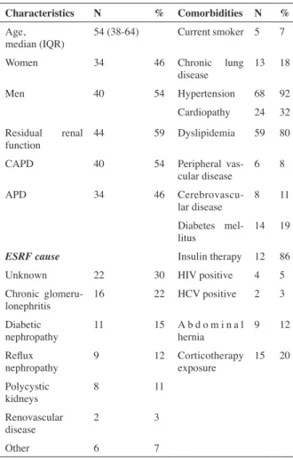
The median age of this cohort were 54 years (38-64) and most of all were men (54%, n=40). The majority were on CAPD modality (59%).
Most of the patients were hypertensive (92%) but only 19% had diabetes mellitus. The other population characteristics were detailed in table 1.
journal officiel du Registr e de D ialyse Péritonéale de Langue Française RDPLF www .rdplf.or g Characteristics N % Comorbidities N % Age,
median (IQR) 54 (38-64) Current smoker 5 7
Women 34 46 Chronic lung
disease 13 18
Men 40 54 Hypertension 68 92
Cardiopathy 24 32 Residual renal
function 44 59 Dyslipidemia 59 80
CAPD 40 54 Peripheral
vas-cular disease 6 8
APD 34 46
Cerebrovascu-lar disease 8 11 Diabetes
mel-litus 14 19
ESRF cause Insulin therapy 12 86
Unknown 22 30 HIV positive 4 5
Chronic
glomeru-lonephritis 16 22 HCV positive 2 3
Diabetic
nephropathy 11 15 A b d o m i n a l hernia 9 12 Reflux
nephropathy 9 12 Corticotherapy exposure 15 20 Polycystic
kidneys 8 11
Renovascular
disease 2 3
Other 6 7
Identified microorganisms in ESI
The most common isolated organisms (table II) were Gram positive (n=81) included Staphylococcus aureus (n=43), Corynebacterium species (n=27), other Staphy-lococcus than aureus (n=6), Enterococcus faecalis (n=3) and Streptococcus species (n=2). Gram negative were identified in 36 cases, including Pseudomonas aerugi-nosa (n=15), Proteus mirabilis (n=13), Serratia marces-cens (n=4), Escherichia coli (n=3) and Haemophilus parainfluenzae (n=1). In 4 cases, ESI were caused by both Gram positive and negative agents, in 2 by fungal and in 11 the agent was not identified. Only in 60% of IOS the microorganisms were multisensitive, while in the others cases the agents had at least one antimicrobial resistance. From all SA identified, only 4 were methi-cilin-resistant (MRSA). Two of the Pseudomonas iso-lated has extended-spectrum B-lactamases (ESBL) and 1 were carbapenemase producing bacteria (KPC). In all cases the antibiotic therapy took at least 2 weeks.
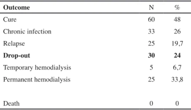
Outcomes
The cure was achieved in almost 48% of cases (n=60), 26% (n=33) failed in cure and 26% (n=33) were res-ponsible for the drop out of technique. When tunnel was involved the drop out reached 60%. Shaving of the ex-ternal cuff was performed in 24 refractory ESI episodes but 12 (50%) still ended in catheter removal.
All the outcomes were described in table III
Predictors of adverse outcomes in ESI
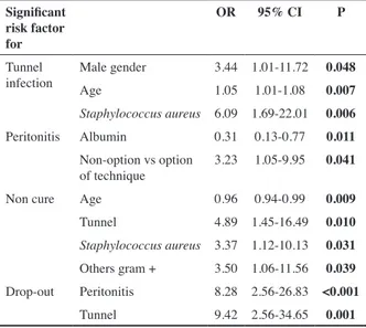
In multivariate logistic regression male sex (0.048), ol-der age (0.007) and Staphylococcus aureus agent (0.006) were predictive of TI, while non-optional PD (PD due to vascular access failure) and lower levels of serum albu-min were predictive of peritonitis (Table IV). Diabetes, anuria, and PD modality were not predictive.
After grouping the ESI events according to the date of the occurrence of infection (group 1:2008 to 2012, group 2: 2013 to 2017 and group 3:2018) a substantial increase of tunnel infections in 2018 was evident (P <0.001 when comparing group 3 vs 1 and 0.005 when comparing group 2 and 3) (table V). There was no significant diffe-rence in patients’ characteristics between the 3 groups and no cause has been identified for this occurrence. When ESI occurs simultaneous with tunnel infection, the probability of not reaching cure is 65%. Drop out occurred in 50% of ESI without peritonitis vs 86% with peritonitis, P <0.001). Staphylococcus aureus is the mi-croorganism most implicated in the failure to heal (P 0.002) and drop out (P 0.010).
DISCUSSION
Skin infection at the catheter exit-site remains a relevant problem in PD patients. The wide variations in is appea-rance leads to inconsistent monitoring and difficulties in
journal officiel du Registr e de D ialyse Péritonéale de Langue Française RDPLF www .rdplf.or g N =130 Multi-sensible (n=76) G r a m
positive Staphylococcus aureusCorynebacterium 43 39 (4 MRSA)
species 27 12
Other Staphylo than aureus (coagulase negative, lugdunensis, epidermidis) 6 4 Enterococcus faecalis 3 0 Streptococcus species (pyogenes, viridans) 2 1 G r a m
negative Pseudomonas aerugi-nosa 15 11 (2 ESBL, 1 KPC)
Proteus mirabilis 13 6
Serratia marcescens 4 2
Escherichia coli 3 1
Haemophilus
parain-fluenzae 1 0
Fungal Candida albicans 2
Non-iden-tified 11
Mixed
ESI Proteus mirabilis + enterococcus faecalis 0 Proteus mirabilis +
Co-rynebacterium species 0 Corynebacterium spe-cies + Streptococcus pyogenes 0 Corynebacterium spe-cies + Enterococcus faecalis 0 Table II : Identified microorganisms in ESI
Table III. Events and outcomes of all ESI in our center
Outcome N % Cure 60 48 Chronic infection 33 26 Relapse 25 19,7 Drop-out 30 24 Temporary hemodialysis 5 6,7 Permanent hemodialysis 25 33,8 Death 0 0
was the most frequent gram-negative agent, followed by other Enterobacteriaceae, according to the literature evidence [5,6].
Implantation protocol
In our center the double-cuffed Tenckhoff catheters were placed in all patients by an expert team (a nephro-logist and a surgeon) using mini-laparotomy and the Moncrief–Popovich method, in an operating room under sterile conditions.
Several randomized trials have compared laparoscopic or peritoneoscopic catheter placement with standard laparotomy, but none of them reported catheter-related infection as a secondary outcome [7]. There are two stu-dies that compare midline and lateral incision but neither found any difference in the risk of catheter-related infec-tion [8,9]. Although the best strategy for catheter place-ment has been questioned, several studies have shown that with appropriate training there is no difference in the rate of ESI in what concerns to catheter placement (by nephrologists or surgeons) or different techniques or incisions [7, 10-18]. Although an uncontrolled study suggests that the technique of burying the PD catheter in subcutaneous tissue for 4 to 6 weeks after implanta-tion is associated with a lower rate of catheter-related in-fections [19], two randomized controlled studies found no difference as compared with the standard technique [20,21].
In all cases we administered intravenous prophylactic cephazolin immediately before implantation and before the exteriorization of external segment of the catheter. Nowadays, it is widely recommended to do prophylactic antibiotics before catheter insertion. However, several prospective trials found that prophylactic perioperative intravenous antibiotics had no significant effect on the rate of early catheter-related infections, although it si-gnificantly reduces the risk of early peritonitis [22-26]. A break-in period of more than 4 weeks before exteriori-zation of the external segment of catheter was standard, usually extended to additional months until dialysis was needed [16]. It remains controversial whether immediate commencement of PD after catheter insertion is asso-ciated with a higher risk of catheter-related infections [27-30].
Nasal carriage of Staphylococcus aureus is seen as a ma-jor risk factor of catheter-related infections. Besides, one prospective study showed that intranasal mupirocin re-duced SA ESI but not tunnel infection [31], there are no data on efficacy of its routine screening and eradication interpreting study results. Although there has been no
change in our unit protocols, there has been a stagge-ring increase in ESI, particularly TI in 2018 whose cause was unclear to us. We then made a quality assessment brainstorming in order to disguise opportunities of im-provement.
Relative to incidence rates, most of studies reports a range from 0.05 to 1.02 episodes/patient-year. We report the incident rate of this affected population, which for itself is more susceptible to ESI. Although it might be a negative methodological bias, our rates of ESI per pa-tient-year are relatively low: 0.58 for ESI (126 events), 0,12 for tunnel infection (27 events) and 0.13 for perito-nitis (28 events). Gram-positive agents were responsible for most peri-catheter infectious episodes, and SA was the primary cause of ESI. Pseudomonas aeruginosa
journal officiel du Registr e de D ialyse Péritonéale de Langue Française RDPLF www .rdplf.or g
Table V : ESI adverse outcome events according to date of occur-rence Group 1 (2008-2012) Patient-time (days): 25572 Group 2 (2013-2017) Patient-time (days): 44535 Group 3 (2018) Patient-time (days): 9235 P Group 1vs2 P Group 1vs3 P Group 2vs3 Tunnel infection rate patient-year 0.04
(3 events) (14 events)0.11 (10 events)0.39 0.107 <0.001 0.005
Peritonitis infection rate patient-year 0.21 (15 events) 0.06 (8 events) (5 events)0.20 0.006 0.910 0.072
Table IV : Multivariate logistic analysis of predictors of adverse outcomes in ESI, adjusted to variables with P<0.02
Significant risk factor for
OR 95% CI P Tunnel
infection Male genderAge 3.44 1.01-11.72 0.0481.05 1.01-1.08 0.007
Staphylococcus aureus 6.09 1.69-22.01 0.006
Peritonitis Albumin 0.31 0.13-0.77 0.011 Non-option vs option
of technique 3.23 1.05-9.95 0.041
Non cure Age 0.96 0.94-0.99 0.009
Tunnel 4.89 1.45-16.49 0.010
Staphylococcus aureus 3.37 1.12-10.13 0.031
Others gram + 3.50 1.06-11.56 0.039 Drop-out Peritonitis 8.28 2.56-26.83 <0.001
in patients prior to insertion of the peritoneal dialysis catheter. In our unit we use nasal mupirocin as part of the pre-implantation protocol in nasal carriers of S. aureus. Facing the increase of ESI with tunnel infection we de-cided to 1) change the implantation procedure with a soaking step the catheter in cefazoline before introdu-cing it in the pelvis and in the subcutaneous tunnel and 2) change the procedure of catheter exteriorization by using a skin biopsy needle to do the exit side to reduce trauma in the early cicatrization process and avoid early exist side colonization.
Exite site care
After exteriorization of the catheter’s external segment, patients were taught to clean the exit site every day with saline solution (0.9% NaCl) and to keep it dry. They were prescribed 2% mupirocin cream to be used at the exit site once daily.
Guidelines recommend daily topical application of anti-biotic cream or ointment on the catheter exit side since it prevents ESI caused by SA. This strategy is proved to be effective by a number of observational studies, ran-domized controlled trials, and meta-analyses [7,22,32-38] and has also been shown to be cost-effective [39]. Xu et al demonstrated that topical mupirocin over the exit-site reduced the risk of SA ESI by 72% [37]. The optimal frequency, however, is not well stablish, but mupirocin resistance has been reported predominantly with intermittent but not daily administration [15, 32-42]. The long-term implication of mupirocin resistance, however, remains unclear and may have been overstated [43]. Daily application of gentamicin cream to the exit site was used in order to try to reduce the ESI caused by Pseudomonas species, but no superiority to mupirocin was described and it was associated with an increase in ESI caused by Enterobacteriaceae, Pseudomonas spe-cies and probably non-tuberculous mycobacteria [33, 35, 44-46]. The incidence and implications of gentami-cin resistance are uncertain [47]. Thokhonelidze et al, in a small randomized trial reported that topical application of 3% hypertonic saline is as effective as topical mupiro-cin cream for the prevention of ESI [48].
No cleansing agent has been shown to be superior with respect to preventing catheter-related infections. Stu-dies with head-to-head comparison of hypochlorite, chlorhexidine or povidone-iodine reported conflicting advantage of one agent over another [49-51].
General measures on exit-site care and meticulous hand hygiene are generally recommended, but none has been proved by randomized controlled trial to reduce the rate of catheter-related infections [52]. In general, the exit
site should be cleansed at least twice weekly and every time after a shower [53, 54]. Although gauze is com-monly used for exit-site dressing and protection, a recent study suggested that regular dressing may not be neces-sary [53] and is what we recommend in our DP unit.
Exite site infection
At each clinic visit, an expert nurse classified the exit-site as “infected,” “equivocal,” or “good” according to a classification adapted from Twardowski [55]. A dia-gnosis of ESI was made when clinical signs of infection led to an exit-site swab and a positive culture. Equivocal exits were kept under surveillance, with topical antibio-tic, saline soak, or silver nitrate granuloma cauterization. Exits that did not improve within 1 month were clas-sified as “infected” and a systemic oral antibiotic was prescribed.
The first choice of empiric antibiotic was cotrimoxazole, usually taken for 2 weeks or until a week had passed since the cessation of signs of ESI. Once a culture re-port became available, the patient was switched to an appropriate antibiotic (if necessary). Pseudomonas ESI were treated with two antipseudomonal antibiotics: oral ciprofloxacin and intraperitoneal ceftazidime. Slow-res-ponding SA ESI were treated with the addition of oral rifampicin. Prophylaxis against fungal peritonitis was undertaken by adding oral fluconazole in cases of recur-rent or prolonged antibiotic prescriptions for ESI. A recurrence of ESI caused by the same organism 30 days or more after appropriate therapy was considered chronic [24]. The presence of peritonitis caused by the same organism or by a fungus within 1 month after diagnosis of an ESI was considered an ESI-related peritonitis.
If prolonged therapy with appropriate antibiotics failed to resolve the infection, external cuff shaving was per-formed. The peritoneal catheter was removed after un-successful cuff shaving in patients with persistent chro-nic ESI, when the ESI progressed to peritonitis, when there was concomitant tunnel infection or when ESI occurred in conjunction with a peritonitis caused by the same infectious agent (with the exception of coagulase negative Staphylococcus). Catheter removal was consi-dered to be related to ESI if it was performed within 3 months after the ESI diagnosis.
Rates of ESI have decreased substantially over the years through improvements in equipment, techniques, and prophylactic measures. It was required a multifaceted process, starting with extensive patient training and
fo-journal officiel du Registr e de D ialyse Péritonéale de Langue Française RDPLF www .rdplf.or g
cusing on proper technique [56].
In our unit however, the increase on tunnel infection rate induced a more aggressive empirical antibiotic protocol with intravenous vancomycin and oral ciprofloxacin, soon adjusted after the agent is diagnosed.
CONCLUSION
The natural history of ESI and timely strategies to pro-mote cure remain challenging. In spite of a number of efforts to reduce ESI (prophylactic antibiotic adminis-tration at catheter implantation, nasal MRSA eradication in the carriers, topical use of mupirocin/gentamicin, im-proved connective systems) continuous monitoring of infection protocols, together with routine microbiologic assessment and quality control, is mandatory for indivi-dualized strategies.
Clinical trials are required on the primary and seconda-ry prevention of ESI, specifically the optimal method of exit-site care and the fundamental strategies for a good patient-training program. Furthermore, the biology and management of catheter biofilm is another area which should be explored in the near future.
CONFLITS D’INTERET
Les auteurs déclarent ne pas avoir de conflit d’intérêt pour cet article.
BIBLIOGRAPHIE
[1] Abraham G, Savin E, Ayiomamitis A, Izatt S, Vas SI, Matthews RE, et al. Natural history of exit-site infection in patients on continuous ambulatory peritoneal dialysis (CAPD). Perit Dial Bull 1988; 8:211–6.
[2] Flanigan MJ, Hochstetler LA, Langholdt D, Lim VS. Continuous ambulatory peritoneal dialysis cathe-ter infections: diagnosis and management. Perit Dial Int 1994; 14:248–54.
[3] Plum J, Sudkamp S, Grabensee B. Results of ultra-sound-assisted diagnosis of tunnel infections in conti-nuous ambulatory peritoneal dialysis. Am J Kidney Dis 1994; 23:99-104.
[4] Holley JL, Bernardini J, Piraino B. Risk factors for tunnel infections in continuous peritoneal dialysis. Am J Kidney Dis 1991; 18:344–8.
[5] Luzar MA. Exit-site infections in continuous ambu-latory peritoneal dialysis: a review. Perit Dial Int 1991; 11:333–40.
[6] Scalamogna A, Castelnovo C, De Vecchi A, Ponticel-li C. Exit-site and tunnel infections in continuous am-bulatory peritoneal dialysis patients. Am J Kidney Dis
1991;18:674–77.
[7] Strippoli GF, Tong A, Johnson D, Schena FP, Craig JC. Catheter-related interventions to prevent peritonitis in peritoneal dialysis: a systematic review of randomized controlled trials. J Am Soc Nephrol 2004; 15:2735–46. [8] Ejlersen E, Steven K, Lokkegaard H. Paramedian versus midline incision for the insertion of permanent peritoneal dialysis catheters. A randomized clinical trial. Scand J Urol Nephrol 1990; 24:151–4.
[9] Rubin J, Didlake R, Raju S, Hsu H. A prospective randomized evaluation of chronic peritoneal catheters. Insertion site and intraperitoneal segment. ASAIO Trans 1990; 36: M497–500.
[10] Restrepo CA, Buitrago CA, Holguin C. Implanta-tion of peritoneal catheters by laparotomy: nephrolo-gists obtained similar results to general surgeons. Int J Nephrol Renovasc Dis 2014; 7:383–90.
[11] de Moraes TP, Campos RP, de Alcântara MT, Chula D, Vieira MA, Riella MC, et al. Similar outcomes of ca-theters implanted by nephrologists and surgeons: analy-sis of the Brazilian peritoneal dialyanaly-sis multicentric study. Semin Dial 2012; 25:565–8.
[12] Cox TC, Blair LJ, Huntington CR, Prasad T, Ker-cher KW, Heniford BT, et al. Laparoscopic versus open peritoneal dialysis catheter placement. Surg Endosc 2016; 30:899–905.
[13] Xie H, Zhang W, Cheng J, He Q. Laparoscopic versus open catheter placement in peritoneal dialysis patients: a systematic review and meta- analysis. BMC Nephrol 2012; 13:69.
[14] Chula DC, Campos RP, de Alcântara MT, Riella MC, do Nascimento MM. Percutaneous and surgical in-sertion of peritoneal catheter in patients starting in chro-nic dialysis therapy: a comparative study. Semin Dial 2014; 27:E32–7.
[15] Al-Hwiesh AK. Percutaneous versus laparoscopic placement of peritoneal dialysis catheters: simplicity and favorable outcome. Saudi J Kidney Dis Transpl 2014; 25:1194–201.
[16] Sun C, Zhang M, Jiang C. Vertical tunnel-based low-site peritoneal dialysis catheter implantation de-creases the incidence of catheter malfunction. Am Surg 2015; 81:1157–62.
[17] Ejlersen E, Steven K, Lokkegaard H. Paramedian versus midline incision for the insertion of permanent peritoneal dialysis catheters. A randomized clinical trial. Scand J Urol Nephrol 1990; 24:151–4.
[18] Rubin J, Didlake R, Raju S, Hsu H. A prospective randomized evaluation of chronic peritoneal catheters. Insertion site and intraperitoneal segment. ASAIO Trans 1990; 36: M497–500.
[19] Brum S, Rodrigues A, Rocha S, Carvalho MJ, No-gueira C, Magalhães C, et al. Moncrief-Popovich
tech-journal officiel du Registr e de D ialyse Péritonéale de Langue Française RDPLF www .rdplf.or g
nique is an advantageous method of peritoneal dialysis catheter implantation. Nephrol Dial Transplant 2010; 25:3070–5.
[20] Park MS, Yim AS, Chung SH, Lee EY, Cha MK, Kim JH, et al. Effect of pro- longed subcutaneous im-plantation of peritoneal catheter on peritonitis rate du-ring CAPD: a prospective randomized study. Blood Pu-rif 1998; 16:171–8.
[21] Danielsson A, Blohme L, Tranaeus A, Hylander B. A prospective randomized study of the effect of a subcu-taneously ‘buried’ peritoneal dialysis catheter technique versus standard technique on the incidence of peritonitis and exit-site infection. Perit Dial Int 2002; 22:211–9. [22] Strippoli GF, Tong A, Johnson D, Schena FP, Craig JC. Antimicrobial agents to prevent peritonitis in pe-ritoneal dialysis: a systematic review of randomized controlled trials. Am JKidneyDis2004;44:591–603. [23] Wikdahl AM, Engman U, Stegmayr BG, Sorenssen JG. One-dose cefuroxime i.v. and i.p. reduces microbial growth in PD patients after catheter insertion. Nephrol Dial Transplant 1997; 12:157–60.
[24] Lye WC, Lee EJ, Tan CC. Prophylactic antibiotics in the insertion of Tenckhoff catheters. Scand J Urol Nephrol 1992; 26:177–80.
[25] Bennet-Jones DN, Martin JB, Barratt AJ, Duffy TJ, Naish PF, Aber GM. Prophylactic gentamicin in the prevention of early exit-site infections and peritonitis in CAPD. Adv PeritDial1988;4:147–50.
[26] Gadallah MF, Ramdeen G, Mignone J, Patel D, Mitchell L, Tatro S. Role of preoperative antibiotic pro-phylaxis in preventing postoperative peritonitis in newly placed peritoneal dialysis catheters. Am J Kidney Dis 2000; 36:1014–9.
[27] Liu Y, Zhang L, Lin A, Ni Z, Qian J, Fang W. Impact of break-in period on the short-term outcomes of patients started on peritoneal dialysis. Perit Dial Int 2014; 34:49–56.
[28] Povlsen JV, Ivarsen P. How to start the late referred ESRD patient urgently on chronic APD. Nephrol Dial Transplant 2006; 21(Suppl 2):ii56–9.
[29] Sharma AP, Mandhani A, Daniel SP, Filler G. Shor-ter break-in period is a viable option with tighShor-ter PD catheter securing during the insertion. Nephrology (Car-lton) 2008; 13:672–6.
[30] Yang YF, Wang HJ, Yeh CC, Lin HH, Huang CC. Early initiation of continuous ambulatory peritoneal dialysis in patients undergoing surgical implantation of Tenckhoff catheters. Perit Dial Int 2011; 31:551–7. [31] Mupirocin Study Group. Nasal mupirocin pre-vents Staphylococcus aureus exit-site infection during peritoneal dialysis. Mupirocin Study Group. J Am Soc Nephrol 1996; 7:2403–8.
[32] Tacconelli E, Carmeli Y, Aizer A, Ferreira G,
Fo-reman MG, D’Agata EM. Mupirocin prophylaxis to prevent Staphylococcus aureus infection in patients un-dergoing dialysis: a meta-analysis. Clin Infect Dis 2003; 37:1629–38.
[33] Bernardini J, Piraino B, Holley J, Johnston JR, Lutes R. A randomized trial of Staphylococcus aureus prophylaxis in peritoneal dialysis patients: mupirocin calcium ointment 2% applied to the exit site versus cy-clic oral rifampin. Am J Kidney Dis 1996; 27:695–700. [34] Chu KH, Choy WY, Cheung CC, Fung KS, Tang HL, Lee W, et al. A prospective study of the efficacy of local application of gentamicin versus mupirocin in the prevention of peritoneal dialysis catheter-related infec-tions. Perit Dial Int 2008; 28:505–8.
[35] Xu G, Tu W, Xu C. Mupirocin for preventing exit-site infection and peritonitis in patients undergoing peri-toneal dialysis. Nephrol Dial Transplant 2010; 25:587– 92.
[36] Mahajan S, Tiwari SC, Kalra V, Bhowmik DM, Agarwal SK, Dash SC, et al. Effect of local mupirocin application on exit-site infection and peritonitis in an In-dian peritoneal dialysis population. Perit Dial Int 2005; 25:473–7.
[37] Lim CT, Wong KS, Foo MW. The impact of topi-cal mupirocin on peritoneal dialysis infection in Singa-pore General Hospital. Nephrol Dial Transplant 2005; 20:2202–6.
[38] Davenport A. Do topical antibiotics reduce exit-site infection rates and peritonitis episodes in peritoneal dia-lysis patients? The Pan Thames Renal Audit. J Nephrol 2012; 25:819–24.
[39] Wong C, Luk IW, Ip M, You JH. Prevention of gram-positive infections in peritoneal dialysis patients in Hong Kong: a cost-effectiveness analysis. Am J Infect Control 2014; 42:412–6.
[40] Lobbedez T, Gardam M, Dedier H, Burdzy D, Chu M, Izatt S, et al. Rou- tine use of mupirocin at the pe-ritoneal catheter exit site and mupirocin resistance: still low after 7 years. Nephrol Dial Tranplant 2004; 19:3140–3.
[41] Perez-Fontan M, Rosales M, Rodriguez-Carmo-na A, Falcon TG, Valdes F. Mupirocin resistance after long-term use for Staphylococcus aureus colonization in patients undergoing chronic peritoneal dialysis. Am J Kidney Dis 2002; 39:337–41.
[42] Annigeri R, Conly J, Vas S, Dedier H, Prakashan KP, Bargman JM, et al. Emergence of mupirocin-resis-tant Staphylococcus aureus in chronic peritoneal dialy-sis patients using mupirocin prophylaxis to prevent exit-site infection. Perit Dial Int 2001; 21:554–9.
[43] Piraino B. Mupirocin for preventing peritonitis and exit site infections in patients undergoing peritoneal dia-lysis. Was it effective? Nephrol Dial Transplant 2010;
journal officiel du Registr e de D ialyse Péritonéale de Langue Française RDPLF www .rdplf.or g
Open Access This article is licensed under a Creative Commons Attribution 4.0 International License, which permits use, sharing, adaptation, distribution and reproduction in any medium or
format, as long as you give appropriate credit to the original author(s) and the source, provide a link to the Creative Commons license, and indicate if changes were made. The images or other third party material in this
article are included in the article’s Creative Commons license, unless indicated otherwise in a credit line to the material. If material is not included in the article’s Creative Commons license and your intended use is not permitted by statutory regulation or exceeds the permitted use, you will need to obtain permission directly from the
copyright holder. To view a copy of this license, visit http://creativecommons.org/licenses/by/4.0/.
25:349–52.
[44] Mahaldar A, Weisz M, Kathuria P. Comparison of gentamicin and mupirocin in the prevention of exit-site infection and peritonitis in peritoneal dialysis. Adv Perit Dial 2009; 25:56–9.
[45] Pierce DA, Williamson JC, Mauck VS, Russell GB, Palavecino E, Burkart JM. The effect on peritoneal dia-lysis pathogens of changing topical antibiotic prophy-laxis. Perit Dial Int 2012; 32:525–30.
[46] Lo MW, Mak SK, Wong YY, Lo KC, Chan SF, Tong GM, et al. Atypical mycobacterial exit-site infection and peritonitis in peritoneal dialysis patients on prophylactic exit-site gentamicin cream. Perit Dial Int 2013; 33:267– 72.
[47] Chen SS, Sheth H, Piraino B, Bender F. Long-term exit-site gentamicin prophylaxis and gentamicin resis-tance in a peritoneal dialysis program. Perit Dial Int 2016; 36(4):387–9.
[48] Thokhonelidze I, Maglakelidze N, Sarishvili N, Kasradze T, Dalakishvili K. Single-center experience in successful prevention of exit-site infection in patients on peritoneal dialysis. Georgian Med News 2015; 241:54– 8.
[49] Fuchs J, Gallagher E, Jackson-Bey D, Krawtz D, Schreiber MJ. A prospective randomized study of peri-toneal catheter exit-site care. Nephrol Hypertens 1990; 19:81–4.
[50] Jones LL, Tweedy L, Warady BA. The impact of exit-site care and catheter design on the incidence of ca-theter-related infections. Adv Perit Dial 1995; 11:302–5.
[51] Shelton DM. A comparison of the effects of two antiseptic agents on Staphylococcus epidermidis colony forming units at the peritoneal dialysis catheter exit site. Adv Perit Dial 1991; 7:120–4.
[52] Firanek C, Guest S. Hand hygiene in peritoneal dia-lysis. Perit Dial Int 2011; 31:399–408.
[53] Mushahar L, Mei LW, Yusuf WS, Sivathasan S, Ka-maruddin N, Idzham NJ. Exit-site dressing and infec-tion in peritoneal dialysis: a randomized controlled pilot trial. Perit Dial Int 2016; 36:135–9.
[54] Prowant BF, Warady BA, Nolph KD. Peritoneal dialysis catheter exit-site care: results of an international survey. Perit Dial Int 1993; 13:149–54.
[55] Twardowski ZJ, Prowant BF. Current approach to exit-site infection in patients on peritoneal dialysis. Nephrol Dial Transplant 1997;12:1284–95.
[56] Figueiredo AE, Bernardini J, Bowes E, et al. A syl-labus for teaching peritoneal dialysis patients and care-givers. Perit Dial Int. 2016, 36:592–605. 94. Piraino B, Bernardini J, Brown E, et al. ISPD position statement on reducing the risks of peritoneal dialysis-related infec-tions. Perit Dial Int. 2011;31:614–630).
Received on 2019/08/10, accepted after revision on 2019/08/30, published on 2019/09/19 journal officiel du Registr e de D ialyse Péritonéale de Langue Française RDPLF www .rdplf.or g