Introduction
Vogt-Koyanagi-Harada (VKH) disease is a nontraumatic, bilateral, granulomatous panuveitis accompanied by a set
of systemic symptoms including meningismus, tinnitus, alo-pecia, poliosis, and vitiligo.1,2
The most commonly observed ocular involvements are iridocyclitis, multifocal areas of serous retinal detachment, and redness and swelling of the optic disc. VKH disease is considered to be an autoimmune disease, and specifi c targets in the posterior segment are stromal choroidal melanocytes. There is increasing evi-dence that the disease mechanism is an autoimmune T-helper type 1 reaction directed against proteins related to stromal choroidal melanocytes.3–5
Therefore, the initial
CLINICAL INVESTIGATION
Indocyanine Green Angiography Findings in Initial
Acute Pretreatment Vogt-Koyanagi-Harada Disease
in Japanese Patients
Masaru Miyanaga
1,2, Tatsushi Kawaguchi
1, Kazunori Miyata
2, Shintaro Horie
1,
Manabu Mochizuki
1, and Carl P. Herbort
1,3,41
Department of Ophthalmology and Visual Science, Tokyo Medical and Dental University Graduate School of Medical and Dental Sciences, Tokyo, Japan; 2
Miyata Eye Hospital, Miyazaki, Japan; 3
Infl ammatory and Retinal Eye Diseases, Centre for Ophthalmic Specialized Care, Lausanne, Switzerland; 4
University of Lausanne, Lausanne, Switzerland
Abstract
Purpose: Indocyanine green angiography (IA) is a highly sensitive method to evaluate choroidal infl
am-matory lesions. We present standardized IA fi ndings of initial acute Vogt-Koyanagi-Harada (VKH) disease in Japanese patients before therapeutical intervention.
Methods: Medical records of patients with VKH disease at Tokyo Medical and Dental University
Hospital and Miyata Eye Hospital were retrospectively analyzed. We analyzed six IA signs: choroidal perfusion inhomogeneity, early hyperfl uorescent stromal vessels, hypofl uorescent dark dots (HDDs), fuzzy or lost pattern of large stromal vessels, disc hyperfl uorescence, and diffuse late choroidal hyperfl uorescence.
Results: Ten patients from the two hospitals were studied. The most constant fi ndings present in all eyes
were early hyperfl uorescent stromal vessels, HDDs, and either fuzzy or lost pattern of large stromal vessels. Disc hyperfl uorescence was present in 18 eyes. Choroidal perfusion inhomogeneity was seen in six patients, and diffuse late choroidal hyperfl uorescence was seen to a certain degree in all eyes.
Conclusions: Four of the analyzed signs, including early hyperfl uorescent stromal vessels, HDDs, fuzzy
or lost pattern of large stromal vessels, and disc hyperfl uorescence were consistent fi ndings in Japanese VKH patients. Because the primary lesion is situated in the choroid, IA is the method of choice to monitor disease activity in VKH disease. Jpn J Ophthalmol 2010;54:377–382 © Japanese Ophthalmo-logical Society 2010
Keywords: choroidal infl ammation, indocyanine green angiography, Japanese patients,
Vogt-Koyanagi-Harada disease
Received: January 12, 2010 / Accepted: April 26, 2010
Correspondence and reprint requests to: Carl P. Herbort, Infl amma-tory and Retinal Eye Disease, Centre for Ophthalmic Specialized Care, 6 Rue de la Grotte, CH-1003, Lausanne, Switzerland
posterior ocular infl ammatory events in VKH disease selec-tively involve the choroid.
Indocyanine green angiography (IA) is an established and highly sensitive method for the evaluation of choroidal infl ammatory lesions.6
For posterior uveitis with choroidal involvement such as VKH disease, IA has been shown to be the most sensitive method to assess and follow choroidal infl ammation.7
The fl uorescence of the indocyanine green (ICG) dye occurs in the near-infrared wavelengths and can be detected through the retinal pigment epithelium, allow-ing imagallow-ing access to the choroidal vascular structures and the stroma.8
Since IA has made detection of choroidal lesions possible, several reports have been published on the choroidal involvement in VKH disease.9–14
Several IA signs have been presented in different series, but their practical value for clinical use has not been clearly determined as the patients analyzed were at different stages of disease and therapy. Diverse IA protocols and methods have been used to analyze VKH patients at more or less advanced stages of disease to describe fi ndings in VKH patients; thus, system-atization of IA signs has not been possible. Systemsystem-atization of IA signs in VKH disease has been undertaken recently, in which a set of IA signs consistently found in VKH patients was put forward, which proved very useful for the follow-up of the choroidal infl ammation.13,14
Submaximal doses of infl ammation suppressive therapy can suppress a clinically apparent disease but not the localized choroidal infl amma-tion that only IA can detect.15
This explains the propensity for sunset glow fundus in seemingly controlled disease.
In the current study, we retrospectively analyzed IA fi ndings using a standardized IA protocol6
in a homoge-neous group of Japanese patients presenting an initial acute episode of VKH disease at presentation before therapeuti-cal intervention to determine whether previously proposed IA signs13,14
are suffi ciently constant, easily recordable, and quantifi able to be useful for the evaluation and monitoring of choroidal infl ammation in Japanese patients.
Materials and Methods
Medical records of VKH patients that had a standard IA performed before treatment either at Tokyo Medical and Dental University Hospital Ophthalmology Department or at Miyata Eye Hospital from 2003 to 2006 were retrospec-tively analyzed. The study followed the tenets of the Dec-laration of Helsinki. All patients provided informed consent after receiving a full explanation of the nature and possible consequences of the study.
Inclusion Criteria
The diagnosis was based on clinical symptomatology, cere-brospinal fl uid examination, and ophthalmoscopic and fl uo-rescein angiography (FA) fi ndings. Patients who met the Revised International Diagnostic Criteria for VKH disease16 as either complete VKH disease or incomplete VKH disease
were included in this study. Further inclusion criteria were the following: (1) patients with an initial acute infl ammatory episode of VKH disease who had an IA performed before systemic therapy was introduced, and (2) patients with an interval of less than 1 month between onset of symptoms and an IA examination.
IA Procedure
A standard IA protocol to analyze choroiditis designed and reported earlier6
was used. In Tokyo Medical and Dental University Hospital Ophthalmology Department, a Topcon TRC-50IA fundus camera coupled to an analogical video system was used, and images were later digitalized using a commercially available video digitalizing system (WinTV-PVR2). In Miyata Eye Hospital, a Kowa VX-10i fundus camera that was coupled to a digitalizing system (VK-2) was used. The angiographic procedure comprises three main phases. The early phase of up to 2–3 min shows superim-posed retinal and choroidal large vessels and physiological incipient exudation of the dye through the choriocapillaris into the choroidal stroma. The intermediate phase lasts about 10 min and shows maximum choroidal stromal back-ground ICG fl uorescence. The late phase, about 20–28 min or more, depending on fundus pigmentation, shows wash-out of the dye from the general circulation with the large choroidal vessels appearing dark against the background stromal fl uorescence.
IA Signs
We chose to evaluate IA signs that were estimated to be most consistent as well as most useful and easy to evaluate and record for the initial evaluation of VKH disease, as previously described.13,14
Major IA signs, (1) early choroidal perfusion inhomogeneity, which is the same as disturbance/ delay in early choriocapillaris circulation; (2) early hyper-fl uorescent stromal vessels; (3) hypohyper-fl uorescent dark dots (HDDs) seen in the intermediate and late phases; (4) fuzzy or lost vascular patterns of large stromal vessels seen in the intermediate to late angiographic phases; (5) disc hyperfl uo-rescence; and (6) diffuse late choroidal hyperfl uorescence, were analyzed and correlated with clinical fi ndings, fundus pictures, FA fi ndings, and optical coherence tomography (OCT) fi ndings in some patients. Decimal visual acuity data were converted to logarithm of the minimum angle of reso-lution equivalents for statistical analysis.
Results
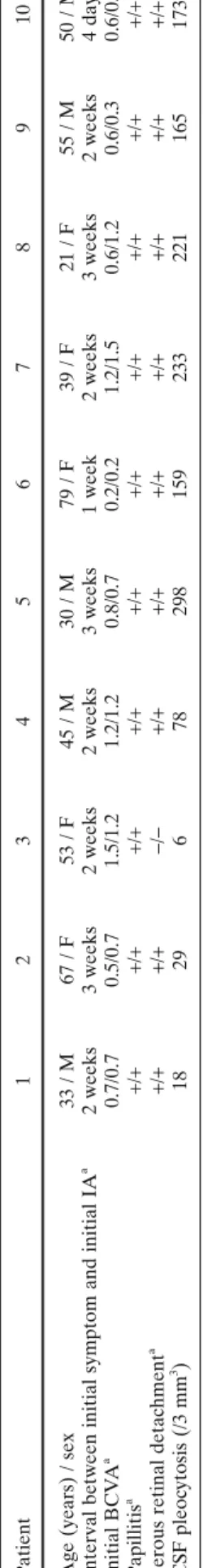
Ten patients from the two centers fulfi lled the inclusion criteria. Patient data and clinical fi ndings at presentation are shown in Table 1. There were fi ve men and fi ve women aged between 21 and 79 years (mean 47.2 ± 17.5 years). Cerebrospinal fl uid pleocytosis and papillitis were seen in
all cases. Mean best-corrected visual acuity (20 eyes) at presentation was 0.72 (0.2–1.5). Anterior segment examina-tion showed anterior uveitis in nine of ten patients (18 of 20 eyes), and this uveitis was typically granulomatous except in one patient. Fundus examination showed bilateral papil-litis in all patients and multiple serous retinal detachments in 18 of 20 eyes. FA showed disc hyperfl uorescence in all patients and subretinal fl uid in 18 of 20 eyes. In four of ten patients OCT was available at the acute stage and showed serous retinal detachments in all patients.
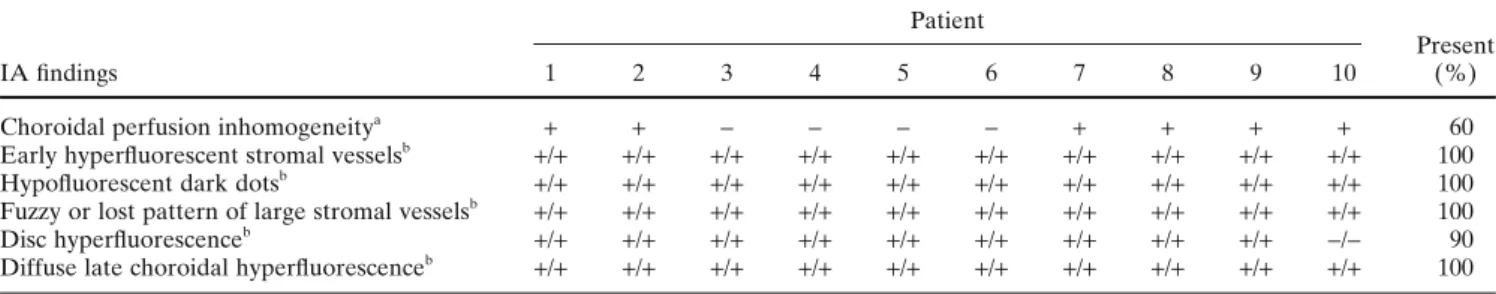
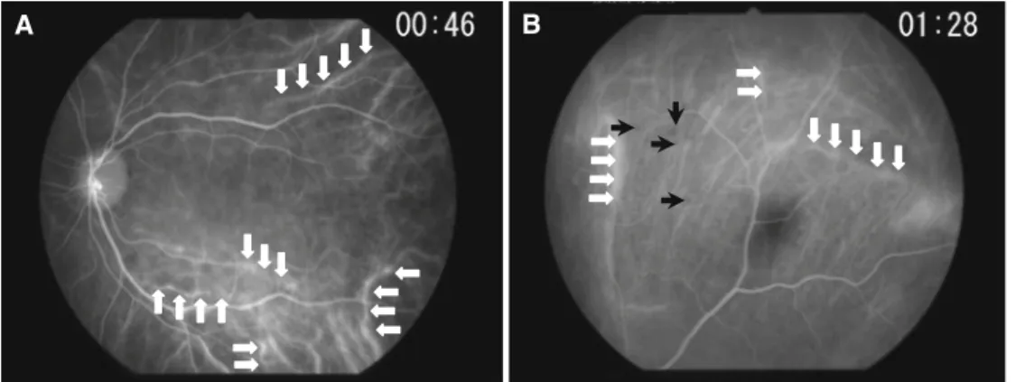
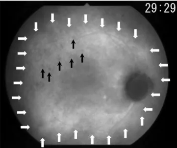
IA fi ndings at presentation are shown in Table 2. Cho-roidal perfusion inhomogeneity (Fig. 1) was seen in six of ten patients presenting some degree of choroidal fi lling delay in the early angiographic phase. The results are expressed for each patient because one eye of each patient had to be chosen for the early frames. Early hyperfl uores-cent stromal vessels (Fig. 2A, B) were present in all cases. HDDs (Fig. 3) were also present in all cases. A fuzzy or lost vascular pattern of large stromal vessels in the intermediate to late phase (Fig. 4) was also very consistent and present in all cases. Disc hyperfl uorescence (Fig. 5A, B) was seen in nine of ten cases (18 of 20 eyes). Diffuse late choroidal hyperfl uorescence (Fig. 6) was found in all patients. The most consistent fi ndings, present in all cases, were early hyperfl uorescent stromal vessels, HDDs, fuzzy or lost vas-cular pattern of large stromal vessels in the intermediate to late phase, and diffuse late choroidal hyperfl uorescence.
Discussion
Among the IA signs looked for in VKH disease, early hyperfl uorescent stromal vessels, HDDs, fuzzy or lost pattern of large stromal vessels in the intermediate to late angiographic phase, and disc hyperfl uorescence were most consistent and therefore represent useful parameters for evaluating choroidal infl ammatory involvement in Japanese patients. This set of universally found signs makes IA a reliable and precise method to assess and follow VKH patients. Analyzing these signs in chronologic order follow-ing the angiographic time sequence, the detailed fi ndfollow-ings can be summarized as follows with some physiopathological explanations.
Choroidal perfusion inhomogeneity (Fig. 1) has been described by most reports on IA in VKH disease and put forward as an important and specifi c sign.9–11 We found, however, that although this was sometimes a very clear sign when present, it was less consistent than other IA signs, as well as rather nonspecifi c and diffi cult to evaluate and quantify.
Early hyperfl uorescent stromal vessels (Fig. 2A, B), which indicate severe choroidal stromal infl ammatory vas-culopathy, are a sign that appears in the early angiographic phase (up to 3 min). In later phases, it gives way to diffuse late hyperfl uorescence. This sign was easy to evaluate and the number of early hyperfl uorescent vessel segments could even be counted, making it a quantitative or at least a semi-quantitative parameter. T a ble 1. Clinical fi ndings of patients Patient 1 2 3 4 5 6 7 8 9 10
Age (years) / sex
33 / M 67 / F 53 / F 45 / M 30 / M 79 / F 39 / F 21 / F 55 / M 50 / M
Interval between initial symptom and initial IA
a 2 weeks 3 weeks 2 weeks 2 weeks 3 weeks 1 week 2 weeks 3 weeks 2 weeks 4 days Initial BCV A a 0.7/0.7 0.5/0.7 1.5/1.2 1.2/1.2 0.8/0.7 0.2/0.2 1.2/1.5 0.6/1.2 0.6/0.3 0.6/0.9 Papillitis a + / ++ / ++ / ++ / ++ / ++ / ++ / ++ / ++ / ++ / +
Serous retinal detachment
a + / ++ / +− / −+ / ++ / ++ / ++ / ++ / ++ / ++ / + CSF pleoc ytosis (/3 mm 3 ) 18 29 6 78 298 159 233 221 165 173 M, male; F , female; IA, indoc
yanine green angiography;
BCV
A,
best-corrected visual acuity;
CSF
, cerebrospinal fl
uid.
aT
he results are shown for each eye:
right eye/left eye
Table 2. IA fi ndings of patients IA fi ndings Patient Present (%) 1 2 3 4 5 6 7 8 9 10
Choroidal perfusion inhomogeneitya + + − − − − + + + + 60
Early hyperfl uorescent stromal vesselsb +/+ +/+ +/+ +/+ +/+ +/+ +/+ +/+ +/+ +/+ 100
Hypofl uorescent dark dotsb +/+ +/+ +/+ +/+ +/+ +/+ +/+ +/+ +/+ +/+ 100
Fuzzy or lost pattern of large stromal vesselsb +/+ +/+ +/+ +/+ +/+ +/+ +/+ +/+ +/+ +/+ 100
Disc hyperfl uorescenceb +/+ +/+ +/+ +/+ +/+ +/+ +/+ +/+ +/+ −/− 90
Diffuse late choroidal hyperfl uorescenceb +/+ +/+ +/+ +/+ +/+ +/+ +/+ +/+ +/+ +/+ 100
aIn each patient (rather than in each eye) as one eye had to be chosen for the early frames. bRight eye/left eye.
Figure 1. Choroidal perfusion inhomogeneity (patient 10). White arrows show choriocapillary hypoperfusion. This sign is found
incon-sistently and is diffi cult to standardize.
Hypofl uorescent dark dots (HDDs) (Fig. 3), which are thought to correspond to choroidal granulomas, were the most consistent, most readily recordable angiographic sign, allowing also the assessment of choroidal infl ammatory activity in a semi-quantitative way. HDDs can be evaluated by using an arbitrary semi-quantitative score from 0 to 4 based on the extension and number of dots, as described earlier.17
Fuzzy or lost vascular pattern of large stromal vessels (Fig. 4) indicate diffuse infl ammatory vasculopathy of stromal vessels. This sign should be looked for in the inter-mediate to late phase, and it gives way in the late phase to diffuse stromal hyperfl uorescence. In case of severe infl am-mation, the usual pattern of choroidal vessels can even become completely lost, preventing identifi cation of indi-vidual vessels.
The optic disc usually remains dark and nonfl uorescent in normal IA. Disc hyperfl uorescence (Fig. 5A, B), rarely seen on IA, indicates severe papillitis with a blood–ocular barrier rupture suffi ciently important to allow leakage of the macromolecular ICG–protein complex from disc
capil-laries. Disc hyperfl uorescence indicates very severe disease and usually regresses rapidly after introduction of initial induction therapy. It is therefore a good sign for evaluating the initial infl ammatory activity of the disease and its response to therapy.13
Diffuse late choroidal hyperfl uorescence (Fig. 6), although a consistent IA sign found in all our patients, is diffi cult to evaluate. The appearance of this sign is too dependent on technical factors such as the quantity of gain used. It is diffi cult to record and not suffi ciently precise to be used in both assessment and follow-up of choroidal involvement.
In the present work, our aim was to study whether the IA signs previously identifi ed and systematized could be applied to Japanese VKH patients and could be considered universally acceptable. We chose to evaluate IA signs that had been established as the most consistent as well as the most useful and easy to evaluate in previously published reports.6,13,14
All examined patients showed extensive cho-roidal involvement even at a very early stage of the disease. As stated earlier, the diagnosis of VKH disease is essen-tially clinical, based on bilaterality, multiple serous retinal detachments, and papillitis, together with prodromal sys-temic symptoms such as malaise and headaches as well as lymphomonocytic pleocytosis of the cerebrospinal fl uid. FA reveals better evidence of these clinical signs, showing disc hyperfl uorescence and allowing exact delineation of the serous retinal detachments with their leaking points. Simi-larly, OCT allows clear depiction of the height and extension of serous retinal detachments in the macular area, but apart from the high-quality images obtained, it provides no essen-tial additional information that is not already available by clinical examination.18
The explanation for this is that clinical signs, FA, and OCT, all give merely information on second-ary infl ammatory events that occur when infl ammation is spilling over from the choroid. IA has been found to be par-ticularly useful for early disease detection when no secondary spill-over infl ammation is present as yet.19
The advantage of IA is that it allows localized choroidal infl ammation to be detected when it has not yet affected adjacent structures and become clinically apparent. It has to be kept in mind that in VKH disease the primary and main infl ammatory events take place and originate within the choroid, where the target of the autoimmune infl ammatory reaction is situated.
B A
Figure 2A, B. Early hyperfl uorescent stromal vessels (patient 10), a consistent indocyanine green angiography (IA) sign found in all our patients. A Some of the hyperfl uorescent stromal vessels are outlined by the white arrows. Most stromal vessels are hyperfl uorescent between the horizontal arrows in the lower part of the picture. B Early hyperfl uorescent stromal vessels in early phase of angiography (up to 3 min); some are outlined
by the white arrows. Numerous hypofl uorescent dark dots start to become visible; some are indicated by the black arrows.
Figure 3. Hypofl uorescent dark dots (HDDs) in late angiographic
phase (patient 4). Numerous HDDs are seen in the late angiographic phase. Persistence of HDDs in the late angiographic phase indicates that the choroidal infl ammatory foci (granulomas) occupy the whole thickness of the choroid and thus do not allow the IA dye to diffuse to these areas. Note also that no pattern of stromal vessels can be recog-nized any more. In this case the stromal vessels are so fuzzy that their network cannot be identifi ed any more.
Figure 4. Fuzzy or lost pattern of large stromal vessels (patient 10).
The stromal vascular pattern is still recognizable but the choroidal vessels have a fuzzy appearance (white arrows).
B A
Figure 5A, B. Disc hyperfl uorescence (patient 9). The optic disc usually remains hypofl uorescent unless severe infl ammation is present. A
Late-phase frame showing an extremely hyperfl uorescent disc (white arrows). The black arrows delineate a zone of diffuse late choroidal hyperfl uo-rescence. B Same case as in A after 1 month of treatment: disc hyperfl uorescence has completely regressed (white arrows), whereas diffuse late hyperfl uorescence is still present (black arrows).
Figure 6. Diffuse late choroidal hyperfl uorescence (patient 10). The
areas of late diffuse hyperfl uorescence are delineated by the white
arrows. Within these areas numerous HDDs are visible (black arrows).
Diffuse late choroidal hyperfl uorescence is diffi cult to standardize and is less practical to use as a follow-up sign.
References
1. Sugiura S. Vogt-Koyanagi-Harada disease. Jpn J Ophthalmol 1978;22:9–35.
2. Moorthy RS, Inomata H, Rao NA. Vogt-Koyanagi-Harada syn-drome. Surv Ophthalmol 1995;39:265–292.
3. Gocho K, Kondo I, Yamaki K. Identifi cation of autoreactive T cells in Vogt-Koyanagi-Harada disease. Invest Ophthalmol Vis Sci 2001;42:2004–2009.
4. Damico FM, Cunha-Neto E, Goldberg AC, et al. T-cell recognition and cytokine profi le induced by melanocyte epitopes in patients with HLA-DRB1*0405-positive and -negative Vogt-Koyanagi-Harada uveitis. Invest Ophthalmol Vis Sci 2005;46:2465–2471. 5. Sugita S, Takase H, Taguchi C, et al. Ocular infi ltrating CD4+ T
cells from patients with Vogt-Koyanagi-Harada disease recognize human melanocyte antigens. Invest Ophthalmol Vis Sci 2006; 47:2547–2554.
6. Herbort CP, LeHoang P, Guex-Crosier Y. Schematic interpreta-tion of indocyanine green angiography in posterior uveitis using a standard protocol. Ophthalmology 1998;105:432–440.
7. Bouchenaki N, Herbort CP. Stromal choroiditis. In: Pleyer U, Mondino B, editors. Essentials in ophthalmology: uveitis and immunological disorders. Berlin, Heidelberg, New York: Springer; 2004. p. 234–253.
8. Bouchenaki N, Cimino L, Auer C, Tran VT, Herbort CP. Assess-ment and classifi cation of choroidal vasculitis in posterior uveitis using indocyanine green angiography. Klin Monatsbl Augenheilk 2002;219:243–249.
9. Yuzawa M, Kawamura A, Matsui M. Indocyanine green video-angiographic fi ndings in Harada’s disease. Jpn J Ophthalmol 1993;37:456–466.
10. Oshima Y, Harino S, Hara Y, Tano Y. Indocyanine green angio-graphic fi ndings in Vogt-Koyanagi-Harada disease. Am J Ophthal-mol 1996;122:58–66.
11. Okada AA, Mizusawa T, Sakai J, Usui M. Videofunduscopy and videoangiography using the scanning laser ophthalmoscope in Vogt-Koyanagi-Harada syndrome. Br J Ophthalmol 1998;82: 1175–1181.
12. Kohno T, Miki T, Shiraki K, et al. Subtraction ICG angiography in Harada’s disease. Br J Ophthalmol 1999;83:822–833.
13. Bouchenaki N, Herbort CP. The contribution of indocyanine green angiography to the appraisal and management of Vogt-Koyanagi-Harada. Ophthalmology 2001;108:54–64.
14. Herbort CP, Mantovani A, Bouchenaki N. Indocyanine green angiography in Vogt-Koyanagi-Harada disease: angiographic signs and utility in patient follow-up. Int Ophthalmol 2007;27:173– 182.
15. Kawaguchi T, Horie S, Bouchenaki N, et al. Suboptimal therapy controls clinically apparent disease but not subclinical progression of Vogt-Koyanagi-Harada disease. Int Ophthalmol 2010;30: 41–50.
16. Read RW, Holland GN, Rao NA, et al. Revised diagnostic criteria for Vogt-Koyanagi-Harada disease: report of an international committee on nomenclature. Am J Ophthalmol 2001;131: 647–652.
17. Altan-Yaycioglu R, Akova YA, Akca S, Yilmaz G. Infl ammation of the posterior uvea: fi ndings on fundus fl uorescein and indocya-nine green angiography. Ocul Immunol Infl amm 2006;14:171– 179.
18. Mantovani A, Resta A, Herbort CP, et al. Work-up, diagnosis and management of acute Vogt-Koyanagi-Harada disease: a case of acute myopization with granulomatous uveitis. Int Ophthalmol 2007;27:105–115.
19. Howe L, Stanford M, Graham E, Marshall J. Indocyanine green angiography in infl ammatory eye diseases. Eye 1998;12:761–767.