RETINAL DISORDERS
Prospective study evaluating the predictability of need
for retreatment with intravitreal ranibizumab for age-related
macular degeneration
Irmela Mantel&Angeliki Deli&
Katia Iglesias&Aude Ambresin
Received: 9 March 2012 / Revised: 7 June 2012 / Accepted: 11 June 2012 / Published online: 26 June 2012 # Springer-Verlag 2012
Abstract
Purpose To investigate the rhythm and predictability of the need for retreatment with intravitreal injections of ranibizu-mab for neovascular age-related macular degeneration (nAMD).
Methods This prospective study enrolled 39 patients with treatment-naïve nAMD. After three loading doses of intra-vitreal ranibizumab, patients underwent an intensified follow-up for 12 months (initially weekly, then with step-wise increases to every 2 weeks and to monthly after each injection). Patients were retreated on an as-needed basis if any fluid or increased central retinal thickness (CRT) (>50μm) was found on spectral domain optical coherence tomography (OCT). Statistical analysis included patients who received at least two retreatments (five injections). Results A mean of 7.5 injections (range 0–12) were given between months 3 and 15. The mean visual acuity increased by 13.1 and 12.6 ETDRS letters at months 12 and 15 respec-tively. Two or more injection–retreatment intervals were found in 31 patients. The variability of their intra-individual
intervals up to 14 weeks was small (SD 0–2.13 weeks),
re-vealing a high regularity of the retreatment rhythm. The SD was correlated with the mean interval duration (r00.89, p<
0.001). The first interval was a good predictor of the following
intervals (regression coefficient00.81). One retreatment
cri-terion was stable in 97 % of patients (cysts or subretinal fluid). Conclusion The results of this study demonstrate a high intra-individual predictability of retreatment need with rani-bizumab injections for nAMD. These findings may be help-ful for developing individualized treatment plans for maintained suppression of disease activity with a minimum of injections and visits.
Keywords Neovascular age-related macular degeneration . Ranibizumab . Intravitreal injection . P.R.N. treatment regimen . Predictability . Optical coherence tomography
Introduction
Age-related macular degeneration (AMD) is the leading cause of severe and irreversible vision loss in people aged
50 years and older in developed countries [1, 2]. Recent
therapeutic gains have been made following the demonstra-tion that the inhibidemonstra-tion of vascular endothelial growth factor A (VEGF-A) is an effective and safe therapy of neovascular age-related macular degeneration (nAMD) when using monthly injections of ranibizumab, a recombinant,
human-ized, monoclonal antibody fragment [3–5]. The MARINA
and ANCHOR trials used monthly treatments of ranibizu-mab, while the PIER study employed quarterly injections
after three monthly loading doses [6, 7]. Although the
overall results of the PIER study demonstrated a smaller benefit in mean visual acuity (VA), a subgroup of patients did well on quarterly injections. Therefore, it appears that some patients may not require monthly injections.
In an attempt to reduce the number of injections accord-ing to individual need, two different approaches have been developed. The pro re nata (p.r.n.) regimen, which involves Funding / Support: none
I. Mantel
:
A. Deli:
A. AmbresinOphthalmology Department of the University of Lausanne, Jules-Gonin Eye Hospital,
Lausanne, Switzerland K. Iglesias
Centre for Clinical Epidemiology, University Hospital Lausanne, Lausanne, Switzerland
I. Mantel (*)
University Eye Hospital Jules Gonin, 15 Av. de France– Case postale 133, CH– 1000 Lausanne 7, Switzerland e-mail: [email protected] DOI 10.1007/s00417-012-2090-9
monthly visits and retreatment according to predefined
cri-teria, has shown good functional outcomes [8,9].“Treat and
extend,” another flexible retreatment regimen (published
after initiation of the present study), showed a similar
func-tional outcome [10, 11]. This approach is based on the
progressive lengthening of treatment intervals until signs of recurrence indicate a shortening of the treatment interval. The underlying concept assumes a regular rhythmic pattern in need of retreatment, although this concept remains un-proven. The results of a recent study also suggests a regular rhythmic pattern of recurrences, although the study’s
retro-spective design has inherent weaknesses [12].
The aim of the present study was to prospectively explore the rhythm and predictability of the need for retreatment with intravitreal injections of ranibizumab for nAMD. Such predictability could allow for the future creation of an indi-vidualised treatment plan with fewer visits and lower costs.
Methods
This prospective study was designed and carried out at a single centre (University Eye Hospital Jules Gonin, Lau-sanne, Switzerland). The study enrolled a consecutive series of patients, aged 50 years or older, with newly diagnosed treatment-naïve nAMD, with active subfoveal CNV as con-firmed on fluorescein angiography by a retinal specialist. In an attempt to recruit patients comparable to those in the MARINA, ANCHOR, and PIER studies, we applied the following inclusion criteria: baseline best-corrected visual acuity (BCVA) between 0.5 and 0.06; an angiographically identifiable CNV (both occult and classic) of 50 % or more of the total retinal lesion; a total lesion size of no more than 12 disc diameters; and absence of any central atrophy or fibrosis. In cases of bilateral nAMD, only one eye was included in the study. Exclusion criteria were: pigment epithelium tears; foveal atrophy, fibrosis or hemorrhage; hemorrhage in more than 50 % of the lesion; confounding retinopathy or vitreous pathology; and the inability to obtain OCT images or fluorescein angiograms of sufficient quality. Baseline examination included a complete ocular and systemic history, BCVA measured at 4 m with standard ETDRS charts, a complete ophthalmic examination with slit-lamp biomicroscopy, intraocular tonometry, dilated fun-doscopy, color fundus photographs, fluorescein and indoc-yanine green angiography (FA and ICGA), fundus autofluorescence, and spectral domain optical coherence tomography (SD-OCT; Cirrus, Carl Zeiss Meditec, Inc., Oberkochen, Germany) with a cube 512 × 126.
The initial treatment consisted of three intravitreal injec-tions of ranibizumab (0.5 mg) at monthly intervals. All injections were performed by a retina specialist according to the internationally standard technique.
From injection 3, the intervals between injection and need for the next injection were measured. To detect signs of exudative activity early and to increase the precision of the study, follow-up visits were stepped up between month 3 and month 15: With the previous injection serving as a time reference, the visits occurred on a weekly basis from the earliest time point of possible retreatment (week 4, 5, 6, 7, 8 post injection), followed by visits every 2 weeks until week 16 (post injection), and then monthly visits after each injection, until retreatment criteria were met (explained be-low). Then, the interval count was reset to 0 at the date of retreatment, and visits were again performed at week 4,5,6, etc as described above. Visit intervals were increased grad-ually in order to maintain compliance, and with the assump-tion that longer recurrence-free intervals would be associated with slower progression from dry retina to clin-ically detectable exudative recurrence, due to lower under-lying disease activity.
The examinations at follow-up visits included BCVA, in-traocular tonometry, fundoscopy, and SD-OCT with a 512 × 126 cube. If fixation was extrafoveal, the fovea was manually centred into the cube measurement. Dilatation was not necessarily performed, if the quality of SD-OCT was at least 7/10, as indicated by the integrated software, and if fundoscopy showed no hemorrhage. FA and fundus autofluor-escence were repeated at month 3 and at the final visit, and also in between, if judged necessary. Criteria for retreatment were the presence of any intraretinal cysts or subretinal fluid (SRF), or an increase of central retinal thickness (CRT) of at least 50μm, as compared with the smallest previous measure-ment. In an attempt to differentiate between exudative and degenerative cysts, the cysts were accepted as retreatment criterion only if they were associated with some exudation on FA, or if they re-appeared on SD-OCT after transient absence, or if they were associated with an increased CRT of >50μm. In unclear cases, one investigator (IM), blinded to the previous evolution, decided whether retreatment criteria were met, based on the OCT scans and FA images. If retreatment criteria were met, a single re-injection with intravitreal ranibi-zumab was performed within 3 days after the visit, and the interval count was reset to 0 at the date of injection. The intervals between the previous injection and the visit that revealed exudative signs on SD-OCT were determined. These
intervals were defined as“retreatment intervals” because of
evidence indicating the need for retreatment, even though the re-injection may have been given up to 3 days later. The retreatment intervals were determined from injection 3 through month 15, including one additional interval (if ascer-tainable) up to month 18. However, the last injection after month 15 was not included in the count of injections given during the 12 months of p.r.n. follow-up.
If a patient missed a visit but had a dry macula on the next visit, the patient was allowed to continue in the study.
However, if a patient missed a visit and had exudative signs on the SD-OCT of the next visit, the corresponding interval was excluded from the study.
Statistical analyses (software: Stata 11, StataCorp LP, College Station, TX, USA) were performed by the Centre of Clinical Epidemiology of the Institute of Social and Preventive Medicine (Lausanne, Switzerland). Statistical analysis of the intervals included all patients in whom we observed and documented at least two intervals of retreat-ment after the loading phase. The standard deviation (SD) of the intra-individual intervals was used to describe the vari-ability of the intervals. To test the relation between the variability of intervals (SD) and the mean intra-individual intervals, a correlation test was performed with Spearman’s rho test (non-normal distribution). To test the predictive capacity of the first interval with regards to the following intervals, a multilevel model was used with patients as random effect, and with measures nested into the patients, in order to take into account the dependence of the measure-ments of the same patient. A p-value <0.05 was considered statistically significant.
The study was approved by the local ethics committee, and it adhered to the tenets of the declaration of Helsinki. All patients gave written informed consent.
Results
A total of 39 patients were included in the study. Their mean age was 79.4 years (SD 7.2); 30 were female and nine male. The study eye was the right eye in 23 patients and the left eye in 16 patients. Predominantly classic CNV was found in nine patients (including six purely classic), minimally clas-sic CNV in one patient, occult CNV in 24 patients, and retinal angiomatous proliferation (RAP) in five patients. Some pigment epithelium detachment was observed in six patients (four occult CNV, one minimally classic CNV, one RAP), and 12 patients had lesions larger than 4 disc diameters.
Patients received a mean of 7.5 injections during the 12 months of p.r.n. follow-up. The mean BCVA improved by 6.4 ETDRS letters at month 3, and continued to improve to a mean gain of 13.1 and 12.6 ETDRS letters at month 12
and month 15 respectively (Fig.1). At month 12 and 15, no
patient had lost 3 lines or more of BCVA, 92 % had stable or improved BCVA, and 36 % / 33 % of patients respectively had BCVA improvement from baseline of more than three lines ETDRS. Mean CRT rapidly diminished by 103
micro-meters at month 3, and remained stable thereafter (Fig.1).
There were 31 patients with at least two retreatment intervals during the p.r.n. follow-up (including the last in-terval measurement after month 15, if available), which
ranged between 4 and 14 weeks (Table1). One patient had
a much higher mean intra-individual value (32 weeks). Our statistical consultant reckoned that a single measurement in a different range would not allow any conclusions regarding this interval range, and recommended not including that patient in the statistical analysis.
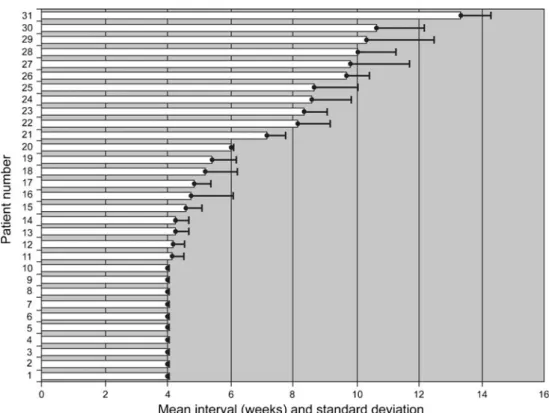
The results of mean intervals and SD are shown in Fig.2.
SD ranged between 0 and 2.13 weeks in all patients, and between 0 and 1.97 weeks in 95 % of the patients, with a 95 % confidence interval for the upper limit of 1.36–2.13. A correlation analysis of the intra-individual interval means and the associated SD values showed a strong and statisti-cally significant correlation with Spearman’s rho00.89 (p< 0.0001); that is, the SD increased with the intra-individual mean intervals.
The rhythm of 11 eyes (35 %) was highly regular, with an SD of zero: ten of them showed signs of liquid on SD-OCT as defined in the Methods section at each 4-week visit, indicating a regular 4-weekly rhythm of need for retreat-ment. One patient showed recurrences exclusively at 6 weeks after each injection. The remaining 20 patients (65 %), with a mean interval between 4 and 14 weeks, had some variation in their intervals with a relatively small SD, between 0.3 and 2.13 weeks. In eight patients, the variability was limited to the two most proximate interval durations (weeks 4+5, or weeks 8+10, or weeks 12+14). Their SD was between 0.36 and 0.49 for measurements with a preci-sion of 1-week units (weeks 4+5), and ranged between 0.75 and 0.94 for the longer intervals, influenced by the 2-week visit interval (weeks 8+10, weeks 12+14). Thus, a total of 19 patients (61 %) had either constant or two adjacent interval durations.
The first measured interval after the three loading doses was a good predictor of the following intervals. The regres-sion coefficient of the multilevel model was 0.81 (95 % confidence interval 0.67–0.95), which is significantly dis-tanced from zero. The first interval was predictive of 70 % of the variance of the following intervals. For each unit of duration of the first interval, the following intervals are equal to 0.81 times the first interval, added to a constant of 1.21 (statistical equation: probable future interval01.21+ 0.81*first interval).
We analyzed factors that could potentially influence the mean interval duration, including age, baseline VA, baseline CRT, the angiographic lesion type, and the baseline lesion size. None of these factors, however, showed any associa-tion with the mean retreatment interval. Furthermore, no particular characteristic was found for the patients with only one or no recurrence.
The retreatment criteria involved SRF in 24 eyes, cysts (re-appearing or associated with exudation of FA or
associ-ated with CRT increase >50 μm) in 24 eyes, and CRT
increases of at least 50 μm in 11 eyes. In the 32 eyes with
a revenant retreatment criterion in all subsequent recurren-ces in 17 eyes, and cysts in a further 14 eyes; therefore, one of these two criteria remained stable in 31 (97 %) out of 32 eyes. One eye showed, alternatively, cysts or SRF. However,
a CRT increase of at least 50μm was observed exclusively
in association with cysts or subretinal fluid, but never as an isolated retreatment criterion.
Discussion
The present study prospectively explored the rhythm and predictability of the need for retreatment with intravitreal injections of ranibizumab for nAMD. The results showed a relatively high consistency of the intra-individual intervals over time (in the range of means of 4-14 weeks), as reflected by very small standard deviations, particularly in association with smaller individual means. Because the small standard deviation indicates high stability over time, we may con-clude that there is a relatively good predictability of the need for retreatment. In fact, the first measured interval duration was shown to have a good predictive value, explaining 70 % of the variance of the following intervals. In most cases, the interval corresponds with a true recurrence (21 patients out of 31 had transiently dry macula, 68 %). Also, slightly earlier regular retreatment may make it possible to keep the macula mostly dry.
To the best of our knowledge, no previous prospective study has measured the degree or regularity and predictabil-ity of the intra-individual need for retreatment for patients with nAMD treated with intravitreal ranibizumab. However, a recent retrospective study has found at least two periodic injection-recurrence intervals in 76 % of eyes with 2–6
recurrences [12]. Furthermore, a recently described p.r.n.
regimen, called“treat and extend”, was shown to result in
good functional results [10, 11, 13]. In this regimen, the
treatment interval was progressively increased by incre-ments of 2 weeks, until the follow-up visit disclosed signs of exudation, justifying a subsequent reduction to the
previ-ous interval. Thus, the“treat and extend” regimen was based
on the assumption of a relatively regular rhythm of the need for retreatment. The results of our current study support that underlying assumption.
A regular rhythm of recurrences is not surprising if we assume that a continuous and stable production of VEGF by the underlying nAMD (and a stable elimination rate) results in a stable level of VEGF in the retina, and also assume stable individual pharmacokinetics. These assumptions could explain that the point of clinically relevant concentra-tion of free VEGF is reached always after the same period of time following the latest standard injected dose of anti-VEGF. Furthermore, the intravitreal half-life of ranibizumab has been measured to be approximately 3 days in rabbits and
monkeys [14,15], and may be up to 9–10 days in humans
Fig. 1 Mean best-corrected vi-sual acuity change (ETDRS letters) and mean central retinal thickness change (micrometers) from baseline of all study eyes treated with intravitreal ranibi-zumab for neovascular age-related macular degeneration (error bars indicate standard error of change)
T able 1 Baseline characteristics of the studied eyes with neovascular age-related macular degeneration, and injection –recurrence intervals during the p.r .n. regimen with intravitreal ranibizumab between months 3 and 15 Patient, gender , age, eye CNV type Lesion size Injection –retreatment intervals Mean intra-individual interval (weeks) SD W eeks of p.r .n. follow-up N (intervals) Re-treatment criteria 1, F , 67, R occ >4 DA 4,4,4,4,4,4,4,4,4,4,4,4,4 4.00 0 > 52 13 SRF 2, F , 86, L p c < 4 D A 4,4,4,4,4,4,4,4,4,4,4,4,4 4.00 0 > 52 13 C+SRF 3, F , 64, L occ <4 DA 4,4,4,4,4,4,4,4,4,4,4,4 4.00 0 5 2 1 2 SRF+Th 4, M, 82, R occ <4 DA 4,4,4,4,4,4,4,4,4,4,4,4,4 4.00 0 > 52 13 SRF 5, F , 82, R occ <4 DA 4,4 4.00 0 8 2 SRF+C 6, F , 80, R p c > 4 D A 4,4,4,4,4,4,4,4,4,4,4,4 4.00 0 5 2 1 2 SRF+C+Th 7, F , 83, R RAP >4 DA 4,4,4,4,4,4,4,4,4,4,4,4 4.00 0 5 2 1 2 C+SRF 8, F , 84, L occ+PED <4 DA 4,4,4,4,4,4,4,4,4,4,4,4,4 4.00 0 > 52 13 SRF 9, F , 92, R RAP <4 DA 4,4,4,4,4,4,4,4,4,4,4,4 4.00 0 5 1 1 2 C+SRF 10, F , 81, L mc+PED <4 DA 4,4,4,4,4,4,4,4,4,4,4,4 4.00 0 5 2 1 2 C or SRF 1 1 , F , 68, R occ+PED >4 DA 5,5,4,4,4,4,4,4,4,4,4,4,4 4.15 0.36 > 5 2 1 3 C+SRF+Th 12, M, 77, R c <4 DA 4,5,5,4,4,4,4,4,4,4,4,4 4.17 0.37 52 12 SRF+C 13, F , 82, L p c < 4 D A 4,4,4,5,5,4,4,4,4,5,4,4 4.25 0.43 52 12 C+SRF 14, F , 77, L RAP+PED <4 DA 4,5,5,5,4,4,4,4,4,4,4,4 4.25 0.43 52 12 SRF 15, F , 84, L occ >4 DA 4,5,5,5,5,4,5,4,4,5,4,5 4.58 0.49 > 5 2 1 2 C 16, F , 81, L c <4 DA 4,8,7,5,4,4,4,4,4,4,4 4.73 1.35 > 5 2 1 1 SRF 17, F , 84, R occ <4 DA 4,5,5,4,5,5,5,5,5,4,5,6 4.83 0.55 > 5 2 1 1 C 18, M, 61, R occ <4 DA 5,6,7,6,6,6,5,4,4,4,4 5.18 1.03 > 5 2 1 1 SRF+Th 19, F , 78, R c >4 DA 4,5,6,5,5,5,5,7,6,6 5.40 0.8 > 5 2 1 0 SRF 20, F , 80, L occ <4 DA 6,6,6,6,6,6,6,6 6.00 0 5 1 8 C+Th 21, F , 79, R occ <4 DA 7,7,7,6,8,8,7,7 7.13 0.6 > 5 2 8 C+Th 22, F , 84, R RAP >4 DA 8 8.00* N/A* 8 1 * C 23, M, 75, R c <4 DA 8,7,10,7,7,10,8 8.14 1.03 > 5 2 7 SRF+C+Th 24, F , 86, L occ >4 DA 10,8,8,8,8,8 8.33 0.75 52 6 C 25, F , 84, L occ <4 DA 7,7,8,8,10,10,10 8.57 1.29 > 5 2 7 C 26, F , 84, R occ+PED <4 DA 10,7,10,10,8,7 8.67 1.37 > 5 2 6 SRF 27, F , 90, R occ <4 DA 10,10,8,10,10,10 9.67 0.75 > 5 2 6 C+SRF+Th 28, F , 74, R occ <4 DA 7,8,12,10,10,12 9.83 1.86 > 5 2 6 C+Th 29, M, 84, R occ >4 DA 10,10,12,10,8 10.00 1.26 52 5 C+Th 30, M, 74, R c <4 DA 12,8,10,10,8,14 10.33 2.13 > 5 2 6 SRF 31, F , 82, L RAP <4 DA 14,10,10,10,10,10 10.67 1.49 > 5 2 6 C 32, F , 84, L occ <4 DA 12,14,14 13.33 0.94 42 3 C+Th 33, M, 88, R occ <4 DA 28,36 32.00 4 > 52 2 * SRF 34, F , 87, L occ <4 DA 48 48.00* N/A* 52 1* C
[16], which is sufficiently short to obviate any accumulation of the drug due to repeated injections.
The intervals of a subset of patients showed an SD of 0 (n011, 35 %) — a perfectly regular rhythm and perfect predictability of future need for treatment. Apart from one patient with a regular 6-week interval, these were mostly patients with persistent exudative signs at their week 4 visits (n010, 32 %). These exudative signs may be unresponsive to treatment or may be very early recurrences before week 4.
Studies like PrONTO [8] have established that a subgroup
of patients showed signs of active nAMD on each monthly visit, requiring monthly anti-VEGF treatment. The number of these patients was relatively high in our study, probably reflecting both our choice of mild changes as being suffi-ciently indicative of active disease, and the high sensitivity of SD-OCT to detect these changes. However, cystic changes on SD-OCT may be degenerative rather than exu-dative. In an attempt to differentiate them and exclude purely degenerative cysts, we required an association with new appearance or CRT increase or exudation on FA for acceptance as retreatment criterion.
A second subset of patients (n021, 68 %) showed a mean individual retreatment interval of >4 weeks, i.e., transiently dry macula on OCT and subsequent recurrence of exuda-tion. The standard deviation of the mean intra-individual intervals was small in this group, indicating moderate fluc-tuation of the intervals over time. The observed correlation with the mean intervals may be explained by three mecha-nisms. First, it might be related to a certain imprecision in
the measurement of intervals— initially weekly visits
(dur-ing month 2 post injection) changed to biweekly visits (during months 3 and 4 post injection) and finally to month-ly visits after each injection. This would lead to higher standard deviations associated with later recurrences, once an interval differs. Second, longer intervals lead to smaller numbers of intervals measured and therefore to larger SD. Third, the underlying disease activity may not always be exactly the same, and the pharmacokinetics may also be subject to some changes over time. If this is the case, we would expect a bigger impact of mild changes on the vari-ability of intra-individual intervals in patients with relatively low retinal concentrations of VEGF and later recurrences. The drug elimination curve becomes flatter over time, and relatively small fluctuations in VEGF production may lead to significantly earlier or later time points at which free VEGF is present and causes an exudative recurrence.
Nevertheless, the first measured interval after the initial three loading doses showed a good predictability of the following intervals, explaining 70 % of their variance. This may allow quite reliable estimation of future need for retreatment, based on only one early interval measurement. The regression formula found for the relationship between first interval and the following intervals (probable future
T able 1 (continued) Patient, gender , age, eye CNV type Lesion size Injection –retreatment intervals Mean intra-individual interval (weeks) SD W eeks of p.r .n. follow-up N (intervals) Re-treatment criteria 35, F , 75, R occ >4 DA 52 52.00* N/A* 52 1* C 36, M, 68, R c <4 DA 52 52.00* N/A* 52 1* SRF 37, F , 67, L occ+PED <4 DA 52 52.00* N/A* 52 1* SRF 38, M, 80, L occ >4 DA >52 > 52.00* N/A* > 5 2 0 * N/A 39, F , 77, R occ >4 DA >52 > 52.00* N/A* > 5 2 0 * N/A *Excluded from statistical analysis Abbreviations: CNV 0 choroidal neovascularisation; SD 0 standard deviation; p.r .n. 0 pro re nata (dosing according to recurrence criteria), M 0 male; F 0 female, R 0 right eye; L 0 left eye; c 0 classic CNV ; p c 0 predominantly classic; mc 0 minimally classic; occ 0 occult; RAP 0 retinal angiomatous proliferation; PED 0 pigment epithelium detachment; DA 0 disc areas; N/A 0 not applicable; C 0 cysts; SRF 0 subretinal fluid; Th 0 central retinal thickness; if linked by +, the first mentioned criterion remained stable, “C o r SRF ” indicate criterion changing
interval0 1.21+0.81*first interval) describes that for longer intervals the first interval tends to be longer than the fol-lowing intervals. Since the first interval is the one that follows three loading injections, it may be that loading has a more profound effect on the new vessel formation and exudation than a single injection for exudative recurrence thereafter.
We tried to evaluate possible associations of baseline characteristics with the mean interval duration. Although no such association was found for age, baseline VA, base-line CRT, the angiographic lesion type, or the basebase-line lesion size, no firm conclusion can be drawn, because the power of the study was insufficient to address this question. The functional and structural results (BCVA and CRT) were excellent in this study. We found a continuous im-provement of the mean BCVA (+6.4 letters at month 3, +13.1 letters at month 12), to a degree that the functional results appear to be just as good as in the MARINA and ANCHOR studies under monthly retreatment (+5.1 to +10.0
letters at month 3, +6.5 to +11.3 letters at month 12) [3–5].
Although p.r.n. regimens often have been associated with
secondary functional losses after the loading dose [17–19],
it has been shown that strict monthly visits and sensitive retreatment criteria may achieve good and durable
function-al results [8,20,21]. Our mean of 7.5 injections during the
12 months p.r.n. follow-up is relatively high, reflecting early retreatment due to sensitive recurrence criteria and increased number of follow-up visits. This may have had a beneficial effect on the functional results. However, the rationale
behind the intensified follow-up was consistent with the study’s aim to determine retreatment intervals; we do not make any recommendation as to follow-up frequency.
We acknowledge that the present study has some weak-nesses. The number of patients was limited. No follow-up visit was performed before week 4 after each injection, resulting in the absence of information about complete or incomplete absorption of liquid during these periods. However, because re-injection was not permitted before the 4-weeks interval, we chose to focus on the subsequent weeks in our protocol, in an attempt to conserve the patients’ motivations for the more important time period. For the same reason, we decided to slowly decrease the visit frequency. A theoretical justification for this decision may be explained by the pharmacokinetics of ranibizumab, assuming that the parabolic curve of drug con-centration leads to slower and milder exudative recurrences the longer it takes for free VEGF to appear (lower VEGF secretion). This, however, caused some statistical heterogene-ity of the interval measurements, and influenced the standard deviations (see explanation above). Furthermore, the transi-tion from dry macular to exudative recurrence, as judged on OCT by the presence of SRF or cysts, may not be sharply demarcated. Therefore, subjective judgements are likely to have played a role. We attempted to offset this risk by using a masked reader (see Methods section). Finally, the study was limited to 12 months of p.r.n. follow-up, resulting in a lack of information about the predictability of longer intervals. The natural time course of nAMD may influence the need for and timing of treatment in the long term.
Fig. 2 Graphic presentation of the individual mean values (between 4 and 14 weeks) of retreatment intervals of study eyes with at least two measured intervals (n031 eyes) with exudative activity in age-related macular degeneration after treatment with intravitreal in-jection of ranibizumab. The mean values are shown as bar with a dark dot at the end, and positive standard deviation is added as a T-Line
The results of the present study do not yet allow us to recommend new treatment regimens. However, the study results provide important information that may contribute to the future development of a secure treatment plan with a
minimum number of visits and injections— still allowing
the same good functional outcome, while reducing the workload and cost: The presence of a regular rhythm and good predictability of need for retreatment may allow an individual treatment plan. However, the variability of the intervals, although small, requires the ideal future regimen to be dynamic, sensitive and combined with a feedback system.
In summary, the present study demonstrates that patients with nAMD, given three initial loading doses of intravitreal ranibizumab, show relatively stable and predictable need for
retreatment— for those with intervals up to 14 weeks, and
during the first year of follow-up. The findings suggest that measurements of the initial interval may play a role in developing future treatment regimens with optimal timing of injections and reduction of costs, while still allowing excellent outcome. Further studies will be needed, however, to apply these results in the clinical care of patients with nAMD
References
1. Bressler NM (2004) Age-related macular degeneration is the lead-ing cause of blindness. JAMA 291:1900–1901
2. Friedman DS, O’Colmain BJ, Munoz B, Tomany SC, McCarty C, de Jong PT, Nemesure B, Mitchell P, Kempen J (2004) Prevalence of age-related macular degeneration in the United States. Arch Ophthalmol 122:564–572
3. Brown DM, Kaiser PK, Michels M, Soubrane G, Heier JS, Kim RY, Sy JP, Schneider S (2006) Ranibizumab versus verteporfin for neovascular age-related macular degeneration. N Engl J Med 355:1432–1444 4. Brown DM, Michels M, Kaiser PK, Heier JS, Sy JP, Ianchulev T
(2009) Ranibizumab versus verteporfin photodynamic therapy for neovascular age-related macular degeneration: Two-year results of the ANCHOR study. Ophthalmology 116:57–65
5. Rosenfeld PJ, Brown DM, Heier JS, Boyer DS, Kaiser PK, Chung CY, Kim RY (2006) Ranibizumab for neovascular age-related macular degeneration. N Engl J Med 355:1419–1431
6. Abraham P, Yue H, Wilson L (2010) Randomized, double-masked, sham-controlled trial of ranibizumab for neovascular age-related macular degeneration: PIER study year 2. Am J Ophthalmol 150:315–324
7. Regillo CD, Brown DM, Abraham P, Yue H, Ianchulev T, Schneider S, Shams N (2008) Randomized, double-masked, sham-controlled trial of ranibizumab for neovascular age-related macular degeneration: PIER Study year 1. Am J Ophthalmol 145:239–248
8. Fung AE, Lalwani GA, Rosenfeld PJ, Dubovy SR, Michels S, Feuer WJ, Puliafito CA, Davis JL, Flynn HW Jr, Esquiabro M (2007) An optical coherence tomography-guided, variable dosing regimen with intravitreal ranibizumab (Lucentis) for neovascular age-related macular degeneration. Am J Ophthalmol 143:566–583 9. Mantel I, Zografos L, Ambresin A (2008) Early clinical experience with ranibizumab for occult and minimally classic neovascular membranes in age-related macular degeneration. Ophthalmologica 222:321–323
10. Engelbert M, Zweifel SA, Freund KB (2010) Long-term follow-up for type 1 (subretinal pigment epithelium) neovascularization us-ing a modified "treat and extend" dosus-ing regimen of intravitreal antivascular endothelial growth factor therapy. Retina 30:1368– 1375
11. Gupta OP, Shienbaum G, Patel AH, Fecarotta C, Kaiser RS, Regillo CD (2010) A treat and extend regimen using ranibizumab for neovascular age-related macular degeneration clinical and eco-nomic impact. Ophthalmology 117:2134–2140
12. Horster R, Ristau T, Sadda SR, Liakopoulos S (2011) Individual recurrence intervals after anti-VEGF therapy for age-related mac-ular degeneration. Graefes Arch Clin Exp Ophthalmol 249:645– 652
13. Oubraham H, Cohen SY, Samimi S, Marotte D, Bouzaher I, Bonicel P, Fajnkuchen F, Tadayoni R (2011) Inject and extend dosing versus dosing as needed: a comparative retrospective study of ranibizumab in exudative age-related macular degeneration. Retina 31:26–30
14. Gaudreault J, Fei D, Rusit J, Suboc P, Shiu V (2005) Preclinical pharmacokinetics of Ranibizumab (rhuFabV2) after a single intra-vitreal administration. Invest Ophthalmol Vis Sci 46:726–733 15. Mordenti J, Cuthbertson RA, Ferrara N, Thomsen K, Berleau L,
Licko V, Allen PC, Valverde CR, Meng YG, Fei DT, Fourre KM, Ryan AM (1999) Comparisons of the intraocular tissue distribu-tion, pharmacokinetics, and safety of 125I-labeled full-length and Fab antibodies in rhesus monkeys following intravitreal adminis-tration. Toxicol Pathol 27:536–544
16. Blick SK, Keating GM, Wagstaff AJ (2007) Ranibizumab. Drugs 67:1199–1206
17. Dadgostar H, Ventura AA, Chung JY, Sharma S, Kaiser PK (2009) Evaluation of injection frequency and visual acuity outcomes for ranibizumab monotherapy in exudative age-related macular degen-eration. Ophthalmology 116:1740–1747
18. Gerding H, Loukopoulos V, Riese J, Hefner L, Timmermann M (2011) Results of flexible ranibizumab treatment in age-related macular degeneration and search for parameters with impact on outcome. Graefes Arch Clin Exp Ophthalmol 249:653–662 19. Holz FG, Meyer C, Eter N, on behalf of the SUSTAIN study group
(2009) Safety and efficacy of ranibizumab treatment in patients with neovascular age-related macular degeneration: 12-Month results of the SUSTAIN study. Invest Ophthalmol Vis Sci 50: E-Abstract 3095
20. Lalwani GA, Rosenfeld PJ, Fung AE, Dubovy SR, Michels S, Feuer W, Davis JL, Flynn HW Jr, Esquiabro M (2009) A variable-dosing regimen with intravitreal ranibizumab for neovascular age-related macular degeneration: year 2 of the PrONTO Study. Am J Ophthalmol 148:43–58
21. Martin DF, Maguire MG, Ying GS, Grunwald JE, Fine SL, Jaffe GJ (2011) Ranibizumab and bevacizumab for neovascular age-related macular degeneration. N Engl J Med 364:1897–1908