Molecular Neul oblology
Copyright 9 1998 Humana Press Inc. All rights of any nature whatsoever reserved. 1SSN0893-7648/98/16(1): 01-20/$13.00
Acetylcholine Release and the Cholinergic Genomic Locus
M a u r i c e Israel *'1 a n d Yves D u n a n t 2
l Laboratoire de Neurobiologie Cellulaire et Mol~culaire, C.N.R.S. F-91198 Gif-sur- Yvette, France; and 2D~partement de Pharmacologie, Universit~ de Gen~ve, Centre M~dical Universitaire,
CH- 1211 Gen6ve-4, Switzerland
Abstract
Choline acetyltransferase and vesicular acetylcholine-transporter genes are adjacent and coreg- ulated. They define a cholinergic locus that can be turned on under the control of several factors, including the neurotrophins and the cytokines. Hirschprung's disease, or congenital megacolon, is characterized by agenesis of intramural cholinergic ganglia in the colorectal region. It results from mutations of the RET (GDNF-activated) and the endothelin-receptor genes, causing a disregula- tion in the cholinergic locus.
Using cultured cells, it was shown that the cholinergic locus and the proteins involved in acetylcholine (ACh) release can be expressed separately ACh release could be demonstrated by means of biochemical and electrophysiological assays even in noncholinergic cells following preloading with the transmitter. Some noncholinergic or even nonneuronal cell types were found to be capable of releasing ACh quanta. In contrast, other cells were incompetent for ACh release. Among them, neuroblastoma N18TG-2 cells were rendered release-competent by transfection with the mediatophore gene. Mediatophore is an ACh-translocating protein that has been puri- fied from plasma membranes of Torpedo nerve terminal; it confers a specificity for ACh to the release process.
The mediatophores are activated by Ca2+; but with a slower time course, they can be desensi- tized by Ca -"+. A strictly regulated calcium microdomain controls the synchronized release of ACh quanta at the active zone. In addition to ACh and ATP, synaptic vesicles have an ATP-dependent Ca 2+ uptake system; they transiently accumulate Ca R+ after a brief period of stimulation. Those vesicles that are docked close to Ca 2+ channels are therefore in the best position to control the profile and dynamics of the Ca R+ microdomains. Thus, vesicles and their whole set of associated proteins (SNAREs and others) are essential for the regulation of the release mechanism in which the mediatophore seems to play a key role.
Index Entries: Cholinergic neurons; gene regulation; choline acetyltransferase; vesicular acetyl-
choline transporter; acetylcholine release, mediatophore; presynaptic proteins.
*Author to whom all correspondence and reprint requests should be addressed.
2
Israel and Dunant
The Vesicular Acetylcholine
Transporter (VAChT)
VAChT was first identified at the genomic level by Alfonso et al. (1993) who showed that impaired cholinergic function in
Coenorhabditis
elegans
mutants was linked to the UNC17 gene.The protein encoded showed good homology with the monoamine vesicular transporters VMAT1 and VMAT2 characterized earlier (Erikson et al., 1992; Liu et al., 1992; Schuldiner, 1992). Taking advantage of the homogenous population of cholinergic motoneurons of the
Torpedo
electric lobe, Varoqui et al. (1994)screened the corresponding cDNA library using a UNC17-radiolabeled probe and iso- lated a full-length clone. Demonstrating that the encoded putative cholinergic transporter was indeed the real acetylcholine transporter (VAChT) came from transfection experiments. Cells transfected with a plasmid encoding this transporter bound the specific blocker vesami- col with an affinity similar to that of synaptic vesicles (Varoqui et al., 1994); after loading with ACh as described by Isra61 et al. (1994), the transfected cells accumulated the neuro- transmitter in a bound pool and this was in- hibited by vesamicol (Erickson et al., 1994). The range of vesamicol concentration that inhibited transport was in agreement with previous work on isolated synaptic vesicles (Bahr and Persons, 1986a,b; Diebler and Talarmain, 1989). The kinetic parameters of ACh transport mea- sured in PC12 cells transfected with h u m a n VAChT corresponded to those of synaptic vesicles (Varoqui and Erickson, 1996) that were purified from
Torpedo
electric organ by classical techniques (Isra61 et al., 1968, 1970, 1980; Whitlaker et al., 1972).A Cholinergic Genomic Locus
It was interesting that the VAChT gene was located at the 5' end of the noncoding region of the choline acetyltransferase (CHAT) gene (Erickson et al., 1994; B6janin et al., 1994) at a
site that was initially thought to be an intron. In neurotransmitter systems, this colocalization of two genes involved in complementary func- tions seems to be unique; it suggests that syn- thesis and storage of ACh are tightly controlled and coregulated (Berrard et al., 1995; Berse and Blusztajn, 1995). In humans the two genes are located on chromosomes 10 at 10qll 2 (Fig. 1). Five possible promoters for ChAT were identi- fied in rat and a diversity of mRNA species have been characterized (B6janin et al., 1992; Misawa et al., 1992; Wu and Hersh, 1994; Cervini et al., 1995). Such a diversity might be important for the coexpression of different phenotypes controlled by complex splicing patterns for ChAT at the level of three exons, R, N, and M, in the noncoding sequence. The VAChT gene is located between R and N, and shares at least two R promoters with CHAT. In addition, VAChT has two specific promoters, V1 and V2 (Cervini et al., 1995).
The adjacent localization of these two genes seems typical for the cholinergic system. The VMATs genes and the genes encoding cate- cholamine synthesizing enzymes are not linked in a comparable way. From a general point of view, VAChT and VMATs accomplish a homol- ogous function: They concentrate the respec- tive transmitters in vesicles or granules by exchange for protons that were accumulated by V-ATPase. However, it should be empha- sized that they operate at very different cyto- plasmic concentrations. The K~ of VMATs for monoamines is in the ~M range, whereas that of VAChT for is quite high, in the mM range, which corresponds to the elevated ACh con- centration (20-30 mM) in the cytoplasm (Dunant et al., 1974; Katz and Miledi, 1977; Morel et al., 1978). In addition, the dynamics of dopamine and noradrenaline are different since dopa-decarboxylase is in the cytoplasmic compartment whereas dopamine-~-hydroxy- lase in intragranular. The significance of the elevated cytosolic ACh level is certainly related to its mode of release, which might differ con- siderably from that of monoamines
(see
below). In summary, the adjacent localization of VAChT and ChAT genes defines acholinergic
Regulation of Cholinergic Function
3
Re#ease + Ca Channel + / I ~ ' - ~ l
t /
/
l-;67/ Iv'~
\
' STATS
# tyrosine kinase it, /
T l t t retinoic acid
Endothelin NGF GDNF LIF
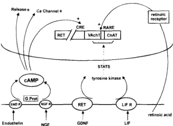
Fig. 1. Regulation of tile cholinergic locus. The cholinergic locus is depicted as two adjacent genes of the human chromosome 10 (10ql 1.2) that also carries the RET gene At the cell membrane, endothelin, NGF, GDNF, and LIF interact with their respective receptors, activating either cAMP-dependent phosphorylation, or tyrosine kinase phosphorylation followed by a cascade of kinases (STATS) As a result, the cholinergic locus, the genes involved in transmitter release, and those of Ca2+-channel expression are turned on. In parallel, retinoic acid, after binding to its receptor, turns on the cholinergic locus. CRE and RARE sequences are seen at the genomic level. Mutations at the level of the GDNF/RET and of the endothelin system were found in Hirschprung's disease.
operon
that is expected to be switched on in acoordinated manner in order to regulate the compartmentation of ACh within the neuron and adapt ACh metabolism to environmental changes.
The physiological role of VACht would be to increase the concentration of the transmitter and build up a store that can be mobilized in two ways.
First, if the cytoplasm becomes acidic, an immediate leakage of vesicular ACh and ATP takes place supporting ACh release from the cytoplasm but also the synthesis of ACh that depends of ATP. Several metabolic conditions (glycolysis for example) may acidify the cyto- plasm, and signal the mobilization of vesicular ATP and ACh stores.
Second, a docked vesicle on the site of release would feed the "releasing gate" with a
much higher transmitter concentration than in the cytoplasm leading to larger mepps. The essential role of VACht may be observed after vesamicol action, w h e n the ACh stores are exhausted, the mechanism looses its efficacy but is still at work. Similar observations come from studies on
C. elegans
mutants for VACht, the ACh storage in vesicles seems essential to maintain release, but VACht itself is not nec- essary for the translocation step, that depends of a membrane gate that we discuss in a later section.Regulation
of the Cholinergic Operon
A turning point in the research of the cholin- ergic phenotype was made possible by the use of sympathetic neurons in culture. The factor CDF-LIF (cholinergic differentiation factor/ leukemia inhibitory factor), present in muscle conditioned-culture medium, was seen to switch off the adrenergic phenotype (i.e., a decrease in tyrosine hydroxylase) and to turn on the ChAT gene (Yamamori et al., 1989). The role of CDF-LIF was recently confirmed with the additional demonstration that VAChT was coexpressed with ChAT (Berrard et al., 1995; Berse and Blusztajn, 1995; Misawa et al., 1995; Varoqui et al., 1996). Other neuropoietic factors such as CNTF (ciliary neurotrophic factor) are also able to reroute the developmental fate of adrenergic neurons towards the cholinergic phenotype. Apparently, CDF-LIF and CNTF may seem to be unrelated cholinergic differen- tiation factors. However, they were shown to share a common n-helical framework that is also present in other compounds, such as hematopoietic cytokines (interleukins 6 and 3, interferons ct and ~); this structural homology has been recently documented by Bazan (1991). An example in which a perturbation of the cholinergic locus leads to a severe disease is Hirschprung's congenital megacolon. Normally, cells of the neural crest migrate towards the terminal intestine forming cholinergic ganglia
4 Israel and Dunant
that innervate smooth muscle cells, these gan- glia are absent in Hirschprung's disease. It was recently demonstrated that familial forms of Hirschprung's disease result from a mutation of the oncogene RET (Angrist et al., 1993; Lyonnet et al., 1993; Mulligan et al., 1993; Edery et al., 1996, 1994; Scuchardt et al., 1994; Pasini et al., 1995). RET is localized on human chromosome tO (10qll 2) like VAChT and CHAT. It would therefore be interesting to know the distance separating RET from the cholinergic locus. Whatever the distance, a crucial question is why RET deletion leads to degeneration of the cholin- ergic ganglia of the colon. The ligand that acti- vates RET is a factor called GDNF (glial cell line-derived neurotrophic factor) (Ibanez and Persson, 1991; Lin et al., 1993; Trupp et al., 1996). GDNF exerts a t r o p h i c influence on different classes of peripheral and central neurons, in- cluding motoneurons (Henderson et al., 1994; Oppenheim et al., 1995; Sargot et al., 1996). Mice lacking GDNF or RET do not survive since they have renal agenesis, a decreased number of neu- rons in several peripheral ganglia and absence of enteric neurons like in Hirschprung's disease (Moore et al., 1996; Pichel et al., 1996; Sanchez et al., 1996). The activation of RET by GDNF involves a complex multicomponent receptor (Treanor et al., 1996); it triggers tyrosine-kinase activity, leading to a cascade of phosphorylation events that results in neuron survival. This effect has been demonstrated for dopaminergic neurons and also for cholinergic motoneurons as measured by an increase in ChAT activity (Oppenheim et al., 1995; Zurn et al., 1994).
Factors Controlling Presynaptic
Expression of Cholinergic Genes
Figure 1 shows that the cholinergic locus can be activated by signals coming from several different signaling pathways. As mentioned above, cytokines (CDF-LIF, CNTF) and GDNF exert their action initially through a receptor dimerization followed by the activation of a tyrosine kinase. The following cascade of bio-
chemical reaction results in activation of tran- scription factors such as STATS (signal trans- ducers and activators of transcription) (Curtis et al., 1994). In Hirschprung's disease, the RET tyrosine kinase link has already been men- tioned, but the disease is also associated with gene defects in a different pathway, involving the endothelin-endothelin receptor system, here, the regulation occurs through a G protein- coupled phosphorylation involving cAMP and protein kinase C. The expression of cholinergic genes might also respond to this signaling sys- tem (Salomon et al., 1996). Such a response is of particular interest since it is known that protein kinase C and cAMP regulate the expression of proteins implicated in ACh release (see below).
Neurotrophins like NGF and BDNF are believed to act through other phosphorylation pathways, one of which involves the cAMP- responding system CREB-CRE (cAMP respon- sive element [protein]). It has been shown that cAMP up regulates both ACh synthesis and release in some cell lines (Blusztajn et al., 1992) and the release process in other lines (Falk- Vairant et al., 1996).
Still another pathway capable of switching on the cholinergic locus involves retinoic acid that binds to intracellular receptors similar to steroid or thyroxin receptors. The complex acti- vates the expression of ChAT and VAChT at the genomic level by reacting with RARE (retinoic acid responsive elements) (Berrard et al., 1995; Berse and Blusztajn, 1995).
Is Expression of the Cholinergic
Locus Coordinated
with That of the Release Process?
It may be expected that the expression of ChAT and VAChT is coordinated with the expression of proteins supporting release. To test this, ACh synthesis and storage, on one side, and ACh release, on the other, must be measured separately. This experiment was re- cently performed by artificially filling with ACh cells that normally do not contain the neu-
Regulation of Cholinergic Function
5
rotransmitter (Isra61 et al., 1994). ACh release from these cells was tested either biochemically (Isra61 and Lesbats, 1981) or in real-time by an electrophysiological assay. It was found in this way that many cell types that do not express ChAT and VAChT are indeed able to produce Ca2§ quantal ACh release (Isra61 et al., 1994; Falk-Vairant et al., 1996a,b,c,d, 1994; Morimoto et al., 1995). This implies that the release machinery can be expressed inde- pendently of ChAT and VAChT. However, cholinergic neurons can both synthesize and release ACh; there the two systems have to be turned on by some coordinated regulation.
For the release and its regulation, the prob- lem is complex since many proteins are in- volved. The approach can be simplified by separately considering the proteins that are more specifically related to the release of ACh and those that are common to all releasing cells. The latter include voltage-gated Ca 2+ channels and the components of the T- and V-SNAREs (Rothman, 1994) that govern the traffic of intra- cellular membranes at the active zone as they do in the Golgi apparatus. Obviously, these proteins (the T-SNAREs syntaxin and SNAP-25 at the plasma membrane, and the V-SNARE VAMP/synaptobrevin on synaptic vesicles) must be expressed in every type of cell. The same is true for V-ATPase, which functions to accumulate protons in intracellular organelles; in neurons this process is responsible for the driving force for concentrating transmitters into granules or vesicles.
In cholinergic neurons, one would expect that the expression of ChAT and VAChT is coordinated in some way to a component of the release machinery that would confer a certain specificity for ACh. A protein support- ing such ACh specificity has been identified and named mediatophore
(see
description below). However, the mediatophore was found to be a homo-oligomer of a 15-kDa proteolipid that is also a component of the membrane sector of the V-ATPase. This 15-kDa element in itself cannot therefore be considered as a cholinergic-specific protein. However, what may be more characteristic for cholinergicneurons is the mechanism that directs the 15 kDa into the plasma membrane. This ques- tion was recently addressed by Leroy and Meunier (1995) who expressed the 15 kDa in the
Xenopus
oocyte. They showed that injecting themRNA of the 15 kDa alone was less efficient in inducing ACh release than injecting the whole set of poly(A*) mRNAs extracted from choliner- gic neurons. In the former, the 15-kDa protein accumulated chiefly in intracellular mem- branes. Therefore, another protein is probably required for directing the 15 kDa subunit at the plasma membrane where it is assembled to form the mediatophore homo-oligomer. This process may coordinate the events associated with expression of the cholinergic locus and ex- pression of the specific step of ACh release. In this respect, cAMP-dependent systems may be an important regulation pathway since they can upregulate both ACh synthesis and ACh re- lease, as well as CaR+-channels in some cells (Isra6l et al., 1994; Berse and Blusztajn, 1995; Falk-Vairant et al., 1996a).
Calcium Microdomains
and Transmitter Release
Calcium has long been k n o w n to be the trigger for ACh release. By opening voltage- dependent Ca 2+ channels, the action potential leads to an abrupt increase of free Ca 2+ at the cytoplasmic orifice of the channels. For a brief instant and in restricted areas (microdomains), Ca 2+ reaches a submillimolar concentration. These Ca ~+ microdomains were visualized by Llinas et al. (1992) in the squid giant synapse by using a modified, low affinity, aequorin dye. It is expected that the mechanism of neurotransmitter release that would be close to C a 2+ channels, would thus operate within microseconds in response to a relatively high Ca 2+ concentration.
A second action of Ca 2+ is the desensitiza- t i o n - o r fatigue--of release Desensitization takes place at a slow rate but it requires micro- molar C a 2§ concentrations. Both the C a 2+ acti-
6
Israfl and Dunant
vation (low affinity, high speed) and the Ca 2+ desensitization (high affinity, low speed) of ACh release have been demonstrated by using either intact nerve terminals, or synaptosomes, or reconstituted presynaptic membranes, or purified mediatophore incorporated in ACh- filled proteoliposomes (Isra61 et al., 1987; Isra61 and Dunant, 1993; Dunant and Isra61, 1995).
The kinetics and the diffusion volume of Ca -"+ microdomains must be strictly controlled at the active zone. In fact, the synaptic vesicles that are docked at the membrane are in the best position to buffer local calcium. Synaptic vesi- cles are indeed able to actively take up Ca 2§ in the presence of ATP, they can accumulate large amounts of Ca 2+, to an average concentration of 20 mM (isra61 et al., 1980; Michaelson et al., 1980; Dunant et al., 1980). After a brief nerve stimulation, calcium transiently accumulates in vesicles (Fig. 2) (Parducz et al., 1987, 1994; Parducz and Dunant, 1993; Buchs and Muller, 1996; Blaustein et al., 1996). The number of exo- cytotic events increases significantly during the first few minutes following a period of synaptic activity (Parducz et al., 1994). This postactivity exocytosis may efficiently contribute to the process of Ca 2+ extrusion. By finely tuning the local Ca 2+ kinetics at the active zone, the vesi- cles may modulate the proportion of release sites that are open, closed, or desensitized. Conditions that impair C a R+ sequestration are therefore expected to cause desensitization and desynchronization of release.
The perturbation of C a 2+ microdomains can have other consequences on quantal ACh release. Careful electrophysiological investiga- tion using neuromuscular and nerve-electro- plaque junctions revealed that the classical quantum of transmitter (approx 10,000 ACh molecules) is composed of a mean of 10 sub- units (Kriebel and Gross, 1974; Kriebel, et al., 1976; Muller and Dunant, 1987; Girod et al., 1995). Individual asynchronous release of subunits (subminiature potentials) becomes prominent under conditions that alter the Ca 2+ microdomain (energy shortage, exhausting stimulation, clostridial neurotoxins, and so on; (Dunant et al., 1988).
Under physiological conditions, one can hypothesize that one microdomain synchro- nizes the release of a mean of 10 subunits at each site. At the arrival of a nerve impulse, the simultaneous opening of all the calcium chan- nels causes simultaneous release at the different sites. This interpretation implies that the quanta are produced instantly at the membrane by channel-like proteins synchronized by the Ca 2+ microdomains. This is fundamentally different from the current theories whereby the quanta arise from the stochastic evacuation of pre- formed packets and the role of Ca 2+ is only to increase the probability of release. One question remained: Is there in the presynaptic membrane a channel protein that would be activated at high Ca 2§ slowly desensitized at low Ca 2+, and would release ACh in a quantal manner?
Mediatophore, the Acetylcholine
Releasing Unit
The starting materials for this approach were synaptosomes from the
Torpedo
electric organ. This population of purely cholinergic nerve terminals have been particularly useful for investigating Ca2+-dependent ACh release (Morel et al., 1977). In usual experiments, ACh release by synaptosomes is measured by bio- chemical techniques (Isra61 and Lesbats, 1981, 1982). It was suspected, but not established, that synaptosomes isolated from their natural environment retain the essential property of physiological release, which is to emit quanta of neurotransmitter. We addressed this ques- tion by using embryonicXenopus
myocytes as ACh detectors for real-time recording of quan- tal ACh release. A suspension ofTorpedo
elec- tric organ synaptosomes was applied to the myocyte culture. Soon after application, we were lucky enough to record bursts of minia- ture potentials. These arose from pulses of ACh since they were sensitive to curare. Miniatures generated byTorpedo
synaptosomes closely resembled those occurring normally during the early phase of synaptogenesis inXenopus
nerve-muscle cultures (Girod et al., 1992).
Regulation of Cholinergic Function
7
"6 E 0 iH
| i i , | i i i iH
I ' I ' I ' I ' 0 0 0 0 0 ll~ 0 la~ ~w~i i selo!se^ o ! l d e u , ~ s r 0 ( 0 o ~ c 0 H E " 6 c- O o 0 ~D ~ r~ N N ~;3 r r r IZ.. T - 0 0 r q , - ~ ' ~ ~ b o U G h " ~ ~ C . O h r- {U ~- : ~ 8 ~ ' - ~ - o 0 , U ~ ~ X 5 0 ' 7 ~ - c , , ~ "~- > ~ ' . ~ 6 ~ ~ " 0 0 N r ~-- ~-- (U M L q ,_.__. 0 - - O . m 0 o | ~ ~ o ~ ~ - ' ~ 2 " - ~ ~ . - - L ) 9 o '~ ,- E - o = ~ ~ _ ~ _ . - ~ ~ - % " ~ 0 (j rZ *'~ '-- " e l " : r - ' : ~ 0 L ~ > o ~ co - > - c ~J U r r ~ ~ _O > ":.~ " - ~ E o - o {- - - G~ r o . X ~ > ~ ._ ~ ~ C o . ~ ,- ~ - - ~ O " t ~ ~ . _ ~ 0 _ . ~ "F- G~ <.; L. G:l -~- kd . ~ ' ~ - - . ~ o ~ r .-r ~- L J . _ Q u ~ o ~ X . q : r ~- ,---- . _ ~ "~- .-- ~ w - - "-- 9 C O _ _ : " ,, ~ . ~ _ _ u - ~ > ~ ,-8
Israidl and Dunant
On this basis, a long series of reconstitution experiments was undertaken with the aim to isolate the putative ACh-releasing protein. A first approach was to examine whether or not synaptosomal ghosts depleted from their vesicle and cytosolic content were still able to release the transmitter. Such ghosts were prepared from
Torpedo
nerve terminals and refilled with ACh.Indeed, they were found to release the transmit- ter upon C a R+ entry. The amounts released were in proportion to the ACh content of the ghosts (Isra61 et al., 1981). Reconstitution experiments of this kind were also successfully performed by using mammalian brain synaptosomes (Meyer and Cooper, 1983; Isra61 et al., 1988) and rat neuromuscular junction (Isra61 et al., 1990).
Then, the proteoliposome approach was used as a functional assay to purify the very component of the presynaptic membrane that would support Ca2§ transmitter release. Fractions were prepared from the plasma membrane of
Torpedo
nerve terminals, reincorporated into proteoliposome mem- brane, and tested for release. This resulted in the identification and characterization of a lipoprotein that carried the expected ACh- translocating properties. This protein was named mediatophore (Isra61 et al., 1984, 1986). Electron microscopic observation using neg- ative staining revealed that the native medi- atophore is a pentameric structure of 8-nm diameter with a central hole. Its molecular weight is approx 220 kDa. Since it presents itself as a homo-oligomer of single 15-kDa subunit (Isra61 et al., 1984; Birman et al., 1986), one can expect that the native protein associates 15 sub- units. Each branch of the pentamer would therefore be composed of three subunits.From a partial sequence of the 15-kDa sub- unit, a nucleotide probe was synthesized. By screening a cDNA library derived from
Torpedo
electric lobe, a clone corresponding to the full- length insert of the 15-kDa protein was selected and sequenced (Birman et al., 1990). It turned out that the 15-kDa subunit of the
Torpedo
me- diatophore showed a high degree of homology with a subunit found in other transmembrane systems, such as the protonophore (c-subunit) ofthe bovine V-ATPase (Nelson, 1992), and a pro- tein found in invertebrate gap junctions (Finbow et al., 1991). This 15-kDa protein appears there- fore as a highly conserved element included in several different protein complexes, all involved in membrane translocation processes (Finbow et al., 1995). By using immunochemical tech- niques, it was seen that the oligomeric form of
the
Torpedo
mediatophore is localized at the"active zones" of the presynaptic membrane (Fig. 3) (Brochier et al., 1993).
As mentioned above, the proteoliposomes containing the mediatophore released ACh with several typical features of the natural nerve terminals. Like natural synaptosomes, proteo- liposomes containing mediato, phore as unique protein released ACh on a Ca ~+ challenge (low affinity), but release was desensitized w h e n the proteoliposome interior was exposed to Ca 2+ (high affinity) for several seconds or min- utes (Isra01 et al., 1987). In addition, release was accompanied by the transient occurrence of large intramembrane particles in the proteolipo- somes (Brochier et al., 1992), as was also ob- served in native synaptosomes (Isra61 et al., 1981) and in the natural synapses (Garcia- Segura et al., 1986; Muller et al., 1987).
When the whole set of mRNAs extracted from nerve cells is injected into a
Xenopus
oocytes, the latter should inherit typical neu- ronal components such as membrane receptors and channels (Barnard and Bilbe, 1986), as well as the equipment for neurotransmitter synthe- sis and release. Successful expression of ACh synthesis and of neuronal ionic conductances were obtained by Gundersen et al. (1984, 1985) by injecting oocytes with mRNAs from electric lobes. The electric lobes are paired nuclei of the
Torpedo
CNS containing more than 100,000electromotoneurons, that is, cells that are homol- ogous to the well-known motoneurons.
We injected electric-lobe mRNAs into oocytes and demonstrated that Ca2§ ACh release was also expressed in the primed oocytes (Cavalli et al., 1991). Like in proteolipo- somes, the precise dependency of ACh release
u p o n C a 2+ concentration and the pharmacology of release closely matched the characteristics
10 IsraJl and Dunant
When ACh-loaded ceils were transfected with antisense oligonucleotides against the rat 15-kDa mRNA proteolipid, ACh release was inhibited (Isra61 et al., 1994). Antisense experiments indeed suggested that the 15 kDa is necessary for release, but many other pro- teins are also necessary. We were aware that the function of V-ATPase might also have been impaired, resulting in a depression of release through a vesicular mechanism. The key ques- tion was whether the mediatophore by itself would support release in a living cell, as sug- gested by proteoliposome experiments. To this end, we decided to transfect cells with medi- atophore cDNA in order to render them able to release ACh.
In preliminary experiments, we found that most cells can be filled by ACh from the ex- ternal medium under appropriate conditions (Isra61 et al., 1994). ACh-filled cells were tested for Ca2+-dependent ACh release by using the luminescence method (Isra61 and Lesbats, 1981, 1982). In parallel experiments, some cells from the same culture were transferred into a culture of Xenopus embryonic myocytes (myoballs) that were used as sensitive and rapid detectors for the released ACh (Evers et al., 1989). Individual ACh-filled cells were moved into contact with the patched myoball and held by a fine-tipped glass pipet through which they could be elec- trically stimulated. Evoked ACh release was detected as nicotinic inward currents recorded from the voltage-clamped myoball. Under these conditions, certain cell lines (NG108-15; C6BU-1; L-M(TK-) fibroblasts) released the neurotransmitter in a quantal and pulse-like manner (Fig. 4). As expected the responses were curare-sensitive and Ca2+-dependent (Falk-Vairant et al., 1996d).
Other cell lines, however, such as the mouse N18TG-2 and NIE-115 neuroblastoma cells, were unable to release ACh (Isra61 et al., 1994; Zhong et al., 1995a,b). Actually, N18TG-2 cells were found to be deficient in both ChAT and VAChT, the proteins controlled by the cholin- ergic operon (Varoqui et al., 1996; Falk-Vairant et al., 1996d). Also they contained only low levels of mediatophore in their membranes
(Isra61 et al., 1994). Were they release-deficient because they lacked the mediatophore? To test this, transfection of the N18TG-2 cells was per- formed by using a plasmid coding for the Torpedo mediatophore 15-kDa subunit. In the transfected cells the Torpedo protein was ex- pressed in the plasma membrane and ACh release was re-established, as assessed by both biochemical and electrophysiological determi- nations. On electrical stimulation, the trans- fected cells generated in the adjacent myoball evoked currents that were curare-sensitive, Ca2+-dependent, and displayed discrete am- plitude levels, like in naturally occurring synapses (Fig. 4) (Falk-Vairant et al., 1996b,c,d). Since N18TG-2 cells did not contain either ChAT and VAChT, it was clear that medi- atophore-induced quantal release could take place in the absence of the vesicular ACh trans- porter. Another cell line (Neuro2A) was investi- gated in coculture with myocytes, the release capability being tested by the frequency of miniature potentials. Native Neuro2A cells were inefficient for release, but they became efficient after transfection with CHAT. In this case, transmitter release was also probably operative in the absence of VAChT, since the two genes of the cholinergic locus are coregu- lated (Zhong et al., 1995a,b).
It is concluded that provided that cells are filled with the transmitter, quantal ACh release can be demonstrated even in the absence of ChAT and VAChT. When a cell is not competent for release, transfection of m~diatophore cDNA is sufficient to restore the function, including the production of pulse-like quanta.
Dissociation Between Transmitter
Release and Vesicle Fusion
A time coincidence between transmitter release and vesicle fusion was only observed in exceptional situations (Parducz and Dunant, 1993; Heuser et al., 1979; Tarelli et al., 1992). In other experiments, the two processes were frankly dissociated in time (Garcia-Segura et al.,
Regulation of Cholinergic Function
9
L,
A
B
P
0.25 p --'7'"- m.---~*
Fig. 3. Immunocytochemical localization of the vesicular acetylcholine transporter and of mediatophore in nerve terminals of the
Torpedo
electric organ. (A) A first anti-VAChT antibody was revealed by a gold-labeled second antibody VAChT is localized mainly in synaptic vesicles (Varoqui et al., 1995). (B) Immunocytochemical localization of mediatophore by an antibody directed against the homo-oligomer. The mediatophores form aggregates at the active zones of the plasma membrane (Brochier et al., 1993).seen in native synapses. Importantly, the 15-kDa subunit of the mediatophore was expressed in the membranes of the mRNA-primed oocytes but not in controls. Thus, when the release process was expressed, the mediatophore was expressed as well (Cavalli et al., 1991). Antisense oligonucleotides directed against segments of
the
Torpedo
15-kDa mRNA were injected intooocytes together with the total poly(A § mRNAs of electric lobes. Strikingly, they abolished both ACh release and the expression of the 15-kDa mediatophore subunit. In contrast, a 15-kDa bovine-specific antisense probe did not affect either ACh release or the expression of the
Torpedo
15-kDa subunit (Cavalli et al., 1993).It was also shown in these experiments that the primed oocytes utilized cytosolic ACh for release. Actually, as much as 99% of the ACh content was hydrolyzed when the oocytes were disrupted in the presence of cholinesterase, demonstrating that almost all their ACh was cytosolic. This confirmed previous observations with the
Torpedo
nerve terminals, that thecytosolic pool of ACh is used and renewed before the vesicular pool (Dunant et al., 1972; lsra61 et al., 1979).
Reconstitution of ACh release in oocytes was confirmed by Alder et al. (1992) and extended to glutamate release. As we have discussed above, Leroy and Meunier (1995) obtained evidence that another protein is probably required for di- recting the 15-kDa subunit at the plasma mem- brane where it can assemble and form the mediatophore homo-oligomer.
Cell Lines Competent
and Incompetent for Quantal ACh
Release; Correction of a Release-
Deficient Cell by Mediatophore
Transfection
The antisense experiments have been re- peated using a cell line (NG108-15) known to express the cholinergic release mechanism.
Regulation of Cholinergic Function
11
NG108 Cells
/ 2 m~nN 18TG-2 Cells
controls E o_'
J il
N18TG-2 Cells
transfectedwith mediatophore cDNA
D u ,// 8 4: b E CZ
~ 6
f-
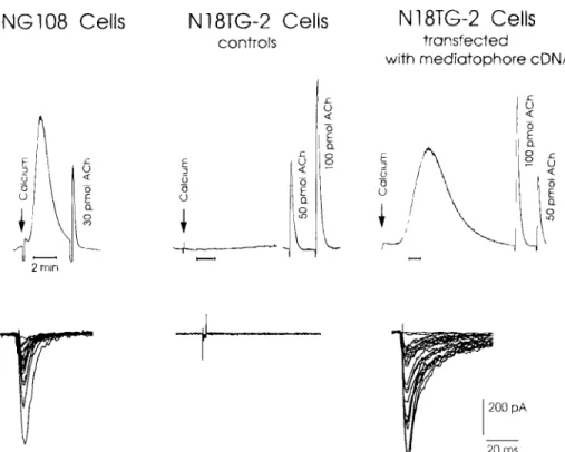
20 msFig. 4. Restoration of quantal release in mediatophore-transfected cells. Cells were filled overnight with ACh, washed, and then harvested to be tested for transmitter release. In the first test (upper traces), ACh release was elicited by application of the ionophore A23187 followed by calcium and.measured by a luminescence reaction (Isra61 and Lesbats, 1981). In parallel, cells were applied onto a culture of embryonic
Xenopus
myocytes. Individual cells were pushed with an electrode against a patch-clamped myocyte and stimulated electrically. Evoked transmitter release was recorded in real time from the myocyte. Neuroblastoma x glioma hybrid cells NG108 efficiently released the transmitter, but neuroblastoma N18TG-2 cells were incompetent for release. However, release could be re-established in N18TG-2 cells upon transfection with the mediatophore subunit cDNA. The electrophysiological tests showed that ACh release from transfected cells was quantal as in naturally occurring synapses (Falk-Vairant et al., 1996b,c,d).1986; Muller et al., 1987). An example of such a dissociation was demonstrated in the
Torpedo
electric organ submitted to a brief nerve stimu- lation (100 Hz during 12 s). Transmitter release took place during the first 10 s of the tetanus but images of vesicle fusion started to occur only after the end of activity, culminating at i min of the posttetanic recovery period (Parducz et al., 1994).
A still more dramatic dissociation between transmitter release a n d vesicle fusion was recently reported at the frog n e u r o m u s c u l a r junction. Synaptic vesicles were stained with the fluorescent marker FMI-43, then the rate of
"destaining" (taken as a marker for exocytosis) was m o n i t o r e d together w i t h the release of transmitter during the course of nerve stimula- tion. U n d e r control conditions, the kinetics of destaining was roughly parallel to the rate of release for 1-2 m i n (Betz and Bewick, 1993). H o w e v e r , staurosporine, a protein kinase C inhibitor, fully abolished destaining but left synaptic transmission unaltered (Henkel and Betz, 1995).
Another example of dissociation was found in Zn2+-treated synapses of
Torpedo
electric organ. Incubation of small pieces of tissue in 250 ~tM ZnC12 irreversibly blocked the release12 Israf/ and Dunant
of ACh, but did not cause any significant alter- ation in the fine structure of resting nerve- electroplate junctions. Electrical stimulation at this stage resulted in C a 2+ accumulation and massive vesicle fusion despite the fact that no transmitter was released. This experimental schedule created an unusual situation where the exo-endocytotic activity of synaptic vesicles could be activated independently from the release of transmitter (Parducz et al., 1997).
The SNARE Proteins
and Quantal Release
As seen above, a host of proteins associated either with synaptic vesicles or with the plasma membrane are widely distributed among neural and nonneural cells. Synaptophysin is the most abundant protein associated with vesicle mem- branes, but surprisingly, suppression of synap- tophysin causes only a modest alteration, if any, of evoked and spontaneous quantal transmitter release (Alder et al.; 1992; Eshkind and Leube, 1995; McMahon et al., 1996). When release is reconstituted in oocytes, however, antisynap- tophysin antisense oligonucleotides cause a reductiofl in release (Alder et al., 1992). On the other hand, alteration of proteins of the V- and T-SNARE family (Sollner and Rothman, 1994) affects transmission more seriously. Among these, VAMP/synaptobrevin, SNAP-25, and syntaxin are of special interest since they can be cleaved by clostridial toxins (Schiavo et al., 1992; Blasi et al., 1993), which results in interruption of the neurally evoked release of transmitter. However, some quantal transmitter release persists during the whole period of clostridial intoxication that can last for several weeks (Kriebel et al., 1976; Harris and Miledi, 1971; Thesleff et al., 1983). Moreover, evoked trans- mitter release can be partially restored by increasing C a R+ entry or by using agents like latrotoxin (Molgo et al., 1990; Gansel et al., 1987). Hence, by inactivating VAMP, SNAP-25, syntaxin, and possibly other proteins, clostridial toxins uncouple the release mechanism from its
natural trigger, i.e., the abrupt elevation of calcium at the above-described microdomains. The very process involved in quantal release is still operational but its Ca "-+ trigger is perturbed.
Expression of the SNARE proteins was also modified or suppressed in transgenic animals. In the absence of synaptotagmin, the sensitivity of neurally evoked release was strongly inhibi- ted, but spontaneous quantal release subsisted; it could still be greatly accelerated by hyper- tonicity or other techniques (Nonet et al., 1993; DiAntonio et al., 1993; Bommert et al., 1993; Broadie et al., 1995; Littleton and Bellen, 1995). A similar picture was obtained by suppressing VAMP/synaptobrevin or syntaxin (Broadie et al., 1995; Sweeney et al., 1995). The picture was therefore very similar to that of clostridial in- toxication. The mechanism supporting quantal release was still present, but its Caa+-dependent trigger was strongly perturbed. This family of proteins is therefore essential for ensuring the physiological regulation and synchronization of quantal release, but not for supporting the most elementary step of release, i.e., the forma- tion of neurotransmitter quanta.
Vesicular Docking Mechanisms
The three proteins syntaxin, SNAP-25, and VAMP/synaptobrevin constitute a clostridial toxin-sensitive anchor. This complex has been purified by fractionation and immunoprecipi- tation experiments (Sollner et al., 1993; Shift et al., 1996). There are, in addition, two possible systems that may anchor the vesicles to the plasma membrane, the <, C2 ~ attachment and the synaptophysin-mediatophore link (Fig. 5).
Several proteins share a homologous stretch of sequence, which is the C2 domain. It is found in the vesicular protein synaptotagmin, in protein kinase C, and in the cytoplasmic protein, rabphilin. The latter can become asso- ciated to Rab3-GTP, and thereby to vesicle membranes. The C2 anchor binds directly or indirectly to the calcium channel as shown by immunoprecipitation experiments (Leveque et al., 1992; Horikawa et al., 1993). Decoy
Regulation of Cholinergic Function
13
C2 Docking Clostidrial docking Mediatophore docking
VAMP
Synaptotagmin Synaptobrevm Synaptophysin
Ca ++Channel Ca ++Channel Ca ++Channel
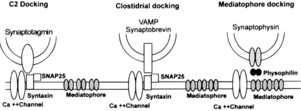
Fig. 5. Three docking links between synaptic vesicles and the plasma membrane. The "C2 docking" complex involves proteins with a C2 sequence (such as synaptotagmin, rabphilin, protein kinase C, and others) and a site at, or close to, the Ca ~+ channel. The "clostridial docking" complex refers to proteins that can be cleaved by botulism or tetanus toxins. They are Vamp/synaptobrevin, SNAP-25 and syntaxin. A third putative docking system involves synaptophysin, mediatophore, and physophilin.
peptides homologous to the C2 domain or protein kinase C itself, perturb this C2 docking and alter transmission (Bommert et al., 1993). Similarly, interference with Rab-rabphilin attachment affects release in pituitary cells (Lledo et al., 1993).
The third type of docking may involve two channel-forming proteins, synaptophysin and the mediatophore This possibility was sug- gested by the fact that synaptophysin (and also synaptobrevin) is immunoprecipitated with the 15-kDa protein (Galli et al., 1996), which is common to mediatophore and to the c-subunit of the V-ATPase membrane sector (Vo). In addition, another subunit of V-ATPase (39 kDa) was previously identified as physophilin, a synaptophysin-binding protein (Thomas and Betz, 1990; Siebert et al., 1994). This docking system might link, in series through media- tophore and synaptophysin-Vo, the vesicle lumen to the extracellular space. This link is expected to be resistant to clostridial toxins. It may still function, like the C2 anchor, when the clostridial anchor (T- and V-SNAREs) is absent or cut. In fact, vesicles still dock to the membrane when the clostridial link is damaged (Broadie et al., 1995). However, a physiologi- cally intact clostridial link is likely to be essen- tial for controlling the shape and kinetics of the Ca 2+ microdomains, ensuring synchroniza-
tion of release, and protecting mediatophore against desensitization.
Conclusions
In order to control a cholinergic phenotype, a neuron has to turn on the cholinergic locus but also must express the mediatophore at the plasma membrane in order to support quantal release of ACh. Synchronization of release with respect to the calcium trigger is regulated by a set of proteins that dock the vesicles at the active zones (Couteaux and P6cot- Dechavassine, 1970). The docked vesicles are expected to control the shape and the kinetics
o f C a 2+ microdomains. The latter regulation is independent of the cholinergic phenotype since it is found in all synapses.
From the work of Zimmermann and Denston (1977) we may consider that vesicle recycling is an essential feature of synaptic function since transmission would rapidly stop if recycling was blocked. Moreover knowledge of protein-protein interactions that support the dynamics of endo-exocytosis are now well documented by the work of Sudhof (1995). These essential contributions are compatible with our own views on the role of an inevitable gate, probably mediatophore, that supports
14 Israel and Dunant
the final t r a n s l o c a t i o n s t e p s of release: the d e l i v e r y of the s m a l l e s t q u a n t u m of A C h .
Acknowledgments
Real-time r e c o r d i n g of t r a n s f e c t e d cells w a s i n i t i a t e d b y o u r f r i e n d a n d c o l l e a g u e , J e a n F a l k - V a i r a n t w h o d i e d in a car a c c i d e n t o n N o v e m b e r 21, 1994. W e are g r a t e f u l to A n n a n d G a b o r K a t o for h e l p f u l c r i t i c i s m a n d to o u r c o w o r k e r s for s u p p o r t , d i s c u s s i o n s , a n d s o m e u n p u b l i s h e d d a t a . W o r k in o u r l a b o r a t o r i e s w a s s u p p o r t e d b y the Swiss F.N.R.S. (Grant N o 31.47286.96) to Y.D. a n d F r e n c h A.F.M. a n d D.R.E.T. g r a n t s to M.I.References
Alder J., Xie X., Valtorta F., Greengard P., and Poo M. M. (1992) Antibodies of synaptophysin inter- fere with transmitter secretion at neuromuscular synapses. Neuron 9, 759-768.
Alder J., Lu B., Valtorta F., Greengard P., and Poo M. M. (1992) Calcium-dependent transmitter secretion reconstituted in Xenopus oocytes: requirement for synaptophysin. Science 257, 657-661.
Alfonso A., Grundahl K., Duerr J. S., Han H. P., and Rand J. B. (1993) The Caenorhabditis elegans unc-] 7 gene: a putative vesicular acetylcholine trans- porter. Science 261, 617-619.
Angrist M., Kauffman E., S l a u g e n h a u p t S. A., Matise T. C., Puffenberger E. G., Washington S. S., Lipson A., Cass D. T., Reyna T., Weeks D. E., et al. (1993) A gene for H i r s c h s p r u n g disease (megacolon) in the pericentromeric region of h u m a n chromosome 10. Nat. Genet. 4, 351-356. B6janin S., Habert E., Berrard S., Edwards J. B.,
Loeffler J. P., and Mallet J. (1992) Promoter elements of the rat choline acetyltransferase gene allowing nerve g r o w t h factor inducibility in transfected primary cultured cells. J. Neurochem.
58, 1580-1583.
BOjanin S., Cervini R., Mallet J., and Berrard S. (1994) A unique gene organization for two cholinergic markers, choline acetyltransferase and a putative vesicular transporter of acetylcholine. J. Biol. Chem. 269, 21,944-21,947.
Bahr B. A. and Parsons S. M. (1986a) Acetylcholine transport and drug inhibition kinetics in Torpedo
synaptic vesicles. J. Neuroclzem. 46, 1214-1218. Bahr B. A. and Parsons S. M. (1986b) Demonstration
of a receptor in Torpedo synaptic vesicles for the acetylcholine storage blocker L-trans-2-(phenyl [3,4 3H] piperidino) cyclohexanol. Proc. Natl. Acad. ScL USA 83, 2267-2270.
Barnard E. A. and Bilbe G. (1986) Functional expression in the Xenopus oocyte of the mRNAs for receptors and ion channels, in Neurochemistry, A Practical Approach (Turner, A. J. and Bachelard H. S., eds.) IRL Press, Oxford, pp. 243-270.
Bazan J. F. (1991) Neuropoietic cytokines in the hematopoietic fold. Neuron. 7, 197-208.
Berrard S., Varoqui H., Cervini R., Isra61 M., Mallet J., and Diebler M.-F. (1995) Coregulation of two e m b e d d e d gene products, choline acetyltrans- ferase and the vesicular acetylcholine trans- porter. J. Neurochem. 65, 939-942.
Berse B. and Blusztajn J. K. (1995) C o o r d i n a t e d up-regulation of choline acetyltransferase and vesicular acetylcholine transporter gene expres- sion by the retinoic acid receptor alpha, cAMP, and leukemia inhibitory factor/ciliary neuro- trophic factor signaling p a t h w a y s in a m u r i n e septal cell line. J. Biol. Chem. 270, 22,101-22,104. Betz W. J. and Bewick G. S. (1993) Optical monitor-
ing of transmitter release and synaptic vesicle recycling at the frog n e u r o m u s c u l a r junction.
J. Physiol. Lond. 460, 287-309.
Birman S., Isra61 M., Lesbats B., and Morel N. (1986) Solubilization and partial purification of a presy- naptic m e m b r a n e protein ensuring calcium- dependent acetylcholine release from proteolipo- somes. J. Neurochem. 47, 433-444.
Birman S., Meunier F. M., Lesbats B., LeCaer J. P. LeCaer, Rossier, J. and Isra61 M. (1990) A 15 kD proteolipid found in mediatophore preparations from Torpedo presents high sequence homology with the bovine chromaffin granule pro- tonophore. FEBS Lett. 261, 303-306.
Blasi J., C h a p m a n E. R., Link E., Binz T., Yamasaki S., De Camilli P., Sudhof T. C., Nieman H., and Jahn R. (1993) Botulinum neurotoxin A selec- tively cleaves the synaptic protein SNAP-25.
Nature 365, 160-163.
Blaustein M. P., Church P. J., and Stanley E. F. (1996) Localization of ion transporters involved in cal- cium control in presynaptic nerve terminals. Soc. for Neurosc. Abstr. 22, 323.
Regulation of Cholinergic Function 15
Blusztajn J. K., Venturini A., Jackson D. A., Lee H. J., and Wainer B. H. (1992) Acetylcholine synthe- sis and release is enhanced by dibutyryl cyclic AMP in a neuronal cell line derived from mouse septum. ]. Neurosci. 12, 793-799.
Bommert K., Charlton M. P., DeBello W. M., Chin G. J., Betz H., and Augustine G. J. (1993) Inhibition of neurotransmitter release by C2-domain pep- tides implicates synaptotagmin in exocytosis.
Nature 363, 163-165.
Broadie K., Prokop A., Bellen H. J., O'Kane C. J., Schulze K. L., and Sweeney S. T. (1995) Syntaxin and synaptobrevin function d o w n s t r e a m of vesicle docking in Drosophila. Neuron 15,
663-673.
Brochier G., Gulik-Krzywicki T., Lesbats B., Dedieu J., and [sra61 M. (1992) Calcium-induced acetylcholine release and intramembrane parti- cle occurrence in proteoliposomes equipped with mediatophore. Biol. Cell 74, 225-230.
Brochier G., lsra61 M., and Lesbats B. (1993) Immunolabelling of the presynaptic membrane of Torpedo electric organ nerve terminals with an
antiserum towards the acetylcholine releasing protein mediatophore. Biol. Cell 78, 145-154.
Buchs P. A. and Muller D. (1996) Induction of long- term potentiation is associated with major ultra- structural changes of activated synapses. Proc. Natl. Acad. Sci. LISA 93, 8040-8045.
Cavalli A., Eder-Colli L., Dunant Y., Loctin F., and Morel N. (1991) Release of acetylcholine Xenopus
oocytes injected with nRNAs from cholinergic neurons. EMBO J. 10, 1671-1675.
Cavalli A., Dunant Y., Leroy C., Meunier F. M., Morel N., and Israel M. (1993) Antisense probes against mediatophore block transmitter release in oocytes primed with neuronal mRNAs. Eur. J. Neurosci. 5, 1539-1544.
Cervini R., H o u h o u L., Pradat P. F., B6janin S., Mallet J., and Berrard S. (1995) Specific vesicular acetylcholine transporter promoters lie within the first intron of the rat choline acetyltransferase gene. J. Biol. Chem. 270, 24,654-24,657.
Couteaux R. and P6cot-Dechavassine M. (1970) V6sicules synaptiques et poches au niveau des "zones actives" de la jonction neuromusculaire.
C.R. Acad. Sci. Paris 271, 2346-2349.
Curtis R., Scherer S. S., Somogyi R., A d r y a n K. M., Ip N. Y., Zhu Y., Lindsay R. M., and DiStefano P. S. (1994) Retrograde axonal transport of LIF is increased by peripheral nerve injury: correla-
tion with increased LIF expression in distal nerve. Neuron. 12, 191-204.
DiAntonio A., Parfitt K. D., and Schwarz T. L. (1993) Synaptic transmission persists in synaptotagmin mutants of Drosophila. Cell 73, 1281-1290.
Diebler M. F. and Morot G a u d r y Talarmain Y. (1989) AH5183 and cetiedil: two potent inhibitors of acetylcholine uptake into isolated synaptic vesicles from Torpedo marmorata. J. Neurochem. 52,
813-821.
Dunant Y. and Isra61 M. (1995) Mediatophore and other presynaptic proteins. A cybernetic linking at the active zone. ]. Physiol. Paris 89,
147-156.
Dunant Y., Gautron J., Isra61 M., Lesbats B., and Manaranche R. (1972) Les compartiments d'ac6tylcholine de l'organe 61ectrique de la Torpille et leurs modifications par la stimulation.
]. Neurodwm. 19, 1987-2002.
Dunant Y., Gautron J., Isra61 M., Lesbats B., and Manaranche R. (1974) Evolution de la d6charge de l'organe 61ectrique de la Torpille et variations simultan6es de l'ac6tylcholine au cours de la stimulation. J. Neurochem. 23, 635-643.
Dunant Y., Babel-Gu6rin E., and Droz B. (1980) Calcium metabolism and acetylcholine release at the nerve-electroplaque junction. ]. Physiol. Paris
76, 471-478.
D u n a n t Y., Loctin F., Marsal J., Muller D., Parducz A., and Rabasseda X. (1988) Energy metabolism and quantal acetylcholine release. Effects of b o t u l i n u m toxin, fluorodinitroben- zene and diamide in the Torpedo electric organ. J. Neuroehem. 50, 431-439.
Edery P., Pelet A., Mulligan L. M., Abel L., Attie T., Dow E., Bonneau D., David A., Flintoff W., Jan D., et al. (1994) Long segment and short segment famil- ial Hirschsprung's disease: variable clinical expres- sion at the RET locus. J. Med. Genet. 31, 602-606.
Edery P., Attie T., Amiel J., Pelet A., Eng C., Hofstra R. M., Martelli H., Bidaud C., Munnich A., and Lyonnet S. (1996) Mutation of the endothelin-3 gene in the Waardenburg-Hirschsprung disease (Shah-Waardenburg syndrome). Nat. Genet. 12,
442-444.
Erickson J. D., Eiden L. E., and Hoffman B. J. (1992) Expression cloning of a reserpine-sensitive vesic- ular monoamine transporter Proc. Natl. Acad. Sci. USA 89, 10,993-10,997.
Erickson J. D., Varoqui H., Schafer M. K., Modi W., Diebler M. F., Weihe E., Rand J., Eiden L. E.,
16 Isra[;l and D u n a n t
Bonner T. l., and Usdin T. B. (1994) Functional identification of a vesicular acetylcholine trans- porter and its expression from a "cholinergic" gene locus. ]. Biol. Chem. 269, 21,929-21,932. Eshkind L. G. and Leube R. E. (1995) Mice lacking
synaptophysin reproduce and form typical synaptic vesicles. Cell Tissue Res. 282, 423-433. Evers J., Laser M., Sun Y., Xie Z., and Poo M. M.
(1989) Studies of nerve-muscle interactions in
Xenopus cell culture: analysis of early synaptic currents. J. Neurosci. 9, 1523-1539.
Falk-Vairant J., Dunant Y., and Isra61 M. (1994) Quantal acetylcholine release in reconstituted systems. J. Neurochem. 63, $90.
Falk-Vairant J., Isra61 M., Bruner J., Stinnakre J., Meunier F. M., Gaultier P., Meunier F. A., Lesbats B., Synguelakis M., Corr6ges P., and Dunant, Y. (1996a) Evoked transmitter release from fibrob- lasts loaded with acetylcholine, enhancement by cAMP. Neuroscience 75, 353-360.
Falk-Vairant J., Meunier F. M., Lesbats B., Corr6ges P., Eder-Colli L., Salem N., Synguelakis M., Dunant Y., and Isra61 M. (1996b) Cell lines expressing an acetylcholine release mechanism, correction of a release-deficient cell by media- tophore transfection. J. Neurosc. Res. 45, 195-201. Falk-Vairant J., Corr6ges P., Eder-Colli L., Salem
N., Meunier F., Lesbats B., Loctin F., Synguelakis M., Isra61 M., and D u n a n t Y. (1996c) Evoked acetylcholine release expressed by transfection of m e d i a t o p h o r e cDNA. J. Neurochenz. 66, 1322-1325.
Falk-Vairant J., Corr6ges P., Eder-Colli L., Salem N., Roulet E., Bloc A., Meunier F., Lesbats B., Loctin F., Synguelakis M., Isra61 M., and Dunant Y. (1996d) Quantal acetylcholine release induced by mediatophore transfection. Proc. Natl. Acad. Sci. USA 93, 5203-5207.
Finbow M. E., Pitts J. D, Goldstein D. J, Schlegel R., and Findlay B. C. (1991) The E5 oncoprotein target: a 16-kDa channel- forming protein with diverse functions. Molec. Carcinog. 4, 441-444. Finbow M. E., Harrison M., and Jones P. (1995)
Ductin--a proton pump component, a gap junc- tion channel and a neurotransmitter release channel. Bioessays 17, 247-255.
Galli T., McPherson P. S., and De Camilli P. (1996) The Vo sector of the VATPase, synaptobrevin, and synaptophysin are associated on synaptic vesicles in a Triton X-100-resistant, freeze-thaw- ing sensitive, complex. J. Biol. Chem. 271, 2193-2198.
Gansel M., Penner R., and Dreyer F. (1987) Distinct sites of action of clostridial neurotoxins revealed by double-poisoning of mouse motor nerve terminals. Pflfigers Arch. 409, 533-539.
Garcia-Segura L. M., Muller D., and D u n a n t Y. (1986) Increase in the n u m b e r of presynaptic large intramembrane particles during synaptic transmission at the Torpedo nerve-electroplaque junction. Neuroscience 19, 63-79.
Girod R., Eder-Colli L., Meditanski J., Dunant Y., Tabti N., and Poo M. M. (1992) Pulsatile release of acetylcholine by nerve terminals (synapto- somes) isolated from the Torpedo electric organ.
I. Physiol. Lond. 450, 325-340.
Girod R., Corr6ges P., Jacquet J., and Dunant Y. (1993) Space and time characteristics of transmit- ter release at the nerve-electroplaque junction of Torpedo. J. Physiol. Lond. 471, 129-157.
Gundersen C. B., Miledi R. B., and Parker I. (1984) Slowly inactivating potassium channels induced in Xenopus oocytes by messenger ribonucleic acid from Torpedo brain. J. Physiol. Lond. 353, 231-248.
Gundersen C. B., Jenden D. J., and Miledi R. B. (1985) Choline acetyltransferase and acetyl- choline in Xenopus oocytes injected with mRNA from the electric lobe of Torpedo. Proc. Natl. Acad. Sci. LISA 82, 608-611.
Harris A. J. and Miledi R. B. (1971) The effect of type D botulinum toxin on frog neuromuscular junc- tions. J. Physiol. Lond. 217, 497-515.
Henderson C. E., Phillips H. S., Pollock R. A., Davies A. M., Lemeulle C., Armanini M., Simmons L., Moffet B., Vandlen R. A., Simpson L. C., Koliastos V. E., and Rosenthal A. (1994) GDNF: a potent survival factor for motoneurons present in peripheral nerve and muscle. Science 266, 1062-1064.
Henkel A. W. and Betz W. J. (1995) Staurosporine blocks evoked release of FM1-43 but not acetyl- choline from frog motor terminals. J. Neurosc. 15, 8246-8258.
Heuser J. E., Reese T. S., Dennis M. J., Jan Y., Jan L., and Evans L. (1979) Synaptic vesicle exo- cytosis captured by quick freezing and correlated with quantal transmitter release. J. Cell. Biol. 81, 275-300.
Horikawa H. P., Saisu H., Ishizuka T., Sekine Y., Tsugita A., Odani S., and Abe T. (1993) A complex of rab3A, SNAP-25, V A M P / s y n a p t o - brevin-2 and syntaxins in brain presynaptic terminals. FEBS Lett. 330, 236-240.
Regulation of Cholinergic Function 17
Ibanez C. F. and Persson H. (1991) Localization of sequences determining cell type specificity and NGF responsiveness in the promoter region of the rat choline acetyltransferase gene. Eur. J. Neurosci. 3, 1309-1315.
Isra61 M. and Dunant Y. (1993) Acetylcholine release, from molecules to function. Prog. Brain Res. 98, 219-233.
Isra61 M. and Lesbats B. (1981) Continuous determi- nation by a chemiluminescent method of acetyl- choline release and compartmentation in Torpedo
electric organ synaptosomes J. Neurochem. 37,
1475-1483.
Isra61 M. and Lesbats B. (1982) Application to m a m m a l i a n tissues of the chemiluminescent method for detecting acetylcholine. J. Neurochem.
39, 248-250.
Isra61 M., Gautron J., and Lesbats B. (1968) Isolement des v6sicules synaptiques de l'organe 61ectrique de la Torpille et localisation de l'ac6tyl- choline a leur niveau. C.R. Acad. Sci. Paris 266,
273-275.
Isra61 M., Gautron J., and Lesbats B. (1970) Fractionnement de l'organe 61ectrique de la Torpille: localisation subcellulaire de l'ac6tyl- choline. J. Neurochem. 17, 1441-1450.
Isra61 M., Dunant Y., and Manaranche R. (1979) The present status of the vesicular hypothesis. Prog. Neurobiol. 13, 237-275.
lsra61 M., Manaranche R. F. M., Morel N., Frachon P., and Lesbats B. (1980) ATP-dependent calcium uptake by cholinergic synaptic vesicles isolated from Torpedo electric organ. J. Membr. Biol. 54,
115-126.
Isra61 M., Lesbats B., and Manaranche R. (1981) ACh release from osmotically shocked synaptosomes refilled with transmitter. Nature 294, 474-475.
Isra61 M., Manaranche R., Morel N., Dedieu J., Gulik-Krzywicki T., and Lesbats B. (1981) Redistribution of intramembrane particles related to acetylcholine release by cholin- ergic synaptosomes J. Ultrastruct. Res. 75,
162-178.
Isra61 M., Lesbats B., Morel N., Manaranche R., Gulik-Krzywicki T., and Dedieu J. (1984) Reconstitution of a functional synaptosomal membrane possessing the protein constituents involved in acetylcholine translocation. Proc. Natl. Acad. Sci. USA 81, 277-281.
Isra61 M., Morel N., Lesbats B., Birman S., and Manaranche R. (1986) Purification of a presynap- tic membrane protein that mediates a calcium-
dependent translocation of acetylcholine. Proc. Natl. Acad. Sci. USA 83, 9226-9230.
Isra61 M., Meunier F. M., Morel N., and Lesbats B. (1987) Calcium-induced desensitization of acetyl- choline release from synaptosomes or proteolipo- somes equipped with mediatophore, a presynaptic membrane protein. J. Neurochem. 49, 975-982.
Isra61 M., Lesbats B., Morel N., Manaranche R., and Le Gal la Salle G. (1988) Is the acetylcholine releasing protein mediatophore present in rat brain? FEBS Lett. 233, 421-426.
Isra61 M., Lesbats B., Sbia M., and Morel N. (1990) Acetylcholine translocating protein: mediatophore at rat neuromuscular synapses. J. Neurocllem. 55,
1758-1762.
Isra61 M., Lesbats B., Synguelakis M., and Joliot A. (1994) Acetylcholine accumulation and release by hybrid NG108-15, glioma and neuroblastoma cells--Role of a 16 kDa m e m b r a n e protein in release. Neurochem. Int. 25, 103-109.
Katz B. and Miledi R. B. (1977) Transmitter leakage from motor nerve endings. Proc. R. Soc. London. B.
196, 59-72.
Kriebel M. E. and Gross C. E. (1974) Multimodal dis- tribution of frog miniature endplate potentials in adult, denervated and tadpole leg muscle.
J. Gen. Physiol. 64, 85-103.
Kriebel M., Llados E. F., and Matteson D. R. (1976) Spontaneous subminiature end-plate potentials in mouse diaphragm muscle: evidence for syn- chronous release. J. Physiol. Lond. 262, 553-581.
Leroy C. and Meunier F. M. (1995) Differential targeting to the plasma m e m b r a n e of the Torpedo 15-kDa proteolipid expressed in oocytes. J. Neurochenl. 65, 1789-1797.
Leveque C., Hoshino T., David P., Shoji-Kasai Y., Leys K., Omori A., Lang B., E1 Far O., Sato K., Martin Moutot N., et al. (1992) The synaptic vesicle protein synaptotagmin associates with calcium channels and is a putative Lambert- Eaton myasthenic syndrome antigen. Proc. Natl. Acad. Sci. USA 89, 3625-3629.
Lin L. F., Doherty D. H., Lile J. D., Bektesh S., and Collins F. (1993) GDNF: a glial cell line-derived neurotrophic factor for midbrain dopaminergic neurons. Science 260, 1130-1132.
Littleton J. T. and Bellen H. J. (1995) Synaptotagmin controls and modulates synaptic-vesicle fusion in a Ca(2+)-dependent manner. Trends Neurosci. 18,
177-183.
Liu Y., Peter D., Roghani A., Schuldiner S., Prive G. G., Eisenberg D., Brecha N., and Edwards R. H.