HAL Id: inserm-00150829
https://www.hal.inserm.fr/inserm-00150829
Submitted on 15 Jan 2010
HAL is a multi-disciplinary open access
archive for the deposit and dissemination of
sci-entific research documents, whether they are
pub-lished or not. The documents may come from
teaching and research institutions in France or
abroad, or from public or private research centers.
L’archive ouverte pluridisciplinaire HAL, est
destinée au dépôt et à la diffusion de documents
scientifiques de niveau recherche, publiés ou non,
émanant des établissements d’enseignement et de
recherche français ou étrangers, des laboratoires
publics ou privés.
Hyperoxic exposure leads to nitrative stress and ensuing
microvascular degeneration and diminished brain mass
and function in the immature subject.
Mirna Sirinyan, Florian Sennlaub, Allison Dorfman, Przemyslaw Sapieha,
Fernand Gobeil, Pierre Hardy, Pierre Lachapelle, Sylvain Chemtob
To cite this version:
Mirna Sirinyan, Florian Sennlaub, Allison Dorfman, Przemyslaw Sapieha, Fernand Gobeil, et al..
Hyperoxic exposure leads to nitrative stress and ensuing microvascular degeneration and diminished
brain mass and function in the immature subject.. Stroke, American Heart Association, 2006, 37 (11),
pp.2807-15. �10.1161/01.STR.0000245082.19294.ff�. �inserm-00150829�
ISSN: 1524-4628
Copyright © 2006 American Heart Association. All rights reserved. Print ISSN: 0039-2499. Online
Stroke is published by the American Heart Association. 7272 Greenville Avenue, Dallas, TX 72514
DOI: 10.1161/01.STR.0000245082.19294.ff
published online Sep 28, 2006;
Stroke
Gobeil, Jr, Pierre Hardy, Pierre Lachapelle and Sylvain Chemtob
Mirna Sirinyan, Florian Sennlaub, Allison Dorfman, Przemyslaw Sapieha, Fernand
Subject
Degeneration and Diminished Brain Mass and Function in the Immature
http://stroke.ahajournals.org
located on the World Wide Web at:
The online version of this article, along with updated information and services, is
http://www.lww.com/static/html/reprints.html
Reprints: Information about reprints can be found online at
journalpermissions@lww.com
Street, Baltimore, MD 21202-2436. Phone 410-5280-4050. Fax: 410-528-8550. Email:
Permissions: Permissions & Rights Desk, Lippincott Williams & Wilkins, 351 West Camden
http://stroke.ahajournals.org/subsriptions/
Subscriptions: Information about subscribing to Stroke is online at
at Universite de Montreal on October 3, 2006
stroke.ahajournals.org
Hyperoxic Exposure Leads to Nitrative Stress and Ensuing
Microvascular Degeneration and Diminished Brain Mass
and Function in the Immature Subject
Mirna Sirinyan, MSc; Florian Sennlaub, MD, PhD; Allison Dorfman, BSc;
Przemyslaw Sapieha, PhD; Fernand Gobeil, Jr, PhD; Pierre Hardy, MD, PhD;
Pierre Lachapelle, PhD; Sylvain Chemtob, MD, PhD
Background and Purpose—Neonates that survive very preterm birth have a high prevalence of cognitive impairment in
later life. A common factor detected in premature infants is their postnatal exposure to high oxygen tension relative to
that in utero. Hyperoxia is known to elicit injury to premature lung and retina. Because data on the exposure of the brain
to hyperoxia are limited, we studied the effects of high oxygen on this tissue.
Methods—Rat pups were exposed from birth until day 6 to 21% or 80% O
2. Cerebral vascular density was quantified by
lectin immunohistochemistry. Immunoblots for several proteins were performed on brain extracts. We assessed cerebral
functional deficits by visual evoked potentials.
Results—Exposure of pups to hyperoxia leads to cerebral microvascular degeneration, diminished brain mass, and cerebral
functional deficits. These effects are preceded by an upregulation of endothelial nitric oxide synthase (eNOS) in cerebral
capillaries and a downregulation of Cu/Zn superoxide dismutase (SOD). The imbalance in nitric oxide (NO) production
and antioxidant defenses favors the formation of nitrating agents in the microvessels revealed by increased nitrotyrosine
(3-nt) immunoreactivity and decreased expression of NF-
B and the dependent vascular endothelial growth factor
receptor 2. NOS inhibitors and eNOS deletion as well as an SOD mimetic (CuDIPS) restore vascular endothelial growth
factor receptor-2 levels and nearly abolish the vasoobliteration. NOS inhibitors and SOD mimetic also prevent
O
2-induced diminished brain mass and functional deficit.
Conclusions—Data identify NO and nitrating agents as major mediators of cerebral microvascular damage, ensuing
impaired brain development and function in immature subjects exposed to hyperoxia. (Stroke. 2006;37:000-000.)
Key Words: antioxidant
䡲 brain 䡲 hyperoxia 䡲 nitric oxide 䡲 vasoobliteration
N
ewborns that survive very preterm birth have a high
prevalence of cognitive impairment in later life.
1,2Only
part of such neurodevelopmental deficits can be attributed to
major neurologic insults.
3A factor of significant
pathophys-iological relevance shared by preterm infants is exposure
to high oxygen tension relative to that in utero. It is well
recognized that supraphysiological oxygen concentrations
exert toxicity to the developing lungs and retina of the
premature subject, in particular to the susceptible vasculature
of these tissues.
4 – 6Oxygen-induced damage can largely be attributed to
reac-tive oxygen species.
4,7Premature subjects transition from an
intrauterine environment with arterial oxygen tension levels
markedly lower than those encountered ex utero and
accord-ingly are particularly prone to insult from reactive oxygen
species because their antioxidant defenses are not yet
devel-oped. Specifically, preterm babies have lower levels of the
nonenzymatic antioxidants vitamins E, A, and C compared
with term ones,
8and the lungs and retina of immature subjects
are deficient in superoxide dismutase,
9,10because this enzyme
along with catalase and glutathione peroxidase mature at term
gestation.
11,12In the lung, exposure to high oxygen induces an increase in
antioxidant enzymes
10and a decrease in nitric oxide synthase
(NOS) activity, possibly as a means of attenuating O
2toxicity.
13In contrast, in the retina, oxygen induces a microvascular
degeneration, which is mediated to a large extent by an
endothelial NOS (eNOS)-dependent increase in nitric oxide
Received April 3, 2006; accepted August 9, 2006.
From the Department of Pharmacology and Therapeutics (M.S., A.D., S.C.), McGill University, Montreal, Quebec, Canada); the Department of Pediatrics and Pharmacology (M.S., F.S., P.S., P.H., S.C.), Research Center of Hoˆpital Sainte-Justine, Universite´ de Montre´al, Quebec Canada; INSERM (F.S.), Unite´ 598, Institut Biome´dical des Cordeliers, Paris, France; the Departments of Ophthalmology and Neurology–Neurosurgery (A.D., P.L.), McGill University, Montreal Children’s Hospital Research Institute, Montreal, Quebec, Canada; and the Department of Pharmacology (F.G.), Universite´ de Sherbrooke, Sherbrooke, Que´bec, Canada.
Correspondence to Sylvain Chemtob, MD, PhD, FRCPC, Departments of Pediatrics, Ophthalmology, and Pharmacology, Research Center, Hoˆpital Sainte-Justine, 3175 Coˆte Sainte-Catherine, Montreal, Quebec H3T 1C5, Canada. E-mail sylvain.chemtob@umontreal.ca
© 2006 American Heart Association, Inc.
Stroke is available at http://www.strokeaha.org DOI: 10.1161/01.STR.0000245082.19294.ff
1
(NO).
7,14Although NO can exhibit either cytoprotective or
cytotoxic properties in retina (and other tissues),
15,16NO-mediated retinal vasoobliteration
14has recently been shown
to be dependent on the redox potential of the tissue favoring
oxidation and resulting in detrimental nitrative/nitrosative
stress and microvascular degeneration.
7At this point, whether
the molecular responses of the brain to hyperoxia resemble
those of the lung or conversely of the retina remains
unknown.
In the brain, hyperoxia has been found to lead to neuronal
cell death and a delay in brain growth in animal models.
17In
the present study, we proceeded to explore underlying
mech-anisms of hyperoxia-induced brain injury in the developing
subject. We used an exposure to 80% O
2, equivalent to that
used to develop a retinal vasculopathy as seen in retinopathy
of prematurity.
7,14We hereby show that hyperoxia diminishes
cerebral antioxidant defenses and increases NO production
resulting in increased nitration and in turn microvascular
degeneration and diminished brain mass and function.
Materials and Methods
Sprague-Dawley rats (Charles River; Que´bec, Canada) were used according to a protocol of the Hoˆpital Sainte-Justine Animal Care Committee.
Oxygen-Induced Microvessel Degeneration
Effects of hyperoxia on neurovascular degeneration were studied by exposing pups from birth until postnatal (P) day 6 to 80% oxygen in chambers controlled by an Oxycycler (BioSpherix Ltd) consistent with an approximately 4-fold increase in PaO2value normally detected in
utero and as used to develop a model of retinopathy of prematurity,7,14
exposure toⱕ50% O2yields a less severe vasculopathy as reported for
the retina.18Control animals were maintained in room air (21% O 2);
PaO2 blood values taken from the left cardiac ventricle were
81⫾3 mm Hg in pups exposed to 21% O2and 332⫾26 mm Hg in
animals exposed to 80% O2(n⫽3 per group). We exposed animals to O2
at this stage of development because at this stage, their postnatal brain development corresponds to that of an infant born at approximately 24 weeks gestation.19Long-term effects of O
2were studied by exposing rat
pups to hyperoxia until P6 and returning them to 21% O2until P30;
animals were otherwise exposed to identical lighting conditions (12-hour daily light cycles) and fed the same rodent chow. Rats were killed at either P6 or P30. Effects of hyperoxia exposure on cerebral micro-vascular density were also investigated in early postnatal eNOS⫺/⫺mice (provided by Dr P. D’Orle´ans-Juste, Universite´ de Sherbrooke, Canada) by exposing them to O2for 2 days. eNOS⫺/⫺(KO) and eNOS⫹/⫹(WT)
mice were bred on a C57BL/6 background. Isolated brains were weighed and fixed in 4% v/v formalin. Brain sections of 50m were permeabilized with methanol for 10 minutes (⫺20°C). These were in-cubated with TRITC-conjugated lectin Griffonia simplicifolia (endothe-lial cell marker; Sigma-Aldrich) (1:100) in phosphate-buffered saline overnight at room temperature. Sections were washed, visualized by epifluorescent microscopy, and vasculature quantified using Image-Pro Plus 4.5 software and normalized relative to 21% O2-exposed rats;7
given the specificity of the lectin and our focus on vasculature, background densitometry was not a concern. Quantification was aver-aged on 5 to 10 sections per animal and varied by⬍5%. This approach to measure vascular density has been abundantly used on another neural tissue, namely the retina.5,7,14,16,20 The microvessel density in the
cortical area of 80% O2-exposed pups was compared with that in
age-matched 21% O2-raised pups, which was assigned a value of 100%.
We measured vascular density as capillary length per surface area (mm/mm2).
Effects of Antinitrating Agents in
Hyperoxia-Exposed Rat Brains
Animals exposed to normoxia or hyperoxia for 6 days were injected intraperitoneally daily with vehicle or antinitrating drugs, namely N-nitroL-arginine methyl ester (L-NAME 20 mg/kg; Cayman Chemi-cal), N-(3-[aminomethyl]benzyl) acetamidine dihydrochloride (1400W 10 mg/kg; Cayman Chemical), 1-(2-trifluoromethylphenyl) imidazole (Trim 10 mg/kg; Cayman Chemical), or Cu(II) (3,5-diisopropyl-salicylate) (CuDIPs 10 mg/kg; Calbiochem). Rats were killed by decapitation at P1 or P6 and isolated brains were weighed and processed for vascular endothelial growth factor receptor-2 (VEGFR2) western blot at P1 or stained with lectin at P6 to quantify vessel density (see previously).
Visual Evoked Potential
Visual evoked potential (VEP) is a reliable and sensitive param-eter to evaluate neurologic functional alterations. VEPs were recorded at P30 from control rats (n⫽5), rats formerly exposed to hyperoxia from P1 to P6 days (n⫽5), and rats exposed to hyperoxia and concomitantly treated with L-NAME, 1400W, Trim, or CuDIPs (n⫽5 for each drug-treated group; concentra-tions as described previously). Animals were anesthetized using a mixture of 85 mg/kg ketamine and 6 mg/kg xylazine and the pupils dilated with 1% cyclopentolate hydrochloride (Mydriacyl solution; Alcon Laboratories). Animals were placed in a record-ing chamber that included both flash stimulus as well as back-ground light. A subdermal needle electrode (Grass model E2) was inserted under the scalp at the lambda suture and served as the active electrode, whereas reference (Grass model E6GH; Grass Instruments) and ground (Grass model E2) electrodes were placed in mouth and tail, respectively. VEPs were evoked to flashes of white light (0.9 log cd/sec/m⫺2) presented against a background light of 30 cd/m⫺2. Each response represents an average of 100 flashes (performed with Acknowledge data acquisition system; BIOPAC Systems Inc).
Western Blotting
Brains were isolated over a time course spanning from 6 hours to 6 days. Standard SDS-PAGE techniques were followed as previously described.7Primary antibodies were used according to the following
conditions: eNOS (1:1000 dilution), nNOS (1:1000), iNOS (1:500) (BD Biosciences Pharmingen), Cu/Zn SOD (1:1000) (Calbiochem), nuclear factor kappa B (NF-B) (1:500) (Zymed), or VEGFR2 (1:250) (Chemicon International). NF-B detection was performed on nuclei isolated from rat brains at 4°C.21Equal protein loading was
ensured by probing with 1:40 000-actin antibody (Novus Biologi-cals). Densitometry was measured in pixel intensity by Image-Pro Plus.
NADPH-Diaphorase Histochemistry
NADPH-diaphorase (NADPH-d), which reflects the activity of NOS isoforms, was performed on brain sections as previously reported.7,16
Immunohistochemical Analysis
Brains from O2and room air-exposed rats at P1 were fixed in 4%
formalin and transferred to 30% sucrose overnight. Cryosections (10 m) were fixed with methanol for 10 minutes (⫺20°C). Immunohistochemical analysis was performed as described16,20,21
using TRITC-labeled lectin and antibodies against eNOS (poly-clonal; Transduction Laboratories), 3-nitrotyrosine (3-nt, monoclo-nal; Transduction Laboratories), and VEGFR2 (polyclomonoclo-nal; Chemi-con International). Alexa-Chemi-conjugated seChemi-condary IgGs were then applied to slides (Molecular Probes), and nuclei were counterstained with DAPI (Molecular Probes). Sections were assessed using epif-luorescent microscopy.
Immunoprecipitation of VEGFR2
Rat brains were isolated, homogenized in lysis buffer, and centrifuged at 8000 g for 10 minutes, and 3 mg of the resulting supernatant was reacted with anti-nt antibody (1:200) overnight at 4°C with the exception of controls. Protein A agarose beads were added to the cell lysate/antibody mixture, as well as the negative control, and rotated for 2 hours. Beads
were washed with lysis buffer and samples were resolved by SDS-PAGE and probed for VEGFR2 as described previously.
Statistical Analysis
Data were analyzed by Student t test, one- or 2-way ANOVA, followed by post hoc Bonferroni test for comparison among means.
Figure 1. Effects of hyperoxia exposure on brain microvascular integrity and VEPs. (a) Lectin-stained brains from pups raised in normoxia
(21% O2) and those exposed to hyperoxia (80% O2) until P6; also, lectin-stained brains for pups exposed to normoxia for 30 days versus
those exposed to hyperoxia until P6 and returned to normoxia until P30. Scale bar⫽100m. Values in histogram are mean⫾SEM of vessel density in cortical region relative to that of 21% O2-exposed rats, n⫽5 to 7 rats per group; ***P⬍0.001 compared with control (CTL). (b) Brain
weights of CTL and O2-exposed from birth to P6 after which exposure to normoxia was resumed until P30. Values are mean⫾SEM of 5 to 7
animals per group; ***P⬍0.001 compared with P6 CTL; ⫹⫹⫹P⬍0.001 compared with P30 CTL. (c) VEPs at P30 of normoxia-exposed rats (CTL) and of those exposed to 80% O2until P6 and then returned to normoxia until P30; records are VEPs of white light flashes. Values in
histogram are mean⫾SEM of P3amplitude; 5 rats per group; **P⬍0.05 compared with CTL.
Values are presented as mean⫾SEM. Statistically significance was set at P⬍0.05.
Results
Microvascular Degeneration, Diminished Mass,
and Functional Deficit in the Brain of
Hyperoxia-Exposed Rat Pups
Exposure to 80% O
2from birth to P6 led to significant
microvascular degeneration throughout the brain, more
pro-nounced in the cortex, which began to be detected by 24
hours after exposure to hyperoxia (Figure 1a). The loss of
vasculature was associated with a decrease in brain weight
(Figure 1b). The decrease in microvascular density and brain
weight persisted at P30 for pups exposed to hyperoxia for the
first 6 postnatal days (Figure 1a, V). However, vessel density
increased by P30, suggestive of reparative angiogenesis
during the normoxic period (P7 to P30) (Figure 1a). Brain
function at P30 (assessed by VEP, difficult to detect at earlier
age) revealed decreased amplitude of the late component P
3in
the hyperoxia-exposed animals (Figure 1c), whereas early
VEP components (N
1, P
1, N
2, P
2) were unaffected.
Expression of NOS, Cu/Zn SOD,
NF-
B and VEGFR2 in Brains Exposed
to Hyperoxia
NO from different NOS isoforms can exert cytotoxicity under
certain circumstances.
7We analyzed the expression of the 3
NOS isoforms. eNOS increased markedly by 6 hours on
exposure to O
2and decreased below control levels by 24
hours (Figure 2). nNOS exhibited an increase by 24 hours,
which was not as pronounced as that seen after 6 hours for
eNOS, but remained elevated until P4. By P6, eNOS and
nNOS returned to control levels. iNOS expression remained
unchanged (Figure 2).
The antioxidant enzyme Cu/Zn SOD catalyzes the
con-version of O
2䡠-anion into hydrogen peroxide. Cu/Zn SOD
protein expression was downregulated at 6 hours and more
markedly so 1 day after exposure to hyperoxia (Figure 2) and
normalized subsequently. The acute reduction in SOD in the
brain is consistent with that reported in the retina
22and
lungs.
10VEGFR2, which mediates vasoprotective effects of VEGF
on neurovascular endothelium,
5started to decrease during O
2
exposure by 6 hours and was heavily suppressed by 1 day
Figure 2. Expression of eNOS, nNOS,
iNOS, Cu/Zn SOD, NF-B, and VEGFR2 in rat brains after exposure to 80% O2. a,
Representative Western blots of eNOS, nNOS, iNOS, Cu/Zn SOD, NF-B, VEGFR2, and-actin (normalization standard) in brains (n⫽5 to 7) of pups exposed to 80% O2or room air from
birth until P6. b, Values in histogram are mean⫾SEM of densitometry relative to that for-actin; ***P⬍0.001 compared with corresponding time-dependent con-trol (CTL) value.
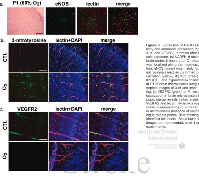
(Figure 2). Immunohistology confirmed this marked
reduc-tion in VEGFR2 in microvasculature on O
2exposure (Figure
3c); it should also be pointed out that VEGFR2 was largely
localized to the endothelium (Figure 3c). By day 6, VEGFR2
expression returned to control levels. Interestingly, changes
in levels of NF-
B, the transcription factor that regulates
VEGFR2,
23paralleled those of the receptor (Figure 2).
NADPH-Diaphorase Reactivity and
Immunolocalization of eNOS and Nitrotyrosine
Strong NADPH-diaphorase reactivity was detected along the
microvascular network of the brain cortex 6 hours after O
2exposure (Figure 3a). This pattern matched eNOS
immmunolo-calization specifically to the endothelium (Figure 3a). Twenty-four
hours after O
2exposure, 3-nt levels (nitrative stress marker
24) were
markedly stronger in brain cortex microvasculature (Figure 3b).
Prevention of O
2-Induced Nitration and
Microvascular Degeneration, Diminished Brain
Mass, and VEGFR2 Expression
We determined the role of nitrative stress on brain microvascular
degeneration and diminished brain mass by treating
hyperoxia-exposed rat pups to NOS inhibitors or SOD mimetic. The NOS
inhibitors (
L-NAME inhibits all NOS isoforms, whereas Trim
inhibits iNOS and nNOS but not eNOS) as well as SOD mimetic
CuDIPs significantly attenuated O
2-induced 3-nt
immunoreac-tivity, diminished microvascular degeneration, and preserved
brain weight, whereas the iNOS-specific inhibitor 1400W was
ineffective (Figure 4a through 4d). This presumed role of eNOS
was corroborated in O
2-exposed eNOS
⫺/⫺mice, which were
protected against microvascular degeneration compared with
eNOS
⫹/⫹congeners (Figure 4d, bottom).
VEGFR2 plays an important role in microvascular survival
during hyperoxia
5and is affected by hyperoxic-induced
nitrative stress.
7We determined whether this major factor is
nitrated under hyperoxic conditions and studied its expression
in O
2-exposed animals treated with
L-NAME, Trim, and
CuDIPs. VEGFR2 was specifically nitrated; this effect was
blocked by
L-NAME (Figure 5a). VEGFR2 nitration was
associated with its decreased expression, which was also
prevented by
L-NAME, Trim, and CuDIPS (Figure 5b). These
observations were corroborated in eNOS
⫺/⫺mice which,
contrary to their wild-type counterparts, did not exhibit
decreased VEGFR2 expression (Figure 5c).
Figure 3. Expression of NADPH-d
reac-tivity and immunofluorescence for eNOS, 3-nt, and VEGFR2 in brains after hyper-oxic exposure. (a) NADPH-d staining in brain cortex 6 hours after O2exposure
was localized along the microvascula-ture; eNOS (green) was mainly found on microvessels (red) as confirmed by colo-calization (yellow). (b) 3-nt (green) in con-trol (CTL) and hyperoxia-exposed pups at P1 in brain microvessels (red); merge depicts images of 3-nt and lectin stain-ing. (c) VEGFR2 (green) at P1 reveals its localization in brain microvessels in CTL pups; merge reveals yellow staining of VEGFR2 and lectin. Hyperoxia resulted in virtual disappearance of VEGFR2 staining in microvessels (absence of yellow stain-ing in middle panel). Blue stainstain-ing (DAPI) identifies cell nuclei. Scale bar⫽10m. Images are representatives of 4 separate experiments.
Inhibition of Nitrative Stress Prevents Altered
Visual Evoked Potential
Finally, administration of
L-NAME, Trim, and CuDIPs (but
not 1400W) for the first 6 postnatal days during O
2exposure
maintained normal P
3amplitudes (Table).
Discussion
The present study reveals that exposure of premature brains
to hyperoxia leads to severe microvascular degeneration,
diminished brain mass, and cerebral functional deficits.
Hy-peroxia is of significant pathophysiological relevance for
Figure 4. Effects of antioxidant treatment on O2-induced brain vasoobliteration. (a) 3-nt immunoreactivity of hyperoxia-exposed pups treated
with 20 mg/kgL-NAME, 10 mg/kg Trim, 10 mg/kg CuDIPs, and 10 mg/kg 1400W from birth to P6. (b) Brain weights of hyperoxia-exposed pups treated like in (a). (c) Representative photomicrographs of lectin-stained brain vascular of rat pups exposed to 80% O2from birth to P6
after treatment with vehicle,L-NAME, Trim, 1400W, or CuDIPS. Scale bar⫽20m. (d) Quantification of vessel density in cortical region of control (CTL) and O2-exposed brains treated with drugs cited in (c) and in eNOS⫺/⫺and congener wild-type mice. Values in histograms are
mean⫾SEM of 3-nt immunoreactivity (a), brain weight (b), and vessel density (d) relative to that in 21% O2-exposed animals; n⫽8 brains per
group. **P⬍0.05 compared with CTL. Drug treatments in normoxia-exposed animals did not affect brain weight and vascular density when com-pared with untreated animals (not shown); the same applies to eNOS⫺/⫺normoxia-exposed mice compared with their wild-type congeners.
preterm infants that prematurely switch from an in utero
envi-ronment of moderately low O
2tension to an extrauterine milieu
of relatively high O
2concentration. Through its autoregulatory
effects, hyperoxia leads to cerebral vasoconstriction in the
developed subject, but this response is curtailed in the
new-born.
25However, hyperoxia leads to neuronal cell death and a
delay in brain growth in animal models,
17,26but the mechanisms
underlying the neuropathology have not been investigated.
Because NO is an important signaling molecule produced in
various cell types in the brain, including cerebral endothelial
cells
27and exerts opposing effects on cell survival depending on
the redox state,
7we explored its role in brain injury after
exposure to hyperoxia. Our data point to a prominent role for
eNOS in hyperoxia-induced brain injury resulting in
microvas-cular obliteration, brain cell death, diminished brain mass, and
cerebral functional deficit.
Figure 5. Effects of drug treatment or eNOS deletion on brain VEGFR2 nitration and expression after exposure to hyperoxia. (a)
VEGFR2 expression in 3-nt immunoprecipitate in P6 animals treated or not withL-NAME (20 mg/kg intraperitoneal). Positive control
(CTL) represents brain lysate, which did not undergo immunoprecipitation, whereas negative CTL was subjected to immunoprecipitation in the absence of 3-nt antibody. (b) VEGFR2 expression and corresponding densitometric analysis of rat pups injected intraperitoneally withL-NAME, Trim, CuDIPs, or 1400W while under hyperoxia as described in Figure 4 or of (c) CTL and O2-exposed eNOS⫹/⫹or
eNOS⫺/⫺mice. Blots were normalized to-actin expression. Values in histograms are mean⫾SEM of 5 experiments. ***P⬍0.001 com-pared with CTL. Drug treatments in normoxia-exposed animals did not affect VEGFR2 expression when comcom-pared with untreated ani-mals (not shown); the same applies to eNOS⫺/⫺normoxia-exposed mice compared with their wild-type congeners.
Evidence for a significant involvement for eNOS during
hyperoxia-induced brain injury includes: (1) an early increase
(within 6 hours) in eNOS expression and NADPH-d
reactiv-ity, which reflects in situ NOS activity mostly confined to the
microvasculature (Figures 2 and 3); findings are consistent
with oxidative stress-inducing changes after ischemia
28and
more specifically with hyperoxia-induced increase in eNOS
reactivity in brain and other tissues.
7,29(2) NO-elicited
cytotoxicity is largely redox-dependent
7resulting in
forma-tion of peroxynitrite. Generaforma-tion of the latter requires
oxida-tion of NO by superoxide, which is favored by decreased
levels of SOD. Indeed, an early decrease in Cu/Zn SOD
expression in brain was observed in response to hyperoxia
(Figure 2) as reported in other tissues.
10,22(3) Accordingly, an
early rise in indicators of nitrative stress (3-nt) mostly
localized on endothelium and associated with cell death
followed the corresponding early augmentation in eNOS
(Figure 3 and supplemental Figure I, available online at
http://stroke.ahajournals.org). (4) Pharmacological inhibition
of eNOS and nNOS, but not of iNOS, or supplementation
with SOD mimetic prevented hyperoxia-induced 3-nt
reac-tivity, brain microvessel degeneration, as well as diminished
brain mass and function (Figure 4, Table). The VEP P
3wave
arises from the visual cortex and enables to assess cortical
function.
30Our VEP data at P30 support the anatomic
changes observed and modulated by effective treatments. (5)
Finally, the specific role of eNOS in the early microvascular
obliteration was confirmed in eNOS
⫺/⫺mice (Figure 4)
despite a “compensatory” increase in nNOS activity observed
in these animals.
31Nonetheless, the increase in nNOS
be-tween P1 and P4 (Figure 2), and the beneficial effect of Trim
(Figures 3 and 5), does not exclude a contribution of nNOS in
hyperoxia-induced injury.
An interesting feature in this study is the downregulation of
the prosurvival factor VEGFR2, which preceded cell death
and vasoobliteration after exposure to hyperoxia (Figures 1
and 2, supplemental Figure I). There is increasing evidence
that nitrating agents lead to extracellular death not only by
directly inhibiting the respiratory chain,
32but by acting as
molecules that negatively regulate the expression of signaling
events.
5,7,33For instance, tyrosine nitration has been described
to downregulate plasma membrane receptors by enhancing
susceptibility and targeting for proteasome degradation in
endothelial cells.
34A similar paradigm appears to apply to
VEGFR2, whereby its hyperoxia-induced downregulation
was prevented by eNOS inhibitors and was undetected in
eNOS
⫺/⫺mice (Figure 5).
The poorer neurodevelopmental outcome observed in
pre-mature relative to term infants cannot for the most part be
only attributed to specific major neurologic insults occurring
during the perinatal period.
3Compelling evidence reveals
more generalized diminished brain volume
2and point to more
subtle structural changes that are likely involved such as
alterations in neuritic extensions and in synaptogenesis,
cerebellar injuries, and possibly cell migration.
4,35However,
these changes intimately depend on a functional vascular
structure.
36The neural vasculature of the developing subject
is particularly susceptible to oxidative stress.
6,7,20Our data
suggest that early severe microvessel loss in response to
hyperoxia is an important contributing factor to decreased
brain function. The results uncover an important mechanism,
specifically involving nitration-elicited vasoobliteration,
which contributes to elucidate the impact of subtle cortical
changes on neurocognitive functional outcome of former
premature subjects.
1Therapeutic strategies aimed at
dimin-ishing nitrative stress may have the potential of dimindimin-ishing
brain injury evoked by hyperoxic stress.
Acknowledgments
The authors thank Hendrika Fernandez for valuable technical assistance.
Sources of Funding
Supported by grants from the Canadian Institutes of Health Re-search, the Heart and Stroke Foundation of Canada, and the Fonds de la Recherche en Sante´ du Que´bec (FRSQ). M.S. holds a studentship from the Heart and Stroke Foundation of Canada; F.S. and S.C. are recipients respectively of fellowship and scientist awards from the Canadian Institutes of Health Research; S.C. also holds a Canada Research Chair (perinatology). P.H. is a recipient of a scholarship from the FRSQ, and F.G. is a recipient of a Junior 1 scholarship from the FRSQ and a researcher of the Canada Foundation for Innovation.
Disclosures
None.Amplitude and Peak Time of VEP Components
Parameters Control Control (composite) O2 (composite) O2⫹L-NAME (composite) O2⫹1400W (composite) O2⫹Trim (composite) O2⫹CuDIPs (composite) N1 Amp 8.91⫾4.83 7.27 7.27 7.27 7.27 10.90 16.36 PT 36.75⫾7.42 35.55 35.55 35.55 35.55 35.55 35.55 P1 Amp 18.94⫾7.02 18.18 14.55 25.45 16.36 20.00 21.82 PT 46.63⫾3.35 46.67 52.22 46.67 46.67 46.67 46.67 P3 Amp 32.45⫾5.30 27.27 19.64* 32.72 20.00* 27.27 29.09 PT 113.62⫾10.64 124.4 107.78 113.33 107.78 124.4 124.4
Amplitude and peak time recordings of VEP parameters (N1, P1, P3) at P30 in normoxia control rats, hyperoxia-treated rats, and rats exposed to hyperoxia while receiving drug treatment. Amplitude (Amp) values in microvolts (V); peak time (PT) values in milliseconds. Significantly different from control (*P⬍0.05).
References
1. Stewart AL, Rifkin L, Amess PN, Kirkbride V, Townsend JP, Miller DH, Lewis SW, Kingsley DP, Moseley IF, Foster O, Murray RM. Brain structure and neurocognitive and behavioural function in adolescents who were born very preterm. Lancet. 1999;353:1653–1657.
2. Abernethy LJ, Cooke RW, Foulder-Hughes L. Caudate and hippocampal volumes, intelligence, and motor impairment in 7-year-old children who were born preterm. Pediatr Res. 2004;55:884 – 893.
3. Hintz SR, Kendrick DE, Vohr BR, Poole WK, Higgins RD. National Institute of Child Health and Human Development Neonatal Research Network. Changes in neurodevelopmental outcomes at 18 to 22 months corrected age among infants of less than 25 weeks gestational age born in 1993–1999. Pediatrics. 2005;115:1645–1651.
4. Freeman BA, Crapo JD. Hyperoxia increases oxygen radical production in rat lungs and lung mitochondria. J Biol Chem. 1981;256: 10986 –10992.
5. Alon T, Hemo I, Itin A, Pe’er J, Stone J, Keshet E. Vascular endothelial growth factor acts as a survival factor for newly formed retinal vessels and has implications for retinopathy of prematurity. Nat Med. 1995;1: 1024 –1028.
6. Madan A, Penn JS. Animal models of oxygen-induced retinopathy. Front
Biosci. 2003;8:d1030 – d1043.
7. Beauchamp MH, Sennlaub F, Speranza G, Gobeil F Jr, Checchin D, Kermorvant-Duchemin E, Abran D, Hardy P, Lachapelle P, Varma DR, Chemtob S. Redox-dependent effects of nitric oxide on microvascular integrity in oxygen-induced retinopathy. Free Radic Biol Med. 2004;37: 1885–1894.
8. Baydas G, Karatas F, Gursu MF, Bozkurt HA, Ilhan N, Yasar A, Canatan H. Antioxidant vitamin levels in term and preterm infants and their relation to maternal vitamin status. Arch Med Res. 2002;33: 276 –280.
9. Ogawa T, Ohira A, Amemiya T. Manganese and copper–zinc superoxide dismutases in the developing rat retina. Acta Histochem. 1997;99:1–12. 10. Morton RL, Das KC, Guo XL, Ikle DN, White CW. Effect of oxygen on
lung superoxide dismutase activities in premature baboons with broncho-pulmonary dysplasia. Am J Physiol. 1999;276:L64 –L74.
11. Gerdin E, Tyden O, Eriksson UJ. The development of antioxidant enzymatic defense in the perinatal rat lung: activities of superoxide dismutase, glutathione peroxidase, and catalase. Pediatr Res. 1985;19: 687– 691.
12. Fujii T, Ikeda Y, Yamashita H, Fujii J. Transient elevation of glutathione peroxidase 1 around the time of eyelid opening in the neonatal rat. J Ocul
Pharmacol Ther. 2003;19:361–369.
13. Arkovitz MS, Szabo C, Garcia VF, Wong HR, Wispe JR. Differential effects of hyperoxia on the inducible and constitutive isoforms of nitric oxide synthase in the lung. Shock. 1997;7:345–350.
14. Brooks SE, Gu X, Samuel S, Marcus DM, Bartoli M, Huang PL, Caldwell RB. Reduced severity of oxygen-induced retinopathy in eNOS-deficient mice. Invest Ophthalmol Vis Sci. 2001;42:222–228.
15. Murohara T, Asahara T, Silver M, Bauters C, Masuda H, Kalka C, Kearney M, Chen D, Symes JF, Fishman MC, Huang PL, Isner JM. Nitric oxide synthase modulates angiogenesis in response to tissue ischemia.
J Clin Invest. 1998;101:2567–2578.
16. Sennlaub F, Courtois Y, Goureau O. Inducible nitric oxide synthase mediates the change from retinal to vitreal neovascularization in ischemic retinopathy. J Clin Invest. 2001;107:717–725.
17. Felderhoff-Mueser U, Bittigau P, Sifringer M, Jarosz B, Korobowicz E, Mahler L, Piening T, Moysich A, Grune T, Thor F, Heumann R, Buhrer C, Ikonomidou C. Oxygen causes cell death in the developing brain.
Neurobiol Dis. 2004;17:273–282.
18. Penn JS, Henry MM, Wall PT, Tolman BL. The range of PaO2 variation determines the severity of oxygen-induced retinopathy in newborn rats.
Invest Ophthalmol Vis Sci. 1995;36:2063–2070.
19. Dobbing J, Sands J, Gratrix CA. Cell size and cell number: a reconsideration of organ growth and catch-up potential. Proc Nutr Soc. 1979;38:99A. 20. Kermorvant-Duchemin E, Sennlaub F, Sirinyan M, Brault S, Andelfinger
G, Kooli A, Germain S, Ong H, d’Orleans-Juste P, Gobeil F Jr, Zhu T, Boisvert C, Hardy P, Jain K, Falck JR, Balazy M, Chemtob S. Trans-ar-achidonic acids generated during nitrative stress induce a thrombospondin-1-dependent microvascular degeneration. Nat Med. 2005;11:1339 –1345. 21. Gobeil F Jr, Dumont I, Marrache AM, Vazquez-Tello A, Bernier SG,
Abran D, Hou X, Beauchamp MH, Quiniou C, Bouayad A, Choufani S, Bhattacharya M, Molotchnikoff S, Ribeiro-da-Silva A, Varma DR, Bkaily G, Chemtob S. Regulation of eNOS expression in brain endothe-lial cells of perinuclear EP3receptors. Circ Res. 2002;90:682– 689.
22. Niesman MR, Johnson KA, Penn JS. Therapeutic effect of liposomal superoxide dismutase in an animal model of retinopathy of prematurity.
Neurochem Res. 1997;22:597– 605.
23. Illi B, Puri P, Morgante L, Capogrossi MC, Gaetano C. Nuclear factor-kappaB and cAMP response element binding protein mediate opposite transcriptional effects on the Flk-1/KDR gene promoter. Circ Res. 2000; 86:E110 –E117.
24. Greenacre SA, Ischiropoulos H. Tyrosine nitration: localisation, quanti-fication, consequences for protein function and signal transduction. Free
Radic Res. 2001;34:541–581.
25. Hardy P, Peri KG, Lahaie I, Varma DR, Chemtob S. Increased nitric oxide synthesis and action preclude choroidal vasoconstriction to hyperoxia in newborn pigs. Circ Res. 1996;79:504 –511.
26. Taglialatela G, Perez-Polo JR, Rassin DK. Induction of apoptosis in the CNS during development by the combination of hyperoxia and inhi-bition of glutathione synthesis. Free Radic Biol Med. 1998;25: 936 –942.
27. Garthwaite J, Charles SL, Chess-Williams R. Endothelium-derived relaxing factor release on activation of NMDA receptors suggests role as intercellular messenger in the brain. Nature. 1988;336:385–388. 28. Wei G, Dawson VL, Zweier JL. Role of neuronal and endothelial nitric
oxide synthase in nitric oxide generation in the brain following cerebral ischemia. Biochim Biophys Acta. 1999;1455:23–34.
29. Atochin DN, Demchenko IT, Astern J, Boso AE, Piantadosi CA, Huang PL. Contributions of endothelial and neuronal nitric oxide synthases to cerebrovascular responses to hyperoxia. J Cereb Blood Flow Metab. 2003;23:1219 –1226.
30. Ekert PG, Keenan NK, Whyte HE, Boulton J, Taylor MJ. Visual evoked potentials for prediction of neurodevelopmental outcome in preterm infants. Biol Neonate. 1997;71:148 –155.
31. Al-Shabrawey M, El-Remessy A, Gu X, Brooks SS, Hamed MS, Huang P, Caldwell RB. Normal vascular development in mice deficient in endothelial NO synthase: possible role of neuronal NO synthase. Mol Vis. 2003;9:549 –558.
32. Shiva S, Oh JY, Landar AL, Ulasova E, Venkatraman A, Bailey SM, Darley-Usmar VM. Nitroxia: the pathological consequence of dys-function in the nitric oxide– cytochrome c oxidase signaling pathway.
Free Radic Biol Med. 2005;38:297–306.
33. el-Remessy AB, Bartoli M, Platt DH, Fulton D, Caldwell RB. Oxidative stress inactivates VEGF survival signaling in retinal endothelial cells via PI 3-kinase tyrosine nitration. J Cell Sci. 2005;118:243–252.
34. Kotamraju S, Tampo Y, Keszler A, Chitambar CR, Joseph J, Haas AL, Kalyanaraman B. Nitric oxide inhibits H2O2-induced transferrin receptor-dependent apoptosis in endothelial cells: role of ubiquitin–pro-teasome pathway. Proc Natl Acad Sci U S A. 2003;100:10653–10658. 35. Miller SP, Ferriero DM, Leonard C, Piecuch R, Glidden DV, Partridge
JC, Perez M, Mukherjee P, Vigneron DB, Barkovich AJ. Early brain injury in premature newborns detected with magnetic resonance imaging is associated with adverse early neurodevelopmental outcome. J Pediatr. 2005;147:609 – 616.
36. Vogel G. Developmental biology. The unexpected brains behind blood vessel growth. Science. 2005;307:665– 667.
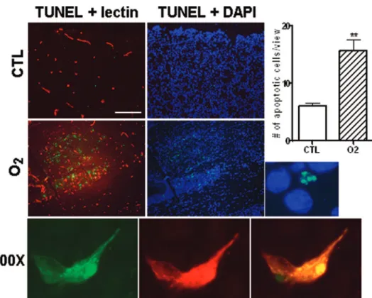
Figure I. Apoptosis in the cortex of 1-day-old brains detected by TUNEL staining. TUNEL-positivity (green) was scarce in the cortical
region of CTL pups, whereas TUNEL-positive cells were abundant in O2-exposed subjects. Blue staining (DAPI) identifies cell nuclei.
TUNEL staining colocalized with lectin (red) in hyperoxia-exposed brains. 100⫻ magnification (lower panel) shows a TUNEL-positive (green) endothelial cell (red), merge in yellow; TUNEL positivity was not only confined to vasculature. Images are representative of 3 independent experiments. Quantification (histogram) of the apoptotic cells was assessed by comparing the number of TUNEL-positive cells in the cortical region of CTL and O2pups in 10 cross-sections/group. Values are mean⫾SEM; **P⬍0.05 compared to CTL values.